Báo cáo y học: "Is there any cardioprotective role of Taurine during cold ischemic period following global myocardial ischemia" pps
Bạn đang xem bản rút gọn của tài liệu. Xem và tải ngay bản đầy đủ của tài liệu tại đây (1.02 MB, 7 trang )
RESEARCH ARTICLE Open Access
Is there any cardioprotective role of Taurine
during cold ischemic period following global
myocardial ischemia?
Mehmet A Sahin
1*
, Orhan Yucel
2
, Adem Guler
1
, Suat Doganci
1
, Artan Jahollari
1
, Faruk Cingoz
1
,Sıddık Arslan
3
,
Mehmet Gamsizkan
4
, Halil Yaman
5
, Ufuk Demirkilic
1
Abstract
Background: The aim of the present study was to investigate the cardioprotective effect of Taurine on the donor
hearts during cold ischemic period.
Methods: 32 rats were divided into four groups (sham, taurine, ischemia, treatment group, 8 rats in each). All rats
were fed with rat food for three weeks. Taurine and treatment groups were given a 200 mg/kg/day dose of
Taurine by oral gavage besides ra t feed. Cardiectomy was performed in all rats after three weeks. In ischemia and
treatment groups, harvested hearts were kept in 0.9% sodium chloride at +4 degrees C for 5 hours. Tissue samples
were taken from left ventricle in all groups. These samples were evaluated by histopathologic and biochemical
examination.
Results: In the present study results of the biochemical and histopathological examination reveals the protective
effects of Taurine. As a marker of lipid peroxidation, Malondialdehyde (MDA) levels in ischemia group were
significantly higher than both Sham and Taurine groups. MDA values were recorded; 3.62 ± 0.197 in the sham
group, 2.07 ± 0.751 in the Taurine group, 9.71 ± 1.439 in the ischemia group and 7.68 ± 1.365 in the treatment
group. MDA levels decreased in treatment group. (p < 0.05) In accordance with MDA findings, while superoxide
dismutase and glutathione peroxidase levels decreased in ischemia group, they increased in treatment group. (p <
0.05) There was no differences in Catalase (CAT) enzyme level between treatment and ischemia group (p = 1.000).
CAT level results were recorded; 7.08 ± 0.609 in the sham group, 6.15 ± 0.119 in the Taurine group, 5.02 ± 0.62 in
the ischemia group, and 5.36 ± 0.384 in the treatment group. Less intracellular edema and inflammatory cell
reaction were observed in histologic examination in favor of treatment group. (p < 0.01)
Conclusion: Taurine decreased myocardial damage during cold ischemic period following global myocardial
ischemia.
Background
Maintaining cardiac functions in explanted hearts within
ischemic time needs good preservation. Hypoxic,
hypothermic, cardioplegic arrest followed by cold trans-
port is a common procedure for preservation of
explanted hearts. This procedure is the main practical
method used for preserving donor organs in many
transplant centers [1].
Unfortunately, there is no perfect protection method
for donor organs currently. With the increase in the
ischemic time following explantation, tissue and the
organ damage are almost inevitable. Organ functions
can be improved by minimizing the myocardial function
during ischemia. For this purpose many studies have
been performed to prolong this ischemic time or protect
the organs in this deleterious process.
Taurine (2-amino ethane sulfonic acid) is a potent
antioxidant agent. It is shown that Taurine has benefi-
cial effects on myocardial ischemia-reperfusion injury,
* Correspondence:
1
Gülhane Military Medical Academy, Department of Cardiovascular Surgery,
06010, Etlik, Ankara, Turkey
Full list of author information is available at the end of the article
Sahin et al . Journal of Cardiothoracic Surgery 2011, 6:31
/>© 2011 Sahin et al; licensee Bi oMed Central Ltd. Th is i s an Open Access article distributed under the terms of the Creative Commons
Attribution License (http://creativecomm ons.org/licenses/by/2.0), which permits unrestricted use, distribution, and reproductio n in
any medium, provided the original work is prop erly cited.
[2-6] cardiomyopathy, congestive heart failure [7,8] and
pulmonary edema [9].
The aim of this study was to investigate the cardiopro-
tective role o f oral Taurine administration in explanted
ischemic hearts which were kept in cold isotonic solu-
tion for 5 hours.
Methods
This study was conducted in compliance with “Principles
of Laboratory Animal Care” determin ed by National
Institutes of Health (National Institutes of Health, publi-
cation No: 85-23, revised 1985). The experiment and ani-
mal care protocol wa s approved by Gülhane Military
Medical Academy local ethical committee of animals use.
Animals
Thirty-two male rats (Rattus norvegicus) approximately
17-19weeksofageandweighing330±10.25gwere
used in this study. Animals were obtained from licensed
suppliers and quarantined for a minimum of seven days
before entering into the study. All animals were main-
tained in the Gülhane Military Medical Academy fully
accredited Animal Care Facility under the rules and reg-
ulations of the Care and use of Laboratory animals.
Study Design
Following quarantine period, rats were put in wire cages
for three days before the study. They were fed with stan-
dard rat feed (Bil-Yem Food Industry, Yenikent-
ANKARA/TURKEY) and tap water was placed near the
cage. Four groups, including randomly chosen 8 rats in
each of them, were constituted. Sham group rats were fed
with standard rat feed. Taurine group rats had additional
Taurinetothefeed.Ischemiagroupratswerefedwith
standard feed and ischemia was established. Treatment
group rats were fed with Taurine and ischemia was estab-
lished. Taurine was given with dose 200 mg/kg/day via
oral gavage method in addition to standard feed to provide
standardization. The primary characteristics of the groups
were shown in Table 1. All animals were cared for three
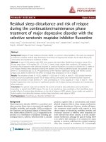
weeks before the experimental procedures. The consort
diagram of the study was shown in Figure 1.
Anesthesia and Surgery
Animals were anesthetized with intraperitoneal ketamine
(75mg/kg)andxylazine(10mg/kg).Heparin(5IU/g
body weight) was given intraperitoneally for 30 minutes
before explantation of heart to prevent the microem-
bolic events. Chests were scrubbed with alcohol and
betadine. Median sternotomy was performed. Aorta was
cannulated and inferior vena cava was cut. Cross clamp
was placed to the aorta and plegisol (Plegisol Cardiople-
gic Solution, Sanofi Synthelabo Industry, Turkey)
infused to t he heart to wash the intracardiac vascular
bed, while blood was rem oving from inferior vena cava.
Hearts were removed after cardiac arrest. In sham and
Taurine groups, following the explantation of the heart,
samples were immediately taken for analysis from left
ventricle. However, in Ischemia an d Treatment groups
explanted hearts were kept in a cold solution (0. 9% iso-
tonic solution, +4 degrees C). For these groups, samples
from left venticles were taken after 5 hours of cold
ischemic period.
Tissue Preparation
Biochemical samples were placed in liquid nitrogen in
polypropylene tubes and kept in deep freeze (-80
degrees C). His topathological samples were fixed in 10%
formaldehyde.
Histopatological Analysis
The paraffin-emb edded tissues were sectioned and
stained with hematoxylin-eosin. The histological slides
were evaluated by a pathologist who was blinded to
experiment protocol. The following morphological cri-
teria were used to determine the histopathological
damage: score 0, no damage; score 1 (mild), interstitial
edema and focal necrosis; score 2 (moderate), diffuse
myocardial cell swelling and necrosis; score 3 (severe),
necrosis with the presence of contraction bands, neutro-
phil infiltration and the capillaries were compressed; and
score 4 (highly severe ), widespread necrosis w ith the
presence of contraction bands, neutrophil infiltration,
compressing capillaries and hemorrhage [10,11].
Biochemical analysis
The frozen tissues were homogenized at a concentration
of 100 mg tis sue per ml of 25 mM phosphate buffer (pH
7.4) on an ice cube using a homogenizer (Heidolph Diax
900; Heidolph Electro GmbH, Kelheim, Germany) at a set-
ting of 8 (out of 10) for 30-s bursts. The homogenates
were centrifuged for 10 min at 2500 g,andthepellet
Table 1 Primary characteristics of groups
Groups (n) Nutrition Nutrition Time Sampling Time
Sham 8 Standard feed Three weeks Immediately after cardiectomy
Taurine 8 Standard feed+Taurine Three weeks Immediately after cardiectomy
Ischemia 8 Standard feed Three weeks 5 hours after cardiectomy
Treatment 8 Standard feed+Taurine Three weeks 5 hours after cardiectomy
Sahin et al . Journal of Cardiothoracic Surgery 2011, 6:31
/>Page 2 of 7
(cellular debris) discarded. The supernatant was allocated
into 2-3 separate tubes and used for biochemical assays.
Tissue lipid peroxidation
The lipid peroxidation level was measured by using Dra-
per and Hadley’sMethod[12].Thismethodusesspec-
trophotometric measurements of the color produced
during the reaction of thiobarbituric acid with malon-
dialdehyde (MDA). The absorbance of the final solution
was measured at 532 nm, and MDA levels were
expressed as MDA (mmol)/protein (g).
Superoxide dismutase (SOD)
SOD level was assayed using the nitroblue tetrazolium
(NBT) method of Sun et al. [13]. NBT was reduced to
blue formazan by superoxide which has a strong abso r-
bance of 560 nm. One unit (U) of SOD is defined as the
amount of protein that inhibits the rate of NBT reduc-
tion by 50%. The calculated SOD level was expressed as
SOD (U)/protein (g).
Glutathione peroxidase (GPx)
GPx level was measured by using the method described
by Paglia and Valentine in wh ich GPx level was coupled
with the oxidation of NADPH by glutathione reductase
[14]. The oxidation of NADPH was spectrophotometri-
cally followed up at 340 nm at 37 degrees C. The absor-
bance at 340 nm was recorded for 5 min. The level was
the slope of the lines (mmol) of oxidized NADPH/min.
GPx level was presented as GPx (U)/protein (g).
Catalase (CAT)
CAT level was determined spectrophotometrically, by
direct measurement of the decrease of light a bsorption
at 240 nm caused by the decomposition of hydrogen
peroxide by Catalase [15].
Statistical Analysis
SPSS for Windows Version 15.00 (Statistical Package for
the Social Sciences, SPSS Inc., Chicago, IL., USA)
package program was used for all statistical analyses and
measurements. Compliance of biochemical measurement
values to normal distribution was examined graphically
and statistically through the Shapiro-Wilk test. Among
the variables, it was determined that MDA and SOD
variables were not in compliance with normal distribu-
tion. For definitive statistics, mean values were given
with the average standard deviation. One way va riance
analysis (One Wa y ANOVA) w as used for comparison
of GPx and CAT measurements; and Kruskal-Wallis
variance analysis was applied for MDA and SOD para-
meters. The Bonferroni andMann-WhitneyUtestwas
used for b ilateral comparisons within the groups. p <
0.05 value was accepted as statistically significant.
Results
Biochemical examination results
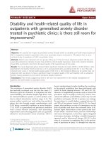
MDA Results (nmol/g)
MDA values were recor ded accordingly; 3.62 ± 0.197 in
the sham group, 2.07 ± 0.751 in the Taurine group,
9.71 ± 1.439 in the ischemia group and 7.68 ± 1.365 in
the treatment group. (Figure 2) The bilateral difference
between all groups was found to be statistically signifi-
cant (p < 0.05). When average values were examined,
the lowest value of MDA level was recorded in Taurine
group and the highest value was recorded in the ische-
mia group.
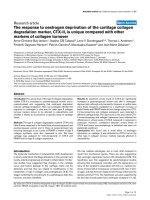
SOD Results (U/g)
SOD level was r ecorded accordingly; 90.11 ± 5.222 in
the sham group, 106.75 ± 3.449 in the T aurine group,
58.01 ± 4.244 in the ischemia group, and 96.12 ± 7.886
in the treatment g roup (Figure 3). The difference
between the sham group and treatment group was sta-
tistically insignificant and bilateral differences between
Groups
Sha
m
Tau
r
ine Ischemia Treatment
Surgery Surgery Surgery Surgery
Sampling Sampling
Ischemia (5 hr, Cold Isotonic)
Analyse Analyse Sampling Sampling
Analyse Analyse
Figure 1 Consort diagram of the study.
*
p
<0.001
* *
Figure 2 MDA levels in rat myocard tissue.
Sahin et al . Journal of Cardiothoracic Surgery 2011, 6:31
/>Page 3 of 7
other groups were found statistically significant. SOD
values that decreased in the sham Group were incre ased
in the Treatment group to which Taurine was adminis-
tered, and this difference between the ischemia group
and the treatment group was found to be statistically
significant (p < 0.001). The lowest SOD value was
observed in the ischemia group and the highest SOD
value was recorded in the Taurine group.
GPx Results (U/g)
GPx values were recorded accordingly; 22.77 ± 1 .308 in the
sham group, 23.42 ± 2.031 in the Taurine group, 16.23 ±
1.131 in the ischemia group, and 21.84 ± 3.298 in the treat-
ment group (Figure 4). T he difference between the ischemia
and the treatment groups and the ischemia and the sham
groups was found to be statisti cally significant (p < 0,001).
CAT Results (KU/g)
CAT level results were recorded accordingly; 7.08 ±
0.609 in the sham group, 6.15 ± 0.119 in the Taurine
group, 5.02 ± 0.62 in the ischemia group, and 5.36 ±
0.384 in the treatment group. (Figure 5) The difference
between ischemia and treatment groups was found to be
statistically insignificant (p > 0.05), a nd bilateral differ-
ences between the other groups were found significant.
When compared to the sham group, there was not a sig-
nificant increase in ischemia group (p = 1,000).
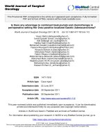
Histopathological results
Muscle fibers in sham and Taurine groups were in nor-
mal limit s. (Figure 6A and 6B) In ischemia group, myo-
fibril s were relativ ely insignificant with intense acidophil
cytoplasm, pyknotic-dark or light nucleus. Bes ides, the
muscle fibers were disorganized and swelling. They were
separated due to interstitial edema. PMN leukocyte
groups were observed in the vessel walls or by penetrat-
ing into the connective tissue. (Figure 6C) Degranulation
was also obser ved from mast cells to the connective tis-
sue. In the treatment group, the distribution of the mus-
cle fibers was better preserved when compared to
ischemic group. In addition, the level of interstitial
edema and inflamma tory cell infiltration was lower than
the ischemia group. (Figure 6D) The mean histopatholo-
gical damage in treat ment group and ischemia group
were scored 1.8 ± 0.8 vs 2.3 ± 0.7. (p < 0.01)
Discussion
The primary mission during ischemic period is to pro-
vide micro-vascular, cellular and functional integrity o f
the myocardium as much as possible. This needs cellu-
lar energy. Heart should be immediately stopped after
placing cross clamp in order to protect cardiac energy
storages. Cold preservation solutions are commonly
*
p
<0.001
**
Figure 3 SOD enzyme levels in rat myocard tissue.
*
p
<0.001
*
*
Figure 4 GPx enzyme levels in rat myocard tissue.
*
p
<0.001 **
p
>0.05
*
**
Figure 5 CAT enzyme levels in rat myocard tissue.
Sahin et al . Journal of Cardiothoracic Surgery 2011, 6:31
/>Page 4 of 7
used protective media to keep the donor organs in good
condition during whole ischemic time. Good preserva-
tion prevents ischemic damages and reperfusion injury
and minimizes cellular damage [16].
Taurine is a semi-essential amino acid that supports
neurological and musculoskeletal system deve lopment.
Taurine comprises 50% of the cardiac free amino acid
pool and is present in the myocardial tissue in the
concentration of 11-38 μM/g [5]. It plays an important
role in the regulation of sodium, potassium, calcium,
and ion flow along with cardiac contractility, regula-
tion of membrane excitability, osmolality and the
volume content [17,18]. Diet is the main source of
Taurine in humans. Taurine occurs naturally in food,
especially in seafood and meat. The mean daily taurine
intake for adult human has been estimated between
40-400 mg [19]. Although various doses of Taurine
(25 mg/kg/day to 6 g/day, p.o. or i.v.) in human and
animal studies reported, [19,20] we preferred to use a
dose of 200 mg/kg/day administered orally (with the
help of gavage).
There is a strong connection between Taurine excre-
tion levels and ischemic heart disease mortality [21]. It
is shown that preoperative Taurine infusion decreases
reperfusion inju ry in coronary artery bypass surgery
[22]. Taurine that was given as a dietary supplement to
the rats’ meal before inducting myocardial infarction
decreases infarct size and improves heart functions after
myocardial infarct [23].
Some structural changes occur in the myocardial cells
during the cold ischemic period. High energy phosphate
synthesis decreases as a result of decreasing oxidative
phosphorilation. Na
+
-K
+
-ATP-ase pump in the cell
membrane deteriorates and the energy storage of the
cell decreases. Na
+
and Ca
2+
ions accumulate in the cell.
The accumulation of Ca
2+
ions in the cell results in
cytotoxicity and subsequently antioxidant enzyme levels
are reduced in cells. Ultimately; swoll en cells, extracellu-
lar edema, acidosis, calcium accumulation, and endothe-
lial damage occur. This situation makes myocardial cell
more sensitive to oxidative damage during reperfusion
period [24-26]. This study histopathologically and bio-
chemically proves that taurine administration decreases
the myocardial damage occured during the cold
ischemic period. In this study, significant swollen cell
and intense inflammatory reaction were observed in the
donor hearts preserved in +4 degrees C and exposed to
ischemia. Swollen cell number a nd inflammatory reac-
tion were much less in the treatment group than others.
It was found that Taurine decreases histopathologic
Figure 6 Histopathological view of the myocardial tissue samples from each group. Muscle fibers in normal appearance are seen in sham
(A) and Taurine (B) groups (HEx400). Muscle fibers are separated in ischemia group due to interstitial edema and muscle fibers are in more
acidophilic appearance. PMN leukocyte infiltration between the muscle fibers is seen (arrow) (C) (HEx400). Distribution of muscle fibers in
treatment group seems better preserved when compared to ischemia group. Inflammatory cell infiltration is observed in the arrowed area. (D)
(HEx400).
Sahin et al . Journal of Cardiothoracic Surgery 2011, 6:31
/>Page 5 of 7
changes that might occur during cold ischemic time.
(Figure 6)
Free oxygen radicals are produ ced in all body cells in a
limited number under normal conditions and are neutra-
lized by endogenous anti oxidants such as superoxide dis-
mutase, glutathione peroxidase and catalase (Scavenging
Enzyme Systems). Free oxygen radicals cause tissue
damage through the peroxidation of the lipids present in
the cell membranes.
Increasing lipid peroxidation might be u sed as a sign
of the tissue damage caused by free oxygen radicals.
MDA is the final product of lipid peroxidation . Mea-
surement of the MDA level in serum might b e used as
an indicator of tissue damage caused by in vivo free
oxygen radicals [27,28 ]. Kaplan and colleagues showed
that taurine deficiency caused an increase in MDA
levels.InourstudywealsofoundthatMDAvalues
were very high in the ischemia group, and decreased in
the treatment group (p < 0.05).
Cells are highly affected by oxidative damage if antioxi-
dant enzymes decr ease in the tissue. Superoxide dismu-
tase enzyme system is the first and the most important
defe nse mechanism of the body against free oxygen radi-
cals [29]. If there is enough superoxide dismutase activity,
cell damage occurs at minimum level. In a study b y Bol-
cal et al, [30] cardioprotective role of antioxidant medica-
tions was researched. In this study there were protective
incre ases in SOD and GPx levels and a decrease in MDA
levels were reported. In our study, although we studied
Taurine as antioxidant medication, there were similar
results. SOD enzyme levels in the ischemia group
decreased when compared to the sham group, but
increased in the Taurine administered treatment group.
This increase is found to be stat istically significant (p < 0,
05) and this raising in the treatment group is found to be
close in the sham and Taurine group.
Catalase is an antioxidant enzyme. It degrades hydro-
gen peroxide (H
2
O
2
) to oxygen and water. Catalase acts
together with GPx in that process. H
2
O
2
concentration
is diminished by Catalase [31,32]. In our study, when
Catalase levels were examined, no statistically significant
difference was found between ischemia and treatment
groups. The probable mechani sm of this could be unin-
volvement of the cells with high CAT e nzyme levels in
the process. The CAT enzyme levels were realized to
have been decreased probably due to the processed
hyd rogen peroxides. There was not a remarkable differ-
ence between ischemia and treatment group since the
treatment group did not have high CAT level obtained
by Taurine.
Study Limitations
Main limitation of this study is the administration way
of Taurine and its clinical impact. In the literature there
are many studies with very large range of administration
periods (5 min before ischemia to 7 weeks before the
study). Also there are very different study doses of Taur-
ine. In our study we tried to use a mean value and dura-
tion according to the literature. Although the Taurine
cardiac effects are well known there are limited reports
related to the ischemia of the donour hearts. It is not
practical to use Taurine three weeks before an unpre-
dicted ischemia, but our aim was only to show if there
is any beneficial effect of supplemental Taurine in such
situations. We think that it can pla y an important role
in heart explantation operations. Detailed protocols of
Taurine usage prior to explantation ischemia has yet to
be established and different administration ways and
dosages just before the predicted ischemia may be sub-
ject of other studies.
Conclusion
This study demonstrated that Taurine decreased
ischemic cellular damage in rat hearts that were kept
under ischemic and cold circumstances for 5 hours. We
believe that the se beneficial effects of Taurine may be
related to its antioxidant effect.
List of abbreviations
CAT: Catalase; GPx: Glutathione peroxidase; H
2
O
2
: Hydrogen peroxide; MDA:
Malondialdehyde; NBT: Nitroblue tetrazolium; SOD: Superoxide dismutase;
SPSS: Statistical Package for the Social Sciences; U: Unit
Author details
1
Gülhane Military Medical Academy, Department of Cardiovascular Surgery,
06010, Etlik, Ankara, Turkey.
2
Gülhane Military Medical Academy, Department
of Thoracic Surgery, 06010, Etlik, Ankara, Turkey.
3
Gazi University, Faculty of
Commerce and Tourism Education, Department of Computer Applications
Training, 06830, Gölbaşı, Ankara, Turkey.
4
Gülhane Military Medical Academy,
Department of Pathology, 06010, Etlik, Ankara, Turkey.
5
Gülhane Military
Medical Academy, Department of Biochemistry, 06010, Etlik, Ankara, Turkey.
Authors’ contributions
MAS, OY, AG and UD were both involved in the conception of the study
design as well as drafting and revising the article. SD, AJ and FC contributed
to the surgical procedures. MG and HY were involved in acquisition of
pathologic and biochemical data. SA was involved in statistical analysis of
data. All authors have approved the manuscript.
Competing interests
The authors declare that they have no competing interests.
Received: 7 December 2010 Accepted: 18 March 2011
Published: 18 March 2011
References
1. Jahania MS, Sanchez JA, Narayan P, Lasley RD, Mentzer RM: Heart
Preservation for Transplantation: Principles and Strategies. Ann Thorac
Surg 1999, 68(5):1983-7.
2. Ueno T, Iguro Y, Yotsumoto G, Fukumoto Y, Nakamura K, Miyamoto TA,
Sakata R: Taurine at early reperfusion significantly reduces myocardial
damage and preserves cardiac function in the isolated rat heart.
Resuscitation 2007, 73(2):287-95.
3. Oriyanhan W, Yamazaki K, Miwa S, Takaba K, Ikeda T, Komeda M: Taurine
prevents myocardial ischemia/reperfusion-induced oxidative stress and
apoptosis in prolonged hypothermic rat heart preservation. Heart Vessels
2005, 20(6):278-85.
Sahin et al . Journal of Cardiothoracic Surgery 2011, 6:31
/>Page 6 of 7
4. Miyamoto TA, Ueno T, Iguro Y, Yotsumoto G, Fukumoto Y, Nakamura K,
Sakata R: Taurine-mediated cardioprotection is greater when
administered upon reperfusion than prior to ischemia. Adv Exp Med Biol
2009, 643:27-36.
5. Huxtable RJ: Physiological actions of Taurine. Physiol Rev 1992,
72(1):101-63.
6. Oz E, Erbaş D, Gelir E, Aricioğlu A: Taurine and calcium interaction in
protection of myocardium exposed to ischemic reperfusion injury. Gen
Pharmacol 1999, 33(2):137-141.
7. Pion PD, Kittleson MD, Thomas WP, Delellis LA, Rogers QR: Response of
cats with dilated cardiomyopathy to Taurine supplementation. J Am Vet
Med Assoc 1992, 201(2):275-84.
8. Welty MC, Welty JD, McBroom MJ: Effect of isoproterenol and Taurine on
heart calcium in normal and cardiomyopathic hamsters. J Mol Cell Cardiol
1982, 14(6):353-7.
9. Yucel O, Kunak ZI, Macit E, Gunal A, Gozubuyuk A, Gul H, Genc O:
Protective efficacy of Taurine against pulmonary edema progression:
experimental study. J Cardiothorac Surg 2008, 3:57.
10. Hoffmeyer MR, Scalia R, Ross CR, Jones SP, Lefer DJ: PR-39, a potent
neutrophil inhibitor attenuates myocardial ischemia-reperfusion injury in
mice. Am J Physiol Heart Circ Physiol 2000, 279(6):H2824-8.
11. Zhu J, Qiu Y, Wang Q, Zhu Y, Hu S, Zheng L, Wang L, Zhang Y: Low dose
cyclophosphamide rescues myocardial function from ischemia-
reperfusion in rats. European Journal of Cardio-thoracic Surgery 2008,
34:661-666.
12. Draper HH, Hadley M: Malondialdehyde determination as index of lipid
peroxidation. Methods Enzymol 1990, 186:421-31.
13. Sun Y, Oberley LW, Li Y: A simple method for clinical assay of superoxide
dismutase. Clin Chem 1988, 34(3):497-500.
14. Paglia DE, Valentine WN: Studies on the quantitative and qualitative
characterization of erythrocyte glutathione peroxidase. J Lab Clin Med
1967, 70(1):158-69.
15. Aebi H: Methods of enzymatic analysis.Edited by: Bergmeyer HU.
Academic Press, New York and London; , 2 1974:2:673-684.
16. McCrystal GD, Pepe S, Esmore DS, Rosenfeldt FL: The Challenge of
Improving Donor Heart Preservation. Heart Lung Circ 2004, 13(1):74-83.
17. Redmond HP, Stapleton PP, Neary P, Bouchier-Hayes D: Immunonutrition:
the role of Taurine. Nutrition 1998,
14(7-8):599-604.
18. Schaffer S, Azuma J, Takahashi K, Mozaffari M: Why is Taurine
cytoprotective? Adv Exp Med Biol 2003, 526:307-321.
19. Wójcik OP, Koenig KL, Zeleniuch-Jacquotte A, Costa M, Chen Y: The
potential protective effects of Taurine on coronary heart disease.
Atherosclerosis 2010, 208(1):19-25.
20. Mizushima S, Nara Y, Sawamura M, Yamori Y: Effects of oral taurine
supplementation on lipids and sympathetic nerve tone. Adv Exp Med Biol
1996, 403:615-22.
21. Yamori Y, Liu L, Ikeda K, Miura A, Mizushima S, Miki T, Nara Y, WHO-
Cardiovascular Disease and Alimentary Comparison (CARDIAC) Study Group:
Distribution of twenty-four hour urinary taurine excretion and association
with ischemic heart disease mortality in 24 populations of 16 countries:
results from the WHO-CARDIAC study. Hypertens Res 2001, 24(4):453-7.
22. Milei J, Ferreira R, Llesuy S, Forcada P, Covarrubias J, Boveris A: Reduction
of reperfusion injury with preoperative rapid intravenous infusion of
Taurine during myocardial revascularization. Am Heart J 1992,
123(2):339-345.
23. Briet F, Keith M, Leong-Poi H, Kadakia A, Aba-Alkhail K, Giliberto JP,
Stewart D, Errett L, David Mazer C: Triple nutrient supplementation
improves survival, infarct size and cardiac function following myocardial
infarction in rats. Nutr Metab Cardiovasc Dis 2008, 18(10):691-9.
24. Orrenius S, Burkitt MJ, Kass GE, Dypbukt JM, Nicotera P: Calcium ions and
oxidative cell injury. Ann Neurol 1992, 32(Suppl):S33-42.
25. Homer-Vanniasinkam S, Crinnion JN, Gough MJ: Post-ischaemic organ
dysfunction: A review. Eur J Vasc Endovasc Surg 1997, 14(3):195-203.
26. Jennings RB, Reimer KA: Acute myocardial ischemia: effects of reperfusion
with arterial blood. Artif Cells Blood Substit Immobil Biotechnol 1994,
22(2):253-78.
27. Molina H, García M: Enzymatic defenses of the rat heart against lipid
peroxidation. Mech Ageing Dev 1997, 97(1):1-7.
28. Kim HS, Kwack SJ, Lee BM: Lipid peroxidation, antioxidant enzymes, and
benzoapyrene-quinones in the blood of rats treated with benzoapyrene.
Chemico-Biological Interactions 2000, 127(2):139-150.
29. McCord JM: Oxygen-derived free radicals in postischemic tissue injury. N
Engl J Med 1985, 312(3):159-63.
30. Bolcal C, Yildirim V, Doganci S, Sargin M, Aydin A, Kuralay E, Ozal E,
Demirkilic U, Oz BS, Sayal A, Tatar H: Do N-acetylcystein, beta-glucan, and
coenzyme Q10 mollify myocardial ischemia-reperfusion injury? Heart
Surg Forum 2007, 10(3):E222-7.
31. Ergun Y, Oksuz H, Atli Y, Kilinç M, Darendeli S: Ischemia-Reperfusion Injury
in Skeletal Muscle: Comparison of the Effects of Subanesthetic Doses of
Ketamine, Propofol, and Etomidate. J Surg R 2010, 159(1)
:e1-e10.
32. Ogawa T, Mimura Y: Antioxidant effect of zinc on acute renal failure
induced by ischemia-reperfusion injury in rats. Am J Nephrol 1999,
19(5):609-14.
doi:10.1186/1749-8090-6-31
Cite this article as: Sahin et al.: Is there any cardioprotective role of
Taurine during cold ischemic period following global myocardial
ischemia? Journal of Cardiothoracic Surgery 2011 6:31.
Submit your next manuscript to BioMed Central
and take full advantage of:
• Convenient online submission
• Thorough peer review
• No space constraints or color figure charges
• Immediate publication on acceptance
• Inclusion in PubMed, CAS, Scopus and Google Scholar
• Research which is freely available for redistribution
Submit your manuscript at
www.biomedcentral.com/submit
Sahin et al . Journal of Cardiothoracic Surgery 2011, 6:31
/>Page 7 of 7