Báo cáo y học: "Mesenchymal stem cell transplantation ameliorates motor function deterioration of spinocerebellar ataxia by rescuing cerebellar Purkinje cell" ppsx
Bạn đang xem bản rút gọn của tài liệu. Xem và tải ngay bản đầy đủ của tài liệu tại đây (2.17 MB, 9 trang )
RESEARC H Open Access
Mesenchymal stem cell transplantation ameliorates
motor function deterioration of spinocerebellar
ataxia by rescuing cerebellar Purkinje cells
You-Kang Chang
1,2,3
, Ming-Hsiang Chen
4
, Yi-Hung Chiang
1,5
, Yu-Fan Chen
4
, Wei-Hsien Ma
4
, Chian-You Tseng
4
,
Bin-Wen Soong
6,7
, Jennifer H Ho
8,9,10*
and Oscar K Lee
1,4,11*
Abstract
Background: Spinocerebellar ataxia (SCA) refers to a disease entity in which polyglutamine aggregates are over-
produced in Purkinje cells (PCs) of the cerebellum as well as other neurons in the central nervous system, and the
formation of intracellular polyglutamine aggregates result in the loss of neurons as well as deterioration of motor
functions. So far there is no effective neuroprotective treatment for this debilitating disease although numerous
efforts have been made. Mesenchyma l stem cells (MSCs) possess multi-lineage differentiation potentials as well as
immuno-modulatory properties, and are theoretically good candidates for SCA treatment. The purpose of this
study is to investigate whether transplantation of human MSCs (hMSCs) can rescue cerebellar PCs and ameliorate
motor function deterioration in SCA in a pre-clinical animal model.
Method: Transgenic mice bearing poly-glutamine mutation in ataxin-2 gene (C57BL/6J SCA2 transgenic mice) were
serially transplanted with hMSCs intravenously or intracranially before and after the onset of motor function loss.
Motor function of mice was evaluated by an accelerating protocol of rotarod test every 8 weeks.
Immunohistochemical stain of whole brain sections was adopted to demonstrate the neuroprotective effect of
hMSC transplantation on cerebellar PCs and engraftment of hMSCs into mice brain.
Results: Intravenous transplantation of hMSCs effectively improved rotarod performance of SCA2 transgenic mice
and delayed the onset of motor function deterioration; while intracranial transplantation failed to achieve such
neuroprotective effect. Immunohistochemistry revealed that intravenous transplantation was more effective in the
preservation of the survival of cerebellar PCs and engraftment of hMSCs than intracranial injection, which was
compatible to rotarod performance of transplanted mice.
Conclusion: Intravenous transplantation of hMSCs can indeed delay the onset as well as improve the motor
function of SCA2 transgenic mice. The results of this preclinical study strongly support further exploration of the
feasibility to transplant hMSCs for SCA patients.
Background
Spinocerebellar ataxias (SCAs) are a group of inherited
neurological disorders that are clin ically and genetically
very heterogeneous. They are progressive neurodegen-
erative diseases that are characterised by cerebellar
ataxia, resulting in unstead y gait, clumsiness, and dysar-
thria. The cerebellar syndrome is often associated with
other neurological signs such as pyramidal or extrapyra-
midal signs, ophthalmoplegia, and cognitive impairment
[1]. Pathogenetic mechanism applies to SCAs caused by
expansions of CAG repeats encoding polyglutamine
tracts, as in the genes that underlie SCA1, SCA2, SCA3,
SCA6, SCA7, SCA17, and dentatorubro-pallidoluysian
atrophy, the so-called polyglutamine expansion SCAs
[2,3]. Other SCA subtypes are caused by expansions in
non-coding regions of genes for SCA8, SCA10, SCA12,
and SCA31, and rare conventional mutations in SCA
genes [2,3]. Mutant phe notype in the polyglutamine
* Correspondence: ;
1
Institute of Clinical Medicine, National Yang-Ming University, Taipei, Taiwan
8
Center for Stem Cell Research, Taipei Medical University-Wan Fang Medical
Center, Taipei, Taiwan
Full list of author information is available at the end of the article
Chang et al. Journal of Biomedical Science 2011, 18:54
/>© 2011 Chang et al; licensee BioMed Cen tral Ltd. This is an Open Access article distributed under t he terms of the Creative Commons
Attribution License ( which permits unrestricted use, distribution, and reproduction in
any medium, provided the original work is properly cited.
expansion S CAs has been widely considered to be pri-
marily a result of a toxic gain-of-function in the mutant
proteins in affected neurons [4,5]. Atrophy of the cere-
bellum and brainstem are most often the prominent fea-
tures, but other structures can be affected, leading to a
substantial range of phenotypes [5,6].
So far there is no cure o f polyglutamine expansi on
SCAs although various therapeutic strategies have been
postulated including silencing gene expression [7],
increasin g protein clearance, reducing the toxicity of the
protein, influencing downstream pathways activated by
the mutant protein and transplantation [4]. For symp-
tom treatment, levodopa is temporarily useful for rigid-
ity/bradykinesia and for tremor, and magnesium for
muscle cramps in SCA2 patients [8], but neuroprotec-
tive therapy is not clinically available. In 1999, Low et
al. reported that cerebellar allografts survived and transi-
ently alleviated ataxia in a transgenic mouse model of
SCA1 [9]. Subsequently, grafting murine neural precur-
sor cells promoted cerebellar PCs survival and func-
tional recovery in an SCA1 mouse model [10]. Murine
MSCs (mMSCs) had been shown to be able to rescue
PCs through releasing of neurotrophic factors and
improve motor functions in a mouse model of cerebellar
ataxia [11]. Althou gh the surface phenotype and multili-
neage po tential of mMSCs used in this study [11] was
not demonstrated completely, these results suggested
that MSC transplantation may be beneficial to SCA2
transgenic mice.
MSCs are defined as plate-a dhering, fibroblast-like
cells possessing self-renewal ability with the capacity to
differentiate into m ultiple mesenchymal cell lineages
such as osteoblasts, chondrocytes, and adipocytes. MSCs
are easily accessible and isolated from a variety of tis-
sues such as bone marrow, umbilical cord blood, trabe-
cular bone, synovial membrane, and adipose tissue
[12-16]. MSCs also prov ide the advantage of minimizing
immune reactions because cells can be derived from the
respective patient. Furthermore, several human trials of
MSCs have shown no adverse reactions to allogenic
MSC t ransplants [17,18]. Many studies show that sys-
temically administrative hMSCs home to site of ische-
mia or tissue injury to repair injured tissues [19]. MSCs
transplantation had been adopted in several clinical
trials of neurological disease, including of multiple sys-
tem atrophy [20], Parkinson’s disease [21], amyotrophic
lateral sclerosis [22], and ischemic stroke [23] with
encouraging early or long-term results.
In our previous studies, we showed that clonally
derived human MSCs (hMSCs), under chemically
defined conditions, differentiate into neuroglial-like cells
that not only express neuroglial-specific genes but also
possessed a resting membrane potential and voltage-sen-
sitive calcium channels on the membrane [13]. We also
showed that in utero transplant ation of hMSCs in mice
contributed to numerous tissues, including the brain
and spinal cord [24]. Donor hMSCs engrafted into mur-
ine tissues originating from all three germ layers and
persisted for up to 4 months or more after delivery.
Therefore, the purpose of this study is to investigate
whether transplantation of human MSCs (hMSCs) can res-
cue cerebellar PCs and ameliorate the deterioration of
motor function in SCA in a pre-clinical animal model.
Transgenic mice bearing poly-glutamine mutation in
ataxin-2 gene (C57BL/6J SCA2 transgenic mice) were seri-
ally transplanted with hMSCs intravenously or intracraniall y
before and after the onset of motor functio n loss. Motor
function of mice was evaluated by an accelerating protocol
of rotarod test every 8 weeks. Immunohistochemical stain
of whole brain sections was adopted to demonstrate the
neuroprotective effect of hMSC transplantation on cerebel-
lar PCs and engraftment of hMSCs into mice brain.
Materials and methods
Culture of hMSCs
The isolation and characterization of hMSCs from bone
marrow was performed as reported previously [25,26].
An approval from the Institutional Review Board of the
Taipei Veterans General Hospital has been obtained
prior to commencement of the study. hMSCs used in
this study were cl onally-derived, a nd their surface
immune phenotype as well as multilineage differentia-
tion potentials into osteoblasts, adipocytes, and chon-
drocytes w ere confirmed [25,26]. hMSCs of passage 8-
10 were used for transplantation. Before transplantation,
hMSCs were t rypsinized with trypsin/EDTA 0.25%,
counted for cell number and suspended in 80 μL PBS.
Animal Model
C57BL/6J SCA2 transgenic mice were purchased from
University of Texas Southwestern Medical Center (Dal-
las, Texas, USA) and wild-type C57BL/6J mice w ere
purchased from Tzu Chi University Laboratory Animal
Center (Hualien, Taiwan). All animal experiments were
performed with the approval of the Animal Care Com-
mittee of the Taipei Veterans General Hospital.
MSC Labeling with Superparamagnetic Iron Oxide (SPIO)
nanoparticles for in vivo Cell Tracking
Amine (NH
3
+
) surface modified iron-oxide nanoparticles
of 6 nm diameter without polymer coating were pre-
pared as reported previously [27]. hMSCs were seeded
in culture plates at the density of 4 × 10
4
cells/well and
were allowed for attachment and growth for 24 h.
Before treatment, 50 μg/ml of SPIO were coated by
mixing with 0.75 μg/ml poly-L-lysine (Sigma-Aldrich) in
the cult ure medium at room temperature for 1 h. After
that, hMSCs were incubated in SPIO-containing
Chang et al. Journal of Biomedical Science 2011, 18:54
/>Page 2 of 9
medium for 24 h. After labeling, the cultures were
washed with PBS thoroughly to remove excess SPIO in
the medium for further transplantation.
MR Image of Mice after Intracranial SPIO-labeled hMSC
Transplantation
Before intracranial transplantation, 100 μLtrypanblue
(Sigma-Aldrich) was injected through foramen magnu m
into position of cer ebellum in a wild-type mouse, which
was immediately sacrificed for visual examination of cer-
ebellum to determine target ac curacy. MR imaging was
used to demonstrate the transplant site in living mice
which received i ntracranial hMSCs transplantation. MR
images of three mice were measured in a Bruker BioS-
pec 7T system (Bruker BioSpin MRI, Ettlingen, Ger-
man y). Mice were anesthetized, followed by injection of
8.4 × 10
6
per kg of mice bod y weight S PIO-labe led or
unlabeled hMSCs in PBS through foramen magnum
into cerebellum. Images were taken 24 h later under
anesthesia using T2 weighted MR acquisition sequence
with the following parameters: fast spin echo with TR/
TE = 2500 ms/33 ms, ET = 10 ms.
Intravenous and Intracranial hMSCs Transplantation
To evaluate the neuroprotective effects of hMSCs, 4.2 ×
10
7
or 8.4 × 10
6
hMSCs per kg of mice body weight
were injected via tail vein (IV hMSC-Tg group) or
through foramen magnum into position of cerebellum
(IC hMSC-Tg group) of C57BL/6J SCA2 transgenic
mice. In IV hMSC-Tg group, hMSCs were transplanted
at 12, 23, 33 and 42-week-old (n = 14). In IC hMSC-Tg
group, hMSCs were transplanted at 12, 23, and 33-
week-old (n = 5). Treated mice were compared to con-
trol SCA2 transgenic (Control-Tg) (n = 10) and wild-
type (Control-Wt) (n = 16) mice.
Motor Behavior Assessment: Accelerating Rotarod Test
Since 9 weeks of age, se x and wei ght-matched IV
hMSC-Tg, IC hMSC-Tg, Control-Tg, and Control-Wt
mice were tested on the rotarod (Singa Technology Cor-
poration, T aipei, Taiwan) every 8 weeks, whic h under-
went linear acceleration from 4 to 40 rpm in 300
seconds. Latency to fall from rotarod was recorded in
seconds. Each trial lasted for a maximum of 5 min and
mice were rest ed for minimum 15 min between trials to
avoid fatigue. After rotarod test, the body weights of
mice were recorded. Mice underwent three trials per
day for four consecutive days, and the mean latency to
fall of each day was considered for statistical analysis.
Histological Examination and Immunohistochemistry:
Purkinje Cells
Three mice from each group at > 50 weeks of age were
sacrificed and processed for histological examination
and immunohistochemistry (IHC) of the cerebellar PCs.
Mice whole brain tissues were f ixed in 3.7% formalin
overnight after sacrifice under anesthesia and emb edded
selected samples in paraffin. Sections (4 μm) were cut
and mounted onto microscopic s lides. Sections were
rehydrated by rinsing twice at 5 min intervals in xylene,
100% ethanol, 95% ethanol and 80% ethanol. After
deparaffinization, sections were treated with 3% H
2
O
2
for peroxidase inactivation, heated in 10 mM citrate buf-
fer (with 0.05% Tween20) for antigen retrieval, blocked
with 1% blocking solution (1% BSA and 0.1% Triton X-
100 in PBS). Sections were incubated with anti-calbindin
D-28K monoclonal antibodies (Sigma-Aldrich) diluted in
blocking solution (1:300) for 40 min at room tempera-
ture (RT). After three extensive washes with PBS, sec-
tions were incubated with secondary antibody diluted in
blocking solution (1:1000) for 40 min at RT. Primary
ant ibodies were detected using DAB (3, 3’-Diaminoben-
zidine tetrahydrochloride) Two-co mponent Enhanced
Liquid Substrate System (Sigma-Aldrich), enhanced by
DAB enhancer, and visualized with diaminobenzidine
(DAB; Sigma-Aldrich). We counte rstained with aqueous
haematoxylin (Sigma-Aldrich). For direct comparison we
processed all slides in a single batch to minimize
variability.
Count of Cerebellar Purkinje Cells
To determine whether MSC transplantation rescued PC
loss in cerebellum of C57BL/6J SCA2 transgenic mice,
we counted calbindin-D28K-positive PCs from twelve
mice in IV hMSC-Tg, IC hMSC-Tg, Control-Tg, and
Control-Wt group (three mice in each group). Every 8
th
sections in the consecutive series of each mouse were
selected and selected parasagittal sections were prepared
for the counting from each mouse. Numbers of PCs
under 20 100 × fields which randomly selected from
non-concave area of parasagittal sections were counted
and summed. Then average PC number of each mouse
was calculated.
Immunohistochemistry: hMSCs
Specific antibody which reacted with human beta2
microglobulin (Abcam, code: ab15976) was chosen to
demonstrate hMSCs in murine brain tissue by IHC. The
specificity of the antibody had been ascertained by
crossed immunoelectrophoresis. Murine whole brain
sections which processed for PCs count ing were used
for staining. Sections (4 μm) were cut and mounted
onto microscopic slides. Sections were rehydrated by
rinsi ng twice at 5 min intervals in xylene, 100% ethanol,
95% ethanol and 80% e thanol. After deparaffin ization,
sections were treated with 3% H
2
O
2
for peroxidase inac-
tivation, heated in 10 mM citrate buffer (with 0.05%
Tween20) for antigen retrieval, and bloc ked with 1%
Chang et al. Journal of Biomedical Science 2011, 18:54
/>Page 3 of 9
blocking solution (1% BSA and 0.1% Triton X-100 in
PBS). Sections were incubated with specific anti-human
b2 microglobulin polyclonal antib odies (Abcam) diluted
in blocking solution (1:400) for 40 min at RT. After
three extensive washes with PBS, sections were incu-
bated with secondary antibody diluted in blocking solu-
tion (1:1000) for 40 min at RT. Primary antibodies were
detected using EnVision Detection System (DAKO), and
visualized with diaminobenzidine (DAB; DAKO). We
counterstained with aqueous haematoxylin (Sigma-
Aldrich). For direct comparison we processed all slides
in a single batch to minimize variability.
Statistical analysis
Data are presented as the mean ± standard error of
mean (SE) for at least three times of independent
exp eriments. The result s were compared using one-way
ANOVA, Tukey’stestasPosthoctest,andStudent’sT
test. Statistical significance was d etermined at 95% con-
fidence interval.
Results
Confirmation of Successful Intracranial Delivery of hMSCs
Whole brain tissue of control mouse which was injected
with trypan blue through foramen magnum into posi-
tion of cerebellum was inspected after sacrifice, and
most of the areas staining by trypan blue were located
at cerebellum, medulla and nearby regions (Figure 1A).
MR imaging was used to demonstrate the transplant site
in living mice which received intracranial hMSCs trans-
plantation. No decreased MRI signal intensity was
observed in the medulla or cerebellums of wild-type
mouse after intracranial injection of unlabeled hMSCs
(Figure 2A). As shown in Figure 2B and 2C, a significant
decreased T2 si gnal intensity was detected in the dorsal
site of medulla, which was adjacent to cerebellums o f
wild-type and transgenic mice after intracranial injection
of SPIO-labeled hMSCs. No evidence of major trauma
or intracerebellar hemorrhage was detected in the
medulla or cereb ellums, either. These MR images
further confirmed the injected hMSCs were located in
the dorsal site of medulla, which was adjacent to cere-
bellum, and this invasive pr ocedure didn’t cause major
trauma or intracranial hemorrhage at the injection site,
as well as did not hamper the evaluation of motor func-
tion by rotarod test.
Motor Behavior of SCA2 Transgenic Mice Improved after
hMSC Transplantation Intracranial hMSC injection
Rotarod testing showed that motor performance of
SCA2 transgenic mice was not significantly different
from that of wild-type mice at six weeks and trans-
genic mice started to perform poorly since 16 weeks of
age with progressive deterioration from 26 weeks of
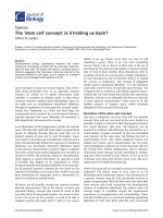
Figure 1 Route of human mesenchymal stem c ells
transplantation and gross pictures of mice brain after trypan
blue injection. (A) 100 μL trypan blue was injected through
foramen magnum into position of cerebellum in a wild-type mouse,
which was immediately sacrificed for visual examination to
determine target accuracy. Most of the areas staining by trypan
blue were located at cerebellum, medulla and nearby regions. (B)
hMSCs were injected intravenously via tail vein or intracranially
through foramen magnum under anesthesia. hMSCs, human
mesenchymal stem cells.
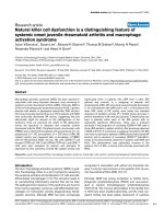
Figure 2 Magnetic resonance images of mice after
superparamagnectic iron oxide nanoparticles (SPIO)-labeled
and unlabeled human mesenchymal stem cells transplantation.
Mice were anesthetized, followed by injection of 8.4 × 10
6
per kg of
mice body weight unlabeled hMSCs (A, wild-type mouse) or SPIO-
labeled hMSCs (B, wide-type mouse; C, SCA2 transgenic mouse) in
PBS through foramen magnum intracranially, and then measured in
a 7-T MR imager 24 h later. (A) No signal was detected in the
medulla or cerebellum of wild-type mouse after intracranial
transplantation of unlabeled hMSCs. (B) A significant decreased T2
signal intensity of the SPIO (white arrow) was detected in the dorsal
site of medulla of wild-type mouse after intracranial transplantation
of SPIO-labeled hMSCs. (C) A significant decreased T2 signal
intensity of the SPIO (white arrow) was detected in the dorsal site
of medulla of transgenic mouse after intracranial transplantation of
SPIO-labeled hMSCs. The length of each small scale was 1 mm. The
letter “P” indicated posterior direction.
Chang et al. Journal of Biomedical Science 2011, 18:54
/>Page 4 of 9
age [28]. In our study, Control-Tg mice started to per-
form poorly since 25 weeks of age with progressive
deterioration from 33 weeks of age (Figure 3) (t test,
p < 0.05). SCA2 transgenic mice which received serial
intracranial hMSC injection for three times had a
trend of better rotarod performance than Control-Tg
mice at 33-40 weeks of age, but the difference was not
significant due to large error bar (one-way ANOVA,
p = 0.055) (Figure 3).
Intravenous hMSC injection
Although the rotarod performance was not improved by
intravenous MSC injection at 25-32 weeks of age, SCA2
transgenic mice which received intravenous MSC injec-
tion for four times had significantly better rotarod per-
formance than Control-Tg mice at 33-40 weeks of age
(Figure 4) (one-way ANOVA, p = 0.012). SCA2 trans-
genic mice which received intravenous hMSC injection
also had similar rotarod performance with wild-type
mice. This result suggested that intravenous transplanta-
tion of hMSCs via tail vein could ameliorate the dete-
rioration of motor function in SCA2 transgenic mice.
Rescue of Purkinje Cells by Transplanted hMSCs
Loss of PCs had been noted by immunohistochemical
stain of calbindin-28K, which was a protein specifically
exp ressed in cytoplasm and dendritic processes of cere-
bellar PCs in SCA2 transgenic mice since age of 4
weeks [28]. Percentage of surviving PCs showed a pro-
gressive decline. At 24-27 weeks, PC number was
reduced by 50-53% in SCA2 transgenic mice [28]. In
our study, PC number (by visual impressions) in cere-
bellar sections of the IC-hMSC-Tg and IV-hMSC-Tg
groups at 33-40 weeks of age was higher than in the
Control-Tg group and similar with number in the Con-
trol-Wt group (Figure 5A). To obtain quantitative data
supporting these visual impressions, the numbers of sur-
viving PCs in the cerebellum of each group were esti-
mated. Residual PCs in Control-Tg group accounted for
66.4 ± 4.7% of wild-type mice (100.0 ± 5.1%), while resi-
dual PCs in the IC-hMSC-Tg and IV-hMSC-Tg groups
accounted for 70.7 ± 3.8% and 86.6 ± 5.9% (Figure 5B)
(one-way ANOVA, p < 0.001). This result suggested
that both serial intravenous and intracranial MSC trans-
plantation had some neuroprotective effects on cerebel-
lar PCs in SCA2 transgenic mice and intravenous MSC
transplantation rescu ed more cerebellar PCs than intra-
cranial transplantation (one-way ANOVA, p = 0.018).
Grafted hMSCs in Murine Cerebellum and Cerebral Cortex
In IV-hMSC-Tg group, hMSCs which were positive for
human b2 microglobulin signals were located in the cer-
ebellar white matter (Figure 6A), molecular layer, and
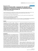
Figure 3 Average of rotarod performance of mouse which
received intracranial human mesenchymal stem cells
transplantation at sequential periods. Average of latency to fall
from rotarod (in seconds) of mice after serial hMSCs implantation
through intracranial injection was compared every 8 weeks. Rotarod
performance of SCA2 transgenic mice (n = 5) was not significantly
improved by serial intracranial hMSCs transplantation at 33-40
weeks of age (p = 0.055). hMSCs, Statistical analysis between each
group was performed by one-way ANOVA (p = 0.055), and between
Control-Wt (n = 16) and Control-Tg group (n = 10) was performed
by t test (p < 0.05).
Figure 4 Average of rot arod perfor mance of mouse which
received intravenous human mesenchymal stem cells
transplantation at sequential periods. Average of latency to fall
from rotarod (in seconds) of mice after serial hMSCs implantation
through intravenous injection was compared every 8 weeks. Rotarod
performance of SCA2 transgenic mice (n = 14) was significantly
improved at 33-40 weeks of age by serial intravenous hMSCs
transplantation (*p = 0.012). The numbers of mice in Control-Wt and
Control-Tg were 16 and 10, respectively. Statistical analysis between
each group was performed by one-way ANOVA (p = 0.012).
Chang et al. Journal of Biomedical Science 2011, 18:54
/>Page 5 of 9
lumens of blood vessels in white matter (Figure 6B).
Large clusters of grafted hMSCs were also detected in
the cerebral cortex as arrows (Figure 6C). These data
suggested that hMSCs which were transplanted via tail
vein injection may extravasate intracranial vessels, and
then migrate through white matter into cerebellar white
matter, molecular layer, and cerebral cortex.
In IC-hMSC-Tg group, positive signals of hMSCs were
not detected over cerebellar white matter, molecular
layer, or Purkinje cell layer (Figure 6D), but limited to a
few lumen of blood vessels (Figure 6E) and a few scat-
tered cells in the cerebral cortex (F igure 6F). Positive
brown IHC signals were also detected at the injection
site beneath the dorsal surface of medulla, which was
adjacent to the cerebellum (Figure 6G). No grafted cell
adopted the morphological and immunohistochemical
characteristics of PCs in either group. No IHC signals
were detected in the cerebellar sections of Control-Wt
(Figure 6H) and Control-Tg mice (Figure 6I), neither.
Besides, no tumor formation was d etected in the serial
sections of cerebellums processed from six SCA2 trans-
genic m ice which received intracranial and intravenous
MSCs transplantation at time of sacrifice.
Discussion
In this study, we investigate whether transplantation of
hMSCs can rescue cerebellar PCs and ameliorate the
deterioration of motor function in SCA in a preclinical
animal model using SCA2 transgenic mice. After pre-
test of intracranial trypan blue in jection (Figure 1A) and
SPIO-labeled hMSCs transplantation (Figure 2), SCA2
transgenic mice were serially transplanted with hMSCs
for three times intracranially or four times intravenously
(Figure 1B). Motor function of mice was evaluated by an
acceleratng protocol of rotarod test every 8 weeks.
Latency to fall on rotarod test of SCA2 transgenic mice
which received serial intracranial hMSC transplantation
of hMSCs failed to show significantly improved motor
function (Figure 3). On the contrary, intravenous
hMSCs transplantation significantly prolonged latency
to fall at 33-40 weeks of age (Figure 4). IHC of serial
cerebellar sections revealed that intravenous hMSC
transplantation effectively rescued more cerebellar PCs
than intracranial transplantation (Figure 5), which was
compatible to rotarod performance of mice. In intra ve-
nous transplantation group, there were also more
hMSCs which were positive for human b2 microglobulin
signals in the cerebellum and cerebral cortex than i n
intracranial transplantation group (Figure 6).
At first, mo use was sacrificed to verify the intracranial
presence of dye after trypan blue injection through fora-
men magnum into position of cerebellum (Figure 1A).
Then SPIO-labeled hMSCs was transplanted intracra-
nially and MR imaging of living mice was arranged to
demon strate the injection site (Figure 2). Low T2-inten-
sit y signals of injected SPIO-labeled hMSCs were found
beneath dorsal surface of medulla, which was adjacent
to cerebellum in MR imaging, and no evidence of major
trauma or intracranial hemorrhage was observed. There-
fore, intracranial and intravenous hMSCs transplanta-
tion proceeded as planned.
We found that rotarod performance of SCA2 trans-
genic mice was not significantly improved by serial
intracranial hMSCs transplantation, and only a trend of
better rotarod performance at 33-40 weeks of age (Fig-
ure 3). The limited number of transgenic mice which
used in intracranial hMSC might probably result in bias
in statistics. Moreover, injection site of intracranial
transplantation was beneath dorsal surface of medulla,
rather than the cerebellum, which made the distance of
hMSCs migration longer.
Rotarod performance of SCA2 transgenic mice was
effectively improved at 33-40 weeks of age by serial
intravenous transplantation of hMSCs via tail vein (Fig-
ure 4). Because previous study had shown that the
majority of intravenously administered MSCs (>80%)
accumulated immediately in the lungs and were cleared
with a half-life of 24 h [29], four times of intravenous
transplantation which delivered larger cell dose of
hMSCs were given in our study. There was no risk of
causing tissue trauma or intracranial hemorrhage for
intravenous transplantation, either. MSCs were also
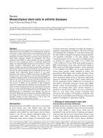
Figure 5 Immunohistochemistry staining for murine Purkinje
cells in cerebellum. (A) Whole brain sections of wild-type mouse,
SCA2 Tg mouse as control, and SCA2 transgenic mouse which
received intravenous and intracranial human mesenchymal stem
cells transplantation (4 μm) were processed by
immunohistochemistry of calbindin D28K for Purkinje cells.
Photographs were taken from the view of 100-folds microscopy and
the scale bar was 40 μm. (B) Quantitative counting of calbindin
D28K+ cells in cerebellum were compared to those of Control-Wt.
Statistical analysis was performed by one-way ANOVA. (* p < 0.05;
** p < 0.001).
Chang et al. Journal of Biomedical Science 2011, 18:54
/>Page 6 of 9
delivered intravenously in animal models of double
toxin-induced multiple system atrophy-parkinsonism
[30], lupus nephritis [31], and clinical trials of ischemic
stroke [23], multiple system atrophy [20], and various
diseases [32] with encouraging results.
IHC showed a marked decline of PC number (66.4%
of wild-type mice) in Control-Tg mice (Figure 5A),
which was previously demonstrated in a mouse model
[28] and an autoposy report [33]. More cerebellar PCs
were found in cerebellar sections of mice which received
intracranial and intravenous hMSCs transplantation by
visual impression (Figure 5A). After counting the num-
bers of surviving PCs, we found that intravenous hMSCs
transplantation significantly rescued more cerebellar PCs
(86.6% of wild-t ype mice) in SCA2 transgenic mice than
intracranial transpl antation (70.7% of wild-type mice, p
= 0. 018) (Figure 5B). This result was compatible to
rotarod performance of transplanted mice. However, the
neuroprotective effects of hMSC transplantation might
be offset by aging effect, since no difference of rotarod
performance among all groups (including wild-type
mice) was noted after 40-47 weeks of age. To elucidate
the aging effect, the histological examinati ons and IHC
at serial time points will be checked in the future
experiments.
To further elucidate the engraftment of transplanted
hMSCs in mice brain, IHC using specific antibodies
against human beta2 microglobulin was performed on
murine whole brain sections (Figure 6). There were
more grafted hMSCs in the cerebellum (Figure 6A and
Figure 6 Immunohistochemistr y staining for human mesenchymal stem cells in whole brain s ections of mice. Whole brain sections of
each mice (4 μm) were proceeded immunohistochemistry staining of b2 microglobulin for hMSCs. Photographs were taken from the view of
100, 200 or 400-folds microscopy and the scale bar was 100 μm. (A-C) In IV-hMSC-Tg group, hMSCs were located over the cerebellar white
matter (A), molecular layer, and the lumens of blood vessels in white matter (B). Large clusters of grafted hMSCs were detected within cerebral
cortex as arrows (C). (D-G) In IC-hMSC-Tg group, positive brown signals were not detected over cerebellar white matter, molecular layer, or the
Purkinje cell layer (D), but limited to a few lumen of blood vessels (E) and a few scattered cells in cerebral cortex (F). Positive signals of hMSCs
were detected over the injection site beneath the dorsal surface of medulla (G), which was adjacent to the cerebellum. (H, I) No signals were
detected in the cerebellar sections of Control-Wt (H) and Control-Tg mice (I).
Chang et al. Journal of Biomedical Science 2011, 18:54
/>Page 7 of 9
6B) a nd cerebral cortex (Figure 6C) in intravenous
transplantation group than in intracranial transplant a-
tion group. Furt hermore, cluster of grafted hMSCs in
the cerebral cortex may also contribute to the better
motor function of mice in intravenous transplantation
group, since degeneration may be encountered in the
cerebral cortex in SCA2 patients [5,6,8]. Local tissue
damages to medulla may be caused by invasive proce-
dures of serial intracranial transplantation (Figure 6G).
Stereotaxic implantation should be co nsidered to
improve target localization and minimize complications
in the future experiments. All these findings suggested
that intravenous hMSCs transplantation was more effec-
tive to ameli orate motor function deterioration of trans-
genic SCA2 mice than intracranial transplantation.
Systemically administered MSCs home to sites of
ischemia or injury and may either transdifferentiate into
exogenous functional neurons or provide neurotrophic
factors for endogenous cells [19,34]. No grafte d cell
adopted the morphological and immunohistochemical
characteristics of cerebellar PCs in this mouse model.
As a result, neuroprotective effects of intravenous
hMSCs transplantation in this study mainly resulted
from neurotrophic factors or direct cell contact with
host cells, not transdifferentiation. Two transgenic
mouse model of SCA1 [10] and cerebellar ataxia [11]
reported the similar findings. Many r ecent clinical stu-
dies which adopt systemically administered MSCs also
implicate paracr ine signaling as the primary mechanism
of action [32].
Although clinical trials of MSC transplantation have
shown no major adverse events over the past 10 years
of testing, recent preclinical studies have stressed poten-
tial long-term risks associated with MSC therapy that
may not be observable in the short follow-up time per-
iod. These long-term risks include potential maldifferen-
tiation, immunosuppression, and instigation of
malignant tumor growth by directly promoting tumor
growth, metastasis, and angiogenesis [32]. For example,
when administered in immunocompromised mice by
systemic injection, MSCs created microemboli and sub-
sequently form osteosarcoma-like pulmonary lesions
[35]. No tumor formation was detected in the serial sec-
tions of cerebellums and medulla processed from six
SCA2 transgenic mice which hMSCs had been trans-
planted at time of sacrifice in our study (Figure 6).
More precl inical and clinical studies are still needed to
evaluate the safety issues of MSC transplantation.
Conclusions
In summary, present study demonstrated that intrave-
nous transplantation of hMSCs effectively improved
rotarod performance of SCA2 transgenic mice and
delayed the onset of motor function loss by better
engraftment of hMSCs in brain tissues and rescuing
cerebellar PCs from cell death, possibly through
release of neurotrophic factors or direct cell contact
with host cells; while intracranial transplantation only
rescued a smaller portion of PCs and failed to
improve motor function. Together, transplantation of
hMSCscanindeeddelaytheonsetaswellasto
improve the motor function of SCA2 transgenic mice.
Results of this preclinical study strongly support
further exploration of the feasibility to transplant
hMSCs for SCA patients.
Acknowledgements
This work was supported in part by the UST-UCSD International Center of
Excellence in Advanced Bio-engineering sponsored by the Taiwan National
Science Council I-RiCE Program under Grant Number: NSC-99-2911-I-009-101.
The authors also acknowledge financial support from the Taipei Veterans
General Hospital (VGH100E1-010, VGH100C-056, VN100-05 and VGH100D-
003-2), the National Science Council, Taiwan (NSC99-2120-M-010-001, NSC99-
2627-B-010-003, NSC99-3111-B-010-002, NSC98-2314-B-010-001-MY3, NSC 99-
2911-I-010-501, and NSC 99-3114-B-002-005), as well as from the Wang Fang
Hospital (100scof03). This study was also supported by a grant from the
Ministry of Education, Aim for the Top University Plan. This work was
assisted in part by the Division of Experimental Surgery of the Department
of Surgery, Taipei Veterans General Hospital.
Author details
1
Institute of Clinical Medicine, National Yang-Ming University, Taipei, Taiwan.
2
Department of Radiation Oncology, Buddhist Tzu Chi General Hospital,
Taipei Branch, New Taipei City, Taiwan.
3
School of Medicine, Tzu Chi
University, Hualien, Taiwan.
4
Stem Cell Research Center, National Yang-Ming
University, Taipei, Taiwan.
5
Department of Orthopaedic Surgery, National
Yang-Ming University Hospital, Yi-Lan, Taiwan.
6
Department of Neurology,
Taipei Veterans General Hospital, Taipei, Taiwan.
7
School of Medicine,
National Yang-Ming University, Taipei, Taiwan.
8
Center for Stem Cell
Research, Taipei Medical University-Wan Fang Medical Center, Taipei, Taiwan.
9
Graduate Institute of Clinical Medicine, Taipei Medical University, Taipei,
Taiwan.
10
Department of Ophthalmology, Taipei Medical University-Wan
Fang Medical Center, Taipei, Taiwan.
11
Department of Orthopaedics and
Traumatology, Taipei Veterans General Hospital, Taipei, Taiwan.
Authors’ contributions
YKC carried out the hMSCs culture, cell transplantation and rotarod test,
performed the statistical analysis and drafted the manuscript. JHH and BWS
provided the transgenic mice and participated in the design of the study.
MHC took care of the animals and carried out the hMSCs culture, cell
transplantation, MRI study and rotarod test. YHC and YFC carried out
immunohistochemical stain of cerebellar sections and counting of Purkinje
cells. WHM and CYT carried out immunohistochemical stain of whole brain
sections and identification of engrafted human cells. OKL conceived of the
study and participated in its design and coordination. All authors read and
approved the final manuscript.
Competing interests
The authors declare that they have no competing interests.
Received: 15 May 2011 Accepted: 8 August 2011
Published: 8 August 2011
References
1. Harding AE: Classification of the hereditary ataxias and paraplegias.
Lancet 1983, , 1: 1151-1155.
2. Durr A: Autosomal dominant cerebellar ataxias: polyglutamine
expansions and beyond. Lancet Neurol 2010, 9:885-894.
3. Soong BW, Paulson HL: Spinocerebellar ataxias: an update. Curr Opin
Neurol 2007, 20:438-446.
Chang et al. Journal of Biomedical Science 2011, 18:54
/>Page 8 of 9
4. Underwood BR, Rubinsztein DC: Spinocerebellar ataxias caused by
polyglutamine expansions: a review of therapeutic strategies. Cerebellum
2008, 7:215-221.
5. Yamada M, Sato T, Tsuji S, Takahashi H: CAG repeat disorder models and
human neuropathology: similarities and differences. Acta Neuropathol
2008, 115:71-86.
6. Taroni F, DiDonato S: Pathways to motor incoordination: the inherited
ataxias. Nat Rev Neurosci 2004, 5:641-655.
7. Gao Y, Zu T, Low WC, Orr HT, McIvor RS: Antisense RNA sequences
modulating the ataxin-1 message: molecular model of gene therapy for
spinocerebellar ataxia type 1, a dominant-acting unstable trinucleotide
repeat disease. Cell Transplant 2008, 17:723-734.
8. Lastres-Becker I, Rub U, Auburger G: Spinocerebellar ataxia 2 (SCA2).
Cerebellum 2008, 7:115-124.
9. Kaemmerer WF, Low WC: Cerebellar allografts survive and transiently
alleviate ataxia in a transgenic model of spinocerebellar ataxia type-1.
Exp Neurol 1999, 158:301-311.
10. Chintawar S, Hourez R, Ravella A, Gall D, Orduz D, Rai M, Bishop DP,
Geuna S, Schiffmann SN, Pandolfo M: Grafting neural precursor cells
promotes functional recovery in an SCA1 mouse model. J Neurosci 2009,
29:13126-13135.
11. Jones J, Jaramillo-Merchan J, Bueno C, Pastor D, Viso-Leon M, Martinez S:
Mesenchymal stem cells rescue Purkinje cells and improve motor
functions in a mouse model of cerebellar ataxia. Neurobiol Dis 2010,
40:415-423.
12. Pittenger MF, Mackay AM, Beck SC, Jaiswal RK, Douglas R, Mosca JD,
Moorman MA, Simonetti DW, Craig S, Marshak DR: Multilineage potential
of adult human mesenchymal stem cells. Science 1999, 284:143-147.
13. Lee OK, Kuo TK, Chen WM, Lee KD, Hsieh SL, Chen TH: Isolation of
multipotent mesenchymal stem cells from umbilical cord blood. Blood
2004, 103:1669-1675.
14. Sottile V, Halleux C, Bassilana F, Keller H, Seuwen K: Stem cell
characteristics of human trabecular bone-derived cells. Bone 2002,
30:699-704.
15. De Bari C, Dell’Accio F, Tylzanowski P, Luyten FP: Multipotent
mesenchymal stem cells from adult human synovial membrane. Arthritis
Rheum 2001, 44:1928-1942.
16. Zuk PA, Zhu M, Ashjian P, De Ugarte DA, Huang JI, Mizuno H, Alfonso ZC,
Fraser JK, Benhaim P, Hedrick MH: Human adipose tissue is a source of
multipotent stem cells. Mol Biol Cell 2002, 13:4279-4295.
17. Fouillard L, Chapel A, Bories D, Bouchet S, Costa JM, Rouard H, Herve P,
Gourmelon P, Thierry D, Lopez M, et al: Infusion of allogeneic-related HLA
mismatched mesenchymal stem cells for the treatment of incomplete
engraftment following autologous haematopoietic stem cell
transplantation. Leukemia 2007, 21:568-570.
18. Marmont AM, Gualandi F, Piaggio G, Podesta M, Teresa van Lint M,
Bacigalupo A, Nobili F: Allogeneic bone marrow transplantation (BMT) for
refractory Behcet’s disease with severe CNS involvement. Bone Marrow
Transplant 2006, 37:1061-1063.
19. Yagi H, Soto-Gutierrez A, Parekkadan B, Kitagawa Y, Tompkins RG,
Kobayashi N, Yarmush ML: Mesenchymal stem cells: mechanisms of
immunomodulation and homing. Cell Transplant 2010, 19:667-679.
20. Lee PH, Kim JW, Bang OY, Ahn YH, Joo IS, Huh K: Autologous
mesenchymal stem cell therapy delays the progression of neurological
deficits in patients with multiple system atrophy. Clin Pharmacol Ther
2008, 83:723-730.
21. Venkataramana NK, Kumar SK, Balaraju S, Radhakrishnan RC, Bansal A,
Dixit A, Rao DK, Das M, Jan M, Gupta PK, et al: Open-labeled study of
unilateral autologous bone-marrow-derived mesenchymal stem cell
transplantation in Parkinson’s disease. Transl Res 2010, 155:62-70.
22. Mazzini L, Ferrero I, Luparello V, Rustichelli D, Gunetti M, Mareschi K, Testa L,
Stecco A, Tarletti R, Miglioretti M, et al: Mesenchymal stem cell
transplantation in amyotrophic lateral sclerosis: A Phase I clinical trial.
Exp Neurol 2010, 223:229-237.
23. Lee JS, Hong JM, Moon GJ, Lee PH, Ahn YH, Bang OY: A long-term follow-
up study of intravenous autologous mesenchymal stem cell
transplantation in patients with ischemic stroke. Stem Cells 2010,
28:1099-1106.
24. Chou SH, Kuo TK, Liu M, Lee OK: In utero transplantation of human bone
marrow-derived multipotent mesenchymal stem cells in mice. J Orthop
Res 2006, 24:301-312.
25. Lee KD, Kuo TK, Whang-Peng J, Chung YF, Lin CT, Chou SH, Chen JR,
Chen YP, Lee OK: In vitro hepatic differentiation of human mesenchymal
stem cells. Hepatology 2004, 40:1275-1284.
26. Lee OK, Ko YC, Kuo TK, Chou SH, Li HJ, Chen WM, Chen TH, Su Y:
Fluvastatin and lovastatin but not pravastatin induce neuroglial
differentiation in human mesenchymal stem cells. J Cell Biochem 2004,
93:917-928.
27. Shieh DB, Cheng FY, Su CH, Yeh CS, Wu MT, Wu YN, Tsai CY, Wu CL,
Chen DH, Chou CH: Aqueous dispersions of magnetite nanoparticles
with NH3+ surfaces for magnetic manipulations of biomolecules and
MRI contrast agents. Biomaterials 2005, 26:7183-7191.
28. Huynh DP, Figueroa K, Hoang N, Pulst SM: Nuclear localization or
inclusion body formation of ataxin-2 are not necessary for SCA2
pathogenesis in mouse or human. Nat Genet 2000, 26:44-50.
29. Lee RH, Pulin AA, Seo MJ, Kota DJ, Ylostalo J, Larson BL, Semprun-Prieto L,
Delafontaine P, Prockop DJ: Intravenous hMSCs improve myocardial
infarction in mice because cells embolized in lung are activated to
secrete the anti-inflammatory protein TSG-6. Cell Stem Cell 2009, 5:54-63.
30. Park HJ, Bang G, Lee BR, Kim HO, Lee PH: Neuroprotective effect of
human mesenchymal stem cells in an animal model of double toxin-
induced multiple system atrophy-parkinsonism. Cell Transplant 2010.
31. Chang JW, Hung SP, Wu HH, Wu WM, Yang AH, Tsai HL, Yang LY, Lee OK:
Therapeutic Effects of Umbilical Cord Blood-Derived Mesenchymal Stem
Cell Transplantation in Experimental Lupus Nephritis. Cell Transplant
2011, 20:245-257.
32. Parekkadan B, Milwid JM: Mesenchymal stem cells as therapeutics. Annu
Rev Biomed Eng 2010, 12:87-117.
33. Estrada R, Galarraga J, Orozco G, Nodarse A, Auburger G: Spinocerebellar
ataxia 2 (SCA2): morphometric analyses in 11 autopsies. Acta Neuropathol
1999, 97:306-310.
34. Torrente Y, Polli E: Mesenchymal stem cell transplantation for
neurodegenerative diseases. Cell Transplant 2008, 17:1103-1113.
35. Aguilar S, Nye E, Chan J, Loebinger M, Spencer-Dene B, Fisk N, Stamp G,
Bonnet D, Janes SM: Murine but not human mesenchymal stem cells
generate osteosarcoma-like lesions in the lung. Stem Cells 2007,
25:1586-1594.
doi:10.1186/1423-0127-18-54
Cite this article as: Chang et al.: Mesenchymal stem cell transplantation
ameliorates motor function deterioration of spinocerebellar ataxia by
rescuing cerebellar Purkinje cells. Journal of Biomedical Science 2011 18:54.
Submit your next manuscript to BioMed Central
and take full advantage of:
• Convenient online submission
• Thorough peer review
• No space constraints or color figure charges
• Immediate publication on acceptance
• Inclusion in PubMed, CAS, Scopus and Google Scholar
• Research which is freely available for redistribution
Submit your manuscript at
www.biomedcentral.com/submit
Chang et al. Journal of Biomedical Science 2011, 18:54
/>Page 9 of 9