Báo cáo y học: " Developmental patterns and characteristics of epicardial cell markers Tbx18 and Wt1 in murine embryonic heart" ppsx
Bạn đang xem bản rút gọn của tài liệu. Xem và tải ngay bản đầy đủ của tài liệu tại đây (4.67 MB, 6 trang )
RESEARC H Open Access
Developmental patterns and characteristics of
epicardial cell markers Tbx18 and Wt1 in murine
embryonic heart
Bin Zeng
1*
, Xiao-feng Ren
2
, Feng Cao
1
, Xiao-yang Zhou
1
and Jing Zhang
1
Abstract
Background: Although recent studies have highlighted the role of epicardial cells during cardiac development
and regeneration, their cardiomyogenic potential is still controversial due to the question of lineage tracing of
epicardial cells. The present study therefore aimed to examine the the expression of Tbx18 and Wt1 in embryonic
heart and to identify whether Tbx18 and Wt1 themselves expressed in the cardiomyocyte.
Methods: Mouse embryonic hearts were collected at different stages for immunofluorescence costaining with
either Tbx18 and the cardiac transcription factor Nkx2.5 or Wilms tumor 1 (Wt1) and Nkx2.5.
Results: Tbx18 and Wt1, but not Nkx2.5, were expressed in the proepicardium and epicardium. Tbx18 was
expressed in cells within the heart from E10.5 to at least E14.5; these Tbx18-expressing cells were Nkx2.5 positive,
except for a few cells that were Nkx2.5 negative at E14.5. Wt1 was expressed in cells within the heart from E12.5 to
at least E14.5, but these Wt1-expressing cells were Nkx2.5 negative.
Conclusion: The data obtained in this study demonstrate that Tbx18 is expressed in the cardiomyocytes from
E10.5 to at least E14.5, and Wt1 is expressed within the heart from E12.5 to at least E14.5, but not in the
cardiomyocyte. These findings may provide new insights on the role of the epicardial cells in cardiac regeneration.
Keywords: Cardiac progenitor, Epicardial cells, Tbx18, Wt1, Development
Background
During embryogenesis, cells from the proepicardium
migrate onto the myocardium to form the epicardium.
The proepicardium is a source of undifferentiated pro-
genitor cells that give rise to endothelial cells, fibroblast
cells, and the smooth muscle cells that form the coronary
vessels during development of the heart [1-3]. The epi-
cardium is the outermost epithelial cell layer overlying
the vertebrate heart and was considered historically to be
a simple derivat ive of the proepicardium. However, a ser-
ies of studies have demonstrated the importance of the
epicardium in the development of the heart and in the
formation of the coronary vascular system [4,5]. Recently,
it was found that the zebrafish epicardium supports car-
diac regeneration during the epithelial-to-mesenchymal
transition (EMT) and subsequent migration into the
myocardium to form new vasculature [6], demonstrating
that it could potentially mediate cardiac regeneration
after injury in lower vertebrates. Most recently, Tbx18-
or Wt1-expressing epicardium was s uggested to provide
a substantial contribution to myocytes in the ventricular
septum (VS) and ventricular walls [7,8], indicating t hat
the epicardium is a potential source for progenitor cells
for cardiovascular the rapeutics. Because of these recent
findings, more and more groups are investigating the
regenerative potential of the epicardium because of its
cardiomyogenic potential. However, Christoffels et al.
show that Tbx18 is expressed in the cardiomyocyte [9],
raising a query whether the epicardium may contribute
to the cardiomyocyte directly. Although the expression of
Tbx18 and Wt1 in heart tissue has been visualized by uti-
lizing genetic knock-in strategies, to the best of our
knowledge, localization analyses of the epicardia l cell
markers Tbx18 and Wt1 in the heart have not been
* Correspondence:
1
Department of Cardiology, Renmin Hospital of Wuhan University, Wuhan,
Hubei, P. R. China
Full list of author information is available at the end of the article
Zeng et al. Journal of Biomedical Science 2011, 18:67
/>© 2011 Zeng et al; licensee BioMed Central Ltd. This is an Open Access article distributed under the terms of the Creative Commons
Attribution License ( which permits unrestricted use, dis tribution, and reproduction in
any medium, provided the original work is properly cited.
published. In this study, we employed immunofluores-
cence staining to investigate the expression of Tbx18 and
Wt1inheartusinganti-Tbx18andWt1antibodiesand
to identify whether Tbx18 and Wt1 themselves expressed
in the cardiomyocyte.
Materials and Methods
Approvement of animal experiments
This study was carried out in strict accordance with the
recommendations in the Guide for the Care and Use of
Labora tory Animals of the National Institutes of Health.
The protocol was approved by the Committee on the
Ethics of Animal Experiments of the University of Min-
nesota (Permit Number: 27-2956). All surgery was per-
formed under sodium pentobarbital anesthesia, and all
efforts were made to minimize suffering [10].
Immunostaining
At least 8 embryos from 5 mice with the same embryo-
nicstages(E9.5,E10.5,E11.5,E12.5,E14.5)werefixed
immediately after dissection in 4% paraformaldehyde for
30-60 min. Tissues were embedded in OCT compound
(Sigma), then quickly frozen in liquid nitrogen and
stored at -80°C. Cryostat sections were cut into 5 μm
thin sections. For coimmunostaining, sections were
incubated with goat polyclonal anti-Tbx18 (SC-17869,
1:100) and rabbit polyclonal anti-Nkx2.5 (SC-25404,
1:300) or rabbit polyclonal anti-Wt1 (SC-192, 1:300) and
goat polyclonal anti-Nkx2.5 (SC-8697, 1:150). The sec-
tions were then incubated with the appropriate second-
ary antibody. The incubation for each primary anti body
was performed at 4°C overnight followed by three
washes with PBS. Finally, the stained sections were
examined using a fluorescence microscope [11,12].
Blocking peptides of Tbx18 (SC-17869 p) and Wt1 (SC-
192 p) were used to tested the specificity of Tbx18 and
Wt1.
Results
Expression of Tbx18 and Wt1 in heart
Tbx18andWt1wereexpressedintheproepicardium
and epicardium. Tbx18 and Wt1 were expressed early in
the proepicardium, and in scattered epicardial cells over
thesurfaceofheartatE9.5(SupplementaryFigure1).
Tbx18 and Wt1 were expressed in the epicardial cells
covering the heart after E10.5 (Figure 1, 2). Tbx18
began to be expressed in some cells within the VS and
left ventricular wall at E10.5, and robust Tbx18 expres-
sion was continuously detectable in the VS and left ven-
tricular wall from E11.5 to at least E14.5 (Figure 1a-d).
Tbx18-expressing cells were observed in the right ven-
tricular wall at E14.5 (Figure 1d4). Wt1 expression was
confined to the epicardium from E9.5 to E11.5 (Supple-
mentaryFigure1andFigure2a-b),butWt1startedto
be expressed in some cells within the VS, left ventricular
wall, and right ventricular w all at E12.5 (Figure 2c).
More Wt1-expressing cells were expressed at E14.5 in
the VS, left ventricular wall, and right ventricular wall
(Figure 2d). Af ter incubated with an excess of peptide
that corresponds to the epitope recognized by Tbx18
and Wt1 antibodies, the staining disappeared (Supple-
mentary Figure 2).
Do Tbx-18- and Wt-1 express in the cardiomyocyte
Coimmunostaining with Tbx18 or Wt1 and the cardiac
transcription factor Nkx2.5 showed that Tbx18- and
Wt1-expressing epicardial cells were not cardiomyocyte
from E9.5 to at least E14.5, because neither were coloca-
lized with Nkx2.5 (Supplementary Figure 2 and Figure 3,
4). Nevertheless, Tbx18-expressing cells in the heart
colocalized with Nkx2.5 from E10.5 to at least E14.5
(Figure 3a-d). Interestingly, at E14.5, some Tb x18-
expressing cells in the VS and LV did not colocalize
with Nkx2.5 (Figure 3d2,3d3, and 3d5,3d6), and Tbx18-
expressing cells, as they began to be detected in the
right ventricular wall, did not colo calize with Nkx2.5
(Figure 3d4, and 3d7). Wt1- expressing cells began to be
detected in some cells within the heart at E12.5 (Figure
4); however, they did not colocalize with Nkx2.5 from
E12.5 to at least E14.5 (Figure 4c1-6 and Figure 4d1-6).
Discussion
The presence of pluripotent cardiac stem cells resident
in the myocardium has received considerable attention
[13,14]. Recently, epicardial progenitors were identified
by expression of Tbx18 and Wt1 due to their cardio-
myogenic potential [7,8], and great expectations rasied
in the field of cardiac regeneration being relevant to epi-
cardial cells and the transcription factors Wt1 and
Tbx18. To obta in better insight into the expression pro-
file and characteristics of the epicardial layer, new
mouse epicardial cell marker genes were identified by
transcriptomics [15]. Tbx18 and Wt1 have been consid-
ered markers of multipotent cardiovascular progenitor
cells [16] . Many studies are focusing on epicardial cells,
Tbx18 and Wt1. Most of these studies were based on
the assumption that the lineage-tracing markers W t1
and Tbx18 are not expressed in the cardiomyocyte.
However, Christoffels et al. showed that cardiomyocytes
themselves express Tbx18, challenging the theory that
Tbx18-expressing epicardial cells have cardiomyoge nic
potential [9].
The results of Cai et al.’s study using genetic knock-in
strategies showed that Tbx18 was expressed e xclusively
in the epicardium from E9.5 to E11.5. After E12.5,
Tbx18 began to be expressed in some cells within the
heart, but it did not colocalize with Nkx2.5. Neverthe-
less, in our study, we found that Tbx18 expression
Zeng et al. Journal of Biomedical Science 2011, 18:67
/>Page 2 of 6
E10.5 E11.5
E12.5 E14.5
Tbx18/DNA
pericardium
LV
RV
RV
RV
RV
VS VS
VS
LV
LV
LV
1
2
3
4
1
2
3
4
12
34
1
2
3
4
E1
E1E1
E
E1
E1
E1
E1
E1
E1E1
E1
E
E
E
E
E1
E1
1
E1
E1
E1
E1
E1
E
E1
E1
1
E1
E1
1
E1
E1
E1
1
E1
E1
E
E
E
E
E
E
E
1
E
E
E
E
E
E
E
E
E
E
E
E
E
E
E
E
E
E
E
1
E
E
E
E
E
2.
2.
2
2.
2.
2
2.
2
2
2
2
2
2
2.
2.
2.
2.
2.2.2.
2.
2
2.
2
2
2
2.
2
2.
2.
2
2
2
2
2
2
2
2
2
2
2
2
2
2
2
2.
2
2
2
2
2
2
2
2
2
2
2
2
2
2
2
2
2
2
2
2
2
2
2
2
2
2
2
2
2
2
2
2
2
2
2
2
2
.
5
5
5
5
5
5
5
5
5
5
5
5
5
5
5
55
5
5
5
5
5
5
5
5
5
5
5
5
5
5
5
5
5
5
5
5
5
5
5
5
5
5
5
5
5
5
5
5
5
5
5
5
5
5
5
5
5
5
RV
RV
RV
RV
RV
RV
RV
RV
RV
RV
RV
RV
RV
RVRV
RV
V
V
RV
RVRV
RV
RV
V
RV
V
RV
RV
RV
RV
V
RV
RV
RV
RV
RV
V
RV
RV
RV
RV
RV
RV
RV
RV
RV
RV
RV
RV
RV
V
V
V
R
V
V
V
V
R
RV
R
RV
RV
R
RV
R
R
RV
R
V
V
V
RV
V
V
V
V
V
R
RV
R
R
R
VS
VSVS
VS
VS
VS
VS
VS
VS
VS
VS
VS
VS
VS
VS
V
V
VS
VS
VS
VS
VS
VS
VS
VS
VS
VS
VS
V
V
VS
V
VS
VS
VS
VS
VS
V
VS
VS
VS
VS
VS
VS
V
VS
VS
V
VS
V
VS
VS
VS
VS
VS
VS
VS
VS
V
V
VS
VS
VS
VS
V
VS
VS
V
VS
VS
V
V
VS
V
S
S
S
VS
VS
S
V
V
V
V
VS
VS
V
S
V
V
V
S
V
S
VS
S
V
V
V
LV
LV
LV
LV
LV
LV
LV
LV
LV
LV
LV
LV
LV
LV
V
V
LV
V
LV
V
V
LV
V
V
LV
LV
LV
LV
LV
LV
LV
V
LV
LV
LV
LV
LV
LV
LV
LV
L
LV
LV
LV
V
LV
LV
L
LV
V
V
V
V
V
V
V
V
V
LV
L
V
V
LV
V
L
L
L
V
V
L
L
L
L
L
1
1
1
1
1
1
1
1
1
1
1
1
1
1
1
1
1
1
1
1
1
1
1
1
1
1
1
1
1
1
1
1
1
1
1
1
1
1
1
1
1
1
1
1
1
1
1
1
1
1
1
1
1
1
1
1
1
1
2
2
2
2
2
2222
2
2
2
2
33
3
3
3
3
3
3
3
3
3
3
3
3
3
3
3
3
3
3
3
3
3
3
3
3
3
3
3
3
3
3
3
3
3
3
3
3
3
3
3
3
3
3
3
3
3
3
3
3
3
3
3
3
3
3
3
3
3
3
3
3
4
4
4
4
4
44
4
4
4
4
4
4
4
4
4
4
4
4
4
4
4
4
4
4
4
4
4
4
4
4
4
4
4
4
4
4
4
4
4
4
4
4
4
4
4
4
4
4
4
4
4
4
E12.5
VS
LV
RV
2
4
1
3
VS
VS
VS VS
VS
a
b
c
d
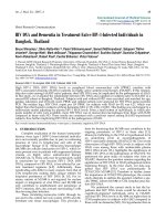
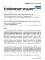
Figure 1 Expression of epicardial cell marker Tbx18 in murine embryonic heart. Tbx18 was found not only in the epicardium (arrowheads)
but also in the VS and LV from E10.5 to at least E14.5 (arrow), and Tbx18 was also found in the RV at E14.5 (arrow) (a-d). a2, b2, c2, and d2 are
higher-magnification views (400×) of a1, b1, c1, and d1 (200×), respectively, and highlight sections from the VS. a3, b3, c3, and d3 are sections
from the LV (200×), and a4, b4, c4, and d4 (200×) are sections from the RV. VS, ventricular septum; LA/RA, left/right atrium; LV/RV, left/right
ventricle.
Wt1/DNA
E10.5
E11.5
E12.5
E14.5
VS
LV
RV
VS
LV
RV
VS
LV
RV
VS
RV
LV
1
1
2
2
3
3
1
22
1
3
3
4
4
5
6
6
5
LV
RV
a
b
c
d
LV
RV
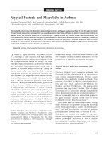
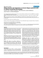
Figure 2 Expression of epicardi al cell marker Wt1 in murine embryonic heart. Expression of Wt1 was confined to the epicardium
(arrowheads) from E10.5 to E11.5 (a, b), but was detected within the heart (arrow) from E12.5 to E14.5 (c, d). a1, b1, c1, and d1 are sections from
the VS (200×); c2 and d2 are higher-magnification views (400×) of c1 and d1, respectively. a2, b2, c3, and d3 are sections from the LV (200×). a3,
b3, c4, and d4 are sections from the RV (200×); c5 and c6 are higher-magnification views (400×) of the areas within the white squares in c3 and
c4, respectively. d5 and d6 are higher-magnification views (400×) of the areas within the white squares in d3 and d4, respectively. VS, ventricular
septum; LA/RA, left/right atrium; LV/RV, left/right ventricle. Blue, DAPI staining of nuclei.
Zeng et al. Journal of Biomedical Science 2011, 18:67
/>Page 3 of 6
began in cells within the VS and left ventricular wall at
E10.5, and robust Tbx18 expression was continuously
detectable in the VS and left ventricular wall from E11.5
to at least E14.5. Furthermore, Tbx18-expressing cells in
the heart colocalized with Nkx2.5 from E10.5 to at least
E14.5. Our data are consistent with those of Christoffels’
group: namely, Tbx18 is expressed in the cardiomy ocyte
fromE10.5toatleastE14.5.Interestingly,wefound
that, at E14.5, some Tbx18-expressing cells in the VS
and LV did not colocalize with Nkx2.5, and Tbx18-
expressing cells, as they began to be detected in the
right ventricular wall, did not colocalize with Nkx2.5.
CFs appear concurrently with ventricular compaction
around embryo nic day E12.5 and increase in number
steadily throughout postnatal day 1 [17]. Those Tbx18-
expressing cells, which do not colocalize with Nkx2.5,
mightbeepicardial-derivedfibroblastcells.Nkx2.5isa
well-characterized marker of early cardiomyocyte lineage
[18]. Though Nkx2.5 expression has been found in pro-
genitors of proepicardium and in coronary vessel
smooth muscle cell progenitors [19,20], these studies
focused on early stage heart, E9.5. In this study, we
found that Tbx18-expressing cells in the heart coloca-
lized with Nkx2.5 from E10.5. The results from Zhou et
al.’s study using genetic knock-in showed that Wt1 was
confined to the proepicardium and epicardium from
E9.5 to E15.5 and not expressed in the cardiomyocyte
[8]. However, we found that Wt1 expression was con-
fined to th e epicardium from E9.5 to E11.5. Wt1 started
to be expressed in some cells within the VS and left
ventricular wall at E12.5, but they did not colocalize
with Nkx2.5 from E12.5 to at least E14.5.
E14.5
E14.5 E14.5
Σ
Τ
Υ
5
6
7
Tbx
1
8 Nkx
2
.5 Merge
E10.5
E11.5 E12.5
E14.5
111
1
22
2
2
3
3
3
3
4
4
4
4
VS
VS VS
VS
LV
LV
LV LV
RV
RV RV
RV
Σ
Τ
Υ
a
b
c
d
d
d
d
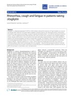
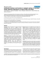
Figure 3 Characteristics of Tbx18 in murine embryonic heart. Expression of Tbx18 was expressed in the epicardi um (arrowheads) and was
also detected in cardiomyocytes in the VS and the LV (white arrow) from E10.5 to E14.5. a1, b1, c1 and d1 are sections from the VS (200×); a2,
b2, c2 and d2 are higher-magnification views (400×) of a1, b1, c1 and d1, respectively; a3, b3, c3 and d3 are sections from the LV (200×); a4, b4,
c4 and d4 are sections from the RV (200×). d5-7 are higher-magnification views of the white square in d2-4, respectively; they highlight that
most of Tbx18-expressing cells are cardiomyocytes, except for a few cells at E14.5 (blue arrow). VS, ventricular septum; LA/RA, left/right atrium;
LV/RV, left/right ventricle.
Zeng et al. Journal of Biomedical Science 2011, 18:67
/>Page 4 of 6
The failure to detect Tbx18 and Wt1 within the heart
may result from the limitations of rely ing solely on line-
age mapping without robust lineage-restricted molecular
markers and clear-cut morphological identification cri-
teria [9,21]. Wt1-expressing cells are descended from
precursor cells that are positive for Nkx2.5 and Isl1 [8].
Wt1 cardiac conditional knockout mice die between
E16.5 and E18.5 due to cardiovascular failure, and in
vitro Wt1-deficient embryoid bodies lack important
markers of endothelial, hematopoietic, and myocardial
cells [22]. Wt1 is a crucial gene for t he development of
epicardial-derived cells; it serves not only to promote
the EMT, but also to regulate the origin of the cardio-
vascular lineage. Though the expression of W t1 in thi s
study is different from Zhou et al.’ s, Wt1 is not
expressed in the cardiomyocyte. The differences between
Tbx 18 and Wt1 express ion pattern need furt her studies
to address.
Conclusions
In conclusion, Tbx18 is expressed in the cardiomyocytes
from E10.5 to at least E14.5; Wt1 is expressed withi n the
heart from E12.5 to at least E14.5, but not in t he cardio-
myocyte. These findings may provide new insights on the
role of the epicardial cells in cardiac regeneration.
Acknowledgements
This work was supported by the Chinese National Nature Science
Foundation (30900609)
Author details
1
Department of Cardiology, Renmin Hospital of Wuhan University, Wuhan,
Hubei, P. R. China.
2
College of Veterinary Medicine, Northeast Agricultural
University, Harbin, Hei Longjiang, P. R. China.
Authors’ contributions
BZ designed, carried out the main experiment and drafted the manuscript.
XFR helped to improve the manuscript. XYZ and FC helped to finish
histological experiments. JZ helped to design the experiment and improve
the manuscript. All authors read and approved the final manuscript.
Competing interests
The authors declare that they have no competing interests.
Received: 5 June 2011 Accepted: 26 August 2011
Published: 26 August 2011
References
1. Nesbitt T, Lemley A, Davis J, Yost MJ, Goodwin RL, Potts JD: Epicardial
development in the rat: a new perspective. Microsc Microanal 2006,
12(5):390-8.
2. Ratajska A, Czarnowska E, Ciszek B: Embryonic development of the
proepicardium and coronary vessels. Int J Dev Biol 2008, 52:229-236.
3. Guadix JA, Carmona R, Muñoz-Chápuli R, Pérez-Pomares JM: In vivo and in
vitro analysis of the vasculogenic potential of avian proepicardial and
epicardial cells. Dev Dyn 2006, 235:1014-1026.
4. Smart N, Risebro CA, Melville AA, Moses K, Schwartz RJ, Chien KR, Rilery PR:
Thymosin beta4 induces adult epicardial progenitor mobilization and
neovascularization. Nature 2007, 445:177-182.
Τ
Τ
E12.5
E14.5
E14.5
Σ
Τ
6
5
6
E10.5
E11.5
1
1
1
1
2
2
2
2
3
3
3
3
4
4
VS
VS
VS
VS
LV
LV
LV
LV
RV
RV
RV
RV
Σ
Σ
Τ
Τ
d
c
a
b
d
c
Σ
5
E12.5
c
d
5
E12.5
E14.5
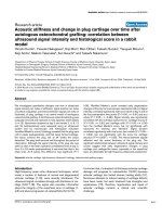
Figure 4 Characteristics of Wt1 in murine embryonic heart. Expression of Wt1 was conf ined to the epicardium (arrowheads) from E10.5 to
E11.5 and was also detected within the heart from E12.5 to E14.5 (arrow). a1, b1, c1 and d1 are sections from the VS (200×); c2 and d2 are
higher-magnification views (400×) of c1 and d1, respectively. a2, b2, and c3, d3 are sections from the LV (200×). a3, b3, and c4, d4 are sections
from the RV (200×). c5,6 and d5,6 are higher-magnification views of the white squares in c3,4 and d3,4 respectively; they highlight that some
Wt1-expressing cells are located within the heart but are not cardiomyocytes. Blue, DAPI staining of nuclei.
Zeng et al. Journal of Biomedical Science 2011, 18:67
/>Page 5 of 6
5. Pennisi DJ, Mikawa T: FGFR-1 is required by epicardium-derived cells for
myocardial invasion and correct coronary vascular lineage
differentiation. Dev Biol 2009, 328:148-159.
6. Lepilina A, Coon AN, Kikuchi K, Holdway JE, Roberts RW, Burns CG, Poss KD:
A dynamic epicardial injury response supports progenitor cell activity
during zebrafish heart regeneration. Cell 2006, 127:607-619.
7. Cai CL, Martin JC, Sun Y, Cui L, Wang L, Ouyang K, Yang L, Bu L, Liang X,
Zhang X, Stallcup WB, Denton CP, McCulloch A, Chen J, Evans SM: A
myocardial lineage derives from Tbx18 epicardial cells. Nature 2008,
454:104-108.
8. Zhou B, Ma Q, Rajagopal S, Wu SM, Domian I, Rivera-Feliciano J, Jiang D,
Von Gise A, Ikeda S, Chien KR, Pu WT: Epicardial progenitors contribute to
the cardiomyocyte lineage in the developing heart. Nature 2008,
454:109-113.
9. Christoffels VM, Grieskamp T, Norden J, Mommersteeg MT, Rudat C,
Kispert A: Tbx18 and the fate of epicardial progenitors. Nature 2009, 16:
E8-9.
10. Zeng B, Lin G, Ren X, Zhang Y, Chen H: Over-expression of HO-1 on
mesenchymal stem cells promotes angiogenesis and improves
myocardial function in infarcted myocardium. J Biomed Sci 2010, 7:80-88.
11. Li Y, Zhang D, Zhang Y, He G, Zhang F: Augmentation of
neovascularization in murine hindlimb ischemia by combined therapy
with simvastatin and bone marrow-derived mesenchymal stem cells
transplantation. J Biomed Sci 2010, 17:75-85.
12. Zeng B, Chen H, Zhu C, Ren X, Lin G, Cao F: Effects of combined
mesenchymal stem cells and heme oxygenase-1 therapy on cardiac
performance. Eur J Cardiothorac Surg 2008, 34:850-6.
13. Wu KH, Liu YL, Zhou B, Han ZC: Cellular therapy and myocardial tissue
engineering: the role of adult stem and progenitor cells. Eur J
Cardiothorac Surg 2006, 30:770-81.
14. Siepe M, Akhyari P, Lichtenberg A, Schlensak C, Beyersdorf F: Stem cells
used for cardiovascular tissue engineering. Eur J Cardiothorac Surg 2008,
34:242-247.
15. Bochmann L, Sarathchandra P, Mori F, Lara-Pezzi E, Lazzaro D, Rosenthal N:
Revealing new mouse epicardial cell markers through transcriptomics.
PloS One 2010, 28:e11429.
16. Lam JT, Moretti A, Laugwitz KL: Multipotent Progenitor Cells in
Regenerative Cardiovascular medicine. Pediiatr Cardiol 2009, 30:690-698.
17. Ieda M, Tsuchihashi T, Ivey KN, Ross RS, Hong TT, Shaw RM, Srivastava D:
Cardiac fibroblasts regulate myocardial proliferation through beta1
integrin signaling. Dev Cell 2009, 16:233-244.
18. Harvey RP: NK-2 homeobox genes and heart development. Dev Biol 1996,
178:203-16.
19. Zhou B, von Gise A, Ma Q, Rivera-Feliciano J, Pu WT: Nkx2-5- and Isl1-
expressing cardiac progenitors contribute to proepicardium.
Biochem
Biophys Res Commun 2008, 375:450-453.
20. Wu SM, Fujiwara Y, Cibulsky SM, Clapham DE, Lien CL, Schultheiss TM,
Orkin SH: Developmental origin of a bipotential myocardial and smooth
muscle cell precursor in the mammalian heart. Cell 2006, 127:1137-50.
21. Snider P, Standley KN, Wang J, Azhar M, Doetschman T, Conway SJ: Origin
of cardiac fibroblasts and the role of periostin. Circ Res 2009, 105:934-947.
22. Martínez-Estrada OM, Lettice LA, Essafi A, Guadix JA, Slight J, Velecela V,
Hall E, Reichmann J, Devenney PS, Hohenstein P, Hosen N, Hill RE, Munoz-
Chapuli R, Hastie ND: Wt1 is required for cardiovascular progenitor cell
formation through transcriptional control of Snail and E-cadherin. Nat
Gent 2010, 42:89-93.
doi:10.1186/1423-0127-18-67
Cite this article as: Zeng et al.: Developmental patterns and
characteristics of epicardial cell markers Tbx18 and Wt1 in murine
embryonic heart. Journal of Biomedical Science 2011 18:67.
Submit your next manuscript to BioMed Central
and take full advantage of:
• Convenient online submission
• Thorough peer review
• No space constraints or color figure charges
• Immediate publication on acceptance
• Inclusion in PubMed, CAS, Scopus and Google Scholar
• Research which is freely available for redistribution
Submit your manuscript at
www.biomedcentral.com/submit
Zeng et al. Journal of Biomedical Science 2011, 18:67
/>Page 6 of 6