Báo cáo y học: "Traumatic vertebral artery dissection in an adult with brachial plexus injury and cervical spinal fracture" pptx
Bạn đang xem bản rút gọn của tài liệu. Xem và tải ngay bản đầy đủ của tài liệu tại đây (338.24 KB, 4 trang )
BioMed Central
Page 1 of 4
(page number not for citation purposes)
Journal of Brachial Plexus and
Peripheral Nerve Injury
Open Access
Case report
Traumatic vertebral artery dissection in an adult with brachial
plexus injury and cervical spinal fractures
Silas NS Motsitsi* and Rian R Steyn
Address: Department of Orthopaedic Surgery, Kalafong Hospital, University of Pretoria, Pretoria, South Africa
Email: Silas NS Motsitsi* - ; Rian R Steyn -
* Corresponding author
Abstract
We present a case of a 32 year-old right-hand dominant woman who sustained a right brachial
plexus injury, ipsilateral fractures of the cervical spine transverse processes, and vertebral artery
dissection. She presented to us four days following the initiating accident. Magnetic Resonance
Imaging showed normal brachial plexus along with vertebral artery dissection with intramural
thrombus and vascular lumen occlusion. The dissection was managed conservatively. A repeat
CAT-SCAN Angiography three months later showed healing of the dissection plus vascular lumen
re-canalization. There were no sequelae due to the dissection.
The details of the case are discussed in this report.
Background
Cervicocerebral dissection is responsible for strokes in
young patients. It accounts for 20% of cerebro-vascular
accidents in patients younger than 45 years [1]. Extra-cra-
nial carotid artery dissection accounts for 70%–80% and
extra-cranial vertebral artery dissection for 15% of strokes
in these young patients. The causes are not completely
understood. Triggers of cervico-cerebral dissection are,
nose blowing, coughing, chiropractic maneuvers, sudden
neck turning, and trauma (minor and major). Genetic
(Ehler-Danlos syndrome) and environmental (smoking,
hypertension, oral contraceptives and migraine) factors
may also be responsible.
Traumatic vertebral artery injury may be occlusive
(thrombosis) or non-occlusive (dissection) [2]. The inci-
dence of vertebral artery injury among patients with blunt
neck trauma is estimated at 0.20%–0.77% [3]. Major
mechanisms of injury are, distraction/extension, distrac-
tion/flexion, and lateral flexion. The vertebral artery is eas-
ily injured by traction [4]. Only about 12%–24% of
unilateral vertebral artery injuries present with signs and
symptoms of vertebro-basilar ischaemia. The majority of
these injuries are missed because clinicians do not think
about them.
Traumatic vertebral artery dissection is common with
major penetrating or blunt neck trauma [5]. A case of ver-
tebral artery dissection (VAD) plus brachial plexus injury
has been reported in a child following a car accident [6].
There has not been such a case reported in an adult. We
report a case of brachial plexus injury, VAD, and ipsilat-
eral five contiguous transverse process fractures of the cer-
vical spine in an adult.
We detail the presentation, physical examination, diag-
nostic work-up, treatment and follow-up.
Published: 6 September 2007
Journal of Brachial Plexus and Peripheral Nerve Injury 2007, 2:17 doi:10.1186/1749-7221-2-
17
Received: 27 June 2007
Accepted: 6 September 2007
This article is available from: />© 2007 Motsitsi and Steyn; licensee BioMed Central Ltd.
This is an Open Access article distributed under the terms of the Creative Commons Attribution License ( />),
which permits unrestricted use, distribution, and reproduction in any medium, provided the original work is properly cited.
Journal of Brachial Plexus and Peripheral Nerve Injury 2007, 2:17 />Page 2 of 4
(page number not for citation purposes)
Case presentation
A 32 year-old right hand dominant secretary was involved
in a car accident. She was a passenger. The car in which she
was traveling was hit from the side by a truck. Her head
was thrown into acute left lateral flexion during impact.
She immediately felt pain in the neck and partial loss of
function of her right upper limb. There was no loss of con-
sciousness. She did not sustain any other injuries. Previ-
ous medical history was unremarkable.
She was referred to our Spinal Clinic four days after the
accident. She was complaining of painful neck, especially
on the right side, and inability to use the right upper limb.
On physical examination, she had torticollis and the
affected limb was supported in a sling. The neck was ten-
der from C1-T1 especially on the right side in the posterior
triangle. Neurological examination of the right upper
limb showed decreased function of the brachial plexus;
motor function; C5 = 0/5, C6 = 2/5, C7 = 2/5, C8 = 3/5,
and T1 = 4/5 according to the modified MRC scale. There
was decreased sensation involving the whole of the bra-
chial plexus distribution. Reflexes were not recorded. The
circulation to the limb was normal compared to the oppo-
site side. There were not any other significant findings.
Plain radiographs of the neck (Antero-posterior, lateral,
and open-mouth) showed loss of cervical lordosis, frac-
ture of the right transverse process of C6 and increased
pre-vertebral soft tissue shadow from C3- C7. Flexion-
extension views were done two weeks later (when she was
pain-free) and did not show any instability. A computer-
ized tomography scan (CT-SCAN) was requested to
exclude other cervical spine fractures. It showed contigu-
ous communited fractures of the cervical transverse proc-
esses of C3-C7 on the right side. There were not any other
fractures detected.
Magnetic Resonance Imaging (MRI) was done to evaluate
brachial plexus injury and to exclude vertebral artery
injury. [It is our policy to exclude vertebral artery injury in
all cases of lateral mass or transverse process fractures of
the cervical spine]. The brachial plexus was normal. MRI
demonstrated high-signal intensity in both T1- and T2-
weighted images of the vertebral artery on the right side.
There was intramural methaemoglobin plus occlusion of
the lumen, but there was no intraluminal thrombus.
There was no intimal flap demonstrated (Figure 1). This
was in keeping with vertebral artery dissection. The spinal
cord was normal.
On the advice of the physicians, she was placed on pro-
phylactic treatment: Aspirin 650 mg orally twice a day for
three months. We were advised to repeat angiography in
three months. She was referred to the brachial plexus
clinic for follow-up.
We repeated angiography three months later. She was
evaluated using a 16-channel multi-detector CT SCAN.
The scan showed complete healing of the dissection and
recanalization of the right vertebral artery (Figure 2). She
continued her further management at the brachial plexus
clinic. They explored the brachial plexus surgically but did
not find any neuromas or pathology needing reconstruc-
tion. They made a decision to manage her conservatively.
Discussion
Our patient presented with a devastating injury involving
her dominant limb. She was referred because of neck pain
and brachial plexus palsy. The brachial plexus injury dom-
inated the clinical picture. There was a potentially devas-
tating injury which was not suspected: vertebral artery
dissection. This injury is commonly overlooked. The clue
to this injury was a transverse process fracture of C6 which
was not diagnosed in the original X-rays. The full extent of
the injuries was only picked up during re-evaluation at
our clinic. The most likely mechanism of fractures of the
transverse processes was avulsion or traction which
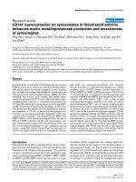
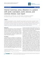
MRI of the cervical spineFigure 1
MRI of the cervical spine. T2-weighted image shows high sig-
nal intensity (white arrow) of the right vertebral artery.
There is an intramural thrombus plus occlusion of the lumen
(Grade four dissection). There is no intraluminal thrombosis.
Journal of Brachial Plexus and Peripheral Nerve Injury 2007, 2:17 />Page 3 of 4
(page number not for citation purposes)
occurred during forceful lateral neck flexion. Prophylactic
treatment with Aspirin was pre-emptive. She made a good
recovery of the vertebral artery dissection: the artery re-
canalized and the dissection healed. There were no neuro-
logical sequelae attributable to VAD.
The areas of the vertebral artery vulnerable to injury dur-
ing blunt neck trauma are, V2 (inside the transverse
foramina) and the V3 (between the C1 and the base of the
skull) [7]. The latter is usually injured in minor trauma.
Clay Cothren et al. [8] in a large series concluded that the
following cervical spine injury patterns mandate screen-
ing for blunt cerebro-vascular injury; fractures extending
into the transverse processes, subluxations, and fractures
of the upper cervical spine. Other authors have argued
that fractures and fracture-dislocations also warrant exclu-
sion of injuries of the vertebral artery. According to Hiro-
shi Taneishi et al. [9] VAD occurs in 20% of patients with
cervical spine fractures or fracture-dislocation. They found
that all their patients who had VAD had spinal cord
injury: there was no significant correlation between the
two. However, there was a statistically significant correla-
tion between unilateral facet dislocation and vertebral
artery occlusion. They also noted that occlusion secondary
to VAD can recanalize in up to 85% of cases within three
months by spontaneous mechanisms. Philip J. Torina et
al. [10] in their series found that vertebral artery occlusion
is significantly more common in motor-complete spinal
cord injury.
One of the most controversial issues in traumatic cerebro-
vascular trauma is what is the best modality for investigat-
ing blunt cerebro-vascular injury. The gold standard is
Digital Subtraction Angiography (DSA). The problem
with DSA is that it is an invasive procedure. Other modal-
ities available are MRI, MRI-Angiography and multi-detec-
tor CT-Angiography (CTA). Lawrence D. Bub et al. [11] in
his series of 32 patients concluded that the accuracy of
CTA in vertebral injury remains in question. It was Alex-
ander L. Eastman et al. [12] in a large series of 162 patient
who demonstrated that CTA is a very good screening tool
for blunt cervical injury. They demonstrated that the over-
all sensitivity, specificity, positive predictive value, nega-
tive predictive value, and accuracy of CTA for the
diagnosis of blunt cerebro-vascular injury were 97.7%,
100%, 100%, 99.3%, and 99.37, respectively.
The natural history of VAD is unknown. It can heal spon-
taneously, it can develop occlusion or it can form a
pseudo-aneurysm. The clinical significance of VAD lies in
its potential to form intra-luminal thrombosis and this
has potential for embolization. Vertebral artery injury
(thrombosis or dissection) can lead to basilar stroke
which has a poor prognosis. The mortality rate due to ver-
tebro-basilar ischaemia can reach 75%–86% [8]. Treat-
ment for VAD is controversial; it not clear whether
patients must be heparinized, be treated with antiplatelets
(Aspirin) or treated at all. Izhar Hasan et al. [13] in their
review of 68 children found that the most common treat-
ment for VAD was antiplatelet therapy. They found that
asymptomatic recovery occurred in 12 of 15 (80%) chil-
dren who received antiplatelets therapy compared to 4 of
15 (27%) who received anticoagulation therapy with or
without antiplatelet. Once thrombosis occurs, it is also
controversial whether anticoagulation or antiplatelet ther-
apy should be the treatment of choice. Vadim Beletsky et
al [14] showed that the recurrence rate for embolization is
decreased significantly (by 8.3%) in patients on anticoag-
ulation compared to those on Aspirin (12.4%). This dif-
ference in outcome at one year was not statistically
significant. It is prudent to seriously consider prophylactic
treatment (unless contra-indications exist) because the
prognosis for brainstem ischaemia is very poor.
Conclusion
Based on the literature and on this case report, we make
the following recommendations;
■Vertebral artery injury must be excluded in high-risk
cases.
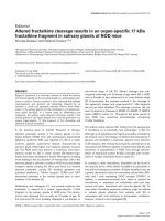
CTA done three months later using a 64-slice coronal recon-structionFigure 2
CTA done three months later using a 64-slice coronal recon-
struction. There is normal blood flow at the level of C4 to
C2 (white arrow).
Publish with BioMed Central and every
scientist can read your work free of charge
"BioMed Central will be the most significant development for
disseminating the results of biomedical research in our lifetime."
Sir Paul Nurse, Cancer Research UK
Your research papers will be:
available free of charge to the entire biomedical community
peer reviewed and published immediately upon acceptance
cited in PubMed and archived on PubMed Central
yours — you keep the copyright
Submit your manuscript here:
/>BioMedcentral
Journal of Brachial Plexus and Peripheral Nerve Injury 2007, 2:17 />Page 4 of 4
(page number not for citation purposes)
■Prophylactic treatment for VAD must be seriously con-
sidered unless there are contra-indications
A randomized control trial is required (if ethically accept-
able) comparing prophylactic treatment versus non-treat-
ment in patients with VAD.
References
1. Thanvi B, Munshi SK, Dawson SL, Robinson TG: Carotid and ver-
tebral artery dissection syndromes. Postgraduate Medical Journal
2005, 81:383-388.
2. Dill-Macky Marcus J, Khangure Mark, Song Swithin: Traumatic cer-
vical distraction complicated by delayed reduction due to
traumatic vertebral artery pseudo-aneurysm. Australasian
Radiology 1999, 43:372-377. 65
3. Inamasu Joji, Guiot Bernard H: Vertebral artery injury after
blunt cervical trauma: an update. Surgical Neurology
2006:238-246.
4. Khurana Divya S, Bonnemann Carsten G, Dooling Elizabeth C, Ouel-
lette Eileen M, Buonanno Ferdinando: Vertebral Artery Dissec-
tion: Issues in Diagnosis and Management. Pediatric Neurology
1996, 14(3):255-258.
5. Mandila Christina G, Koukoulitsios Georgios V, Stathopoulos Geor-
gios T, Karampelas Ioannis, Karydas Georgios, Karabinis Andreas:
Unilateral and bilateral vertebral artery dissection following
motor vehicle injury – two cases and a mini-review. Interna-
tional Journal of Angiology 2006, 14:242-248.
6. Park Seong-Hyun, Sung Joo-Kyung, Hwang Sung-Kyoo: Traumatic
vertebral artery dissection in a child with brachial plexus
injury. Paediatric Neurosurgery 2005, 41:141-144.
7. Peltier J, Toussaint P, Page C, Descenclos C, Deramond H, Le Gars
D: Dissection of the vertebral artery complicating Jefferson
fracture. European Journal of Orthopaedic Surgery and Traumatology
2004, 14:230-233.
8. Clay Cothren C, Moore Ernest E, Ray Charles E JR, Johnson Jeffrey L,
Moore John B, Burch Jon M: Cervical spine fracture patterns
mandating screening to rule out blunt cerebrovascular
injury. Surgery 2007, 141:76-82.
9. Taneichi Hiroshi, Suda Kota, Kajino Tomomichi, Kaneda Kiyoshi:
Traumatically induced vertebral artery occlusion associated
with cervical spine injuries; prospective study using Magnetic
Resonance Imaging Angiography. Spine 2005,
30(17):1955-1963.
10. Torino Philip J, Flanders Adam E, Carrino John A, Burns Anthony S,
friedman David P, Harrop James S, Vaccaro Alexander: Incidence of
vertebral artery thrombosis in cervical spine trauma: corre-
lation with severity of spinal cord injury. AJNR American Journal
of Neuroradiology 26:2645-2651.
11. Bub Lawrence D, Hollingworth William, jarvik Jeffrey G, Hallam
Danial K: Screening for blunt cerebrovascular injury: evaluat-
ing the accuracy of multidetector Computed Tomographic
Angiography. Journal of Trauma 2005, 59:691-697.
12. Eastman Alexander L, Chason David P, Perez Carlos L, McAnulty
Amy L, Minei Joseph P: Computed Tomographic Angiography
for the Diagnosis of Blunt Cervical Vascular Injury: Is It
Ready forPrimetime? J Trauma 2006, 60:925-929.
13. Hasan Izhar, Wapnick Simon, Tenner Michael S, Couldwell William T:
Vertebral Artery Dissection in Children: A Comprehensive
Review. Pediatric Neurosurgery 2002, 37:168-177.
14. Beletsky Vadim, Nadareishvili Zurab, Lynch John, Shuaib Ashfaq,
Woolfenden Andrew, Norris John W: Cervical Arterial Dissec-
tion: Time for a Therapeutic Trial? Stroke 2003, 34:2856-2860.