Báo cáo y học: "Ultrasound in the diagnosis of a median neuropathy in the forearm: case report" doc
Bạn đang xem bản rút gọn của tài liệu. Xem và tải ngay bản đầy đủ của tài liệu tại đây (302.03 KB, 4 trang )
BioMed Central
Page 1 of 4
(page number not for citation purposes)
Journal of Brachial Plexus and
Peripheral Nerve Injury
Open Access
Case report
Ultrasound in the diagnosis of a median neuropathy in the forearm:
case report
Stuart D Ginn*
1
, Michael S Cartwright
2
, George D Chloros
1
,
Francis O Walker
2
, Joon-Shik Yoon
3
, Martin E Brown
2
and Ethan R Wiesler
1
Address:
1
Department of Orthopaedic Surgery, Wake Forest University School of Medicine, Winston-Salam, NC, USA,
2
Department of Neurology,
Wake Forest University School of Medicine, Winston-Salem, NC, USA and
3
Department of Rehabilitation Medicine, Korea University College of
Medicine, Seoul, South Korea
Email: Stuart D Ginn* - ; Michael S Cartwright - ; George D Chloros - ;
Francis O Walker - ; Joon-Shik Yoon - ; Martin E Brown - ;
Ethan R Wiesler -
* Corresponding author
Abstract
Background: Electrodiagnostic studies are traditionally used in the diagnosis of focal
neuropathies, however they lack anatomical information regarding the nerve and its surrounding
structures. The purpose of this case is to show that high-resolution ultrasound used as an adjunct
to electrodiagnostic studies may complement this lack of information and give insight to the cause.
Case presentation: A 60-year-old male patient sustained a forearm traction injury resulting in
progressive weakness and functional loss in the first three digits of the right hand. High-resolution
ultrasound showed the presence of an enlarged nerve and a homogenous soft-tissue structure
appearing to engulf the nerve. The contralateral side was normal. Surgery revealed fibrotic bands
emanating from the flexor digitorum profundus muscle compressing the median nerve thus
confirming the ultrasound findings.
Conclusion: A diagnostically challenging case of median neuropathy in the forearm is presented
in which high-resolution ultrasound was valuable in establishing an anatomic etiology and directing
appropriate management.
Background
The traditional diagnostic approach for focal neuropa-
thies involves a detailed history and physical examina-
tion, augmented by electrodiagnostic studies (nerve
conduction studies and electromyography). [1] While this
approach is effective for localizing the site of pathology
and determining the severity of the condition, it does have
limitations. Electrodiagnostic studies are uninformative
about structures surrounding the nerve and muscle, they
do not allow visualization of intrinsic nerve or muscle
abnormalities, and they are painful. High-resolution
ultrasound (HRUS) is a non-invasive, painless, portable,
and inexpensive modality that has become an attractive
adjunct to electrodiagnostic studies in the evaluation of
entrapment neuropathies. [2]
We present a diagnostically challenging case of median
neuropathy in the forearm in which HRUS was used to
Published: 4 December 2007
Journal of Brachial Plexus and Peripheral Nerve Injury 2007, 2:23 doi:10.1186/1749-7221-2-
23
Received: 14 August 2007
Accepted: 4 December 2007
This article is available from: />© 2007 Ginn et al; licensee BioMed Central Ltd.
This is an Open Access article distributed under the terms of the Creative Commons Attribution License ( />),
which permits unrestricted use, distribution, and reproduction in any medium, provided the original work is properly cited.
Journal of Brachial Plexus and Peripheral Nerve Injury 2007, 2:23 />Page 2 of 4
(page number not for citation purposes)
direct appropriate management. This case illustrates that
HRUS can be a useful complement to electrodiagnostic
studies in the evaluation of focal neuropathies.
Case presentation
A 60 year-old right-handed man with a history of degen-
erative cervical disc disease presented with complaints of
right hand and forearm weakness that started 6 months
earlier following an acute traction injury sustained while
moving a large mattress. The mattress fell and pulled his
right arm, and he immediately felt pain in his shoulder
and elbow. Two hours after the injury he noticed weak-
ness in the first three digits of his right hand.
One month later the weakness persisted, but it had not
worsened. His primary care physician was initially con-
cerned about cervical root trauma given his history of
degenerative disc disease and the nature of the injury, but
an MRI and CT myelogram of the cervical spine showed
no changes compared to his previous cervical spine
images. It was then assumed that he had a brachial plexus
injury, and the plan was to follow his course clinically.
Over the next several months he developed progressive
numbness over the palmar aspect of the first three digits,
and progressive weakness in his hand and forearm. He
also noted atrophy of the muscles in his volar forearm.
Eight months after the initial injury he presented to our
electromyography (EMG) laboratory. On examination he
had profound weakness of the flexor pollicis longus and
flexor digitorum profundus to the index and middle fin-
gers, and mild weakness of the flexor digitorum superfi-
cialis, flexor carpi radialis, and abductor pollicis brevis.
He also had decreased sensation over the palm in the dis-
tribution of the median nerve. Motor and sensory nerve
conduction studies showed no response from the median
nerve, and EMG localized the lesion as a focal neuropathy
of the median nerve distal to the branch to the pronator
teres muscle.
HRUS using a Philips iU22 scanner (Philips Medical Sys-
tems, Bothell, WA) with a 12 MHz linear array transducer
was performed to further explore this focal neuropathy.
The median nerve was shown to be intact throughout the
arm. At the presumed site of neuropathy the cross-sec-
tional area of the nerve was enlarged, from 10.9 mm2 at
the wrist to 17.2 mm2 at the site of maximal enlargement
in the proximal forearm, but it maintained a normal echo-
texture. The soft tissue deep to the median nerve at this
site was hyperechoic and homogenous and appeared to
engulf the nerve (Figure 1). Ultrasound of the correspond-
ing level of the contralateral forearm demonstrated nor-
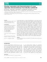
The cross-sectional ultrasound image (A) of the proximal forearm demonstrates the normal echo-texture of the median nerve (arrow)Figure 1
The cross-sectional ultrasound image (A) of the proximal forearm demonstrates the normal echo-texture of the median nerve
(arrow). The hyperechoic and homogenous ground glass appearance of the flexor digitorum profundus muscle (curved arrows)
is also shown. The intra-operative photo (B) depicts a fibrotic band (straight line) across the anterior aspect of the median
nerve (arrow). Arrowheads = arteries, * = pronator teres muscle. The ultrasound image was obtained with a Philips iU22 scan-
ner (Philips Medical Systems, Bothell, WA) with a 12 MHz linear array transducer.
Journal of Brachial Plexus and Peripheral Nerve Injury 2007, 2:23 />Page 3 of 4
(page number not for citation purposes)
mal appearing muscle in clear contrast to the
symptomatic arm.
Approximately one year had passed since the initial injury
and based on the progressive weakness, new sensory find-
ings, and ultrasonographic changes, median nerve explo-
ration in the proximal forearm with planned neurolysis
was pursued. A longitudinal incision was made in the
anterior forearm just distal to the antecubital fossa. The
median nerve was identified, surrounded by healthy pro-
nator teres and flexor digitorum superficialis muscles. Ini-
tial intraoperative nerve conduction studies showed no
response from the median nerve. Deep to the median
nerve the flexor digitorum profundus to the index finger
was found to be atrophic and fibrotic, and multiple rigid
fibrous bands emanated from the muscle. Several of these
bands crossed over and compressed the median nerve,
both proximal and distal to the anterior interosseous
nerve (Figure 1). These bands were released and intraop-
erative nerve conduction studies were repeated, again with
no response from the median nerve.
Tendon transfers were performed to improve function.
AIN reconstruction was foregone due to the low probabil-
ity of functional improvement given the extensive fibrosis
observed in the FDP muscle tissue. The viable flexor digi-
torum profundus to the ring finger was attached to the
flexor digitorum profundus to the index finger with side-
to-side tenodesis, and the flexor carpi radialis was trans-
ferred to the distal flexor pollicis longus through an inci-
sion at the wrist. The post-operative course was
uncomplicated, and two months after the procedure the
patient had improved hand function, consisting of slow,
partial return of his sensory recovery, improved motor
function and grip strength.
Conclusion
The use of HRUS in peripheral nerve surgery is a relatively
novel concept. To date, the majority of the studies using
HRUS in peripheral nerves of the upper extremity have
focused on the entrapment neuropathies of the median
nerve at the wrist and of the ulnar nerve at the elbow.
These studies have shown that HRUS is as a low-cost, non-
invasive, painless adjunct to the nerve conduction studies
in the diagnosis of these entities and have highlighted that
in addition to nerve conduction studies, HRUS may fur-
ther provide anatomic information that might help deter-
mine the cause. [3-6] Furthermore, recent studies have
used HRUS to assess the morphologic changes of the
median nerve after carpal tunnel syndrome release, [7] the
presence of nerve transections, [8] and primary peripheral
nerve repair. [9] In addition, a previous study showed
ultrasound to be helpful in the pre-operative evaluation of
nerve injuries. [10]
There are many potential causes of median nerve com-
pression and injury in the forearm, including masses
extrinsic or intrinsic to the nerve, trauma, anatomic
anomalies, and entrapment. [11-14] HRUS can greatly
improve diagnostic yield by identifying the specific ana-
tomic etiologies responsible for the nerve pathology and
was particularly useful in delineating the nature of
median nerve involvement in this case. The median nerve
was found to be intact throughout the forearm, which
ruled out primary injury or transection of the nerve.
Enlargement of the median nerve at the site of the neurop-
athy was consistent with compression-induced neuropa-
thy, as is seen with entrapment at other sites, and this
finding identified the specific site of neuropathy. [5,6]
There were no ultrasonographic changes to suggest the
presence of a neuroma. Finally, the abnormal appearance
of the soft tissue deep to the median nerve in the anatomic
location of the flexor digitorum profundus was consistent
with an inflammatory or fibrotic process engulfing the
median nerve, which prompted the decision to pursue
surgical exploration and excision of the compressing tis-
sue. The ultrasonographic findings were confirmed during
surgical exploration of the forearm, where the abnormal
soft tissue structure visualized by ultrasound corre-
sponded to fibrous bands originating from the flexor dig-
itorum profundus and entrapping the median nerve.
The mechanism of injury and the sequence of events that
led to median neuropathy in this case are unclear, how-
ever, based on the history and ultrasound findings, we can
make speculations. One possibility is that the initial trac-
tion injury damaged the anterior interosseous nerve,
which resulted in the initial weakness without sensory
changes. The absence of innervation to part of the flexor
digitorum profundus caused this muscle to atrophy and
fibrose, and some of the fibrotic tissue formed rigid bands
that compressed the median nerve. The compression led
to the development of a focal neuropathy, which was
localized with ultrasound as an increase in median nerve
cross-sectional area. Alternatively, the initial injury could
have caused a tear of the flexor digitorum profundus mus-
cle to the index finger, with the development of fibrotic
bands compressing the median nerve during subsequent
healing.
It has been shown that HRUS may be used as an adjunct
to physical examination and electrodiagnostic findings in
the diagnosis of nerve entrapment neuropathies in the
absence of anatomic abnormalities. [3,5] This case dem-
onstrates that it may be valuable in establishing an ana-
tomic etiology and directing appropriate management in
a diagnostically challenging case of median neuropathy in
the forearm. In addition, ultrasound is non-invasive, inex-
pensive, and effective as a pre-operative planning tool for
the surgical treatment of focal neuropathies.
Publish with BioMed Central and every
scientist can read your work free of charge
"BioMed Central will be the most significant development for
disseminating the results of biomedical research in our lifetime."
Sir Paul Nurse, Cancer Research UK
Your research papers will be:
available free of charge to the entire biomedical community
peer reviewed and published immediately upon acceptance
cited in PubMed and archived on PubMed Central
yours — you keep the copyright
Submit your manuscript here:
/>BioMedcentral
Journal of Brachial Plexus and Peripheral Nerve Injury 2007, 2:23 />Page 4 of 4
(page number not for citation purposes)
Competing interests
Dr. Cartwright has a Clinical Research Training Grant
from the Muscular Dystrophy Association to study neu-
romuscular ultrasound; however, this organization does
not have a financial interest or conflict with the content of
the manuscript. The other authors declare that they have
no competing interests.
Authors' contributions
SG performed all pertinent research and drafted the man-
uscript. MC, FW, and EW conceived the case report, per-
formed evaluations and treatments for the patient, and
helped to edit the manuscript. EW performed the patient's
surgery. JY and GC helped to conceive of the study and
participated in the editing process. MB performed the
electrodiagnostic studies in the neurology clinic. All
authors read and approved the final manuscript.
Acknowledgements
The patient was informed that data concerning his case would be submitted
for publication and informed consent was obtained.
References
1. Campion D: Electrodiagnostic testing in hand surgery. J Hand
Surg [Am] 1996, 21(6):947-956.
2. Walker FO, Cartwright MS, Wiesler ER, Caress J: Ultrasound of
nerve and muscle. Clin Neurophysiol 2004, 115(3):495-507.
3. Kele H, Verheggen R, Bittermann HJ, Reimers CD: The potential
value of ultrasonography in the evaluation of carpal tunnel
syndrome. Neurology 2003, 61(3):389-391.
4. Colak A, Kutlay M, Pekkafali Z, Saracoglu M, Demircan N, Simsek H,
Akin ON, Kibici K: Use of sonography in carpal tunnel syn-
drome surgery. A prospective study. Neurol Med Chir (Tokyo)
2007, 47(3):109-115.
5. Wiesler ER, Chloros GD, Cartwright MS, Shin HW, Walker FO:
Ultrasound in the diagnosis of ulnar neuropathy at the
cubital tunnel. J Hand Surg [Am] 2006, 31(7):1088-1093.
6. Wiesler ER, Chloros GD, Cartwright MS, Smith BP, Rushing J, Walker
FO: The use of diagnostic ultrasound in carpal tunnel syn-
drome. J Hand Surg [Am] 2006, 31(5):726-732.
7. El-Karabaty H, Hetzel A, Galla TJ, Horch RE, Lucking CH, Glocker FX:
The effect of carpal tunnel release on median nerve flatten-
ing and nerve conduction. Electromyogr Clin Neurophysiol 2005,
45(4):223-227.
8. Cartwright MS, Chloros GD, Walker FO, Wiesler ER, Campbell
WW: Diagnostic ultrasound for nerve transection. Muscle
Nerve 2007, 35(6):796-799.
9. Peer S, Harpf C, Willeit J, Piza-Katzer H, Bodner G: Sonographic
evaluation of primary peripheral nerve repair. J Ultrasound
Med 2003, 22(12):1317-1322.
10. Cokluk C, Aydin K, Senel A: Presurgical ultrasound-assisted
neuro-examination in the surgical repair of peripheral nerve
injury. Minim Invasive Neurosurg 2004, 47(3):169-172.
11. Flores LP, Carneiro JZ: Peripheral nerve compression second-
ary to adjacent lipomas. Surg Neurol 2007, 67(3):258-262.
12. Hobson-Webb LD, Walker FO: Traumatic neuroma diagnosed
by ultrasonography. Arch Neurol 2004, 61(8):1322-1323.
13. Kuo YL, Yao WJ, Chiu HY: Role of sonography in the preopera-
tive assessment of neurilemmoma. J Clin Ultrasound 2005,
33(2):87-89.
14. Lubahn JD, Cermak MB: Uncommon nerve compression syn-
dromes of the upper extremity. J Am Acad Orthop Surg 1998,
6(6):378-386.