Báo cáo y học: "Bilateral superficial peroneal nerve entrapment secondary to anorexia nervosa: a case repor" doc
Bạn đang xem bản rút gọn của tài liệu. Xem và tải ngay bản đầy đủ của tài liệu tại đây (298.22 KB, 3 trang )
BioMed Central
Page 1 of 3
(page number not for citation purposes)
Journal of Brachial Plexus and
Peripheral Nerve Injury
Open Access
Case report
Bilateral superficial peroneal nerve entrapment secondary to
anorexia nervosa: a case report
Teoman Toni Sevinç, Aydıner Kalacı*, Yunus Doğramacı and
Ahmet Nedim Yanat
Address: Dept. of Orthopaedics and Traumatology, Mustafa Kemal University, Faculty of Medicine, Antakya, Hatay, Turkey
Email: Teoman Toni Sevinç - ; Aydıner Kalacı* - ; Yunus Doğramacı - ;
Ahmet Nedim Yanat -
* Corresponding author
Abstract
We report a case of severe weight loss secondary to anorexia nervosa causing bilateral superficial
peroneal nerve entrapment in a young female patient who was treated successfully by bilateral
surgical decompression.
Background
Among entrapment neuropathies, superficial peroneal
nerve (SPN) entrapment is relatively rare [1-8] and only a
few bilateral cases have been reported in the literature
[9,10].
Severe weight loss, as a result of anorexia nervosa, associ-
ated with common peroneal nerve entrapment is very rare
[11-17] and SPN involvement alone has not been
described in the literature published in English. Bilateral
presentation is always related to systemic cause rather
than local mechanical compression.
Herein we report a case of severe weight loss secondary to
anorexia nervosa causing bilateral SPN entrapment in a
young female patient who was treated successfully by
bilateral surgical decompression.
Case presentation
A 20-year-old, female university student presented to our
outpatient orthopaedic clinic with a two month history of
vague pain on the outer border of both legs, and numb-
ness over the dorsum of the feet and big toes. Her symp-
toms were exacerbated by walking and running and
partially relieved by elevation. She had to stop to rest after
30 minutes of walking because of intolerable pain.
There was neither history of trauma or surgery to the lower
limb nor history of lower back problems. There was, how-
ever, a history of severe weight loss of (30 kg) during the
previous six months and the patient was diagnosed with
anorexia nervosa using criteria from the American Psychi-
atric Association's Diagnostic and Statistical Manual of
Mental Disorders (DSM-IV-TR) and the World Health
Organization's International Statistical Classification of
Diseases and Related Health Problems (ICD).
Physical examination revealed bilateral tender points
approximately 11 cm proximal to the ankle joint on the
outer surface of the leg, Tinel sign was also positive bilat-
erally. There were sensory deficits on the dorsum of both
big toes but no muscle weakness or abnormal reflexes.
Published: 27 April 2008
Journal of Brachial Plexus and Peripheral Nerve Injury 2008, 3:12 doi:10.1186/1749-7221-3-
12
Received: 14 January 2008
Accepted: 27 April 2008
This article is available from: />© 2008 Sevinç et al; licensee BioMed Central Ltd.
This is an Open Access article distributed under the terms of the Creative Commons Attribution License ( />),
which permits unrestricted use, distribution, and reproduction in any medium, provided the original work is properly cited.
Journal of Brachial Plexus and Peripheral Nerve Injury 2008, 3:12 />Page 2 of 3
(page number not for citation purposes)
Examination of the lumbar spine and lower limbs
revealed no clinical abnormalities in the joints and there
was neither suspicion of nerve root compression at the
level of the lumbar spine nor nerve entrapment at the
neck of the fibula.
Radiographic examination of the lumbar spine, legs and
feet were normal and EMG studies were positive for bilat-
eral entrapment neuropathy of the SPN proximal to the
ankle joint with no abnormality of the common peroneal
nerves or of the proximal nerve roots.
After preoperative assessment, the patient was admitted
for surgical treatment with the diagnosis of SPN entrap-
ment. The operation was done under general anaesthesia,
using pneumatic tourniquet. Bilateral explorations of the
site of tenderness revealed adhesions of both SPNs to the
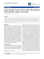
fascia with perineural fibrosis. Careful dissections were
done to free the nerves and neurolysis was successfully
performed (Figure 1). The nerves were freed distally and
proximally by splitting the overlying fascia for a few cen-
timetres above and below the site of entrapment.
Symptoms of bilateral peroneal nerve entrapment were
relieved immediately and completely in the postoperative
period. Physiotherapy was started immediately to prevent
postoperative adhesions. No recurrence was observed in
the first year following the operation.
Discussion
Superficial peroneal nerve syndrome is an entrapment
neuropathy that usually results from mechanical com-
pression of the nerve at or near the point where the nerve
pierces the fascia to travel within the subcutaneous tissue.
A thorough and accurate knowledge of the course of the
SPN and its relationships is essential to understand the
pathophysiology, and a thorough and careful physical
examination is important for diagnosing this condition.
Stephens et al. described a physical sign to identify the dis-
tal subcutaneous course of the SPN below the skin, prima-
rily by means of plantar flexion and inversion of the ankle
and foot and, secondarily by a passive flexion of the
fourth toe [1].
In his study Styf, described 3 provocative tests for nerve
compression at rest at rest following exercise [2]. In the
first test, pressure is applied over the anterior intermuscu-
lar septum while the patient actively dorsiflexes the ankle.
In the second test, the foot is passively plantar flexed and
inverted at the ankle. In the third test, while the patient
maintains the passive stretch, gentle percussion is applied
over the course of the nerve. These tests are useful in com-
petitive athletes who have symptoms suggestive of exer-
cise-induced compartment syndrome.
Electrophysiological studies are helpful for the diagnosis,
however, normal conduction velocity may be found espe-
cially at rest which does not exclude compression of the
superficial peroneal nerve [2].
Injection of the nerve with lidocaine or Marcaine just
above the site of involvement may be the most valuable
diagnostic tool. The patient can define the extent of relief
obtained from such an injection, which can be helpful in
defining the zone of injury and expected relief from surgi-
cal release or excision.
Entrapment of the superficial peroneal nerve has trau-
matic and non traumatic causes. Local trauma and com-
pression are the most common causes of nerve
entrapment. This may be due to recurrent stretch injuries
Photograph at operation showing the superficial peroneal nerveFigure 1
Photograph at operation showing the superficial per-
oneal nerve.
Publish with BioMed Central and every
scientist can read your work free of charge
"BioMed Central will be the most significant development for
disseminating the results of biomedical research in our lifetime."
Sir Paul Nurse, Cancer Research UK
Your research papers will be:
available free of charge to the entire biomedical community
peer reviewed and published immediately upon acceptance
cited in PubMed and archived on PubMed Central
yours — you keep the copyright
Submit your manuscript here:
/>BioMedcentral
Journal of Brachial Plexus and Peripheral Nerve Injury 2008, 3:12 />Page 3 of 3
(page number not for citation purposes)
or certain positions like prolonged kneeling and squat-
ting, which cause perineural fibrosis [17,18]. Oedema
after trauma may result in a mini compartment syndrome
which may occur when the tunnel was fibrotic, of low
compliance and longer than 3 cm [2]. Chronic or exer-
tional lateral compartment syndrome can also cause com-
pression of the superficial peroneal nerve, particularly in
athletes [19,20]. Fasciotomy of the anterior compartment
for chronic anterior compartment syndrome may also
cause compression of the SPN nerve [19].
Nontraumatic causes of SPN entrapment are commonly
due to anatomical variations such as fascial defects, with
or without muscle herniation about the lateral lower leg,
where the nerve is entrapped as it emerges into the subcu-
taneous tissue or a short peroneal tunnel proximally.
Nerve compression in patients with fascial defects is
explained by the normal increase in muscle relaxation
pressure and intramuscular pressure at rest during and
after exercise. This increase is sufficient to cause herniated
muscle tissue and this can impinge upon or compresses
the nerve [20].
Lowdon reported a case of an abnormally long course of
the SPN nerve through the deep fascia which was thought
to have caused compression. Exercise may have exacer-
bated the symptoms by producing mechanical irritation
or by raising the pressure in the peroneal compartment
and thus increasing compression of the nerve [3].
Conclusion
In our case, the bilateral involvement forced us to think
about a systemic cause of SPN entrapment. The patient
had severe loss of weight in a period of few months due to
previously undiagnosed anorexia nervosa which may
have caused changes in the subcutaneous tissues that led
to adhesions and perineural fibrosis. Although the exact
cause is unknown; SPN entrapment should be kept in
mind especially in patients with severe weight loss and
changes in body habits.
Competing interests
The authors declare that they have no competing interests.
Acknowledgements
Written informed consent was obtained from the patient for publication of
this case report and accompanying images. A copy of the written consent
is available for review by the Editor-in-Chief of this journal.
References
1. Stephens MM, Kelly PM: Fourth toe flexion sign: a new clinical
sign for identification of the superficial peroneal nerve. Foot
Ankle Int 2000, 21:860-863.
2. Styf J: Entrapment of the superficial peroneal nerve. Diagno-
sis and results of decompression. J Bone Joint Surg Br 1989,
71:131-135.
3. Lowdon IM: Superficial peroneal nerve entrapment. A case
report. J Bone Joint Surg Br 1985, 67:58-59.
4. Yang LJ, Gala VC, McGillicuddy JE: Superficial peroneal nerve syn-
drome: an unusual nerve entrapment. Case report. J Neuro-
surg 2006, 104:820-823.
5. Styf J, Morberg P: The superficial peroneal tunnel syndrome.
Results of treatment by decompression. J Bone Joint Surg Br
1997, 79:801-803.
6. Daghino W, Pasquali M, Faletti C: Superficial peroneal nerve
entrapment in a young athlete: the diagnostic contribution
of magnetic resonance imaging. J Foot Ankle Surg 1997,
36:170-172.
7. Kernohan J, Levack B, Wilson JN: Entrapment of the superficial
peroneal nerve. Three case reports. J Bone Joint Surg Br 1985,
67:60-61.
8. Banerjee T, Koons DD: Superficial peroneal nerve entrapment.
Report of two cases. J Neurosurg 1981, 55:991-992.
9. Saragaglia D, Farizon F, Drevet JG, Butel J: Peroneal nerve entrap-
ment syndrome of the front of the foot. Treatment by neu-
rolysis. Apropos of a bilateral case. Rev Chir Orthop Reparatrice
Appar Mot 1986, 72:579-581.
10. McAuliffe TB, Fiddian NJ, Browett JP: Entrapment neuropathy of
the superficial peroneal nerve. A bilateral case. J Bone Joint
Surg Br 1985, 67:62-63.
11. Constanty A, Vodoff MV, Gilbert B, Dantoine F, Roche JF, Piguet C,
Tabaraud F, de Lumley L: Peroneal nerve palsy in anorexia ner-
vosa: three cases. Arch Pediatr 2000, 7:316-317.
12. Lutte I, Rhys C, Hubert C, Brion F, Boland B, Peeters A, Van Den
Bergh P, Lambert M: Peroneal nerve palsy in anorexia nervosa.
Acta Neurol Belg 1997, 97:251-254.
13. Kershenbaum A, Jaffa T, Zeman A, Boniface S: Bilateral foot-drop
in a patient with anorexia nervosa. Int J Eat Disord 1997,
22:335-337.
14. MacKenzie JR, LaBan MM, Sackeyfio AH: The prevalence of
peripheral neuropathy in patients with anorexia nervosa.
Arch Phys Med Rehabil 1989, 70:827-830.
15. Schott GD: Anorexia nervosa presenting as foot drop. Postgrad
Med J 1979, 55:58-60.
16. Kopell HP, Thompson WAL: Peripheral entrapment. neuropa-
thies of the lower extremity. N Engl J Med 1960, 262:56-60.
17. Stack RE, Bıanco AJ Jr, Maccarty CS: Compressıon of the com-
mon peroneal nerve by ganglıon cysts: report of nıne cases.
J Bone Joint Surg Am 1965, 47:773-778.
18. Styf J: Diagnosis of exercise-induced pain in the anterior
aspect of the lower leg. Am J Sports Med 1988, 16:165-169.
19. Styf JR, Körner LM: Diagnosis of chronic anterior compartment
syndrome in the lower leg. Acta Orthop Scand 1987, 58:139-144.
20. Styf JR, Körner LM: Microcapillary infusion technique for meas-
urement of intramuscular pressure during exercise. Clin
Orthop Relat Res 1986, 207:253-262.