Báo cáo y học: "Cardiac CT and MRI guide surgery in impending left ventricular rupture after acute myocardial infarction'''' ppt
Bạn đang xem bản rút gọn của tài liệu. Xem và tải ngay bản đầy đủ của tài liệu tại đây (803.77 KB, 5 trang )
BioMed Central
Page 1 of 5
(page number not for citation purposes)
Journal of Cardiothoracic Surgery
Open Access
Case report
Cardiac CT and MRI guide surgery in impending left ventricular
rupture after acute myocardial infarction
Jens Vogel-Claussen*
1
, Jan Skrok
1
, Elliot K Fishman
1
, João AC Lima
2
,
Ashish S Shah
3
and David A Bluemke
4
Address:
1
Johns Hopkins University School of Medicine, Russell H. Morgan Department of Radiology and Radiological Science, Baltimore, MD,
USA,
2
Johns Hopkins University School of Medicine, Department of Cardiology, Baltimore, MD, USA,
3
Johns Hopkins University School of
Medicine, Department of Surgery, Division of Cardiac Surgery, Baltimore, MD, USA and
4
National Institutes of Health, Department of Radiology
and Imaging Sciences, Bethesda, MD, USA
Email: Jens Vogel-Claussen* - ; Jan Skrok - ; Elliot K Fishman - ;
João AC Lima - ; Ashish S Shah - ; David A Bluemke -
* Corresponding author
Abstract
We report the case of a 67 year-old patient who presented with worsening chest pain and
shortness of breath, four days post acute myocardial infarction. Contrast enhanced computed
tomography of the chest ruled out a pulmonary embolus but revealed an unexpected small
subepicardial aneurysm (SEA) in the lateral left ventricular wall which was confirmed on cardiac
magnetic resonance imaging. Intraoperative palpation of the left lateral wall was guided by the
cardiac MRI and CT findings and confirmed the presence of focally thinned and weakened
myocardium, covered by epicardial fat. An aneurysmorrhaphy was subsequently performed in
addition to coronary bypass surgery and a mitral valve repair. The patient was discharged home on
post operative day eight in good condition and is feeling well 2 years after surgery.
Background
The formation of left ventricular (LV) myocardial aneu-
rysms is one of several potentially life-threatening compli-
cations post acute myocardial infarct (AMI). These
aneurysms are traditionally divided into two main groups:
true and false aneurysms. While true aneurysms have a
wide mouth and the wall is comprised of infracted/
fibrous tissue [1], false aneurysms represent complete rup-
tures of the myocardial wall. They have a narrow neck and
are contained by pericardium. In contrast to true aneu-
rysms, false aneurysms have a dismal prognosis. There-
fore, fast and accurate diagnosis and treatment can be life
saving [2].
Impending wall ruptures and thus precursors to false
aneurysms are called subepicardial aneurysms (SEA).
They were first described by Hunter in 1933 as a rare form
of saccular aneurysm [3]. In 1983 Epstein was first to use
the term "subepicardial" aneurysms and described them
as having three distinguishing features: abrupt interrup-
tion of the myocardium at the neck of the aneurysm, a
narrow neck relative to the diameter of the aneurysm, and
a propensity to rupture spontaneously [4]. SEAs are diffi-
cult to diagnose and are often only found post-mortem. In
this case we report an impending rupture of an SEA in a
patient with chest pain 4 days post AMI. This diagnosis
was made using computed tomography (CT) and mag-
Published: 12 August 2009
Journal of Cardiothoracic Surgery 2009, 4:42 doi:10.1186/1749-8090-4-42
Received: 16 March 2009
Accepted: 12 August 2009
This article is available from: />© 2009 Vogel-Claussen et al; licensee BioMed Central Ltd.
This is an Open Access article distributed under the terms of the Creative Commons Attribution License ( />),
which permits unrestricted use, distribution, and reproduction in any medium, provided the original work is properly cited.
Journal of Cardiothoracic Surgery 2009, 4:42 />Page 2 of 5
(page number not for citation purposes)
netic resonance imaging (MRI), which assisted in securing
a favorable patient outcome.
Case presentation
A 67 year-old patient presented to the emergency room
with worsening chest pain and shortness of breath four
days post acute inferolateral myocardial infarction with
subsequent left circumflex coronary artery stent place-
ment. The patient had no history of prior myocardial inf-
arctions. The chest radiograph showed moderate
pulmonary edema and small bilateral pleural effusions
(Fig. 1).
To rule out pulmonary embolism, the patient was referred
for a computed tomography (CT) scan. The CT was nega-
tive for pulmonary embolus; however, an incidental 1.0 ×
1.6 cm blister-like pouch/aneurysm was seen in the lateral
LV wall within a hypoperfused area that extended from
the lateral to the inferoseptal wall and from the base to the
mid-cardiac level (Fig. 2). This finding was concerning for
an SEA/impending myocardial rupture within the suba-
cutely infarcted left ventricular wall. However, it was
deemed necessary to further characterize the anatomy of
the infarct and myocardial outpouching to determine the
urgency for cardiac surgery.
Because the patient was hemodynamically stable, cardiac
magnetic resonance (CMR) imaging was performed. Car-
diac MR images demonstrated a large subacute inferosep-
tal, inferior, and lateral transmural myocardial infarction
with extensive microvascular obstruction on the first pass
perfusion images (Fig. 3a and 3b, see additional files 1
and 2). The first pass perfusion defect persisted on the
delayed enhancement MR images taken 55 minutes after
the gadolinium injection (Fig. 4). At the lateral edge of the
infarction, a small aneurysm with a narrow neck was iden-
tified (see additional file 3), consistent with the CT find-
ings. The aneurysm was covered by only 1 mm of
infarcted myocardium (Fig. 3c). There was no evidence of
rupture into the pericardium.
During surgery, which was performed within 24 hours of
CT/MR imaging, a distinct area of thin and weak myocar-
dium in the lateral left ventricular wall was evident. The
epicardium was intact and the area correlated with the
preoperative imaging. Since the region was very close to
the base of the heart as well as the AV groove, a bovine
pericardial patch was sewn over the region using a contin-
uous prolene suture. The patch was reinforced with a thin
layer of Bioglue
®
adhesive (Cryolife, Inc). At the same
time, coronary bypass grafting and a mitral valve repair
were performed to treat the patient's ischemic heart dis-
ease and severe mitral valve insufficiency. The patient was
discharged home on post operative day eight in good con-
dition and is feeling well 2 years after surgery.
Discussion
After an acute myocardial infarction (AMI), there are sev-
eral potentially life-threatening complications: (1)
Arrhythmias [5], (2) cardiogenic shock, (3) complete free
wall ruptures which account for almost 4% of patients'
deaths after AMI (33% occur within the first 24 hours,
85% within the first week [6]), (4) complete septal rup-
tures (accounting for 1% – 5% of all infarct-related deaths
[7]), and (5) the formation of false aneurysms.
While true aneurysms typically do not require treatment,
false aneurysms, or pseudoaneurysms, are the result of a
complete rupture of the ventricular wall with containment
of the resulting hematoma by adherent pericardium and
thus have a high mortality rate. As SEAs are precursors to
pseudoaneurysms with a high propensity to rupture,
immediate treatment is often life-saving. Although con-
servative management has been reported to be successful
in asymptomatic chronic SEAs [8-10], surgical treatment
is still considered standard of care, especially for sympto-
matic acute SEAs, as in our case [9,11-13]. The options
include aneurysmectomy (resection) or aneurysmor-
rhaphy (patch repair) [11]. In addition to an elevated risk
of death, patients with SEAs are initially difficult to diag-
nose due to a lack of specific symptoms (our patient was
suspected to have a pulmonary embolus). Diagnosis can
be made using ultrasound, MRI, left heart catheter, or CT
[11]. Due to the high risk of rupture in combination with
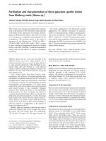
Portable AP chest radiograph of a 67 year old patient with pulmonary edema, small bilateral pleural effusions, and cardi-omegaly five days post myocardial infarctionFigure 1
Portable AP chest radiograph of a 67 year old patient
with pulmonary edema, small bilateral pleural effu-
sions, and cardiomegaly five days post myocardial inf-
arction.
Journal of Cardiothoracic Surgery 2009, 4:42 />Page 3 of 5
(page number not for citation purposes)
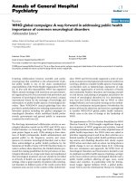
Axial contrast enhanced CT image of the chest (a) shows an area of decreased perfusion in the lateral wall of the left ventricle (arrowheads) with a 1 × 1.6 cm blister-like pouch (arrow)Figure 2
Axial contrast enhanced CT image of the chest (a) shows an area of decreased perfusion in the lateral wall of
the left ventricle (arrowheads) with a 1 × 1.6 cm blister-like pouch (arrow). A volume rendered 3D MDCT image (b)
of the left ventricle shows an area of localized contrast out-pouching with a narrow neck in the lateral left ventricular wall
(arrow).
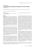
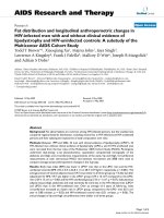
Axial (a) and short axis (b) first pass perfusion SSFP MR images demonstrate a large area of microvascular obstruction in the inferolateral and inferoseptal left ventricular wall (arrowheads) with an area of blister-like contrast pouch covered by a 1 mm thin rim of infarcted myocardial tissue (arrow) compatible with an impending left ventricular ruptureFigure 3
Axial (a) and short axis (b) first pass perfusion SSFP MR images demonstrate a large area of microvascular
obstruction in the inferolateral and inferoseptal left ventricular wall (arrowheads) with an area of blister-like
contrast pouch covered by a 1 mm thin rim of infarcted myocardial tissue (arrow) compatible with an impend-
ing left ventricular rupture. The magnified view (c, the area is indicated by the square in Fig. 3a) of an axial T1 weighted
double inversion FSE MR image confirms the thin myocardial cover (arrow) of this subepicardial aneurysm (arrow), which has
bright signal due to slower flow compared to the left ventricular blood pool. The overlying epicardial fat (arrowhead) and peri-
cardium are normal. Figure 3d represents a drawing of the complex anatomy in figure 3c.
Journal of Cardiothoracic Surgery 2009, 4:42 />Page 4 of 5
(page number not for citation purposes)
the difficult diagnosis, SAEs have a high mortality rate and
diagnosis is often made post-mortem.
SEAs are rare; in 1,814 autopsied hearts with 1,140 MIs
(in 704 hearts), only three SEAs were found (0.2% of inf-
arcts) [4]. Review of literature reveled 36 published cases
to date. As in our case, SEAs typically occur post AMI, usu-
ally within the first few weeks. Additionally, there are
reports of SEAs (1) in an avascular region without history
of AMI or signs of coronary artery disease [14], (2) as a
direct result of apicoaortic bypass [15], and (3) after repair
of a ventricular septal rupture [16].
Conclusion
In a patient with continued chest pain post-AMI, suben-
docardial left ventricular aneurysm/impending rupture
should be considered as an uncommon yet life-threaten-
ing differential diagnosis. In this case, the SEA was visible
on the pulmonary embolism CT scan as an incidental
finding and confirmed on a dedicated cardiac MRI. Emer-
gency surgery guided by these imaging findings most
likely saved the patient's life.
Consent
Written informed consent was obtained from the patient
for publication of this case report and any accompanying
images. A copy of the written consent is available for
review by the Editor-in-Chief of this journal.
Competing interests
The authors declare that they have no competing interests.
J.V.C. was supported by the Radiological Society of North
America Research and Education Foundation.
Authors' contributions
JVC conducted the CT and MRI exams and drafted the
manuscript. JS drafted the manuscript and conducted the
literature search. ASS was directly involved in the patient
care. EKF, JACL, ASS and DAB substantially revised and
edited this manuscript.
Additional material
References
1. Vlodaver Z, Coe JI, Edwards JE: True and false left ventricular
aneurysms. Propensity for the latter to rupture. Circulation
1975, 51:567-572.
2. Roberts WC, Morrow AG: Pseudoaneurysm of the left ventri-
cle. An unusual sequel of myocardial infarction and rupture
of the heart. Am J Med 1967, 43:639-644.
3. Hunter WBR: Rare form of saccular cardiac aneurysm with
spontaneous rupture. Am J Pathol 1933, IX:593-602.
4. Epstein JI, Hutchins GM: Subepicardial aneurysms: a rare com-
plication of myocardial infarction. Am J Med 1983, 75:639-644.
5. Chatterjee K: Complications of acute myocardial infarction.
Curr Probl Cardiol 1993, 18:1-79.
6. Pollak H, Nobis H, Mlczoch J: Frequency of left ventricular free
wall rupture complicating acute myocardial infarction since
the advent of thrombolysis. Am J Cardiol 1994, 74:184-186.
Additional file 1
First pass resting perfusion short axis MRI. Extensive microvascular
obstruction in the inferoseptal and inferolateral left ventricular wall.
Click here for file
[ />8090-4-42-S1.avi]
Additional file 2
First pass resting perfusion long axis MRI. Extensive microvascular
obstruction in the lateral left ventricular wall.
Click here for file
[ />8090-4-42-S2.avi]
Additional file 3
Cine short axis MRI. Akinesia at the inferior wall and moderate hypoki-
nesia at the inferior septum and lateral wall with the focal impending left
ventricular rupture in the lateral wall.
Click here for file
[ />8090-4-42-S3.mpg]
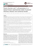
Short axis delayed enhancement inversion recovery MR image with phase correction after 55 minutes (SSFP-GRE) post intravenous gadolinium injection shows the large infero-lateral and inferoseptal acute myocardial infarction (arrow-heads) with a persistent large area of microvascular obstruction (*)Figure 4
Short axis delayed enhancement inversion recovery
MR image with phase correction after 55 minutes
(SSFP-GRE) post intravenous gadolinium injection
shows the large inferolateral and inferoseptal acute
myocardial infarction (arrowheads) with a persistent
large area of microvascular obstruction (*). The
impending rupture site in the lateral left ventricular wall
shows delayed enhancement of the thin overlying cover of
infracted myocardium (arrow).
Publish with BioMed Central and every
scientist can read your work free of charge
"BioMed Central will be the most significant development for
disseminating the results of biomedical research in our lifetime."
Sir Paul Nurse, Cancer Research UK
Your research papers will be:
available free of charge to the entire biomedical community
peer reviewed and published immediately upon acceptance
cited in PubMed and archived on PubMed Central
yours — you keep the copyright
Submit your manuscript here:
/>BioMedcentral
Journal of Cardiothoracic Surgery 2009, 4:42 />Page 5 of 5
(page number not for citation purposes)
7. Fox AC, Glassman E, Isom OW: Surgically remediable complica-
tions of myocardial infarction. Prog Cardiovasc Dis 1979,
21:461-484.
8. Natarajan MK, Salerno TA, Burke B, Chiu B, Armstrong PW:
Chronic false aneurysms of the left ventricle: management
revisited. Can J Cardiol 1994, 10:927-931.
9. Yeo TC, Malouf JF, Reeder GS, Oh JK: Clinical characteristics and
outcome in postinfarction pseudoaneurysm. Am J Cardiol 1999,
84:592-595.
10. Yang HS: Subepicardial Aneurysm Evaluated by Multiplane
2D and Real-Time 3D Volumetric Transesophageal Echocar-
diography. Circulation: Cardiovascular Imaging 2008, 1:171-172.
11. Giltner A, Marelli D, Halpern E, Savage M: Subepicardial aneu-
rysm with impending cardiac rupture: a case of antemortem
diagnosis and review of the literature. Clin Cardiol 2007,
30:44-47.
12. Koito H, Nakamura C, Suzuki J, Kamihata H, Takayama Y, Iwasaka T,
Imamura H: Pseudoaneurysm of the left ventricle progressing
from a subepicardial aneurysm. Jpn Circ J 1999, 63:559-563.
13. Tanabe K, Sugamori T, Yoshitomi H, Asanuma T, Shimada T: Fatal
cardiac rupture: a case of subepicardial aneurysm after myo-
cardial infarction. J Am Soc Echocardiogr 2000, 13:951-952.
14. Tosun R, Kaplan M, Sanioglu S, Olsun A, Oztek I, Duzyol C, Gur AK,
Bilgen F: Subepicardial left ventricular aneurysm with an
intraventricular narrow neck on the avascular apical area:
Report of a case. Surg Today 2008, 38:951-954.
15. Doi A, Takahara Y, Mogi K, Hatakeyama M: Subepicardial aneu-
rysm after apicoaortic bypass. J Thorac Cardiovasc Surg 2007,
134:237-238.
16. Minami H, Mukohara N, Hidefumi O, Yoshida M, Matsuhisa H, Shida
T: Subepicardial aneurysm following ventriculotomy closure
of ventricular septal rupture due to acute myocardial infarc-
tion. Jpn J Thorac Cardiovasc Surg 2004,
52:45-47.