Báo cáo y học: "An occasional diagnosis of myasthenia gravis - a focus on thymus during cardiac surgery: a case repor" ppt
Bạn đang xem bản rút gọn của tài liệu. Xem và tải ngay bản đầy đủ của tài liệu tại đây (190.95 KB, 3 trang )
BioMed Central
Page 1 of 3
(page number not for citation purposes)
Journal of Cardiothoracic Surgery
Open Access
Case report
An occasional diagnosis of myasthenia gravis - a focus on thymus
during cardiac surgery: a case report
Marco Agrifoglio
1
, Fabio Barili*
1,2,3
, Luca Dainese
1
, Antioco Cappai
1
,
Faisal H Cheema
3
and Paolo Biglioli
1
Address:
1
Department of Cardiovascular Surgery, University of Milan, Via Parea 4, 20138 Milan, Italy,
2
Department of Cardiovascular Surgery, S
Croce Hospital, Via M, Coppino 26, 12100 Cuneo, Italy and
3
Division of Cardiothoracic Surgery, College of Physicians and Surgeon of Columbia
University - New York Presbyterian Hospital, New York, USA
Email: Marco Agrifoglio - ; Fabio Barili* - ; Luca Dainese - ;
Antioco Cappai - ; Faisal H Cheema - ; Paolo Biglioli -
* Corresponding author
Abstract
Background: Myasthenia gravis, an uncommon autoimmune syndrome, is commonly associated
with thymus abnormalities. Thymomatous myasthenia gravis is considered to have worst prognosis
and thymectomy can reverse symptoms if precociously performed.
Case report: We describe a case of a patient who underwent mitral valve repair and was found
to have an occasional thymomatous mass during the surgery. A total thymectomy was performed
concomitantly to the mitral valve repair.
Conclusion: The diagnosis of thymomatous myasthenia gravis was confirmed postoperatively.
Following the surgery this patient was strictly monitored and at 1-year follow-up a complete stable
remission had been successfully achieved.
Background
Myasthenia gravis (MG), an uncommon autoimmune
syndrome caused by the failure of neuromuscular trans-
mission, results from binding of autoantibodies to those
proteins that are involved in signaling at the neuromuscu-
lar junction [1].
The role of thymus in the pathogenesis of myasthenia
gravis is not entirely clear, but most patients with
myasthenia gravis are found to have some degree of thy-
mus abnormality. The thymus is hypothesized to be the
site of autoantibody formation and therefore thymectomy
has been proposed as a first line therapy. This is especially
true if a thymoma is present, as thymectomy has been
reported to significantly improve the clinical condition
[2].
Case presentation
We report a case of a 37-year-old white female who pre-
sented with an echocardiographic diagnosis of severe
mitral valve regurgitation and had a history of fatigue,
weakness and dyspnea on exertion for last three months.
Her symptoms were not further investigated considering
the severe mitral valvular disease. She had no other co-
morbidities and the preoperative EuroSCORE was 3. She
was scheduled for an elective mitral valve repair surgery.
The patient underwent routine median sternotomy. At
direct inspection of the retro-sternal space, the superior
Published: 7 October 2009
Journal of Cardiothoracic Surgery 2009, 4:55 doi:10.1186/1749-8090-4-55
Received: 9 July 2009
Accepted: 7 October 2009
This article is available from: />© 2009 Agrifoglio et al; licensee BioMed Central Ltd.
This is an Open Access article distributed under the terms of the Creative Commons Attribution License ( />),
which permits unrestricted use, distribution, and reproduction in any medium, provided the original work is properly cited.
Journal of Cardiothoracic Surgery 2009, 4:55 />Page 2 of 3
(page number not for citation purposes)
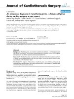
third of the anterior mediastinum was filled with a 2 × 1.5
cm mass arising from left lobe of thymus (Figure 1). The
mass was not invasive and easily resectable. No intraoper-
ative frozen sections were examined. However, A total
thymectomy was performed before opening the pericar-
dium. Thereafter, the classic mitral valve repair was per-
formed without any intraoperative or perioperative
complication.
In the postoperative stay, high titers of anti-acetylcholine
receptor antibodies and anti-striated muscle antibodies
were found (9.1 nmol/L and titer >1:80, respectively) and
myasthenia gravis was diagnosed. MG diagnosis was fur-
ther confirmed by the positive tensilon test. The severity of
MG was retrospectively evaluated in the postoperative
period according to the clinical classification of the Medi-
cal Scientific Advisory Board of the Myasthenia Gravis
Foundation of America (MGFA) [3]. The patient was clas-
sified to be in MGFA class IIa, as mild weakness involved
not only ocular muscles but also axial muscles while
oropharyngeal and respiratory muscles were not con-
cerned. The histopathologic examination of the specimen
revealed a completely excised thymoma. It was classified
as type A accordingly to new World Health Organization
classification of Rosai and Sabin (WHO type A or medul-
lary). Using the staging process described by Masaoka,
this specimen was staged to be as Masaoka Stage I since
macroscopically it was a completely encapsulated thy-
moma with no microscopically determined capsular inva-
sion [4].
The postoperative course was uneventful and the patient
was discharged on postoperative day 6 to home-rehabili-
tation in tele-cardiology without any complications. The
patient was strictly monitored by a multidisciplinary team
composed of a cardiologist, a surgeon and a neurologist
for regular follow-ups. No MG therapy was initiated con-
sidering the recent operation and the good clinical status.
At six-month follow-up, no mitral regurgitation was
detected by transthoracic echocardiography and the
symptoms related to MG were completely remitted. The
anti-acetylcholine receptor antibodies titer decreased to
4.2 nmol/L. At 1-year follow-up a complete stable remis-
sion (CSR) was assessed according to the MGFA Post-
Intervention Status Classification and the acetylcholine
receptor antibodies titer had further decreased to 1.4
nmol/L [3].
Conclusion
Thymomatous MG (T-MG) is considered to have worst
prognosis compared with non-thymomatous MG. The
patho-physiological bases are not clear but clinical data
suggest that patients with T-MG have high-grade symp-
toms with low rate of remission even after therapy [5].
In patients who undergo cardiac surgery, the evaluation of
the thymus is often considered secondary and tumoral
disease of the thymus is only considered when a mass is
found intraoperatively or at a CT scan. Few reports have
focused on the incidental finding of a thymic mass during
cardiac surgery and the management is generally guided
by the type and extension [6,7]. Total thymectomy is
advised if an encapsulated thymic mass or a resectable
invasive thymoma are found. However, histological
examination on frozen section should be performed first
if the malignant mass is unresectable or metastases are evi-
dent [8].
Moreover, the clinical evaluation usually represents the
first diagnostic step for a patient with suspected MG. The
evaluation of MG-related symptoms could be difficult as
they may get masked by the cardiac disease. In this report,
the preoperative clinical status was not correctly addressed
as the cardiac symptoms were predominant. Hence, the
diagnosis of thymomatous MG was guided by the intraop-
erative findings which led us to revaluate the preoperative
clinical conditions.
Although uncommon, MG represents an invalidating dis-
ease which has to be diagnosed as soon as possible in
order to initiate the appropriate therapy thereby increas-
ing the remission rate [1,3,5]. The clinical evaluation
should be more accurate in patients with cardiac disease
as initial MG symptoms could be masked resulting in an
underestimated or incorrect diagnosis. Moreover, the
meticulous evaluation of the thymus gland itself during
cardiac surgery can be an effective step towards finding
even small macroscopic abnormalities of thymus that
could be prophylactically excised. Therefore, a focus on
thymus during cardiac surgery may not only lead to an
The intraoperative finding of the small thymomatous mass which led to the MG diagnosisFigure 1
The intraoperative finding of the small thymomatous
mass which led to the MG diagnosis.
Publish with BioMed Central and every
scientist can read your work free of charge
"BioMed Central will be the most significant development for
disseminating the results of biomedical research in our lifetime."
Sir Paul Nurse, Cancer Research UK
Your research papers will be:
available free of charge to the entire biomedical community
peer reviewed and published immediately upon acceptance
cited in PubMed and archived on PubMed Central
yours — you keep the copyright
Submit your manuscript here:
/>BioMedcentral
Journal of Cardiothoracic Surgery 2009, 4:55 />Page 3 of 3
(page number not for citation purposes)
occasional intraoperative diagnosis of thymus abnormal-
ity but also results in re-evaluation of the clinical status
postoperatively to confirm the suspected concomitant T-
MG.
Consent
The written consent for publication was obtained. A copy
of the written consent is available for review by the Editor-
in-Chief of this journal.
Competing interests
The authors declare that they have no competing interests.
Authors' contributions
MA conceived the study idea, wrote the first draft and led
the project from beginning to end. FB assisted the study in
data collection, draft revision and coordinating with all
co-authors. LD helped with literature review and manu-
script writing. AC helped the study with discussions about
the topic and assistance in manuscript writing. FHC
edited the manuscript and helped with revisions and final
submission. PB provided expert opinion throughout the
study and also operated on this case. All authors read and
approved the final manuscript.
References
1. Hampton T: Novel therapies target myasthenia gravis. JAMA
2007, 298(2):163-4.
2. Jaretzki A, Steinglass KM, Sonett JR: Thymectomy in the manage-
ment of myasthenia gravis. Semin Neurol 2004, 24(1):49-62.
3. Jaretzki A 3rd, Barohn RJ, Ernstoff RM, Kaminski HJ, Keesey JC, Penn
AS, Sanders DB: Myasthenia gravis: recommendations for clin-
ical research standards. Task Force of the Medical Scientific
Advisory Board of the Myasthenia Gravis Foundation of
America. Neurology 2000, 55(1):16-23.
4. Masaoka A, Monden Y, Nakahara K, Tanioka T: Follow-up study of
thymomas with special reference to their clinical stages. Can-
cer 1981, 48(11):2485-92.
5. Kim HK, Park MS, Choi YS, Kim K, Shim YM, Han J, Kim BJ, Kim J:
Neurologic outcomes of thymectomy in myasthenia gravis:
comparative analysis of the effect of thymoma. J Thorac Cardi-
ovasc Surg 2007, 134(3):601-7.
6. Mirsadraee S, Shah SS, Kumar B, Kaul P: Incidental locally infiltrat-
ing malignant thymoma and coronary artery bypass surgery-
excision should always be considered. J Card Surg 2005,
20(3):291-2.
7. Abdullah F, Loon LG: An incidental finding of thymic carcinoid
during urgent CABG operation. Heart Surg Forum 2002,
5(4):E35-6.
8. Nakahara K, Ohno K, Hashimoto J, Maeda H, Miyoshi S, Sakurai M,
Monden Y, Kawashima Y: Thymoma: results with complete
resection and adjuvant postoperative irradiation in 141 con-
secutive patients. J Thorac Cardiovasc Surg 1988, 95(6):1041-7.