Báo cáo y học: "Left ventricular diastolic dysfunction of the cardiac surgery patient; a point of view for the cardiac surgeon and cardio-anesthesiologist" pptx
Bạn đang xem bản rút gọn của tài liệu. Xem và tải ngay bản đầy đủ của tài liệu tại đây (346.6 KB, 10 trang )
BioMed Central
Page 1 of 10
(page number not for citation purposes)
Journal of Cardiothoracic Surgery
Open Access
Review
Left ventricular diastolic dysfunction of the cardiac surgery patient;
a point of view for the cardiac surgeon and cardio-anesthesiologist
Efstratios E Apostolakis
1
, Nikolaos G Baikoussis*
1,2
, Haralabos Parissis
3
,
Stavros N Siminelakis
2
and Georgios S Papadopoulos
4
Address:
1
Cardiothoracic Surgery Department, University of Patras, School of Medicine, Patras, Greece,
2
Cardiac Surgery Department, University
of Ioannina, School of Medicine, Ioannina, Greece,
3
Basildon & Thurrock University Hospital NHS FT, Basildon, Essex, UK and
4
Department of
Clinical Anesthesiology and Intensive Postoperative Care Unit, University of Ioannina, School of Medicine, Ioannina, Greece
Email: Efstratios E Apostolakis - ; Nikolaos G Baikoussis* - ;
Haralabos Parissis - ; Stavros N Siminelakis - ; Georgios S Papadopoulos -
* Corresponding author
Abstract
Background: Left ventricular diastolic dysfunction (DD) is defined as the inability of the ventricle
to fill to a normal end-diastolic volume, both during exercise as well as at rest, while left atrial
pressure does not exceed 12 mm Hg. We examined the concept of left ventricular diastolic
dysfunction in a cardiac surgery setting.
Materials and methods: Literature review was carried out in order to identify the overall
experience of an important and highly underestimated issue: the unexpected adverse outcome due
to ventricular stiffness, following cardiac surgery.
Results: Although diverse group of patients for cardiac surgery could potentially affected from
diastolic dysfunction, there are only few studies looking in to the impact of DD on the
postoperative outcome; Trans-thoracic echo-cardiography (TTE) is the main stay for the diagnosis
of DD. Intraoperative trans-oesophageal (TOE) adds to the management. Subgroups of DD can be
defined with prognostic significance.
Conclusion: DD with elevated left ventricular end-diastolic pressure can predispose to increased
perioperative mortality and morbidity. Furthermore, DD is often associated with systolic
dysfunction, left ventricular hypertrophy or indeed pulmonary hypertension. When the diagnosis
of DD is made, peri-operative attention to this group of patients becomes mandatory.
Introduction
Left ventricular diastolic dysfunction (DD) is defined as
the inability of the ventricle to fill to a normal end-diasto-
lic volume, both during exercise as well as at rest, while
left atrial pressure does not exceed 12 mm Hg [1-3]. It has
been shown that several patients with DD are suffering
from paroxysmal dyspnoea and "unexplained" pulmo-
nary oedema with a normal ejection fraction [4,5]).
Among patients operated for coronary artery disease or
aortic stenosis, the incidence of left ventricular DD ranges
widely between 44%, and 75% [6-10]. The significance
and the severity of ventricular diastolic dysfunction
among these patients are not well elucidated. On the
other hand, estimation of the degree of DD peri-opera-
Published: 24 November 2009
Journal of Cardiothoracic Surgery 2009, 4:67 doi:10.1186/1749-8090-4-67
Received: 10 July 2009
Accepted: 24 November 2009
This article is available from: />© 2009 Apostolakis et al; licensee BioMed Central Ltd.
This is an Open Access article distributed under the terms of the Creative Commons Attribution License ( />),
which permits unrestricted use, distribution, and reproduction in any medium, provided the original work is properly cited.
Journal of Cardiothoracic Surgery 2009, 4:67 />Page 2 of 10
(page number not for citation purposes)
tively, is difficult in up to 20% of cardiac-surgery patients
for several reasons [10,11] including rhythm abnormality,
preload and afterload alterations, coexistence of valvular
disease, age related changes, and inability to obtain
proper Doppler images [12-15]. The diastolic heart failure
annual mortality varies between 9-28% (four-fold that of
disease-free subjects [16], while it has also been linked to
increased incidence of postoperative complications (mor-
tality or morbidity) after cardiac surgery [13,17,18].
Revascularization of ischemic myocardium seems to be
beneficial for DD (if not immediately), some weeks after
revascularization [19]. Potential direct postoperative
improvement in diastolic function may be offset by the
detrimental effect of global ischemia during cardioplegic
arrest in combination with myocardial interstitial oedema
[11,20]. There are only a few studies concerning surgical
outcomes of patients suffering from diastolic dysfunction.
Moreover, intra-operative diagnosis and strategies to
manage patients with left ventricular diastolic dysfunction
are not well clarified. In that sense, diastolic dysfunction
could be considered perioperatively as a "Trojan horse".
Source of Research
Pertinent medical literature in the English language was
identified through a Medline computerized literature
search and a manual search of selected articles using the
key words "left ventricular diastolic dysfunction", "left
ventricular diastolic impairment", "transmitral flow Dop-
pler", "pulmonary venous flow patterns". The search
terms were combined using the Boolean operator term
"or" to find all abstracts pertaining to the chosen search
terms. These individual terms were then combined using
the Boolean operator term "and" to find articles that con-
tained information of all search terms. The reference lists
of articles found through these searches were also
reviewed for relevant articles. Links provided on the web
sites of published articles were searched for relevant arti-
cles.
Pathophysiology
DD is present when an elevated filling pressure is neces-
sary to achieve normal ventricular filling. So, DD is related
to abnormal left ventricular relaxation and filling during
diastolic phase of cardiac cycle [21-24]. During this phase
there are four timely and sequential events: a) isovolemic
relaxation, b) rapid (early) LV filling, c) slow LV filling
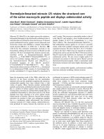
(diastasis) and d) atrial contraction [2,23]. In figure 1 is
shown schematically the pathophysiology of DD. Accord-
ing to echocardiographic depiction, filling of normally
relaxed LV is completed in two phases: the first phase is
due to the passive filling of the LV, is massive and depicted
early in diastole by a high E wave. The second phase is due
to the left atrial contraction, takes place during late diasto-
lic phase, and leads to late LV filling depicted by the wave
A of transmitral inflow Doppler [22,25]. The rate of
decrease of E wave in early diastole depends on the rate of
increase in LV pressure and is represented by the so-called
deceleration time (DT). This time is influenced by a
number of factors such as, a) left atrial-left ventricular
pressure gradient at the time of mitral valve opening, b)
left atrial chamber compliance, c) left ventricular chamber
compliance, d) grade of left ventricle relaxation, e) visco-
elastic forces of the myocardial wall, f) pericardial
restraint and finally g) left-right ventricular interaction.
Left ventricular relaxation-similar to contraction- is an
energy-dependent process, because it requires the re-
uptake of calcium into the sarcoplasmic reticulum [26].
When patients with left ventricular DI are subjected to
stress-as occurs during surgery or during faster heart rates-
due to shorter diastolic filling time available, the ventricle
is not allowed to relax and fill properly; thus, causing
increased left ventricular end-diastolic pressure and pul-
monary congestion [1,2,16]. Furthermore, relaxation of
the left ventricle is determined by visco-elasticity and
restoring forces (recoil). It is believed that impaired
diastolic filling of the left ventricle is the first manifesta-
tion of active ischemia and results in an upward shift of
left ventricular diastolic pressure-volume relationship
[2,26]. Decreased activity of sarcoplasmic reticulum cal-
cium ATPase pump (SERCA) can slow down calcium
removal out of the cytosolic net [27]. In contrast,
increased levels or activity of phospholamban-the natural
SERCA-inhibitory protein-can also impair relaxation.
Hypothyroidism decreases SERCA and increases phos-
pholamban, leading to impaired relaxation, while the
opposite effect occurs in hyperthyroidism [27]. In a simi-
lar way, increasing the action of SERCA by administration
of captopril, and β-agonists (or decreasing the action of
phospholamban), results in improvement of diastolic
relaxation [28].
Pathophysiology and diagnosis of DD
Another aspect of DD is the relationship between systolic
and diastolic left ventricular dysfunction [2,29,30]
Increased left ventricular end systolic volume for example,
affects the rate of left ventricular relaxation, and as a
result, patients with reduced LV ejection fractions are
expected to have a prolonged relaxation time [2]. Loading
conditions, such as inotropic stimulation and neurohu-
moral factors generally affect both systolic and diastolic
function in a parallel way [2]. As it has been shown, ele-
vated left ventricular end-diastolic pressure may or may
not be associated with systolic dysfunction of left ventri-
cle, suggesting left ventricular DD even in the absence of
reduced left ventricular ejection fraction [29]. Indeed,
patients with symptoms of heart failure and normal ejec-
tion fraction have significant abnormalities in active relax-
ation and passive stiffness, which cause increased left
ventricular end-diastolic pressure [30]. Literature review:
"The theory" of DD is presented in table 1.
Journal of Cardiothoracic Surgery 2009, 4:67 />Page 3 of 10
(page number not for citation purposes)
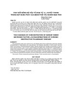
Pathophysiology of DD and its consequencesFigure 1
Pathophysiology of DD and its consequences.
Ischemia, LV strain, Age, Arrhythmia, Systolic LV dysfunction, CPB (Cardio-
Pulmonary Bypass) or ↓ activity of SERCA
Impaired diastolic filling
Increased end diastolic pressure of LV
Upwards and to the left shift of LV pressure/volume relationship
Symptoms of Heart failure with elevated end-diastolic pressure: 24±8
mmHg, normal EF, normal chamber size & LVH
Prolonged relaxation pattern E/A=1
time constant of LV relaxation is longer > 50-55 msec
Pseudonormal pattern
LV passive –stiffness constant is high> 0.025
Restrictive pattern
E/A = 2 and DT=150ms
E-wave deceleration 350±140ms
Stimuli
Diagnosis
non-invasive (Doppler, MR-myocardial tagging,radionuclide ventriculography) and invasive (micromanometry
,
angiography).
Journal of Cardiothoracic Surgery 2009, 4:67 />Page 4 of 10
(page number not for citation purposes)
Diagnosis of DD in a Cardiothoracic setting
Assessment and diagnosis of DD can be performed with
non-invasive (2D and Doppler-echocardiography, colour
Doppler M-mode, Doppler tissue imaging, MR-myocar-
dial tagging, radionuclide ventriculography) and invasive
techniques (micromanometry, angiography, conductance
method). Typical findings of primary DD in chest radio-
graph include absence of cardiomegaly and presence of
pulmonary congestion. Electrocardiogram/ECHO reveals
the presence of excessive concentric hypertrophy in com-
bination with normal ejection fraction [5]. Measurement
of peak early filling wave (E wave: is caused by difference
in atrium-ventricle pressure) and atrial filling wave (A
wave: is caused by atrial contraction) ratio by Doppler
echocardiography, as well as deceleration time (DT: is
caused by left ventricular compliance), are useful screen-
ing tools for abnormal left ventricular relaxation [29]. In
presence of abnormal relaxation, atrial contraction occurs
in an incompletely empty atrium and blood is propelled
into the left ventricle in increased velocity, accounting for
the heightened A wave and consequent decreased E/A
ratio. Blood flow in the pulmonary veins is biphasic, with
peaks of forward flow occurring in both systole and dias-
tole and inverse diastolic flow occurring during atrial con-
traction. There is an inverse relationship between left
atrial pressure and pulmonary venous systolic flow. That
is the reason why determination of systolic pulmonary
venous flow velocity is a rapid method to estimate LV fill-
ing pressures after CABG [30].
Pathological filling is determined from transmitral flow
pattern
1) Prolonged relaxation pattern: characterized by pro-
longed isovolumetric relaxation time and deceleration
time, low E and high A wave velocities with an E/A
wave ratio typically 1. It is related to the remodelling
process including hypertrophy or scarring of an infarct
zone leading to a non-uniform LV relaxation.
Table 1: Articles "investigating the background" of the entity Diastolic Heart Failure (DHF).
Author Year Journal Conclusions
Kessler KM et al [35] 1989 Hosp Pract Introduction of the term DHF
Paulus WJ [67] 1999 Cardiovasc Res. Development of specific diagnostic criteria for DHF
Bruch C et al [68] 2000 Eur Heart J Tei-index: relation to LVEDP, sensitive indicator of overall cardiac dysfunction
Mandinov L et al [69] 2000 Cardiovasc Res Doppler Echo definitions
Vasan and Levy [70] 2000 Circulation Development of criteria for definite, probable and possible DHF
Crossman W [16] 2000 Circulation Thoughtful Editorial
Zile MR et al [71] 2001 Circulation Tested the hypothesis that measurements of the LV relaxation and passive stiffness were
not necessary to make the diagnosis of DHF
Poulsen SH et al [72] 2001 Dan Med Bull DHF following acute MI
Catuzzo B et al [73] 2003 J Card Fail Regarding patients with CHF: BNP plasma levels is related to diastolic restrictive pattern
Hogg K et al [21] 2004 J Am Coll Cardiol Epidemiology of the syndrome of heart failure with preserved LV systolic function: clinical
characteristics
Zile MR et al [30] 2004 N Engl J Med Invasive assessment of DHF. Identification of significant abnormalities in active relaxation
and passive stiffness
Yturralde RF et al [74] 2005 Prog Cardiovasc Dis Review and current recommendations
Zile MR et al [75]) 2005 Prog Cardiovasc Dis Overview of systolic and DHF
Shammas RL et al [76] 2007 Int J Cardiol DHF: "what we don t know"
Scardovi AB et al [77] 2007 Eur J Echocardiogr BNP and advanced DHF
Literature review: "The Theory" of DD
Journal of Cardiothoracic Surgery 2009, 4:67 />Page 5 of 10
(page number not for citation purposes)
2) Pseudonormal pattern: an intermediate stage
between prolonged relaxation and restrictive filling as
a consequence of disease progression. There is an asso-
ciation with atrial dilatation and prominent pulmo-
nary venous wave reversal.
3) Restrictive pattern: associated with shortened iso-
volumetric relaxation time, increased peak E wave
velocity with very short deceleration time and small A
wave, leading to an E/A wave ratio of 2. This pattern
might be due to increasing LV volume and also to
increased myocardial stiffness. DD is severe when the
transmitral filling pattern E/A ratio is 2 and the decel-
eration time is 150 ms.
For patients undergoing cardiac surgery, Doppler assess-
ment of transmitral flow has been used to estimate post-
operative left ventricular filling pressure, relaxation, and
stiffness [31]. The most important problem in evaluating
transmitral flow patterns is their great variation, depend-
ing on many factors such as: heart rate [32,33], preload
[34], afterload [34], positive-pressure mechanical ventila-
tion [21], systolic ventricular function [35,36], use of ino-
tropic or generally vasoactive agents due to their effect on
the afterload [34,37], and hemodilution (higher velocities
due to reduced blood viscosity) [34]. To surmount this, a
new method for diagnosis of LV diastolic dysfunction, the
so called flow propagation velocity (Vp) is applied. It
bears the advantage of being insensitive to heart rate and
preload changes [10]. According to Vp measurement, left
ventricular filling patterns does not change significantly
after cardiopulmonary bypass. Furthermore, newer tech-
niques such as tissue Doppler imaging (TDI) which meas-
ures high intensity, low velocity echo of the myocardium
has been developed. By using TDI, local myocardial relax-
ation can be calculated by obtaining the velocity of early
diastolic wall motion (Em) and it's timing [38]. In other
words, TDI allows assessment of diastolic function
because of its unique ability to assess regional abnormal-
ities in relaxation, in addition to their global effect on ven-
tricular relaxation and filling dynamics. An E/Em ratio >
10 remains the best discriminatory value when it is used
as a single parameter for the prediction of elevated filling
pressures or simply diastolic dysfunction [39]. However,
definite diagnosis of diastolic dysfunction is established
by cardiac catheterization and direct measurement of
pressure at the end of systole and volume loops [40]. This
invasive assessment of diastolic function allows the study
of isovolumic relaxation (time constant of LV relaxation is
longer > 50-55 msec) and evaluation of the passive elastic
properties of the myocardium (LV passive-stiffness con-
stant is high).
Intraoperative diagnosis
Intraoperative diagnosis of diastolic dysfunction is diffi-
cult, [41,42] because: a) most variables measuring diasto-
lic function depend on loading conditions, heart rate and
age [32-34,43], b) no single individual measurement can
fully characterize left ventricular diastolic dysfunction,
and c) ECHO estimation may give different results
whether it is performed with the patient awake and
breathing spontaneously, or anesthetized and receiving
positive pressure ventilation [35]. Diastolic dysfunction
of left ventricle can be intraoperatively diagnosed, esti-
mated and graded by using Trans Oesophageal Echo
(TOE). Moreover, valuable information may be obtained
with the additional use of a Swan-Ganz catheter
[33,34,39]. According to Ranucci [44], first degree of
diastolic dysfunction of the left ventricle is depicted as
impaired relaxation, is usually observed just after discon-
tinuation of cardiopulmonary bypass, and is often revers-
ible (temporary). Second degree mimicking pseudo-
normalization, is a more severe condition, which some-
times is an intermediate step towards, third degree of dys-
function which is characterized by a restrictive pattern. An
increased ratio (> 2) between E and A waves of transmitral
flow, and a blunted systolic waveform of the pulmonary
vein flow is present due to left atrial pressure [34,36,39].
It has been demonstrated that mitral and pulmonary vein
flow indexes correlate with pulmonary capillary wedge
pressure (PCWP) [44,45]. Therefore, additional measure-
ment of PCWP by using a Swan-Ganz catheter may be in
this phase useful in estimating the time course of diastolic
dysfunction and the effect of therapeutic manipulations
[44]. Fluid responsiveness is better defined by TOE
derived variables (left ventricular end-diastolic area, peak
blood velocity variation), but some information can be
derived by the Swan-Ganz catheter as well (PCWP and
peak pulmonary pressure variation) [45,46]. In table 2 we
present the high risk groups for developing DD, while in
table 3, we report articles looking into: the impact of
diastolic dysfunction (DD) on patient's outcome follow-
ing Cardiac Surgery.
Progression of DD following Cardiac surgery
Following coronary artery bypass grafting, DD is tempo-
rarily deteriorated (expressed as a decrease in E-max and
an increase in A-max of transmitral flow) [47]. This dete-
rioration of DD seems to persist, at least for the first three
postoperative hours after coronary artery bypass grafting
[48,49]. In a similar way, Yamamoto et al by using classi-
cal ECHO after coronary artery bypass grafting, showed
that DD was characterized by a decrease in E wave veloc-
ity, prolongation of the E wave DT, and a decrease of E/A
ratio [43]. Potential implicated mechanisms are those of
free oxygen radicals, altered intracellular calcium homeos-
tasis, or both [50,51]. Temporary improvement has been
shown, especially if calcium channels blocking factors like
Journal of Cardiothoracic Surgery 2009, 4:67 />Page 6 of 10
(page number not for citation purposes)
diltiazem were perioperatively administered or added in
the cardioplegic solution [43,50,52,53]. For patients who
underwent off-pump coronary artery bypass grafting
(OPCAB), comparative studies on the postoperative
changes in left ventricular diastolic function, have shown
that, while left ventricular diastolic dysfunction impair-
ment was observed in both groups (conventional CABG
and OPCAB), it was more significantly impaired in the
CABG group [54]. Other studies showed that right ven-
tricular diastolic dysfunction was in a similar way signifi-
cantly impaired after CABG and OPCAB [43,55,56], and
this deterioration persisted in up to one year postopera-
tively [15]. In contrast to this, Shi et al who evaluated
short- and long-term evolution of biventricular diastolic
performance postoperatively in 49 pts who underwent
coronary artery bypass grafting showed that postoperative
deterioration of diastolic dysfunction had an absolute
return to preoperative status at six months postoperatively
[9].
Table 2: High risk groups for developing DD
Systolic dysfunction Only 50% to 60% of patients with clinical findings of congestive heart failure have an abnormal systolic function, which is
indicated by reduced ejection fraction. The remaining 40%-50% of pts, have congestive heart failure with normal systolic
function and represent the patients with diastolic dysfunction [22,23]. For clarification, Sanderson proposed the term "heart
failure with normal ejection fraction" (HFNEF) for left ventricular diastolic dysfunction, and heart failure with reduced
ejection fraction (HFREF) for systolic dysfunction of left ventricle [78]. According to this classification, the main difference
between HFNEF and HFREF is the degree of ventricular remodeling accompanied by increased ventricular volume in HFREF
[78]. In other words, distinction between systolic and diastolic dysfunction is very important because the latter has a lower
mortality (5%-8% annually), and requires different medical management (no inotropes) [22,23].
LVH In patients with AS, preoperative DD is attributable to hypertension, myocardial hypertrophy- fibrosis, and/or to ischemia
[64].
CAD Patients with CAD are prone for the development of postoperative myocardial diastolic dysfunction [39]. Left ventricular
filling abnormalities have been detected in as many as 90% of patients [39]. Possible related factors that were considered
were ischemia, hypertrophy, and hypertension [79].
DM All insulin dependent diabetes mellitus patients with left diastolic dysfunction had evidence of definite autonomic neuropathy
[80]. Moreover, diabetic patients with autonomic neuropathy form a subgroup of particularly high mortality and
cardiovascular event risk [81,82].
Age Aging is correlated to DD through an increase upon wall thickness (secondary to enlargement of cardiac myocytes), and
changes in the vasculature, the diameter, and vascular stiffness of the aorta and large arteries [83]. Up to 60% of geriatric
patients with normal EF, following non-cardiac surgery, had been postoperatively diagnosed with diastolic dysfunction [35].
High risk groups for DD
Table 3: Articles looking into: The impact of diastolic dysfunction (DD) on patient's outcome following Cardiac Surgery.
Authors Year Journal Conclusions
Casthely et al [84] 1997 J Thorac Cardiovasc Surg The effects of myocardial protection on diastolic function after cardiac operations
Bernard F et al [13] 2001 Anesth Analg The significance of diastolic dysfunction perioperatively; Diastolic dysfunction is
associated with difficult weaning from CPB.
Vaskelyte J [18] 2001 Eur J Echocardiogr The interesting concept to subdivide patients with severe LV dysfunction into different
groups according to diastolic filling pattern abnormality. One of the few articles
investigating the relationship between diastolic dysfunction and post-operative mortality.
Drawbacks: All patients had low EF < 35%.
Liu J et al [17] 2003 Am J Cardiol The prognostic value of transmitral flow patterns on patients following CABG; Probably
one of the most important papers on the subject. The study claims that pseudonormal
and restrictive TMF patterns, correlates with short term adverse outcome
Malouf PJ [85] 2006 J Am Soc Echocardiogr Doppler tissue imaging of mitral annular velocity: Lateral segmental velocity has
advantages over the septal segmental velocity
Literature review: the Outcome
Journal of Cardiothoracic Surgery 2009, 4:67 />Page 7 of 10
(page number not for citation purposes)
Management of DD
According to a multivariate analysis by Bernard et al [13],
left ventricular diastolic dysfunction was a better predictor
of hemodynamic instability after cardiac surgery com-
pared to systolic dysfunction. Treatment of the underlying
disease is currently the most important therapeutic
approach. In patients with tachycardia, use of b-blockers
or calcium antagonists, is beneficial so as to prolong
diastolic (filling) time [24,57]. Treatment of atrial fibrilla-
tion by cardioversion or amiodarone infusion is indicated
in patients with diastolic dysfunction [22,24,57]. In addi-
tion, digitalis may decelerate ventricular rate in cases of
permanent atrial fibrillation, and contribute to better ven-
tricular filling [58]. Denault et al [59] developed a diag-
nostic algorithm which they then applied to a group of 74
cardiac surgical patients, to determine whether moderate
to severe left ventricular diastolic dysfunction (LVDD)
and right ventricular diastolic dysfunction (RVDD) can
predict difficult discontinuation of cardiopulmonary
bypass. Patients with moderate to severe LVDD tended to
have higher PCWP compared to those with normal to
mild LVDD. The presence of moderate to severe RVDD
was also associated with lower mean pulmonary artery
pressure and lower cardiac index compared to patients
with normal to mild RVDD. Difficult separation from car-
diopulmonary bypass was present in 65.5% and 72% of
patients with moderate/severe LVDD and RVDD respec-
tively, in contrast to 40.9% and 48% of patients with nor-
mal/mild LVDD/RVDD. They concluded that moderate
and severe degree of LVDD and RVDD can be identified
with very good reproducibility, and both degrees of
diastolic dysfunction are associated to difficult discontin-
uation from cardiopulmonary bypass [59]. During this
effort, transesophageal echo is a needful tool to estimate
the degree of diastolic dysfunction, as well as preload and
afterload. Appropriate increase of volume load is a mile-
stone of timing in order to discontinue cardiopulmonary
bypass. Phosphodiesterase inhibitors seem to be benefi-
cial for diastolic dysfunction improvement, and should be
used in perioperatively [60]. In a similar way, Levosi-
mendan may used in perioperative management of
diastolic dysfunction [61]. It increases cardiac output and
decreases pulmonary capillary wedge pressures. This
mode of enhanced contractile force generation is achieved
without an increase in myocardial oxygen consumption,
intracellular calcium concentrations, or an adverse effect
on diastolic function [61]. For the next postoperative days
milestone of treatment remain diuretics, in doses which
prevent dyspnea and liver congestion on one side, but not
reduce the cardiac output on the other [57]. ACE inhibi-
tors in combination with spironolactone are beneficial
because they prevent excessive activation of rennin-angi-
otensin-aldosterone system, and improve ventricular
relaxation although not yet confirmed [62,63]. In contrast
to systolic dysfunction, use of calcium antagonists alone
or in combination with ACE, contributes effectively in
hypertension control and has a beneficial influence on
hypertrophic myocardium [23,24,58]. In patients with
diastolic dysfunction due to hypertrophic cardiomyopa-
thy (either idiopathic or due to acquired aortic valve sten-
osis), the main problem is to load the left ventricle with
adequate volume (preload) because it is common to
notice an echo-finding of low preload (i.e. very low left
ventricular end-diastolic area), while the measured PCWP
is found high [55]. Such patients need increased volumes,
but each fluid administration should be carefully guided
by constant measurement of PCWP, in order to avoid an
abrupt increase in pulmonary venous pressure and conse-
quent acute pulmonary oedema [55]. Postoperatively, use
of intra-aortic balloon pump in patients with left ventricu-
lar diastolic dysfunction seems to result in a favourable
influence on left ventricular function [34]. Possible expla-
nations for this effect lie on the positive effects of balloon
on coronary flow against ischemia, the favourable effect
on systolic function of left ventricle, and the increase of
left ventricular long axis [34]. For those cases whereby
"restricted pattern" is diagnosed, inotropic agents should
be considered. Maslow et al showed that the use of ino-
tropes in 44 patients, who underwent AVR for stenosis,
was associated with significantly larger increase in right
ventricular ejection fraction and cardiac output after CPB
[64]. Changes in cardiac output and index were more
strongly correlated with changes in RVEF than LVEF.
Lastly, infusion of a new B-natriuretic peptide (BNP)
nesiritide was associated with increased CO in patients
with diastolic dysfunction and low CO syndromes under-
going cardiac surgery, when other measures failed. This
agent seems to offer an additional option to inotropes and
fluid challenges perioperatively [65]. Castellá et al in an
experimental study conducted in pigs in 2006, demon-
strated that temporary LAD ischemia alters the normal
sequential pattern of contraction responsible for ejection
and suction through reduction of the systolic contractile
force, and prolongation of the endocardial contraction
into early diastole to disrupt the normal endocardial-epi-
cardial sequence responsible for ventricular suction [66].
The systolic and diastolic effects of myocardial stunning
were studied to evaluate the role of the endocardial and
epicardial segments and to determine if preconditioning
by Na+-H+ exchange (NHE) inhibition effect post-stun-
ning dysfunction. In this study conducted in Yorkshire-
Duroc pigs, NHE inhibition before ischemia limits pos-
tischemic systolic and diastolic dysfunction by re-estab-
lishing the expected shortening sequences within the
ventricular myocardial band model [66].
Conclusion
There are only few studies looking in to the impact of DD
on the outcome following cardiac surgery. Without doubt
DD with elevated left ventricular end-diastolic pressure
Journal of Cardiothoracic Surgery 2009, 4:67 />Page 8 of 10
(page number not for citation purposes)
can predispose to increased perioperative mortality and
morbidity. Furthermore, DD is often associated with
systolic dysfunction, left ventricular hypertrophy or
indeed pulmonary hypertension. The mainstay of man-
agement of DD starts with the prompt recognition and
diagnosis of this entity and relies on the aggressive man-
agement of the underlie aetiology of this insidious dis-
ease.
Competing interests
The authors declare that they have no competing interests.
Authors' contributions
All authors: 1. have made substantial contributions to
conception and design, or acquisition of data, or analysis
and interpretation of data; 2. have been involved in draft-
ing the manuscript or revisiting it critically for important
intellectual content; 3. have given final approval of the
version to be published.
References
1. Kitzman DW, Little WC, Brubaker PH, Anderson RT, Hundley WG,
Marburger CT, Brosnihan B, Morgan TM, Stewart KP.: Pathophysi-
ological characterization of isolated diastolic heart failure in
comparison to systolic heart failure. JAMA 2002, 288:2144-50.
2. Rodeheffer R, Miller W, Burnett J: Pathophysiology of circulatory
failure. In Mayo Clinic Practice of Clinical Cardiology 3rd edition. Edited
by: Giuliani E, Gersh B, Megoon M, Hayes D, Schaff H. Mosby;
1996:556-58.
3. Vasan R, Benjamin E, Levy D: Prevalence, clinical features and
prognosis of diastolic heart failure: an epidemiologic per-
spective. J Am Coll Cardiol 1995, 26:1565-74.
4. Kramer K, Kirkman P, Kitzman D, Little WC.: Flash pulmonary
edema: association with hypertension and reoccurrence
despite coronary revascularization. Am Heart J 2000,
140:451-5.
5. Givetz M, Colucci W, Braunwald E: Clinical aspects of heart fail-
ure;Pulmonary edema, High-output failure. In Braunwald's
Heart Disease 7th edition. Edited by: Zipes D, Libby P, Bonnow R,
Braunwald E. Elsevier Saunders; 2005:541-43.
6. Lappas DG, Skubas NJ, Lappas GD, Ruocco E, Tambassis E, Pasque M.:
Prevalence of left ventricular diastolic filling abnormalities in
adult cardiac surgical patients: an intraoperative echocardi-
ographic study. Sem Thorac Cardiovasc Surg 1999, 11:125-33.
7. Djaiani GN, McCreath BJ, Ti LK, Mackensen BG, Podgoreanu M, Phil-
lips-Bute B, Mathew JP.: Mitral flow propagation velocity identi-
fies patients with abnormal diastolic function during
coronary artery bypass graft surgery. Anesth Analg 2002,
95:524-30.
8. Mathison M, Edgerton JR, Horswell JL, Akin JJ, Mack MJ.: Analysis of
hemodynamic changes during beating heart surgical proce-
dures. Ann Thorac Surg 2000, 70:1355-61.
9. Shi Y, Denault AY, Couture P, Butnaru A, Carrier M, Tardif JC: Biv-
entricular diastolic filling patterns after coronary artery
bypass graft surgery. J Thorac Cardiovasc Surg 2006, 131:1080-86.
10. Malouf JF, Enriquez-Sarano M, Pellikka PA, Oh JK, Bailey KR, Chan-
drasekaran K, Mullany CJ, Tajik AJ.: Severe pulmonary hyperten-
sion in patients with severe aortic stenosis: clinical profile
and prognostic implications. JACC 2002,
40:789-95.
11. Royse CF, Royse AG, Blake DW, Grigg LE.: Instantaneous end
diastolic stiffness (IEDS): a simple, load independent meas-
urement of left ventricular diastolic function in patients
undergoing cardiacsurgery. Ann Thorac Cardiovasc Surg 2000,
6:203-10.
12. Kim Y-J, Sohn D-W: Mitral annulus velocity in the estimation of
left ventricular filling pressure; prospective study in 200
patients. J Am Soc Echocardiogr 2000, 13:980-5.
13. Bernard F, Denault A, Babin D, Goyer C, Couture P, Couturier A,
Buithieu J.: Diastolic dysfunction is predictive of difficult wean-
ing from cardiopulmonarybypass. Anesth Analg 2001, 92:291-98.
14. Lappas DG, Skubas NJ, Lappas GD, Ruocco E, Tambassis E, Pasque M:
Prevalence of left ventricular diastolic filling abnormalities in
adult cardiac surgical patients: an intraoperative echocardi-
ographicstudy. Sem Thorac Cardiovasc Surg 1999, 11:125-33.
15. Alam M, Hedman A, Nordlander R, Samad B.: Right ventricular
function before and after an uncomplicated coronary artery
bypass graft as assessed by pulsed wave Doppler tissue imag-
ing of the tricuspid annulus. Am Heart J 2003, 146:520-26.
16. Grossman W: Defining diastolic dysfunction. Circulation 2000,
101:2020-1.
17. Liu J, Tanaka N, Murata K, Ueda K, Wada Y, Oyama R, Matsuzaki M.:
Prognostic value of pseudonormal and restrictive filling pat-
terns on left ventricular remodeling and cardiac events after
coronary artery bypass grafting. Am J Cardiol 2003, 91:550-54.
18. Vaskelyte J, Stoskute N, Kinduris S, Ereminiene E.: Coronary artery
bypass grafting in patients with severe left ventricular dys-
function: predictive significance of left ventricular diastolic
filling pattern. Eur J Echocardiogr 2001, 2:62-67.
19. Natsuaki M, Itoh T, Ohteki H, Minato N, Ishii K, Suda H.: Evaluation
of left ventricular early diastolic function after coronary
artery bypass grafting relating to myocardial damage. Jpn Circ
J 1991, 55:117-24.
20. Houltz E, Hellström A, Ricksten SE, Wikh R, Caidahl K:
Early effects
of Coronary artery bypass surgery and cold cardioplegic
ischemia on left ventricular diastolic function: evaluation by
computerassisted transesophageal echocardiography. J Car-
diothorac Vasc Anesth 1996, 10:728-33.
21. Hogg K, Swedberg K, McMurray J: Heart failure with preserved
left ventricular systolic function: Epidemiology, clinical char-
acteristics, and prognosis. JACC 2004, 43:317-327.
22. Zile M, Brutsaerd D: New concepts in diastolic dysfunction and
diastolic heart failure: Part I: Diagnosis, prognosis, and meas-
urement of diastolic function. Circulation 2002, 105:1387-93.
23. Zile M, Brutsaerd D: New concepts in diastolic dysfunction and
diastolic heart failure: Part II: Causal mechanisms and treat-
ment. Circulation 2002, 105:1503-8.
24. Angeja B, Grossman W: Evaluation and management of diasto-
lic heart failure. Circulation 2003, 11; 107(5):659-63.
25. Naqvi T: Diastolic function assessment incorporating new
techniques in Doppler echocardiography. Rev Cardiovasc Med
2003, 4:81-99.
26. Braunwald E, Zipes DP, Libby P: Normal and abnormal cardiac
function: mechanisms of cardiac contraction and relaxation.
In Heart Diseases 6th edition. Edited by: Zohrab R, GeryL, Reilly S etal.
Philadelphia: WB Saunders Company; 2001:451-4.
27. Cain BS, Meldrum DR, Joo KS, Wang JF, Meng X, Cleveland JC Jr, Ban-
erjee A, Harken AH.: Human SERCA2a levels correlate
inversely with age in senescent human myocardium. JAAC
1998, 32:458-67.
28. Lubien E, DeMaria A, Krishnaswamy P, Clopton P, Koon J, Kazanegra
R, Gardetto N, Wanner E, Maisel AS.: Utility of B-natriuretic pep-
tide in detecting diastolic dysfunction: comparison with Dop-
pler velocity recordings. Circulation 2002, 105:595-601.
29. European Study Group on Diastolic Heart Failure. How to
diagnose diastolic heart failure. Eur Heart J 1998, 19:990-1003.
30. Zile M, Baicu C, Gaasch W: Diastolic heart failure-Abnormali-
ties in active relaxation and passive stiffness of the left ven-
tricle. N Engl J Med 2004, 350:1953-59.
31. McKenney PA, Apstein CS, Mendes LA, Connelly GP, Aldea GS,
Shemin RJ, Davidoff R.: Increased left ventricular diastolic
chambers stiffness immediately after coronary artery bypass
surgery. JAAC 1994, 24:1189-94.
32. Nishimura R, Tajik A: Evaluation of diastolic filling of left ventri-
cle in health and disease: Doppler echocardiography is the
clinician's Rosetta stone. JAAC 1997, 30:8-18.
33. Yamamoto K, Masuyama T, Tanouchi J, Doi Y, Kondo H, Hori M,
Kitabatake A, Kamada T.: Effects of heart rate on left ventricular
filling dynamics: Assessment from simultaneous recordings
of pulsed Doppler transmitral flow velocity pattern and
hemodynamic variables. Cardiovasc Res 1993, 27:935-41.
34. Maaten JM van der, de Vries AJ, Henning RH, Epema AH, Berg MP van
den, Lip H: Effects of preoperative treatment with diltiazem
Journal of Cardiothoracic Surgery 2009, 4:67 />Page 9 of 10
(page number not for citation purposes)
on diastolic ventricular function after coronary artery bypass
graft surgery. J CardiothoracVasc Anesth 2001, 15:710-16.
35. Kessler KM: Diastolic heart failure. Diagnosis andmanage-
ment. Hosp Pract (Off Ed) 1989, 24(7):137-41.
36. Tresch D, McGough M: Heart failure with normal systolic func-
tion: a common disorder in old people. J Am Geriatr Soc 1995,
43:1035-42.
37. Ridker PM, Rifai N, Rose L, Buring JE, Cook NR: Comparison of C-
reactive protein and low-density lipoprotein cholesterol lev-
els in the prediction of first cardiovascular events. N Engl J
Med 2002, 347:1557-65.
38. De Boeck BW, Cramer MJ, Oh JK, Aa RP van der, Jaarsma W: Spec-
tral pulsed tissue Doppler imaging in diastole: a tool to
increase our insight in and assessment of diastolic relaxation
of the leftventricle. Am Heart J 2003, 146:411-9.
39. Nagueh SF, Lakkis NM, Middleton KJ, Spencer WH, Zoghbi WA,
Quiñones MA.: Doppler estimation of left ventricular filling
pressures in patients with hypertrophic cardiomyopathy. Cir-
culation 1999, 99:254-61.
40. Bonow R, Udelson J: Left ventricular diastolic dysfunction as a
cause of congestive heart failure: mechanisms and manga-
ment. AnnIntern Med 1992, 117:501-10.
41. del Monte F, Williams E, Lebeche D, Schmidt U, Rosenzweig A,
Gwathmey JK, Lewandowski ED, Hajjar RJ.: Improvement in sur-
vival and cardiac metabolism after gene transfer of sarco-
plasmic reticulum Ca(2+)-ATPase in a rat model of heart
failure. Circulation 2001, 104:1424-9.
42. De Hert SG, Linden PJ Vander, ten Broecke PW, De Mulder PA, Rod-
rigus IE, Adriaensen HF: Assessment of length-dependent regu-
lation of myocardial function in coronary surgery patients
using transmitral flow velocity patterns. Anesthesiology 2000,
93:374-81.
43. Yamamoto K, Nishimura RA, Chaliki HP, Appleton CP, Holmes DR
Jr, Redfield MM: Determination of left ventricular filling pres-
sure by Doppler echocardiography in patients with coronary
artery disease: Critical role of left ventricular systolic func-
tion.
JACC 1997, 30:1819-26.
44. Ranucci M: Which cardiac surgical patients can benefit from
placement of a pulmonary artery catheter? Crit Care 2006,
10(Suppl 3):S6.
45. Lattik R, Couture P, Denault AY, Carrier M, Harel F, Taillefer J, Tardif
JC.: Mitral doppler indices are superior to two-dimensional
echocardiographic and hemodynamic variables in predicting
responsiveness of cardiac output to a rapid intravenous infu-
sion of colloid. Anesth Analg 2002, 94:1092-1099.
46. DiCorte CJ, Latham P, Greilich PE, Cooley MV, Grayburn PA, Jessen
ME.: Esophageal doppler monitor determinations of cardiac
output and preload during cardiac operations. Ann Thorac Surg
2000, 69:1782-1786.
47. McKenney PA, Apstein CS, Mendes LA, Connelly GP, Aldea GS,
Shemin RJ, Davidoff R.: Increased left ventricular diastolic
chambers stiffness immediately after coronary artery bypass
surgery. JAAC 1994, 24:1189-94.
48. Ekery DL, Davidoff R, Orlandi QG, Apstein CS, Hesselvik JF, Shemin
RJ, Shapira OM.: Imaging and diagnostic testing: diastolic dys-
function after coronary artery bypass grafting: a frequent
finding of clinical significance not influenced by intravenous
calcium. Am Heart J 2003, 145:896-902.
49. Skarvan K, Filipovic M, Wang J, Brett W, Seeberger M.: Use of myo-
cardial tissue Doppler imaging for intraoperative monitoring
of left ventricular function. Br J Anesth 2003, 91:473-80.
50. Gardin JM, Arnold AM, Bild DE, Smith VE, Lima JA, Klopfenstein HS,
Kitzman DW: Left ventricular diastolic filling in the elderly:
the cardiovascular health study. Am J Cardiol 1998, 82:345-51.
51. Hess M: Free radicals, calcium homeostasis, heat shock pro-
teins, and myocardial stunning. Ann Thorac Surg 1995,
60:760-66.
52. Malhotra R, Mishra M, Kler TS, Kohli VM, Mehta Y, Trehan N.: Car-
dioprotective effects of diltiazem infusion in the periopera-
tiveperiod. Eur J Cardiothorac Surg 1997, 12:420-27.
53. Seitelberger R, Hannes W, Gleichauf M, Keilich M, Christoph M, Fasol
R.: Effects of diltiazem on perioperative ischemia, arrhyth-
mias, and myocardial function in patients undergoing elec-
tive coronary bypass grafting. J Thorac Cardiovasc Surg 1994,
107:811-21.
54. Ng KK, Popovic ZB, Troughton RW, Navia J, Thomas JD, Garcia MJ.:
Comparison of left ventricular diastolic function after on-
pump versus off-pump coronary artery bypass grafting. Am J
Cardiol 2005, 95:647-50.
55. Michaux I, Filipovic M, Skarvan K, Schneiter S, Schumann R,
Zerkowski HR, Bernet F, Seeberger MD: Effects of on-pump ver-
sus off-pump coronary artery bypass graft surgery on right
ventricularfunction. J Thorac Cardiovasc Surg 2006, 131:1281-88.
56. Kwak YL, Oh YJ, Jung SM, Yoo KJ, Lee JH, Hong YW: Change in
right ventricular function during off-pump coronary artery
bypass graft surgery. Eur J Cardiothorac Surg 2004, 25:572-77.
57. Bristow M, Lowes B: Management of heart failure. In Braunwald's
Heart Disease 7th edition. Edited by: Zipes D, Libby P, Bonnow R,
Braunwald E. Elsevier Saunders; 2005:610.
58. Lee T: Management of heart failure. In Braunwald's Heart Disease
7th edition. Edited by: Zipes D, Libby P, Bonnow R, Braunwald E. Else-
vier Saunders; 2005:623-24.
59. Denault AY, Couture P, Buithieu J, Haddad F, Carrier M, Babin D,
Levesque S, Tardif JC.: Left and right ventricular diastolic dys-
function as a predictors of difficult separation from cardiop-
ulmonary bypass. Can J Anesth 2006, 53:1020-29.
60. Mitrovic V, Strasser R, Berwing K, Thormann J, Schlepper M.: Acute
effects of enoximone after intracoronary administration on
hemodynamics, myocardial perfusion, and regional wall
motion. Z Kardiol 1996, 85:856-67.
61. Ng T, Akhter M: Levosimendan: dual mechanisms for acute
heart failure and beyond? Minerva Cardioangiol 2005, 53:565-84.
62. Yusuf S, Pfeffer MA, Swedberg K, Granger CB, Held P, McMurray JJ,
Michelson EL, Olofsson B, Ostergren J, CHARM Investigators and
Committees: Effects of candesartan in patients with chronic
heart failure and preserved left ventricular ejection fraction.
Lancet 1994, 362:767-71.
63. Okura Y, Nakashima Y, Tojo H, Tashiro E, Saku K.: Valsartan, an
angiotensin II typa-I receptor blocker, and left ventricular
diastolic function. A case report. Angiology
2005, 56:67-73.
64. Maslow AD, Regan MM, Schwartz C, Bert A, Singh A.: Inotropes
improve right heart function in patients undergoing aortic
valve replacement for aortic stenosis. Anesth Analg 2004,
98:891-902.
65. Gordon GR, Schumann R, Rastegar H, Khabbaz K, England MR.:
Nesiritide for treatment of perioperative low cardiac output
syndromes in cardiac surgical patients: an initial experience.
J Anesth 2006, 20:307-11.
66. Castellá M, Buckberg GD, Saleh S: Diastolic dysfunction in
stunned myocardium: a state of abnormal excitation-con-
traction coupling that is limited by Na+-H+ exchange inhibi-
tion. Eur J CardiothoracSurg 2006, 29(Suppl 1):S107-14. Epub 2006
Mar 27.
67. Paulus WJ, Shah AM: NO and cardiac diastolic function. Cardio-
vasc Res 1999, 43(3):595-606.
68. Bruch C, Schmermund A, Marin D, Katz M, Bartel T, Schaar J, Erbel
R: Tei-index in patients with mild-to-moderate congestive-
heart failure. Eur Heart J 2000, 21(22):1888-95.
69. Mandinov L, Eberli FR, Seiler C, Hess OM: Diastolic heartfailure.
Cardiovasc Res 2000, 45(4):813-25.
70. Vasan RS, Levy D: Defining diastolic heart failure: a call for
standardized diagnostic criteria. Circulation 2000,
102:2118-2121.
71. Zile MR, Gaasch WH, Carroll JD, Feldman MD, Aurigemma GP,
Schaer GL, Ghali JK, Liebson PR: Heart failure with a normal
ejection fraction: is measurement of diastolic function neces-
sary to make the diagnosis of diastolic heart failure? Circula-
tion 2001, 104(7):779-82.
72. Poulsen SH: Clinical aspects of left ventricular diastolic func-
tion assessed by Doppler echocardiography following acute
myocardial infarction. Dan Med Bull 2001, 48(4):199-210.
73. Catuzzo B, Ciancamerla F, Bobbio M, Longo M, Trevi GP: In patients
with severe systolic dysfunction, only brain natriuretic pep-
tide is related to diastolic restrictive pattern. J Card Fail
2003,
9(4):303-10.
74. Yturralde RF, Gaasch WH: Diagnostic criteria for diastolic heart
failure. Prog Cardiovasc Dis 2005, 47(5):314-9.
75. Zile MR, Baicu CF, Bonnema DD: Diastolic heart failure. Defini-
tions and terminology. Prog Cardiovasc Dis 2005, 47(5):307-13.
76. Shammas RL, Khan NU, Nekkanti R, Movahed A: Diastolic heart
failure and left ventricular diastolic dysfunction: what we
Publish with Bio Med Central and every
scientist can read your work free of charge
"BioMed Central will be the most significant development for
disseminating the results of biomedical research in our lifetime."
Sir Paul Nurse, Cancer Research UK
Your research papers will be:
available free of charge to the entire biomedical community
peer reviewed and published immediately upon acceptance
cited in PubMed and archived on PubMed Central
yours — you keep the copyright
Submit your manuscript here:
/>BioMedcentral
Journal of Cardiothoracic Surgery 2009, 4:67 />Page 10 of 10
(page number not for citation purposes)
know, and what we don't know! Int J Cardiol 2007,
115(3):284-92. Epub 2006 Aug 14.
77. Scardovi AB, Coletta C, Aspromonte N, Perna S, Greggi M, D'Errigo
P, Sestili A, Ceci V: Brain natriuretic peptide plasma level is a
reliable indicator of advanced diastolic dysfunction in
patients with chronic heart failure. Eur J Echocardiogr 2007,
8(1):30-6.
78. Sanderson J: Diastolic heart failure or heart failure with a nor-
mal ejection fraction. Minerva Cardioangiol 2006, 54:715-24.
79. Aronson S, Boisvert D, Lapp W: Isolated systolic hypertension is
associated with adverse outcomes from coronary artery
bypass grafting surgery. Anesth Analg 2002, 94:1079-84.
80. Rajan S, Gokhale S: Cardiovascular function in patients with
insulin-dependent diabetes mellitus: a study using noninva-
sivemethods. Ann N Y Acad Sci 2002, 958:425-30.
81. Zola B, Kahn J, Juni J: Abnormal cardiac function in diabetic
patients with autonomic neuropathy in the absence of
ischemic heart disease. J Clin Endocrinol Metab 1986, 63:208-14.
82. Ewing D, Campbell I, Clarke B: Heart rate changes in diabetes
mellitus. Lancet 1981, 1:183-6.
83. Yamada H, Goh PP, Sun JP, Odabashian J, Garcia MJ, Thomas JD, Klein
AL: Prevalence of left ventricular diastolic dysfunction by
Doppler echocardiography: clinical application of the Cana-
dian Consensus Guidelines. J Am Soc Echocardiogr 2002,
15:1238-44.
84. Casthely PA, Shah C, Mekhjian H, Swistel D, Yoganathan T, Komer C,
Miguelino RA, Rosales R: Left ventricular diastolic function after
coronary artery bypass grafting: a correlative study with
three different myocardial protection techniques. J Thorac
CardiovascSurg 1997, 114(2):254-60.
85. Malouf PJ, Madani M, Gurudevan S, Waltman TJ, Raisinghani AB,
DeMaria AN, Blanchard DG: Assessment of diastolic function
with Doppler tissue imaging after cardiac surgery: effect of
the "postoperative septum" in on-pump and off-pump pro-
cedures. J Am Soc Echocardiogr
2006, 19(4):464-7.