Báo cáo y học: "Acute Complex Type A Dissection associated with peripheral malperfusion syndrome treated with a staged approach guided by lactate levels" ppt
Bạn đang xem bản rút gọn của tài liệu. Xem và tải ngay bản đầy đủ của tài liệu tại đây (885.54 KB, 5 trang )
CAS E REP O R T Open Access
Acute Complex Type A Dissection associated with
peripheral malperfusion syndrome treated with a
staged approach guided by lactate levels
Amna Suliman
1
, Michael Dialynas
2
, Hutan Ashrafian
1
, Colin Bicknell
2
, Maziar Mireskandari
2
, Mohamad Hamady
2
,
Thanos Athanasiou
1*
Abstract
Acute type A aortic dissection can be complicated by visceral malperfusion and is associated with a significant sur-
gical morbidity and mortality. We describe a case of successful management of a complex acute type A dissection
with mesenteric and lower limb ischemia treated with endovascular thoracic stenting and femoro-femoral cross-
over bypass grafting followed by aortic arch repair. To accomplish this, we applied a staged therapeutic approach
using serial lactate measurements to assess the adequacy of peripheral perfusion and metabolic status prior to sur-
gical repair of the proximal dissection.
Background
Acute aortic dissection is amongst the most lethal surgi-
cal emergencies of the aorta. It results from a tear in
the aortic wall intima that extends into the aortic wall
media to create a false lumen and a dissection flap. Dis-
sections of the ascending aorta are c ategorized as Type
A according to the Stanford classification, and are com-
plicated by visceral malperfusion in 16-33% of cases
[1,2]. This is due to the antegrade propagation of the
dissection from the ascending aorta to the level of the
aortic visceral branches. These complex cases are asso-
ciated with a significant mortality (up to 89% of cases),
particularly in the presence of mesenteric ischemia
(resulting in multi-organ failure) that renders surgical
repair difficult [3,4]. Recent reports have suggested t hat
physiological stabilization through the restoration of
visceral perfusion by endovascular techniques as a bene-
ficial strategy prior to dissection repair [5]. The extent
of malperfusion however remains difficult to assess in
view of the poor clinical signs which typically present at
a late stage. The use of biomarkers such as serum la c-
tate has therefore been suggested as potentially useful
indicators of ischemia [6-8].
We describe a case of successful management of such
a complex acute type A dissection with mesenteric and
lower limb ischemia treated with endovascular thoracic
stenting and femoro-femoral crossover bypass grafting
followed by aortic arch repair. To achieve this, we
applied a staged therapeutic approach using serial lac-
tate measurements to assess the adequacy of peripheral
perfusion and metabolic status prior to surgical repair of
the proximal dissection.
Case Presentation
A 63-year-old Japanese man presented with sudden
onset chest pain radiating to his back and weakness in
both lower limbs. Past medical history included mild
coronary artery disease that did not require intervention,
atrial fibrillation, secondary polycythemia associated
with smoking, psoriasis and degenerative spondyloarthir-
its, and no history of other connective tissue disorders.
There was no previous history of cerebrovascular or
peripheral vascular disease. He was transferred to our
institution over 12 h ours from initial presentation, and
was assessed by our multidisciplinary team (cardiothor-
acic surgeon, vascular surgeon and an interventional
radiologist). On examination his blood pressur e was
225/136 mmHg and there was clear ischemia of both
lower limbs with bilateral absent femoral pulses. The
sensory and motor function in the lower extremities was
* Correspondence:
1
Department of Cardiothoracic Surgery, Imperial College Healthcare NHS
Trust, St Mary’s Hospital, Praed Street, London W2 1NY, UK
Suliman et al. Journal of Cardiothoracic Surgery 2010, 5:4
/>© 2010 Suliman et a l; licensee BioMed Central Ltd. This is an Open Access article distributed under the terms of th e Crea tive Commons
Attribution License ( censes/by/2.0), which permits unrestricted use, distribution, and reproduction in
any medium, provid ed the original wor k i s properly cited.
significantly reduced and abdominal examination was
unremarkable.
Computed Tomographi c Angi ography (CTA) revealed
a complex type-A aortic dissection with the primary
entry in the aortic arch leadi ng to a dissection flap aris-
ing within the inferior aspect of the aortic arch and dis-
tal aorta extending to involve the entire thoracic aorta.
The true lumen was small and severely narrowed
beyond the level of the right renal artery, disappearing
entirely just above the aortic bifurcation (Figure 1a and
Figur e 2a). No contrast could be visualized in the native
iliac arteries and there was reduced blood flow in the
celiac axis and the primary branches of the superior
mesenteric artery which were perfused only by a very
small channel of contrast seen extending from the true
lumen. The transverse colon appeared thick-walled but
both liver and spleen were normal. His left kidney was
well perfused from the false lumen but there was no
enhancement of the right kidney, which received its
arterial supply from the true lumen. There was no invol-
vement of the head and neck vessels or coronary
arteries and there was no pleural or pericardial effusion.
Arterial bloods gas analysis revealed a mild acidosis
(pH 7.34 with a base excess of -5.7) and an elevated lac-
tate level of 11.9 mmol/lt. Blood pressure control was
administered by beta-blockade and gylceryl-trinitrate
infusion. Following stabilization, surgical management
took place in 4 stages:
1) Endovascular insertion of 2 stents: Through a
right axillary and bilateral common femoral
approaches, a 150 mm cove red stent graft (Medtro-
nic, Santa Rosa, USA) was deployed into the thoracic
aorta, distal to the left subclavian artery. A further
covered stent (14 × 14 × 60 mm) (Medtronic, Santa
Rosa, USA) was deployed in the infra-renal aorta,
improving right but not left femoral circulation. The
right axillary wound was temporarily closed with a
conduit for cannulation use in the subsequent repair
of the aorta. This was directly followed by femoro-
femoral bypass grafting.
2) Femoro-femoral bypass grafting: An 8 mm
Dacron graft was used for right to left femoro-
femoral bypass restoring left lower limb perfusion.
This resulted in a full complement of palpable pulses
in both lower limbs.
3) Stabilization in the Intensive Care Unit (ICU):
The patient was observed closely particularly with
regards to any indicators of persisting mesenteric
ischemia. The biomarker lactate played a key role in
our management and was measured by taking regu-
lar peripheral arterial samples. Having previously
been >10 mmol/lt, overnight the lactate fell to 7.2
mmol/lt, then 3.1 mmol/lt and by the next morning
(during 8 hours period) returned to normal levels.
The normalization of the lactate levels indicated the
stabilisation of the patient’s condition with resolu-
tion of the visceral and peripheral ischemia. Based
on biomarker levels and clinical status, a decision
was subsequently made to proceed to surgical repair
of the dissection.
4) Surgical repair of the aortic dissection: Follow-
ing median sternotom y and cannulation via the pre-
vious right-axillary artery conduit, cardiopulmonary
bypass was instituted and the patient was cooled to
22°C. Antegrade cardioplegia and cerebral perfusion
were applied. Total circulatory arrest time was 20
min and total bypass time was 120 min. The entry
point tear was located, the hemi-arch was excised,
the false lumen was obliterated with 6- interrupted
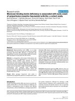
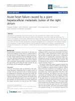
Figure 1 (a) Pre-operative coronal view of the aorta and the aorto-iliac segment showing contrast in the aorta but no flow in the iliac
arteries. The dissection extended into both sides. (b) Post-operative coronal view of the same segment with uncovered stent in-situ
demonstrating increased flow within the iliac system
Suliman et al. Journal of Cardiothoracic Surgery 2010, 5:4
/>Page 2 of 5
Teflon felt pledgetted sutures. We specifically passed
these pledgetted sutures through the proximal stent
in the medial part of the descending thoracic aorta
providing extra strength in these stitches and poten-
tially reducing the risk of stent migration or creation
of endoleak in this weak part of the aortic wall. A 28
mm Dacron conduit was then anastomosed (hemi-
arch replacement) and the patient was rewarmed to
37°C. The chest was packed and left open for
delayed closure, which was performed 48 h later.
The outcome of this staged approach was very suc-
cessful (Figure 1b and Figure 2b) and our patient recov-
ered well. His progress was complicated by a hospital-
acquired pneumonia requiring prolonged intubation and
formation of a tracheostomy. The total ITU stay was 33
days. He was gradually rehabilitated, and was discharged
40 days after admission.
Conclusions
Approximately 25% of aortic dissections have evidence
of peripheral malperfusion at presentation [2]. In cases
of peripheral malperfusion syndrome, particularly invol-
ving the superior mesenteric artery, the operative mor-
tality is s ignificantly increased [9]. In these cases with
such degree of metabolic disturbance, temporary
postponement in surgical repair while peripheral reper-
fusion is re-established may prove beneficial [3,9].
Our patient did not have clinical signs of intestinal
malperfusion (although there was significant peripheral
ischemia). Lack of immediate symptoms in these
patients can delay accurate diagnosis and management
contributing to the high mortality. One possible treat-
ment option includes initial endovascular fenestration of
the infrarenal aorta [10]. In the last few years however,
biomarkers (in particular serum lacta te) have become a
usef ul tool in assessing mesenteric ischemia. Our staged
therapeutic approach (Figure 3) illustrates the diagnos tic
value of biomarkers in malperfusion, particularly where
there is a delayed presentation.
If the initial lactate reading (measured quickly and
simply from an arterial blood sample) is considerably
high with no other cause and there is radiological or
clinical evidence of bowel ischemia, revascularization
using percutaneous endovascula r techniques should first
be carried out [5]. Following this, further serial lactate
measurements should be taken to gauge the success of
the intervention and monitor t he improvement in per-
ipheral malperfusion.
We recommend this method as D-lactate (a stereo-
isomer of phy siological L-lactate) is a sensitive marker
for early mesenteric ischemia produced in large amounts
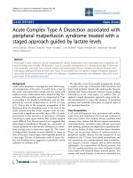
Figure 2 (a) Pre-operative sagittal view of the thoracic aorta showing contrast within the true and false lumina.Noteneartotal
occlusion of the celiac and superior mesenteric arteries (SMA) denoted by white arrows. (b) Post operative sagittal image of the same aortic
segment with stent graft in-situ demonstrating increased flow within the celiac and superior mesenteric arteries (SMA) denoted by white arrows.
Suliman et al. Journal of Cardiothoracic Surgery 2010, 5:4
/>Page 3 of 5
by the overgrowth of gut microbial flora [6,8]. In view of
the slow rate of enzymatic breakdown, it is a very sensi-
tive early marker of the ischemic process (where the lac-
tate levels may be subject to several factors including
ischemia-related hepatic dysfunction) [6-8]. Our
approach recommends that a consistent fall in lactate
during this interim period may represent the ischemia
as resolving. One can therefore perform a delayed repair
of the proximal aortic dissection [11] providing a
decreased intra-operative risk to the patient. Individuals
with persistently high lactate levels may then require a
revascularization procedure at that time rather than
delaying intervention in anticipation of clinical signs.
Differentiating between bowel ischemia and lower limb
ischemia in the absence of clinical signs and a raised
lactate can be based on radiological imaging.
A persistently raised lactate level associated with clini-
cal or radiological evidence of bowel ischemia requires
the treatment of the dynamic or static compression
associated with the Type A dissection. This can be
achieved by surgical revascularization, fenestration of
the dissection flap or covered endovascular stenting of
the thoracic aorta followed by closure of the dissection
entry point with surgical repair (Figure 3).
If it is possible to attend a patient within 6 hours of a
Type A dissection, then primary repair of the dissection
is advised after locating the primary tear on preoperative
CT scan (Figure 3). The optimal management for an
acute type A dissection is entry closure and in cases of
central aortic repair, distal organ ischemia can be mana-
ged through revascularization grafts such as axillary-
femoral bypass. Endovascular stenting without entry clo-
sure for type A dissection has the risk of cardiac tampo-
nade and in our case the entry point was closed during
the arch repair. In more complex Type A dissections, a
tailored multi-disciplinary strategy is required to address
underlying risk in order to provide optimum perfusion
and survival.
Our approach in using the biomarker lac tate to guid e
our management of acute type A aortic dissection allows
the restoration of an improved metabolic status before
the insult of the total circulatory arrest (preserving the
kidneys and bowel during the subsequent surgical dis-
section repair). It also has the potential to be extremely
useful in terms of selecting patients w ho would be able
to tolerate such complex operations and can improve
patient outcomes in terms of morbidity and mortality.
Consent
Written informed consent was obtained from the patient
for publication of this case report and any accompany-
ing images. A co py of the written consent is available
for review by the Editor-in-Chief of this journal.
Author details
1
Department of Cardiothoracic Surgery, Imperial College Healthcare NHS
Trust, St Mary’s Hospital, Praed Street, London W2 1NY, UK.
2
Regional
Vascular Unit, Imperial College Healthcare NHS Trust, St Mary’s Hospital,
Praed Street, London W2 1NY, UK.
Authors’ contributions
AS participated in this case and contributed to its analysis. MD participated
in this case and contributed to its analysis. HA participated in this case and
contributed to its analysis. CB participated in this case and contributed to its
Figure 3 Therapeutic approach in Type A dissection with peripheral malperfusion.
Suliman et al. Journal of Cardiothoracic Surgery 2010, 5:4
/>Page 4 of 5
analysis. MM participated in this case and contributed to its analysis. MH
participated in this case and contributed to its analysis. TA participated in
this case and contributed to its analysis. All authors read and approved the
final manuscript.
Competing interests
The authors declare that they have no competing interests.
Received: 2 December 2009
Accepted: 28 January 2010 Published: 28 January 2010
References
1. Borst HG, Laas J, Heinemann M: Type A aortic dissection: diagnosis and
management of malperfusion phenomena. Semin Thorac Cardiovasc Surg
1991, 3(3):238-241.
2. Fann JI, Sarris GE, Mitchell RS, Shumway NE, Stinson EB, Oyer PE, Miller DC:
Treatment of patients with aortic dissection presenting with peripheral
vascular complications. Ann Surg 1990, 212(6):705-713.
3. Deeb GM, Williams DM, Bolling SF, Quint LE, Monaghan H, Sievers J,
Karavite D, Shea M: Surgical delay for acute type A dissection with
malperfusion. Ann Thorac Surg 1997, 64(6):1669-1675, discussion 1675-1667.
4. Girardi LN, Krieger KH, Lee LY, Mack CA, Tortolani AJ, Isom OW:
Management strategies for type A dissection complicated by peripheral
vascular malperfusion. Ann Thorac Surg 2004, 77(4):1309-1314, discussion
1314.
5. Slonim SM, Nyman U, Semba CP, Miller DC, Mitchell RS, Dake MD: Aortic
dissection: percutaneous management of ischemic complications with
endovascular stents and balloon fenestration. J Vasc Surg 1996,
23(2):241-251, discussion 251-243.
6. Gunel E, Caglayan O, Caglayan F: Serum D-lactate levels as a predictor of
intestinal ischemia-reperfusion injury. Pediatr Surg Int 1998, 14(1-2):59-61.
7. Muraki S, Fukada J, Morishita K, Kawaharada N, Abe T: Acute type A aortic
dissection with intestinal ischemia predicted by serum lactate elevation.
Ann Thorac Cardiovasc Surg 2003, 9(1):79-80.
8. Murray MJ, Gonze MD, Nowak LR, Cobb CF: Serum D(-)-lactate levels as an
aid to diagnosing acute intestinal ischemia. Am J Surg 1994,
167(6):575-578.
9. Patel HJ, Williams DM, Dasika NL, Suzuki Y, Deeb GM: Operative delay for
peripheral malperfusion syndrome in acute type A aortic dissection: a
long-term analysis. J Thorac Cardiovasc Surg 2008, 135(6):1288-1295,
discussion 1295-1286.
10. Fattori R, Botta L, Lovato L, Biagini E, Russo V, Casadei A, Buttazzi K:
Malperfusion syndrome in type B aortic dissection: role of the
endovascular procedures. Acta Chir Belg 2008, 108(2):192-197.
11. Cambria RP, Brewster DC, Gertler J, Moncure AC, Gusberg R, Tilson MD,
Darling RC, Hammond G, Mergerman J, Abbott WM: Vascular
complications associated with spontaneous aortic dissection. J Vasc Surg
1988, 7(2):199-209.
doi:10.1186/1749-8090-5-4
Cite this article as: Suliman et al.: Acute Complex Type A Dissection
associated with peripheral malperfusion syndrome treated with a
staged approach guided by lactate levels. Journal of Cardiothoracic
Surgery 2010 5:4.
Submit your next manuscript to BioMed Central
and take full advantage of:
• Convenient online submission
• Thorough peer review
• No space constraints or color figure charges
• Immediate publication on acceptance
• Inclusion in PubMed, CAS, Scopus and Google Scholar
• Research which is freely available for redistribution
Submit your manuscript at
www.biomedcentral.com/submit
Suliman et al. Journal of Cardiothoracic Surgery 2010, 5:4
/>Page 5 of 5