báo cáo khoa học: " All-trans retinoic acid inhibits KIT activity and induces apoptosis in gastrointestinal stromal tumor GIST-T1 cell line by affecting on the expression of survivin and Bax protein" pdf
Bạn đang xem bản rút gọn của tài liệu. Xem và tải ngay bản đầy đủ của tài liệu tại đây (475.28 KB, 8 trang )
RESEA R C H Open Access
All-trans retinoic acid inhibits KIT activity and
induces apoptosis in gastrointestinal stromal
tumor GIST-T1 cell line by affecting on the
expression of survivin and Bax protein
Hoang Thanh Chi
1,2†
, Bui Thi Kim Ly
1,2†
, Takahiro Taguchi
3
, Toshiki Watanabe
2
, Yuko Sato
1*
Abstract
Background: Imatinib, a selective tyrosine kinase inhibitor, has been used as a standard first-line therapy for
irresectable and metastasized gastrointestinal stromal tumor (GIST) patients. Unfortunately, most patients
responding to imatinib will eventually exhibit imatinib-resistance, the cause of which is not fully understood. The
serious clinical problem of imatinib-resistance demands alternative therapeutic strategy. This study was conducted
to investigate the effect of all-trans retinoic acid (ATRA) on GIST cell lines.
Methods: Cell proliferation was determined by trypan blue dye exclusion test. Western blot analysis was
performed to test the expression of activated KIT, its downstream proteins, and apoptosis associated proteins. The
cytotoxic interactions of imatinib with ATRA were evaluated using the isobologram of Steel and Peckham.
Results and conclusion: In this work, for the first time we have demonstrated that ATRA affected on cell
proliferation of GIST-T1 and GIST-882 cell line through inhibition of cell growth in a dose dependent manner and
induced apoptosis. High dose of ATRA induced morphologic change in GIST-T1 cells, rounded-up cells, and
activated the caspase-3 protein. In further examination, we found that the ATRA-induced apoptosis in GIST-T1 cells
was accompanied by the down-regulated expr ession of survivin and up-regulated expression of Bax protein.
Moreover, ATRA suppressed the activity of KIT protein in GIST-T1 cells and its downstream signal, AKT activity, but
not MAPK activity. We also have demonstrated that combination of ATRA with imatinib showed additive effect by
isobologram, suggesting that the combination of ATRA and imatinib may be a novel potential therapeutic option
for GIST treatment. Furthermore, the scracht assay result suggested that ATRA was a potential reagent to prevent
the invasion or metastasis of GIST cells.
Background
Gastrointestinal stromal tumors (GISTs) are the most
common mesenchym al neoplasms occurring throughout
the entire region of the gastrointestinal tract and are
considered to originate from intestitial cells of Cajal, the
pacemaker cells of the gut [1]. The most likely causative
molecular event in the vast majority of GISTs is a gain-
of-function mutation of KIT or PDGFRA (platelet-
derived growth factor receptor alpha) which activates
these receptor tyrosine kinases (RTKs) by rendering
them constitutively phosphorylated [2-4]. Thereafter, the
downstream signaling pathways are activated promoting
cell proliferation and/or survival.
To date, surgical resection seems to be the only treat-
ment approach for GISTs with resulting in 5 year survival
rates of 48-54% for resec table cases [5] while for irresect-
able or metastasized GIST cases, the median survival per-
iod was only 19 months and 5 year survival rate of 5-10%
[6]. More recently, imatinib (Glivec, Gleevec; Novartis
Pharma AG), a selective inhibitor of KIT, PDGFRA, ABL,
as well as the other certain tyrosine kinases, has been
used as a standard first- line thera py for i rresectable a nd
metastasized GISTs [7-11]. C linical evidenc e suppor ting
* Correspondence:
† Contributed equally
1
Division of Ultrafine Structure, Department of Pathology, Research Institute,
National Center for Global Health and Medicine, Tokyo, Japan
Full list of author information is available at the end of the article
Chi et al. Journal of Experimental & Clinical Cancer Research 2010, 29:165
/>© 2010 Chi et al; licensee BioM ed Central Ltd. T his i s an Open A ccess article distributed under the terms of the Creative Commons
Attribution License ( which permits unrestri cted use, distribution, and reproduction in
any medium, provided the original work is properly cited.
the indication of imatinib for GISTs was obtained from
phase II/III trials in patients with irresectable GISTs [12].
Although imatinib has show n p rominent effects to meta-
static lesions of GIST, serious problems involved in ima-
tinib-resi stance h ave been reported recently [13,14] . The
resistance develops after a median of about 2 years of
treatment with imatinib [15]. Other KIT inhibitors such
as sunitinib, PKC412 or BMS-354825 are reported to be
effective in a subset of patients w ith imatinib -resistant
GISTs. However, none of them have been proven to be
effective to all the known imatinib-resistant mutations of
KIT [16-18]. Therefore, development of novel KIT inhibi-
tors or finding novel therapeutic strategy for GISTs is
demanded.
Vitamin A (retinol) is a fat-soluble vitamin essential
for the formation and maintenance of many body tis-
sues, such as skin, bone, and vasculature, as well as for
the promotion of good vision and i mmune function
[19]. Vitamin A also plays a role in reproduction and in
embryonic growth and development. Vitamin A is con-
verted to more active compounds, such as retinoic acid,
through which it exerts its multiple e ffects on embryo-
nic development and organog ene sis, tissue homeost asis,
cell pro liferation, differentiation, and apoptosis [ 20,21].
Retinol has six known biologically-active isoforms: all-
trans, 11-cis,13-cis,9,13-di-cis,9-cis, and 11,13-di-cis
with all-trans being the predominant physiological form.
Endogenous retinoids with biological activity include all-
trans retinoic acid, 9-cis retinoic acid, 11-cis retinalde-
hyde, 3,4-didehydro retinoic acid [22].
The functions of retinoic acid regulating differentia-
tion, proliferation and apoptosis are mediated by nuclear
receptors, such as retinoic acid rece ptors (RARs) and
retinoic × receptors (RXR) [23]. Although the mechan-
isms of retinoic aci ds on regulating differentiati on, pro-
liferation and apoptosis are not fully elucidated, it has
been suggested that induction of differentiation and
apoptosis by retinoic acids might contribute to treat-
ment of cancers.
In this work, we studied the effect of ATRA on GIST
cells in term of inhibition of cell proliferation, and induc-
tion of apopt osis. For the first time we have demon-
strated that ATRA i nhibited cell proliferation of GIST-
T1 and GIST-882 ce ll line in a d ose depende nt manner
and caused apoptosis. The apoptosis induced by ATRA
may be regulated at least by down-re gulated expression
of survivin and up-regulated expression of Bax.
Materials and methods
Cell lines and culture conditions
The human GIST cell lines, GIST-T1 with 57-nucleotide
(V570-Y57 8) in-flame deletion in KIT exon 11 [24], and
GIS T-882 cells with K642E mutation in exon 13 of KIT
and the human normal diploid fibroblast cells (WI-38)
(IFO 50075, Human Scien ce Research Resource Ba nk,
Osaka, Japan) were used in this study.
The cells were grown in Dulbecco’s modified Eagle’ s
medium (DMEM) with high glucose (Nakalai Tesque,
Kyoto, Japan) supplemented with 10% fet al bovine
serum (FBS) (JRH Biosciences, Lenexa, KS, USA),
100 IU/ml penicillin, and 0.1 mg/ml streptomycin
(Nakalai Tesque) in a humidified incubator of 5% CO
2
at 37°C.
Reagents
Imatinib and all-trans retinoic acid were purchased from
Sequoia Research Products (Oxford, UK) and WAKO
Chemicals (Osaka, Japan), respectively. Both of them are
dissolved in DMSO. The concentration of DMSO was
kept under 0.1% throughout all the experiments to
avoid its cytotoxicity.
Cell proliferation assays
Cell proliferation was determined by trypan blue dye
exclusion test. Cells were seeded in 6-well plates at a den-
sity of 1 × 10
5
cells/ml in the presence of different con-
centrations of ATRA or imatinib for 72 hours in
humidified incubator o f 5% CO
2
at 37°C. After the trea t-
ment, the cells were washed twice with PBS without Ca
2+
and Mg
2+
[PBS(-)] to remove the medium. Then cells
were dissociated with EDTA-trypsin solution. Ten micro
liter of the cell suspension was mixed with 10 μlof0.4%
trypan blue, and ali ve cells were counted manually using
a hemacyto meter. Resu lts were cal culated as the percen-
tage of the values measured when cells were grown in the
absence of reagents.
Western blot analysis
Cells were plated onto 10-cm dishes at a density of
1×10
5
cells/ml in the presence of 180 μMATRA.
After incubation for indicated durations, cells were col-
lected by trypsinization and washed twice with PBS(-).
Cell protein was extracted and western blot analysis was
done as described previously [25]. The followin g antibo-
dies ERK1 (sc-93), total Akt (sc-1618), anti-KIT
antibody (cKIT-E1), survivin (sc-17779), anti-rabbit
IgG-HRP (sc-2317), and anti-mouse IgG-HRP (sc-2031)
were purchased from Santa Cruz Biotechnology (Santa
Cruz, CA, USA). Anti-actin (A2066) was from Sigma-
Aldrich. Ph ospho-p44/42 Map kinase (Thr202/Tyr204),
phospho-Akt (Ser 473), XIAP, caspase-3, phospho-c-Kit
(tyr719) antibodies were from Cell Signaling Technology
Japan (Tokyo, Japan). Anti-PARP antibody was from
WAKO Chemicals (Osaka, Japan).
Cell morphologic assessment
Cellswereplatedatadensityof1×10
5
cells/ml in the
presence of different concentration of ATRA onto
Chi et al. Journal of Experimental & Clinical Cancer Research 2010, 29:165
/>Page 2 of 8
6-well dishes. After 3-day treatment, cell morphology
was observed under an inverted microscope.
Wright-Giemsa staining
For fragmented nuclei and condense d chromatin assess-
ment, cells at a density of 1 × 10
5
cells/ml were treated
with 180 μM ATRA. After indicated durations, cells
were harvested and fixed onto slides by using a cytospin
(Shandon, Shandon Southern Products Ltd., Cheshire,
UK). Cells then were stained with Wright-Giemsa solu-
tion. Morphology of cells was observed under an
inverted microscope.
DNA fragmentation assay
GIST-T1 cells were treated with or without 180 μM
ATRA for different durations. Cells then were collected
and total genomic DNA (gDNA) was extracted with a
standard protocol. For DNA fragmentation assay, 10 μg
gDNA of each sample was blotted and electrophoresed
on 1.2% aga rose gel. DNA fragmen tation was detected
under UV light.
Scratch assay
GIST-T1 cells were seeded in 6-well plates with or with-
out reagent. After 24-hour treatment, a line was scraped
within confluent cells using the fine end of 10 μLpip-
ette tip (time 0). After 24 hours, migration of GIST cells
was observed under an inverted microscope.
Assessment of cytotoxic effect of ATRA in combination
with imatinib
The cytotoxic interactions of imatinib with ATRA were
evaluated using the isobologram of Steel and Peckham
[26]. The IC
50
was defined as the concentration of
reagent that produced 50% cell growth inhibition.
Statistical analysis
All data were expressed as the mean ± standard devia-
tion. Statistical analyses were done using Student’s t-test,
in which p < 0.05 was the minimum requirement for a
statistically significant difference.
Results
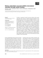
Growth inhibitory effect of ATRA on GIST-T1 cells
ATRA treatment resulted in inhibition of cell prolifera-
tion of GIST-T1 and GIST-882 cells in a dose-dependent
manner but showed nearly no effect on the human nor-
mal fibroblast WI-38 cell (Figure 1A). The adherence of
GIST-T1 cells was m uch inhibited by ATR A-treatment
in a dose-dependent manner (Figure 1B). In addition,
ATRA treatment highly affected on morphology of
GIST-T1 cel ls. ATRA-treated (180 μM, 3 days) GIST-T1
cells changed to rounded-up cells compared with the
control cells (Figure 1C), suggesting that ATRA might
cause inhibition of peripheral attachment in these cells.
The effect of ATRA on morphological changes in GIST-
882 cells was similar to GIST-T1 cells (data not shown).
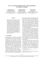
ATRA induced apoptosis in GIST-T1 cells
To confirm whether ATRA induces apoptosis in GIST-
T1 cells, we further investigated apoptotic markers,
nuclei shrinkage, DNA fragmentation and activation of
caspase-3 in GIST-T1 cells after ATRA treatment.
As mentioned above, ATRA not only induced the mor-
phologic change (rounded-up cells) in GIST-T1 cells after
3-day treatment, but also induced detachment of the cells
from the dishes after 6-day treatment (data not shown).
To check whether detached cells show the features of
apoptosis, cells were collected a nd fixed onto slide s by
using a cytospin before performing Wright-Giemsa stain-
ing. The result showed that detached cells showed shrunk
and fragment ed nuclei, the apoptotic features, compared
with control cells (Figure 2A right), the fragmented nuclei
were confirmed by DNA fragmentation assay (Figure 2B).
As expected, DNA fragmentation was observed after 2-day
treatment and increased in a time dependent manner.
Moreover, to clearly demonstrate that ATRA causes
apoptosis in GIST-T1 cells, we assessed the molecular
aspects of apoptosis, such as caspase-3, well recognized
as a marker of apoptosis, and PARP, considered as a
biochemical marker of necrosis when it is hyperactivated
[27], by w estern blot. After 2-day treatment with
180 μM ATRA, cleaved caspase-3 and PARP were
observed (Figure 2C). T his result is consistent with the
data of DNA fragmentation, demonstrating that ATRA
induced apoptosis in GIST-T1 cells.
Overall, our result s demonstrated that ATRA induced
apoptotic cell death in GI ST-T1 cells. The similar result
was also confirmed in GIST-882 cells (data not shown).
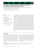
ATRA affected on expression of survivin, XIAP
and Bax protein
It is well known that apoptoti c pro cess is regulated by
many factors. We investigated the expression of inhibitors
of apoptosis, survivin, XIAP, and pro-apoptosis Bax. The
results showed down-regulation of survivin (Figure 3A)
and up-re gulation of Bax (Figure 3B). These results were
consistent with the appearance of cleaved caspase-3 and
PARP in GIST-T1 cells (Figure 2C). However, ATRA did
not affect on XIAP expression in GIST-T1 cells by western
blot analysis (Figure 3C). All together, the apoptosis
induced by ATRA treatment may be regulated at least by
down-regulation of survivin and up-regulation of Bax
proteins.
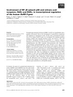
ATRA suppressed the phosphorylation of KIT protein
KIT protein is one of the most important molecules in
the pathogenesis of GISTs. Despite clinicopathological
Chi et al. Journal of Experimental & Clinical Cancer Research 2010, 29:165
/>Page 3 of 8
difference, most GISTs have a similar genetic profile,
gain-of-function mutations of KIT or PDGFRA [2].
Upon the importance of KIT pr otein, we examined
whether ATRA can suppress KIT act ivity in GIST-T1
cell s. We treated GIST-T1 cells with 180 μMATRAfor
the indicated duration. Total cell lysate s were subjected
to western blot analysis.
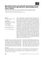
Interestingly, ATRA treatment resulted in suppression
of KIT activity after 4-day treatment in GIST-T1 cells
(Figure 4A the top row) and GIST-882 cells (data not
shown). The suppression of KIT activity in GIST-T1 and
GIST-882 cells by ATRA required longer time compared
with other reagents such as imatinib or EGCG [25].
In addition, ATRA treatment also suppressed the AKT
activity (Figure 4A the middle row) but not MAPK activ-
ity (Figure 4A the bottom row) in GIST-T1 cells.
Interestingly, the suppr ession of KIT and AKT activity
by ATRA treatment was enhanced in serum-free me dia.
However, suppression of MAPK activity was not observed
even in serum-free media (Figure 4B). The similar results
were observed in GIST-882 cells (data not shown).
ATRA prevented the migration of GIST-T1 cells
Next, t o study the migration of GIST-T1 cells in vitro,
the scratch assay was performed. This method is based
on the observation that, upon creation of a new artificial
gap, so called a scratc h on a confluent cell monolayer,
the cell on the edge of the newly created gap will move
toward the opening to close the scratch until cell t o cell
contacts are established again.
In this study, GIST-T1 cells were seeded with or without
ATRA (45, 90 μM) in plates. After 24 hour i ncubation to
Figure 1 Effect of ATRA on cell proliferation of GIST-T1, GIST-882 and human normal fibroblast WI-38 cells. GIST-T1, GIST-882 and
human normal fibroblast WI-38 cells at a density of 1 × 10
5
cells/ml were treated with different concentrations of ATRA dissolved in DMSO or
with DMSO alone (0 μM ATRA as control) for 3 days. Panel A shows cell growth curve which represents the effect of different concentrations of
ATRA. Results were calculated as the percentage of the control values. Panel B shows the effect of ATRA on adherence of GIST-T1 cells at
various concentrations of ATRA. Panel C shows cell morphologic change of GIST-T1 cells after 3-day treatment with 180 μM ATRA.
Chi et al. Journal of Experimental & Clinical Cancer Research 2010, 29:165
/>Page 4 of 8
Figure 2 ATRA induces apoptotic cell death in GIST-T1 c ells. Panel A shows the s hrinkage and fragmentation of nuclei in GIST-T1 cells after
6-day treatment with 180 μM ATRA compared with the control cells. Panel B shows the result of DNA fragmentation after 2-, 4- or 6-day
Figure 3 ATRA affects on the expression of survivin and Bax. Panel A shows the down-regulated expression of survivin after 2-, 4- or 6-day
treatment with 180 μM ATRA. Panel B shows the up-regulated expression of Bax after 2-, 4- or 6-day treatment with 180 μM ATRA. Panel C
shows the effect of ATRA on XIAP expression after 2-, 4- or 6-day treatment with 180 μM ATRA.
Chi et al. Journal of Experimental & Clinical Cancer Research 2010, 29:165
/>Page 5 of 8
get the confluence, a scratch was created. The images of
GIST-T1 cells at the beginning and 24 hour later were
compared to assess the migration of GIST-T1 cells. The
result revealed that 90 μM ATRA inhibited completely
migration of GIST-T1 cells compared with the non-ATRA
treated dishes (Figure 5A). However, at a lower concentra-
tion ( 45 μM), ATRA inhibited but not completely the
migration of these cells (data not shown). All together, the
data suggested that ATRA may be useful to prevent the
invasion or metastasis of GIST cells.
Cytotoxic effect of combination with ATRA and imatinib
The result of isobologram was showed in Figure 5B. All
data points in the combination fell within the envelope
of additivity, the area surrounded by the three lines, sug-
gesting that this combination gave additive effect.
Discussion
ATRA have been reported to show therapeutic effect on
breast and ovarian cancers and APL [28]. However, for the
first time we have demonstrated that ATRA suppressed
the cell proliferation and induced apoptosis in GIST-T1
cells, suggesting anti-cancer effect of ATRA on GISTs.
The cell death inducing mechanism by ATRA in cancers
has not yet been fully clarified. In this report we have
shown that apoptosis induced by ATRA in GIST-T1 cells
are regulated at least by the down-regulation of survivin
and up-regulation of Bax (Figure 3A and 3B). Even though
XIAP and survivin belong to the same family of apoptotic
inhibitors, it is likely that ATRA effected quite differently
on expression of XIAP and survivin. Survivin was sup-
pressed in a time dependentmannerwhereasXIAPwas
not suppressed by ATRA treatment (Figure 3C). It is likely
that survivin may be a target molecule that plays an
important role in ATRA-induced apoptosis in GIST-T1
cells. Further studies a re definitely necessary for better
understanding of the apoptosis-inducing mechanism by
ATRA in GIST-T1 cells.
GISTs can be successfully treated with imatinib with
the response rate of up to 85% [15,29,30]. However,
after a median of 2 years of treatment with imatinib,
resistance can develop [15]. The effect of imatinib is
mainly due to the suppressio n of KIT activ ity. In this
study, we found that the suppression of KIT activity
(Figure 4A) was a lso obtained b y ATRA treat ment.
Moreover, we have demonstrated that combination of
ATRA and imatinib showed additive effect (Figure 5B)
by isobologram, suggesting that the combination of
ATRA and imatinib would be a novel therapeutic poten-
tial for GISTs. The scratch assay result (Figu re 5A) also
suggested the useful of ATRA to prevent the invasion or
metastasis of GIST cells.
Inconclusion,wehavedemonstratedthatATRAhad
an ability to inhibit the cell proliferation and migration,
Figure 4 ATRA suppresses the auto-phosphorylation of KIT and AKT protein but not MAPK activity. Panel A shows the suppression of
KIT and AKT activity after 2-, 4- or 6-day treatment with 180 μM ATRA. Panel B shows the suppression of KIT and AKT activity after 4 hours
treatment with different ATRA concentrations in serum-free media. The results demonstrated that KIT and AKT activity were suppressed by ATRA
treatment in a dose- and time-dependent manner but not MAPK activity.
Chi et al. Journal of Experimental & Clinical Cancer Research 2010, 29:165
/>Page 6 of 8
inducing apoptosis in GIST-T1 cells. Thus ATRA can
have a potential for novel therapeutic agent for GISTs.
Since the combination of ATRA and imatinib showed
additive effect on GIST-T1 cells, ATRA may be used in
combination with imatinib for GISTs treatment.
Acknowledgements
This work was supported by the Japan Foundation for Promotion of
International Medical Research Co-operation (JF-PIMRC).
Author details
1
Division of Ultrafine Structure, Department of Pathology, Research Institute,
National Center for Global Health and Medicine, Tokyo, Japan.
2
Department
Figure 5 Panel A shows the result of scratch assay, GIST-T1 cells were treated with or without ATRA (9 0 μM). Migration was observed
after 24-hour incubation. Panel B shows the isobologram result of drug combination between ATRA and imatinib. This combination resulted in
additive effect.
Chi et al. Journal of Experimental & Clinical Cancer Research 2010, 29:165
/>Page 7 of 8
of Medical Genome Sciences, Graduate School of Frontier Sciences, the
University of Tokyo, Tokyo, Japan.
3
Graduate School of Integrated Arts and
Sciences, Doctoral Course, Kuroshio Science, Kochi University, Kochi-shi,
Kochi-ken, Japan.
Authors’ contributions
HTC and BTKL have carried out the study design, molecular biological work,
and statistical analyses and drafted the manuscript. TT has established GIST-
T1 cell line. TW and YS have carried out the study design, statistical analyses
and drafted the manuscript. All authors read and approved the final
manuscript.
Competing interests
The authors declare that they have no competing interests.
Received: 9 September 2010 Accepted: 16 December 2010
Published: 16 December 2010
References
1. Kindblom LG, Remotti HE, Aldenborg F, Meis-Kindblom JM: Gastrointestinal
pacemaker cell tumor (GIPACT): gastrointestinal stromal tumors show
phenotypic characteristics of the interstitial cells of Cajal. Am J Pathol
1998, 152:1259-1269.
2. Lasota J, Miettinen M: Clinical significance of oncogenic KIT and PDGFRA
mutations in gastrointestinal stromal tumours. Histopathology 2008,
53:245-266.
3. Hirota S, Isozaki K, Moriyama Y, Hashimoto K, Nishida T, Ishiguro S,
Kawano K, Hanada M, Kurata A, Takeda M, et al: Gain-of-function
mutations of c-kit in human gastrointestinal stromal tumors. Science
1998, 279:577-580.
4. Heinrich MC, Corless CL, Duensing A, McGreevey L, Chen CJ, Joseph N,
Singer S, Griffith DJ, Haley A, Town A, et al: PDGFRA activating mutations
in gastrointestinal stromal tumors. Science 2003, 299:708-710.
5. Bauer S, Hartmann JT, de Wit M, Lang H, Grabellus F, Antoch G, Niebel W,
Erhard J, Ebeling P, Zeth M, et al: Resection of residual disease in patients
with metastatic gastrointestinal stromal tumors responding to treatment
with imatinib. Int J Cancer 2005, 117:316-325.
6. DeMatteo RP, Lewis JJ, Leung D, Mudan SS, Woodruff JM, Brennan MF: Two
hundred gastrointestinal stromal tumors: recurrence patterns and
prognostic factors for survival. Ann Surg 2000, 231:51-58.
7. Buchdunger E, Cioffi CL, Law N, Stover D, Ohno-Jones S, Druker BJ,
Lydon NB: Abl protein-tyrosine kinase inhibitor STI571 inhibits in vitro
signal transduction mediated by c-kit and platelet-derived growth factor
receptors. J Pharmacol Exp Ther 2000, 295:139-145.
8. Heinrich MC, Griffith DJ, Druker BJ, Wait CL, Ott KA, Zigler AJ: Inhibition of
c-kit receptor tyrosine kinase activity by STI 571, a selective tyrosine
kinase inhibitor. Blood 2000, 96:925-932.
9. Okuda K, Weisberg E, Gilliland DG, Griffin JD: ARG tyrosine kinase activity
is inhibited by STI571. Blood 2001, 97:2440-2448.
10. Tuveson DA, Willis NA, Jacks T, Griffin JD, Singer S, Fletcher CD, Fletcher JA,
Demetri GD: STI571 inactivation of the gastrointestinal stromal tumor c-
KIT oncoprotein: biological and clinical implications. Oncogene 2001,
20:5054-5058.
11. Dagher R, Cohen M, Williams G, Rothmann M, Gobburu J, Robbie G,
Rahman A, Chen G, Staten A, Griebel D, Pazdur R: Approval summary:
imatinib mesylate in the treatment of metastatic and/or unresectable
malignant gastrointestinal stromal tumors. Clin Cancer Res 2002,
8:3034-3038.
12. Demetri GD, von Mehren M, Blanke CD, Van den Abbeele AD, Eisenberg B,
Roberts PJ, Heinrich MC, Tuveson DA, Singer S, Janicek M, et al: Efficacy
and safety of imatinib mesylate in advanced gastrointestinal stromal
tumors. N Engl J Med 2002, 347:472-480.
13. Heinrich MC, Corless CL, Blanke CD, Demetri GD, Joensuu H, Roberts PJ,
Eisenberg BL, von Mehren M, Fletcher CD, Sandau K, et al: Molecular
correlates of imatinib resistance in gastrointestinal stromal tumors. J Clin
Oncol 2006, 24:4764-4774.
14. Koyama T, Nimura H, Kobayashi K, Marushima H, Odaira H, Kashimura H,
Mitsumori N, Yanaga K: Recurrent gastrointestinal stromal tumor (GIST) of
the stomach associated with a novel c-kit mutation after imatinib
treatment. Gastric Cancer 2006, 9 :235-239.
15. Verweij J, Casali PG, Zalcberg J, LeCesne A, Reichardt P, Blay JY, Issels R, van
Oosterom A, Hogendoorn PC, Van Glabbeke M, et al: Progression-free
survival in gastrointestinal stromal tumours with high-dose imatinib:
randomised trial. Lancet 2004, 364:1127-1134.
16. Demetri GD, van Oosterom AT, Garrett CR, Blackstein ME, Shah MH,
Verweij J, McArthur G, Judson IR, Heinrich MC, Morgan JA, et al: Efficacy
and safety of sunitinib in patients with advanced gastrointestinal
stromal tumour after failure of imatinib: a randomised controlled trial.
Lancet 2006, 368:1329-1338.
17. Shah NP, Tran C, Lee FY, Chen P, Norris D, Sawyers CL: Overriding imatinib
resistance with a novel ABL kinase inhibitor. Science 2004, 305:399-401.
18. Debiec-Rychter M, Cools J, Dumez H, Sciot R, Stul M, Mentens N, Vranckx H,
Wasag B, Prenen H, Roesel J, et al: Mechanisms of resistance to imatinib
mesylate in gastrointestinal stromal tumors and activity of the PKC412
inhibitor against imatinib-resistant mutants. Gastroenterology 2005,
128:270-279.
19. Collins MD, Mao GE: Teratology of retinoids. Annu Rev Pharmacol Toxicol
1999, 39:399-430.
20. Morriss-Kay GM, Ward SJ: Retinoids and mammalian development. Int Rev
Cytol 1999, 188:73-131.
21. Kastner P, Mark M, Chambon P: Nonsteroid nuclear receptors: what are
genetic studies telling us about their role in real life? Cell 1995,
83:859-869.
22. Napoli JL: Biochemical pathways of retinoid transport, metabolism, and
signal transduction. Clin Immunol Immunopathol 1996, 80:S52-62.
23. Bastien J, Rochette-Egly C: Nuclear retinoid receptors and the
transcription of retinoid-target genes. Gene 2004, 328:1-16.
24. Taguchi T, Sonobe H, Toyonaga S, Yamasaki I, Shuin T, Takano A, Araki K,
Akimaru K, Yuri K: Conventional and molecular cytogenetic
characterization of a new human cell line, GIST-T1, established from
gastrointestinal stromal tumor. Lab Invest 2002, 82:663-665.
25. Chi HT, Vu HA, Iwasaki R, Thao le B, Hara Y, Taguchi T, Watanabe T, Sato Y:
Green tea (-)-epigalocatechin-3-gallate inhibits KIT activity and causes
caspase-dependent cell death in gastrointestinal stromal tumor
including imatinib-resistant cells. Cancer Biol Ther
2009, 8:1934-1939.
26. Steel GG, Peckham MJ: Exploitable mechanisms in combined
radiotherapy-chemotherapy: the concept of additivity. Int J Radiat Oncol
Biol Phys 1979, 5:85-91.
27. Kroemer G, Galluzzi L, Vandenabeele P, Abrams J, Alnemri ES, Baehrecke EH,
Blagosklonny MV, El-Deiry WS, Golstein P, Green DR, et al: Classification of
cell death: recommendations of the Nomenclature Committee on Cell
Death 2009. Cell Death Differ 2009, 16:3-11.
28. Fields AL, Soprano DR, Soprano KJ: Retinoids in biological control and
cancer. J Cell Biochem 2007, 102:886-898.
29. van Oosterom AT, Judson IR, Verweij J, Stroobants S, Dumez H, Donato di
Paola E, Sciot R, Van Glabbeke M, Dimitrijevic S, Nielsen OS: Update of
phase I study of imatinib (STI571) in advanced soft tissue sarcomas and
gastrointestinal stromal tumors: a report of the EORTC Soft Tissue and
Bone Sarcoma Group. Eur J Cancer 2002, 38(Suppl 5):S83-87.
30. Blanke CD, Rankin C, Demetri GD, Ryan CW, von Mehren M, Benjamin RS,
Raymond AK, Bramwell VH, Baker LH, Maki RG, et al: Phase III randomized,
intergroup trial assessing imatinib mesylate at two dose levels in
patients with unresectable or metastatic gastrointestinal stromal tumors
expressing the kit receptor tyrosine kinase: S0033. J Clin Oncol 2008,
26:626-632.
doi:10.1186/1756-9966-29-165
Cite this article as: Chi et al.: All-trans retinoic acid inhibits KIT activity
and induces apoptosis in gastrointestinal stromal tumor GIST-T1 cell
line by affecting on the expression of survivin and Bax protein. Journal
of Experimental & Clinical Cancer Research 2010 29:165.
Chi et al. Journal of Experimental & Clinical Cancer Research 2010, 29:165
/>Page 8 of 8