báo cáo khoa học: " Cyclooxygenase-2 up-regulates vascular endothelial growth factor via a protein kinase C pathway in non-small cell lung cancer" pdf
Bạn đang xem bản rút gọn của tài liệu. Xem và tải ngay bản đầy đủ của tài liệu tại đây (1.61 MB, 10 trang )
RESEARC H Open Access
Cyclooxygenase-2 up-regulates vascular
endothelial growth factor via a protein kinase
C pathway in non-small cell lung cancer
Honghe Luo
1†
, Zhenguang Chen
1*†
, Hui Jin
1
, Mei Zhuang
2
, Tao Wang
3
, Chunhua Su
1
, Yiyan Lei
1
,
Jianyong Zou
1
, Beilong Zhong
4
Abstract
Background: Vascular endothelial growth factor (VEGF) expression is up-regulated via a cyclooxygenase-2 (COX-2)-
dependent mechanism in non-small cell lung cancer (NSCLC), but the specific signaling pathway involved is
unclear. Our aim was to investigate the signaling pathway that links COX-2 with VEGF up-regulation in NSCLC.
Material and methods: COX-2 expression in NSCLC samples was detected immunohistochemically, and its
association with VEGF, microvessel density (MVD), and other clinicopathological characteristics was determined. The
effect of COX-2 treatment on the proliferation of NSCLC cells (A549, H460 and A431 cell lines) was assessed using
the tetrazolium-based MTT method, and VEGF expression in tumor cells was evaluated by flow cytometry. COX-2-
induced VEGF expression in tumor cells was monitored after treatment with inhibitors of protein kinase C (PKC),
PKA, prostaglandin E2 (PGE
2
), and an activator of PKC.
Results: COX-2 over-expression correlated with MVD (P = 0.036) and VEGF expression (P = 0.001) in NSCLC
samples, and multivariate analysis demonstrated an association of VEGF with COX-2 expression (P = 0.001).
Exogenously applied COX-2 stimulated the growth of NSCLCs, exhibiting EC
50
values of 8.95 × 10
-3
, 11.20 × 10
-3
,
and 11.20 × 10
-3
μM in A549, H460, and A431 cells, respectively; COX-2 treatment also enhanced tumor-associated
VEGF expression with similar potency. Inhibitors of PKC and PGE
2
attenuated COX-2-induced VEGF expression in
NLCSCs, whereas a PKC activator exerted a potentiating effect.
Conclusion: COX-2 may contr ibute to VEGF expression in NSCLC. PKC and downstream signaling through
prostaglandin may be involve d in these COX-2 actions.
Background
Cyclooxygenase- 1 and -2 (COX-1 and COX-2) are the
rate-limiting enzymes for the synthesis of prostaglandins
from arachidonic acid [1]. These two isoforms play dif-
ferent roles, with COX-2 in particular suggested to con-
tribute to the progression of solid tumors [2]. Generally,
constitutive activation of COX-2 has been demonstrated
in various tumors of the lung, including atyp ical adeno -
matous hyperplasia [3], adenocarcinoma [4], squamous
cell carcinoma [5] and bronchiolar alveolar carcinoma
[6], and its over-expression has been associated with
poor prognosis and short survival of lung cancer
patients [7]. However, although altered COX -2 activity
is associated with malignant progression in non-small
cell lung cancer (NSCLC), the intrinsic linkage has
remained unclear. COX-2 is believed to stimulate
proliferation in lung cancer cells via COX-2-derived
prostaglandin E2 (PGE
2
) and to prevent anticancer
drug-induced apoptosis [8]. COX-2 has also been s ug-
gested to act as an angiogenic stimulator that may
increase the production of angiogenic factors and
enhance the migration of endothelial cells in tumor tis-
sue [9]. Interestingly, COX-2 levels are significantly
higher in adenocarcinoma than in squamous cell carci-
noma, an observation that is difficult to account for
based on the findings noted above [10].
* Correspondence:
† Contributed equally
1
Department of Thoracic Surgery, The First Affiliated Hospital, Sun Yat-sen
University, Guangzhou (510080), Guangdong, People’s Republic of China
Full list of author information is available at the end of the article
Luo et al. Journal of Experimental & Clinical Cancer Research 2011, 30:6
/>© 2011 Luo et al; licensee BioMed Central Ltd. This is an Open Access article distribute d under the terms of the Creative Commons
Attribution License ( which permits unrestricted use, distribution, and reprod uction in
any medium, provided the original work is properly cited.
More importantly, recent evidence has demonstrated
that COX-2-transfected cells exhibit enhanced expres-
sion of VEGF [11], and COX-2-derived PGE
2
has been
found to promote angiogenesis [12]. These results sug-
gest that up-regulation of VEGF in lung cancer by
COX-2 is dependent on downstream metabolites rather
than on the level of COX-2 protein itself. Although
thromboxane A2 had been identified as a potential med-
iator of COX-2-dependent angiogenesis [13], little is
known about the specific downstream signaling path-
ways by which COX-2 up-regulates VEGF in NSCLC.
Here, on the basis of the association of COX-2 expres-
sion with VEGF in both NSCLC tumor tissues and cell
lines, we treated NSCLC cells with concentrations of
COX-2 sufficient to up-regulate VEGF expression and
evaluated the signaling pathway s that linked COX-2 sti-
mulation with VEGF up-regulation.
Material and methods
Patients and specimens
In our study, tissues fr om 84 cases of NSCLC, including
adjacent normal tissues (within 1-2 cm of the tumor
edge), were selected from our tissue d atabase. Patients
had been treated in the Department of Thoracic Surgery
of the First Affiliated Hospital of Sun Yat-sen University
from May 2003 to January 2 004. None of the patients
had received neoadjuvant chemotherapy or radioche-
motherapy. Clinical information was obtained by review-
ing the preoperative and periope rative medical records,
or through telephone or written correspondence. Cases
were staged based on the tumor-node-metastases
(TNM) classifi cati on of the International Union Against
Cancer revised in 2002 [14]. The study has been
approved by the hospital ethics committee. Patient clini-
cal c haracteristics are shown in Table 1. Paraff in speci-
mens of these cases were collected, and 5-mm-thick
tissue sections were cut and fixed onto siliconized slides.
Thehistopathologyofeachsamplewasstudiedusing
hematoxylin and eosin (H&E) staining, and histological
typing was determined according to the W orld Health
Organization (WHO) classification [15]. Tumor size and
metastatic lymph n ode number and locations were
obtained from pathology reports.
Cell culture and experimental agents
The NSCLC lines used in this experiment (A549, H460,
and A431) were obtained from the American Type Cul-
ture Collection; human bro nchi al epithelial cells (HBE)
were used as controls. A549 cells were cultured in 80%
Roswell Park Memorial Institute (RPMI) 1640 medium
supplemented with 20% fetal bovine serum (FBS); H 460,
A431, and HBE cells were cultured in 90% Dulbecco’s
Modified Eagle medium (DMEM) supplemented with
10% FBS. Cells were maintained at 37°C in a humidified
5% CO
2
atmosphere. As cells approached confluence,
they were sp lit following treatment with Trypsin-EDTA;
cells were used after four passages. COX-2, methylthia-
zolyl tetrazol ium (MTT), the PGE
2
receptor (EP1/2)
antagonist AH6809 (catalog number 14050), and selec-
tive inhibitors of PKA (KT5720, catalog number K3761),
and PKC (RO-31 -8425 ) were all purcha sed from Sigma-
Aldrich Co., Ltd (St. Louis, MO, USA). An antibody
against human COX-2 was obta ined from Invitrogen
Biotechnology (catalog number COX 229, Camarillo,
CA, USA), antibody against human VEGF was obtained
from Santa Cruz Biotechnology (catalog number C-1,
Santa Cruz, CA, USA), and antibody against human
CD34 was o btained from Lab Vision (catalog number
MS-363, Fremo nt, CA, USA). The selective PKA activa-
tor phorbol myristate acetate (PMA) was purchased
from Promega (Madison, WI, USA).
Immunohistochemical staining and assessment of COX-2,
VEGF, and MVD
Immunohistochemical staining was carried out using the
streptavidi n-peroxidase meth od. Briefly, each tiss ue sec-
tion was deparaffinized, rehydrated, and then incubated
with fresh 3% hydrogen peroxide in methanol for 15
min. After rinsing with phosphate-buffered saline (PBS),
antigen retrieval was carried out by microwave treat-
men t in 0.01 M sodi um citrate buffer (pH 6.0) at 100°C
for 15 mi n. Next, non-specific binding was blocked with
normal goat serum for 15 min at room temperature,
followed by incubation at 4°C overnight with different
primary antibodies. Antibodies, clones, dilutions, pre-
treatment conditions, and sources are listed in Table 2.
After rinsing with P BS, slides were incubated with bio-
tin-conjugated second ary antibodies for 10 min at room
temperature, followed by incubation with streptavidin-
conjugated peroxidase working solution for 10 min.
Subsequently, sections were stained for 3-5 min with
3,39-di aminobenzidine tetrahydro chlorid e (DAB), coun-
terstained with Mayer’s hematoxylin, dehydrated, and
mounted. Negative contr ols were prepared by substitut-
ing PBS for primary antibody. For this study, the inten-
sity of VEGF and COX-2 staining were scored on a
scaleof0-3:0,negative;1,light staining; 2, moderate
staining; and 3, intense staining. The percentages of
positive tumor cells of different intensities (percentage
of the surface area covered) were calculated as the num-
ber of cells with each intensity score divided by the total
number of tumor cells (x 100). Areas that were negative
were given a value of 0. A total of 10-12 discrete foci in
every section were analyzed to determine average stain-
ing intensity and the percentage of the surface area cov-
ered. The final histoscore was calculated using the
formula: [(1× percentage of weakly positive tumor cells)
+ (2× percentage of m oderately positive tumor cells) +
Luo et al. Journal of Experimental & Clinical Cancer Research 2011, 30:6
/>Page 2 of 10
(3× percentage of intensely positive tumor cells)].
The histoscore was estimated independently by two
investigators by microscopic examination at 400× mag-
nification. If the histoscores determined by the two
investigators differed by m ore than 15%, a re count was
taken to reach agreement. The results of COX-2 and
VEGF immunostaining were classified into high and low
expression using cut-off values based on the median
values of their respective histoscores.
On the othe r hand, I mmunohistochemi cal reactions
for CD34 antigen were observed independently by two
investigators using microscope. The two most vascular-
ized areas within tumor (’hot spots’) were chosen at lo w
magnification (×40) and vessels were counted in a repre-
sentative high magnification (×400; 0.152 mm
2
; 0.44 mm
diameter) field in each of these three areas. The high-
magnification fields were then marked f or subsequent
image cytometric analysis. Single immunoreactive
endothelial cells, or e ndothelial cell clusters separating
from other microvessels, were counted as individual
microvessels. Endothelial stai ning in large vessels with
tunica media and nonspecific staining of non endothelial
structures were excluded in microvessel counts. Mean
visual microvessel density for CD34 was calculated as
the average of six counts (two hot spots and three
microscopic fields). The microvessel counts that were
higher than the median of the microvessel counts were
taken as high MVD, and the microvessel counts that
were lower than the median of the microvessel counts
were taken as low MVD.
Measurement of cell viability of NSCLC cells
treated with COX-2
Adherent cells in culture flasks were washed th ree times
with serum-free medium, and digested with 0.25% tryp-
sin for 3-5 minutes to dislodge cells from the substrate.
Trypsin digestion was stopped by adding medium con-
taining FBS, and a single-cell suspension was obtained
by trituration. Cells were seeded at a density of 8 × 10
3
cells/well in a 96-well plate, and the space surrounding
wells was filled with sterile PBS to prevent dehydration.
Aft er incubating for 12 h, cells were treated with COX-
2 (diluted 0-3000-fold). After 24 h, 20 μL of a 5-mg/mL
MTT solution was added to each well and then cells
were cultured for an additional 4 h. The process was
terminated by aspirating the medium in each well. After
Table 1 Association of COX-2 expression in NSCLC with clinical and pathologic factors (c
2
test)
Total COX-2 low expression n (%) COX-2 high expression n (%) P
Sex
Male 63 33 (52.4) 30 (47.6) 0.803
Female 21 12 (57.1) 9 (42.9)
Age
≤60 years 44 23 (52.3) 21 (47.7) 0.830
> 60 years 40 22 (55.0) 18 (45.0)
Smoking
Yes 38 21 (55.3) 17 (44.7) 0.828
No 46 24 (52.2) 22 (47.8)
Differentiation
Well and moderate 40 20 (50.0) 20 (50.0) 0.662
Poor 44 25 (56.8) 19 (43.2)
TNM stage
I 44 21 (47.7) 23 (52.3) 0.357
II 19 10 (52.6) 9 (47.4)
III + IV 21 14 (66.7) 7 (33.3)
Histology
Adeno 34 18 (52.9) 16 (47.1) 0.561
SCC 45 23 (51.1) 22 (48.9)
Large cell carcinoma 5 4 (80.0) 1 (20.0)
VEGF expression
High 42 12 (28.6) 30 (71.4) 0.000
Low 42 33 (78.6) 9 (21.4)
MVD expression
High 28 10 (35.7) 18 (64.3) 0.036
Low 56 35 (62.5) 21 (37.5)
Abbreviations: Adeno, adenocarcinoma; SCC, squamous cell carcinoma.
Luo et al. Journal of Experimental & Clinical Cancer Research 2011, 30:6
/>Page 3 of 10
adding 150 μL of dimethyl sulfoxide per well, the plate
was agitated by low-speed oscillation for 10 min to
allow the crystals to fully dissolve. Absorbance values
(OD 490 nm) for each well were measured using a n
enzyme-linked immunosorbent assay and a Thermo
Multiskan Spectrum full-wavelength microplate reader
(Thermo Electron Corp., Burlington, ON, Canada).
Blank controls (medium) and untreated control cell con-
ditions were included in each assay. Cell viability is
expressed as a rati o of the absorbance of treated cells to
that of untreated contro ls. The median effective concen-
tration (EC
50
) for COX-2 was determined by linear
regression analysis of the average promotion rate and
chemical concentration using EXCEL (version 2003). All
experiments were p erformed three times and the aver-
age results were calculated.
Measurement of VEGF expression in NSCLC cells treated
with COX-2
NSCLC cells were carefully washed with a serum-free
medium, digested with 0.25% trypsin to generate a sin-
gle-cell suspension, and then seeded in 6-w ell plates at
5×10
5
cells/well. After 12 h of starvation at 37°C and 5%
CO
2
, different concentrations of COX-2 were added, and
cells were incubated at 37°C and 5% CO
2
for 12 h. COX-
2-treated cells were then digested with 0.25% trypsin to
yield a single-cell suspension. The cell suspension was
added to two tubes (experimental and control) at 10
8
cells/mL, and then fixed by adding 100 μL fixation buffer
to each tube and incubating for 15 min. The cells were
then washed twice with permeabilization buffer and the
supernatant was removed. Mouse anti-human VEGF anti-
body (1 μL) and human anti-rabbit IgG (1 μL) was added
to experimental and control tubes, respectively, and tubes
were incubated at room temperature (18°C-25°C) 30 min.
After washing cells twice with 500 μL permeabilization
buffer, 100 μL fluorescein isothiocyanate (FITC)-conju-
gated sheep anti-rabbit antibody (diluted 1:200 in permea-
bilization buffer) was added and tubes were incubated at
room temperature for 30 min. Cells were then washed two
times with 500 μL permeabilization buffer and 300 μLPBS
was added. After preheating a Coulter Elite flow cytometer
(Beckman-Coulter Company, Fullerton, CA, USA) for
30 min, correcting the instrument using fluorescent
microspheres (laser wavelength, 488 nm) and calibrating
using the blank control, 1000 cells were counted and
the percentage of positive cells and mean fluorescence
intensity were calculated.
Comparison of VEGF expression in NSCLC cells treated
with COX-2 and inhibitors or activators of PKC,
PKA, and PGE
2
Adherent cells in culture flasks were washed th ree times
with serum-free medium, and digested with 0.25%
Table 2 Multivariate analysis of VEGF and MVD expression in NSCLC specimens
VEGF expression MVD expression
b HR (95% CI) P b HR (95% CI) P
COX-2 expression
High 2.286 9.836 (3.387 - 28.564) 0.000 1.146 3.147 (1.152 - 8.598) 0.025
Low 1.000 1.000
TNM stage
III + IV 0.061 1.063 (0.493 - 2.289) 0.877 0.025 1.025 (0.493 - 2.132) 0.947
I+II 1.000 1.000
Histology
Adeno -0.300 0.741 (0.303 - 1.810) 0.510 0.400 1.491 (0.649 - 3.425) 0.346
SCC 1.000 1.000
Differentiation
Poor -0.292 0.746 (0.198 - 2.809) 0.665 -0.969 0.379 (0.106 - 1.359) 0.137
Well and moderate 1.000 1.000
Smoking
Yes -0.775 0.461 (0.145 - 1.461) 0.188 -0.481 0.618 (0.214 - 1.785) 0.374
No 1.000 1.000
Sex
Male -1.005 0.366 (0.101 - 1.330) 0.127 -0.511 0.600 (0.170 - 2.110) 0.426
Female 1.000 1.000
Age
≥ 60 yrs 0.316 1.371 (0.413 - 4.551) 0.606 -0.223 0.800 (0.251 - 2.551) 0.706
< 60 yrs 1.000 1.000
Abbreviations: HR, hazard ratio; CI, confidence interval of the estimated HR; Adeno, adenocarcinoma; SCC, squamous cell carcinoma
Luo et al. Journal of Experimental & Clinical Cancer Research 2011, 30:6
/>Page 4 of 10
trypsin as described above to obtain a single-cell suspen-
sion. Cells were seeded in 6-well plates by adding
1.5mLofcellsuspension(3-5×10
5
cells/well), and
then incubated at 37°C in a humidified 5% CO
2
atmo-
sphere until reaching confluence. After serum starvation,
a suitable concentration of COX-2 was added and cells
were incubated for 12 h. Thereafter, AH6809 (50 μM),
KT5720 (10 μM), RO-31-8425 (1 μM), o r PMA (0.1
μM) was added, as indicated in the text, and cells were
incubated for an additional 12 h. Cultures were then
trypsin-digested to yield a single-cell suspension and
evaluated by flow cytometry to obtain the geometric
mean fluorescence intensity of VEGF expression. This
experiment was performed three times.
Statistical analysis
All calculations were done using SPSS v12.0 statistical
software (Chicago, IL, USA). Data were presented as
mean ± standard deviation. Spearman’s coefficient of
correlation, Chi- squared tests, and Mann-Whitney tests
were used as appropriate. A multivariate model employ-
ing logistic regression analysis was used to evaluate the
statistical association among variables. For all tests, a
two-sided P-value less than 0.05 was considered to be
significant. Hazard ratios (HR) and their corresponding
95% confidence intervals (95% CI) were computed to
provide q uantitative information about the relevance of
the results of statistical analyses.
Results
Basic clinical information and tumor characteristics
A total of 84 NSCLC patients (63 male and 21 female)
treated by curative surgical resection were enrolled in
the study; the mean age of the study participants was
58.0 ± 10.3 years (rang, 35-78 years). Of the 84 cases, 34
were lung adenocarcinoma, 45 were squamous cell car-
cinoma, and five were large-cell carcinoma; 40 cases
were well or moderately differentiated and 44 were
poorly differentiation. Using the TNM staging system of
the International Union Against Cancer (2002) [13],
cases were classified as stage I (n = 44), stage II (n =
19), stage III (n = 17), and stage IV (n = 4). Patient data
were analyzed after a 5-year follow-up, and information
was obtained from 91.6% (77 of 84) of patients. The
median overall survival was 26.0 ± 2.4 months; mean
overall survival was 39.3 ± 6.2 months.
COX-2 expression is correlated with VEGF profile in
NSCLC tumors
We first observed the association between COX-2
expression and clinicopathologic factors. As shown in
Table 1 COX-2 expression varied among tumor sam-
ples. Strong COX-2 staining was observed in 45 cases
(53.6%), whereas weak staining or no staining was
detected in 39 cases (46.4%). COX-2 expression in
tumor cells was significantly correlated with MVD (P =
0.036) and V EGF expression ( P = 0.001), b ut was not
correla ted with age, sex, smoking, TNM stage, or histol-
ogy. The strength of the associations between each
individual predictor and VEGF or MVD is shown in
Table2.Whenallofthepredictorswereincludedina
multivariate analysis, COX-2 expres sion in tumor tissue
retained a significant association with both VEGF
expression and MVD (hazard ratio, 9.836; P = 0.001;
hazard ratio, 3.147; P = 0.025), demonstrating that
COX-2 expression in tumor tissue is an independent
predictive factor of VEGF expression and MVD in
NSCLC patients.
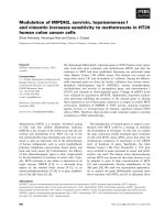
Effects of COX-2 on tumor-associated VEGF expression
We next addressed whether COX-2 enhanced the prolif-
eration of NSCLC cells. As demonstrated in Figure 1
treatment with exogenously applied COX-2 induced a
prominent dose-dependent increase in the proliferation
of the tumor cells used in these assays; in contrast,
COX-2 failed to promote the proliferation of HBE cells,
used as controls. A linear regression analysis of cell via-
bility showed the EC
50
values for enhancement of tumor
cell growth by COX-2 (concentration required to
increase growth by ~50% after a 24-hour treatment)
were 8.95 × 10
-3
, 11.20 × 10
-3
, and 8.44 × 10
-3
μMfor
A549, H460 and A431 cells, respectively.
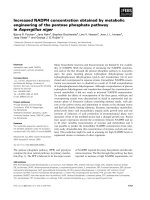
We further addressed whether COX-2 enhanced
tumor-associated VEGF expression in NSCLC cells,
treating tumor ce ll lines with different concentrations of
COX-2 (0.5-, 1-, 1.5-, and 2-times the EC
50
value). As
shown in Figure 2 COX-2 increased the geometric mean
fluorescence intensity of VEGF expression in a dose-
dependent manner. This phenomenon was especially
obvious in A549 and H460 cells. As demonstrated in
Figure 1 and 2, the doses of COX-2 that optimally
induced VEGF expression without causing a cytotoxic
effect were 13.43 × 10
-3
, 16.8 × 10
-3
, and 12.66 × 10
-3
μM in A549, H460, and A431 cells, respectively.
Effect of AH6809, KT5720, and RO-31-8425 on COX-2
stimulation of tumor-associated VEGF expression
To explore the mechanism underlying COX-2 involve-
ment in tumor-associated VEGF expression, we employed
select ive inhib itors of several intracellular signaling path-
ways. As shown in Figure 3 treatment of NSCLC tumor
cells with the PKC inhibitor RO-31-8425 caused a promi-
nent decrease in COX-2-dependent VEGF expression,
reducing COX-2-stimulated VEGF expression by 51.1% in
A549 cells (p < 0.01), 41.2% in H460 cells (p <0.01),and
23.2% in A431 cells (p < 0.01) compared with controls.
Inhibition of PKA with the selective inhibitor KT5720 did
not significantly inhibit COX-2-depend ent, tumor-
Luo et al. Journal of Experimental & Clinical Cancer Research 2011, 30:6
/>Page 5 of 10
associated VEGF expression in NSCLC cells. Notably,
AH680, a selective antagonist of EP1/EP2 receptors,
exerted an inhibitory effect on COX-2-dependent VEGF
expression in NSCLC cells (p < 0.05).
Effect of PMA on COX-2 stimulation of tumor-associated
VEGF expression
To confirm that PKC played a key role in COX-2-
dependent, tumor-associated VEGF expression, we trea-
ted NSCLC cell lines with the PKC activator PMA. As
demonstrated in Figure 4 treatment with both COX-2
and PMA significantly increased the geometric mean
fluorescence intensity of VEGF expression in A549,
H460, and A431 cells compared to treatment with
COX-2 or PMA alone (p < 0.01 for all).
Discussion
Tumor-induced angiogenesis is a ca rdinal attribute of
malignant disease [16]. The microvasculature formed
with new bloo d vessels in tumor stroma me diates trans-
port of nutrients to the tumor cells, and is a prerequisite
for growth of tumors beyond a certain size [17]. It is
known that malignant angiogenesis is induced by speci-
fic angiogenesis-promoting molecules, such as VEGF,
which are highly expressed in various types of solid
tumors and are released by the tumor itself. The result-
ing tumor-induced neovasculature exhibits enhanced
endothel ial cell permeability, and the associated increase
in vascular permeability ma y allow the extravasation of
plasma proteins and formation of extracellular matrix
favorable to endothelial and stromal cell migration [18].
Importantly, certain molecules, such as COX-2, have
been found t o participate in up-regulati on of VEGF in
malignant tissue. COX-2 expression has been imp licated
in the regulation of VEGF in colonic cancer [19], thyr-
oid cancer [20], and nasopharyngeal carcinoma [21].
Previous studie s have demonstrated that COX-2 is able
to induce angiogenesis or promote tumor adhesion and
metastasis [22,23], and also plays a key role in drug
resistance in NSCLC patients [24]. Consistent with this,
COX-2 expression has been detected immunohisto-
chemically in NSCLC specimens, including all squamous
cell lung cancer and 70% of adenocarcinomas [25].
However, the involvement of COX-2 in the angiogenic
response of tumor cells and t he role of COX-2 in up-
regulating VEGF r elease by NSCLC cells has been
unclear. In order to elucidate the relationship between
COX-2 and tumor-associated VEGF expression, we first
investigated the association of COX-2 expression in
NSCLC tissue samples with clinical and pathologic fac-
tors, including VEGF expression and MVD. Our find-
ings indicated a significant difference in VEGF staining
and MVD between NSCLC spec imens with strong and
weak COX-2 expression. When all of the predictors
were included in a multivariate analysis, COX-2 expres-
sion retained its significant association with VEGF stain-
ing and MVD, demonstrating that COX-2 expression is
an independent predictive factor for changes in both
VEGF expression and MVD in NSCLC tissue. These
results sugge st that COX-2 may contribute to ma intain-
ing a high level of VEGF in NSCLC tissue, thereby play-
ing an important role in tumor-induced angiogenesis.
Previo us reports provide no insight into how up-regu -
lating COX-2 might mediate tumor-ass ociated VEGF
expression in NSCLC tissue i n a physiological context.
In order to address this question, we assessed changes
in tumor-associated VEGF expression in NSCLC cells
that accompany changes in COX-2 by treating cells
directly with COX-2 protein. Because this is the first
such study, there was no available information on the
concentrations of COX-2 that are effective in stimulat-
ing proliferation i n NSCLC cells in vitro. Accordingly,
we used an MTT assay to investigate the characteristic
Figure 1 Cell viability (MTT assay) for determination of EC
50
of
COX-2 stimulation in non-small cell lung cancer cell lines. (A)
Prominent increasing in population of A549, H460, and A431 cells
were showed in COX-2 concentration of 0, 3.82 × 10
-13
mol/ml, and
2.29 × 10
-12
mol/ml, respectively (×200). (B) Curves of cell viability
(MTT assay) for determination of EC
50
in A549 (y = 0.0511× +
0.0424), H460 (y = 0.0408× + 0.043), and A431 cells (y = 0.0543× +
0.0415) were showed. Calculated EC
50
were 8.95 nmol/L in A549,
11.2 nmol/L in H460, and 8.44 nmol/L in A431 cells.
Luo et al. Journal of Experimental & Clinical Cancer Research 2011, 30:6
/>Page 6 of 10
Figure 2 Determination of the effective concentration for COX-2 mediated VEGF up-regulation in NSCLC cells.(A) In A549 cells, red,
purple, green and blue curves represented COX-2 concentrations of 0, 9.17 × 10
-12
mol/ml, 1.83 × 10
-11
mol/ml, and 7.34 × 10
-11
mol/ml, with G-
mean fluorescence intensity of 26.32, 32.93, 35.45, and 39.98, respectively. (B) In H460 cells, red, purple and green curves represented COX-2
concentrations of 0, 9.17 × 10
-12
mol/ml, 3.67 × 10
-11
mol/ml, with G-mean fluorescence intensity of 25.33, 29.56, and 34.99, respectively. (C) In
A431 cells, red, purple, green and blue curves represented COX-2 concentrations of 0, 9.17 × 10
-12
mol/ml, 1.83 × 10
-11
mol/ml, and 7.34 × 10
-
11
mol/ml, with G-mean fluorescence intensity of 25.98, 33.23, 36.09, and 38.89, respectively. (D) COX-2 mediated VEGF up-regulation was shown.
G-mean, geometric mean.
Figure 3 COX-2 mediated VEGF up-regulation in NSCLC cells was changed with treatment with several reagents. VE GF expression after
treatment with several reagents was showed in A549 (A), H460 (B), and A431 cells (C). Red curve indicated cells treatment with COX-2, black
curve indicated with COX-2 and AH6809, green curve indicated with COX-2 and KT5720, and blue curve indicated with COX-2 and RO-31-8425.
Comparison of G-mean fluorescence intensity of VEGF was showed (D). G-mean, geometric mean.
Luo et al. Journal of Experimental & Clinical Cancer Research 2011, 30:6
/>Page 7 of 10
Figure 4 Eff ect of COX-2 and PAM on tumor associated VEGF expression in NSCLC cells. VEGF expression after treatment with PMA was
showed in A431, A549, and H460 (A). Red curve indicated no treatment, black curve indicated treatment with PMA. VEGF expression after
treatment with COX-2 and PMA was showed in A431, A549, and H460 (B). Red curve indicated treatment with COX-2, black curve indicated
treatment with COX-2 and PMA. Comparison of G-mean fluorescence intensity of VEGF was showed (C). G-mean, geometric mean.
Luo et al. Journal of Experimental & Clinical Cancer Research 2011, 30:6
/>Page 8 of 10
tumor cell responses to COX-2 as a chemical agent in
three NSCLC cell lines. Crucially, our data demon-
strated that A549, H460, and A431 tumor cells were sti-
mulated to proliferate by exogenously app lied COX-2,
wherea s normal bronchial epithelial cells (HBE) used as
a control were not. The EC
50
values for COX-2 in sti-
mulating proliferation were not substantially different
among the tested tumor cell lines. Based on our data, it
is reasonable to propose that COX-2 is an active agent
in these tested NSCLC cells. We also found using flow
cytometry that COX-2 exposure up-regulated tumor-
associated VEGF expression in NSCLC cells, exhibiting
prominent dose-dependent activity. This phenomenon
was particularly e vident in A549 lung adenocarcinoma
cells. Thus, tumor-associated expression of VEGF may
be promoted by COX-2 in NSCLCs.
Although COX-2-mediated VEGF up-regulation in
NSCLC has been well studied by several groups [26,27 ],
the detailed molecular mech anism underlying this pro-
cess had not been previously demonstrated. To explore
the linkage between COX-2 and tumor-associated VEGF
expression, we employed inhibitors of protein kinase sig-
naling pathways. Our demonstration that COX-2 stimu-
lation of tumor-associated VEGF expression was
decreased in NSCLC cells by treatment with selective
PKC inhibitors, but not by selective PKA inhibitors,
indicates that the contribution of COX-2 to tumor-
associated VEGF expression in NSCLC may involve
the PKC pathway with no involvement of PKA. This
interpretation is supported by results obtained using the
PKC act ivator PMA, which significantly enhanced COX-
2-stimulated, tumor-associated VEGF expression with-
out altering VEGF expressionwhenusedalone.Thus,
the PKC pathway likely plays a rol e in COX-2-mediated
VEGF up-regulation in NSCLC.
Interestingly, our finding that antagonism of the PGE
2
receptor decreased COX-2-mediated VEGF up-regulation
in NSCLC cells, especially in H460 large-cell lung cancer
cells, confirms that PGE
2
, a downstream product of
COX-2 acti vity, may p articipate in COX-2-mediated VEG F
up-regulation. Recently, sequential changes in COX-2,
downstream PGE
2
, and protein kinase signal transduction
pathways have been demonstrated in some tumors [28,29].
PGE
2
binds to four subtypes of G-protein-coupled recep-
tors–EP1, EP2, EP3, EP4–that activate intracellular signal-
ing cascades. These receptors are distributed on the cell
surface and their action depends on PGE
2
concentrat ion
[30]. The EP1 receptor couples to the G
q
subtype and
mediates a rise in intracellular calcium concentration; EP2
and EP4 receptors are coupled to the adenylyl cyclase-
stimulating G protein G
s
, and mediate a rise in cAMP con-
centration; by contrast, the EP3 receptor couples to G
i
,
inhibiting cyclic AMP generation [31]. Results obtained
with AH6809, which inhibits both EP1 and EP2, suggest a
G
q
-orG
s
-mediated mechanism, although additional stu-
dies wil l be re quired to confirm which receptor is th e main
target on the NSCLC cell surface. Another interesting find-
ing of the present study wa s the absence of a pro minent
decrease in COX-2-dependent VEGF activity following
inhibition of PGE
2
receptor(s) in A549 and A431 cells.
This result suggests that other prostaglandin components
may participate in pathways leading from COX-2 to VEGF
expression in different NSCLC cells.
Conclusions
Our findings demonstrate that COX-2 expression in
tumor tissue was an independent predictor of VEGF
expression and MVD in NSCLC patients, and COX-2
may be a stimulator of tumor-associated VEGF activity
in NSCLC tissue. COX-2-dependent VEGF up-regula-
tion in NSCLC may involve the PKC pathway with no
involvement of PKA. Mo reover, different downstream
prostaglandin products of COX-2 activity may partici-
pate in the changes linking COX-2 to VEGF expression
in different NSCLC cells.
Acknowledgements
This study was supported by grants from the Key Scientific and
Technological Projects of Guangdong Province (Grant no. 2008B030 301311
and 2008B030301341).
Author details
1
Department of Thoracic Surgery, The First Affiliated Hospital, Sun Yat-sen
University, Guangzhou (510080), Guangdong, People’s Republic of China.
2
Private Medical Center, The First Affiliated Hospital, Sun Yat-sen University,
Guangzhou (510080), Guangdong, People’s Republic of China.
3
Center for
Stem Cell Biology and Tissue Engineering, Sun Yat-sen University, Key
Laboratory for Stem Cells and Tissue Engineering, Ministry of Education,
Guangdong, People’s Republic of China.
4
Department of Thoracic Surgery,
The Fifth Affiliated Hospital, Sun Yat-sen University, Zhuhai (519000),
Guangdong, People’s Republic of China.
Authors’ contributions
The authors contributed to this study as follows: HL, ZC, and HJ conceived
of the study; HJ, MZ, SC, LY, JZ, and BZ performed experiments; TW analyzed
data and prepared the figures; CZ and HJ drafted the manuscript. All
authors have read and approved the final manuscript.
Competing interests
The authors declare that they have no competing interests.
Received: 18 December 2010 Accepted: 10 January 2011
Published: 10 January 2011
References
1. Smith WL, DeWitt DL, Garavito RM: Cyclooxygenases: structural, cellular,
and molecular biology. Annu Rev Biochem 2000, 69:145-82.
2. Warner TD, Mitchell JA: Cyclooxygenases: new forms, new inhibitors, and
lessons from the clinic. FASEB J 2004, 18:790-804.
3. Hosomi Y, Yokose T, Hirose Y, Nakajima R, Nagai K, Nishiwaki Y, Ochiai A:
Increased cyclooxygenase 2 (COX-2) expression occurs frequently in
precursor lesions of human adenocarcinoma of the lung. Lung Cancer
2000, 30:73-81.
4. Wolff H, Saukkonen K, Anttila S, Karjalainen A, Vainio H, Ristimäki A:
Expression of cyclooxygenase-2 in human lung carcinoma. Cancer Res
1998, 58:4997-5001.
Luo et al. Journal of Experimental & Clinical Cancer Research 2011, 30:6
/>Page 9 of 10
5. Hida T, Yatabe Y, Achiwa H, Muramatsu H, Kozaki K, Nakamura S, Ogawa M,
Mitsudomi T, Sugiura T, Takahashi T: Increased expression of
cyclooxygenase 2 occurs frequently in human lung cancers, specifically
in adenocarcinomas. Cancer Res 1998, 58:3761-4.
6. Diperna CA, Bart RD, Sievers EM, Ma Y, Starnes VA, Bremner RM:
Cyclooxygenase-2 inhibition decreases primary and metastatic tumor
burden in a murine model of orthotopic lung adenocarcinoma. J Thorac
Cardiovasc Surg 2003, 126(4):1129-33.
7. Grimminger PP, Stöhlmacher J, Vallböhmer D, Schneider PM, Hölscher AH,
Metzger R, Danenberg PV, Brabender J: Prognostic significance and
clinicopathological associations of COX-2 SNP in patients with nonsmall
cell lung cancer. J Oncol 2009, 139590, Epub 2009 Nov 22.
8. Soslow RA, Dannenberg AJ, Rush D, Woerner BM, Khan KN, Masferrer J,
Koki AT: COX-2 is expressed in human pulmonary, colonic, and
mammary tumors. Cancer 2000, 89(12):2637-45.
9. Wolff H, Saukkonen K, Anttila S, Karjalainen A, Vainio H, Ristimaki A:
Expression of cyclooxygenase-2 in human lung carcinoma. Cancer
Research 1998, 58(22):4997-5001.
10. Ochiai M, Oguri T, Isobe T, Ishioka S, Yamakido M: Cyclooxygenase-2 (COX-
2) mRNA expression levels in normal lung tissues, and nonsmall cell
lung cancers. Jpn J Cancer Res 1999, 90:1338-43.
11. Tsujii M, Kawano S, DuBois RN: Cyclooxygenase-2 expression in human
colon cancer cells increases metastatic potential. Proc Natl Acad Sci USA
1997, 94:3336-40.
12. Nie D, Honn KV: Cyclooxygenase, lipoxygenase and tumor angiogenesis.
Cell Mol Life Sci 2002, 59:799-807.
13. Nie D, Lamberti M, Zacharek A, Li L, Szekeres K, Tang K, Chen Y, Honn KV:
Thromboxane A(2) regulation of endothelial cell migration,
angiogenesis, and tumor metastasis. Biochem Biophys Res Commun 2000,
267:245-51.
14. Sobin LH, Wittekind C: International Union Against Cancer (UICC) TNM
classification of malignant tumors. New York, NY: Wiley-Liss;, 6 2002,
99-103.
15. Travis WD, Brambilla E, Muller-Hermelink HK: WHO classification of tumors.
Pathology and Genetics. Tumors of lung, pleura, thymus and heart. IARC
Press, Lyon; 2004, 9-124.
16. Samuelsson B, Morgenstern R, Jakobsson PJ: Membrane prostaglandin E
synthase-1: a novel therapeutic target. Pharmacol Rev 2007, 59(3):207-24.
17. Folkman J, Klagsbrun M: Angiogenic factors. Science 1987, 235:442-7.
18. Gupta MK, Qin RY: Mechanism and its regulation of tumor-induced
angiogenesis. World J Gastroenterol 2003, 9(6):1144-55.
19. Garcea G, Sharma RA, Dennison A, Steward WP, Gescher A, Berry DP:
Molecular biomarkers of colorectal carcinogenesis and their role in
surveillance and early intervention. Eur J Cancer 2003, 39:1041-52.
20. Siironen P, Ristimäki A, Narko K, Nordling S, Louhimo J, Andersson S,
Haapiainen R, Haglund C: VEGF-C and COX-2 expression in papillary
thyroid cancer. Endocrine-Related Cancer 2006, 13:465-73.
21. Murono S, Inoue H, Tanabe T, Joab I, Yoshizaki T, Furukawa M, Pagano JS:
Induction of cyclooxygenase-2 by Epstein-Barr virus latent membrane
protein 1 is involved in vascular endothelial growth factor production in
nasopharyngeal carcinoma cells. PNAS 2001, 98(12):6905-10.
22. Petersen C, Baumann M, Petersen S: New targets for the modulation of
radiation response–selective inhibition of the enzyme cyclooxygenase 2.
Curr Med Chem Anticancer Agents 2003, 3(5):354-9.
23. Krysan K, Reckamp KL, Dalwadi H, Sharma S, Rozengurt E, Dohadwala M,
Dubinett SM: Prostaglandin E2 activates mitogen-activated protein
kinase/Erk pathway signaling and cell proliferation in non-small cell lung
cancer cells in an epidermal growth factor receptor-independent
manner. Cancer Res 2005, 65(14):6275-81.
24. Kang HK, Lee E, Pyo H, Lim SJ: Cyclooxygenase-independent down-
regulation of multidrug resistance-associated protein-1 expression by
celecoxib in human lung cancer cells. Mol Cancer Ther 2005, 4(9):1358-63.
25. Wolff H, Saukkonen K, Anttila S, Karjalainen A, Vainio H, Ristimäki A:
Expression of cyclooxygenase-2 in human lung carcinoma. Cancer Res
1998, 58:4997-5001.
26. Leahy KM, Ornberg RL, Wang Y, Zweifel BS, Koki AT, Masferrer JL:
Cyclooxygenase-2 inhibition by celecoxib reduces proliferation and
induces apoptosis in angiogenic endothelial cells in vivo. Cancer Res
2002, 62(3):625-31.
27. Seno H, Oshima M, Ishikawa T, Oshima H, Takaku K, Chiba T, Narumiya S,
Taketo M: Cyclooxygenase 2- and prostaglandin E
2
receptor EP
2
-
dependent angiogenesis in Apc
Δ716
mouse intestinal polyps. Cancer Res
2002, 62:506-511.
28. Zheng Y, Ritzenthaler JD, Sun X, Roman J, Han S: Prostaglandin E2
stimulates human lung carcinoma cell growth through induction of
integrin-linked kinase: the involvement of EP4 and Sp1. Cancer Res 2009,
69(3):896-904.
29. Mayoral R, Fernández-Martínez A, Boscá L, Martín-Sanz P: Prostaglandin E2
promotes migration and adhesion in hepatocellular carcinoma cells.
Carcinogenesis 2005, 26(4):753-61.
30. Okuyama T, Ishihara S, Sato H, Rumi Ma, Kawashima K, Miyaola Y,
Suetsugu H, Kazumori H, Cava CF, Kadowaki Y, Fukuda R, Kinoshita Y:
Activation of prostaglandin E2-receptor EP2 and EP4 pathways induced
growth inhibition in human gastric carcinoma cell lines. J Lab Clin Med
2002, 140:92-102.
31. Dubinett SM, Mao JT, Hazra S: Focusing Downstream in Lung Cancer
Prevention:15-Hydroxyprostaglandin Dehydrogenase. Cancer Prev Res
2008, 1(4):223-5.
doi:10.1186/1756-9966-30-6
Cite this article as: Luo et al.: Cyclooxygenase-2 up-regulates vascular
endothelial growth factor via a protein kinase C pathway in non-small
cell lung cancer. Journal of Experimental & Clinical Cancer Research 2011
30:6.
Submit your next manuscript to BioMed Central
and take full advantage of:
• Convenient online submission
• Thorough peer review
• No space constraints or color figure charges
• Immediate publication on acceptance
• Inclusion in PubMed, CAS, Scopus and Google Scholar
• Research which is freely available for redistribution
Submit your manuscript at
www.biomedcentral.com/submit
Luo et al. Journal of Experimental & Clinical Cancer Research 2011, 30:6
/>Page 10 of 10