báo cáo khoa học: "Overexpression of LCMR1 is significantly associated with clinical stage in human NSCLC" ppt
Bạn đang xem bản rút gọn của tài liệu. Xem và tải ngay bản đầy đủ của tài liệu tại đây (1.92 MB, 8 trang )
RESEARCH Open Access
Overexpression of LCMR1 is significantly
associated with clinical stage in human NSCLC
Liangan Chen
*
, Zhixin Liang
*
, Qing Tian, Chunsun Li, Xiuqing Ma, Yu Zhang, Zhen Yang, Ping Wang, Yanqin Li
Abstract
Background: Lung cancer is one of the most common human cancers and the leading cause of cancer death
worldwide. The identification of lung cancer associated genes is essential for lung cancer diagnosis and treatment.
Methods: Differential Display-PCR technique was used to achieve the novel cDNA, which were then verified by
real-time PCR. Northern blot was utilized to observe the expression of LCMR1 in different human tissues. 84 cases
human NSCLC tissues and normal counterparts were analyzed for the expression of LCMR1 by
immunohistochemistry.
Results: A novel 778-bp cDNA fragment from human large cell lung carcinoma cell lines 95C and 95D was
obtained, and named LCMR1 (Lung Cancer Metastasis Related protein 1). LCMR1 was differentially expressed in
different human tissues. LCMR1 was strongly overexpressed in NSCLC and its expression was significantly
associated with clinical stage.
Conclusion: Our data indicated that LCMR1, strongly overexpressed in NSCLC, might have applications in the
clinical diagnosis and treatment of lung cancer.
Introduction
The development of new therapeutics and diagnostics of
cancer rely on the understanding of carcinogenesis
mechanisms. Genes dysregula ted significantly in tumor
tissues compared with their normal counterparts are
always considered as biomarkers or closely associated
with carcinogenesis. Over the past two decades plentiful
efforts have been devoted to the identification of genes
involved in cancer development [1].
Many approaches have been used to compare gene
expression between two different physiological states.
Differential Display (DD) is a useful method to compare
patterns o f gene expression in RNA samples of different
types or under different biological conditions [2,3]. The
technique produces partial cDNA fragments by a combi-
nation of reverse transcription and PCR of randomly
primed RNA. Changes in the expression level of genes
are identified after separation of the cDNA fragments
produced in an arbitrarily primed polymerase chain
reaction on a sequencing-type gel. Combined with RNA
expression verification, Differential Display is a powerful
method for generating high confidence hits in the
screening of hundreds of potential differentially
expressed transcripts.
Lung cancer is one of the most common human cancers
and the leading cause of cancer death worldwide [4,5].
With the same genetic backgrounds but different meta-
static potential, 95C and 95D cell lines were subcloned
from a poorly differentiated human large cell lung carci-
noma cell line PLA-801 by Dr. Lezhen Chen (Department
of Pathology, Chinese PLA General Hospital), which were
suitable for Differential Display analysis. Nude mice incu-
bated wit h 95D cells showed earlier and more metastasis
than incubated with 95C cells [6,7]. Although the impor-
tance of tumorigenesis has been realized and studied,
limited knowledge is known about its associated genes and
signal networks. Understanding further more players and
intrinsic processes involved in carcinogenesis could lead to
effective, targeted strategies to prevent and treat cancer.
In the present study, we found that LCMR1 was expressed
significantly higher in 95 D cell line compared to 95C using a
combination of DD-PCR and rea l-time PCR. W e then inves-
tigated its expression in various human tissues by northern
blot. Recombinant LCMR1 protein was expressed and its
specific polyclonal antibody was generated. To examine its
* Correspondence: ;
Department of Respiratory Diseases, Chinese PLA General Hospital, Beijing
100853, PR China
Chen et al. Journal of Experimental & Clinical Cancer Research 2011, 30:18
/>© 2011 Chen et al; licensee BioMed Central Ltd. This is an Open Access article distributed under the terms of the Creative Co mmons
Attribution License ( which permits unrestricted use, distribution, and reproduction in
any medium, provided the original work is properly cited.
involvement in carcinogenesis, 84 specimen s of NSCLC
patients were examined for the expression of LCMR1 by
immunohistochemistry analysis. Our results strongly sug-
gested that LCMR1 was significantly overexpressed in
human NSCLC and its expression was closely associated
with c linical stage o f patients w ith NSCLC, which may have
applications in lung cancer dia gnosis and treatment.
Materials and methods
Cell lines
95C and 95D cell lines were subcloned from a poorly
differentiated human large cell lung carcinoma cell line
PLA-801 and kindly provided by Dr. Lezhen Chen
(Department of Pathology, Chinese PLA General H ospi-
tal, China). Both cell lines were cultured in RPMI 1640
medium, supplemented with 10% fetal bovine serum,
100 μg/ml penicillin, and 100 μg/ml streptomyci n at 37°
C in a humidified 5% CO
2
incubator.
RNA extraction and cDNA synthesis
Total RNA was prepared using Trizol reagent (Invitro-
gen, CA, USA) according to the manufacturer’s instruc-
tions. RNA was treated with RNase (Invitrogen) in the
presence of 50 μM T7 (dT12) AP1, T7 ( dT12) AP5 and
T7 (dT12) A P8 primers in 20 μl RT buffer (1× Super-
script II RT buffer, 10 mM DTT, 0.025 mM dNTP), at
25°C for 5 minutes, followed by 50°C for 50 minutes.
Reverse transcriptase was inactivated at 70°C for 15
minutes.
Differential display and full-length gene cloning
Differential display was performed using Hieroglyph
mRNA Profile Kit (Beckman, CA, USA). Briefly, PCR
amplification was don e using 1.5 μl of the cDNA, primed
with arbitrary P primer and anchored T primer. Amplifi-
cation at (95°C 2 minutes) 1 cycle, (92°C for 15 seconds,
50°C for 30 seconds, 72°C for 2 minutes) 4 cycles, (92°C
for 15 seconds, 60°C for 30 seconds, 72°C for 2 minutes)
30 cycles, followed by a final extension at 72°C for 7 min-
utes on a GeneAmp PCR system 9600 (Perkin-Elmer,
Norwalk, USA). Following amplification of randomly
primed mRNAs by RT-PCR, the cDNA products were
heated at 95°C for 2 minutes and separated on a denatur-
ing 5.6% p olyacrylamide gel at 5 5°C for 5 hours using a
Genomyx LR DNA Sequencer (Beckman), under 3000 V.
Bands exclusively present in either of two samples were
considered as candidates of differentially expressed tran-
scripts, which were excised, eluted, re-amplified, and sub-
cloned into the T easy vector (Promega, San Luis Obispo,
CA, USA). The sequence reactions were performed by
Invitrogen. Sequence homology to published database
was analyzed with the BLAST p rogram at the internet
site of NCBI (National Center for Biotechnology Infor-
mation). 5’-RACE (rapid amplification of cDNA 5’ ends)
and 3’-RACEwereusedtoisolatethecompletecDNA.
The human Marathon-ready cDNA (Clontech, Heidel-
berg, Germany) served as the template.
Real-time quantitative reverse transcription polymerase
chain reaction
We measured LCM R1 gene expression in 95C and 95D
cell lines by real-time quantitative RT-PCR in an ABI
PRISM 7500 Sequence Detection System. The real-time
RT-PCR allows, by means of fluorescence emission, the
identification of the cycling point when PCR product is
detectable. The Ct value inversely correlates w ith the
starting quantity of target mRNA. Measurements were
performed in duplicate and the controls were included
in which the reaction mixture contained no cDNA. The
amount of target mRNA after normalized to the loading
control b-actin was calculated by the Ct method. Pri-
mers for b-actin and LCMR1 mRNAs were chosen
using the Primer Express 2.0 software (Applied Bio-
systems, Foster City, USA). Primers for LCMR1 were:
5’-AACAGAGCCGTACCCAGG AT-3’ (F orward) and
5’-GGGTGGTCTGGACATTGTC -3’ (Reverse). Primers
for b-actin were: 5’-CATGTACGTTGCTATCCAGGC-
3’ (Forward) and 5’-CTCCTTAATGTCACGCACGAT-
3’ (Reverse). Primers were synthesized by Invitrogen.
RNA expression analysis by northern blot in human
normal tissues
LCMR1 expression was analyzed by multiple tissue
northern blots (MTN) in a panel of following normal
tissues (Clontech): brain, heart, skeletal muscle, colon,
thymus, spleen, kidney, liver, small intestine, placenta,
lung, and peripheral blood leukocytes. Hybridization was
performed using 25 ng of a gene-specific 32P-labeled
DNA probe derived from LCMR1 cDNA. This gene-spe-
cific cDNA fragment was radiolabelled using a Prime-A-
Gene Labeling System (Promega), hybridized overnight
at 68°C using ExpressHyb Hybridization Solution (Clon-
tech), washed, and exposed to Kodak XAR-5 X-ray film
with an intensifying screen (Eastman Kodak Co, Roche-
ster, NY, US).
Expression and polyclonal antibodies preparation of
LCMR1 protein
The plasmid pGEX-5T-LCMR1 was constructed. The
GST-LCMR1 protein expression was induced by adding
0.6 mM IPTG to the transformed E. coli and the bac-
teriawereincubatedat20°Cfor4hours.Thedegreeof
expression was evaluated by sodium dodecy l sulfate-
polyacrylamide gel electrophoresis (SDS-PAGE). The
GST-LCMR1 fusion protein was purified by affinity
chromatography using glutathione-agarose resin (GE
Healthcare). The New Zealand white rabbits were given
intradermal injections of purified GST-LCMR1 fusion
Chen et al. Journal of Experimental & Clinical Cancer Research 2011, 30:18
/>Page 2 of 8
protein and the antibody against LCMR1 was prepared.
The titer of antiserum was determined by an indirect
ELISA.
Cases and Clinical Data
We studied a consecutive series of 84 cases primary
NSCLC cancers diagnosed and treated between 2005
and 2007 at the Department of thoracic surgery,
Chinese PLA General Hospital, Beijing, China. None of
the patients had received radiotherapy or neoadjuvant
therapy before surgery. Metastatic lymph nodes of
51 cases in this group were also examined for the
expression of LCMR1. The duration of 65 cases follow-
up ranged from 5 to 39 months (median, 3 1 months).
Tumor characteristics, including histologic grade, lymph
node status, and clinical stage, were routinely assessed
by pathologists.
Immunohistochemical analysis
The se ctions were dewaxed with xylene and rehydrated
through an ethanol g radient into water. After endogen-
ous peroxidase activity was quenched with 3% H
2
O
2
for
30 minu tes, sections were digested with 0.1% trypsin at
37°C for 2 0 minutes. After phosphate-buffered saline
(PBS) washing, nonspecific antibody binding was
blocked by i ncubating the slides with 10% normal goat
nonimmune serum for 30 minutes at 37°C. Sections
were incubated at 4°C overnight with the self-made rab-
bit polyclonal primary antibody against human LCMR1
at a 1:200 dilution. After PBS washing, sections were
incubated wit h biotinylated secondary antibody for
30 minutes at 37°C and then with horseradish peroxi-
dase-labeled streptavidin for 30 minutes at 37°C.
After PBS washing, sections were developed using 3,3V-
diaminobenzidine (Sigma-Aldrich). Sections were washed
in running tap water and lightly counterstained with
hematoxylin, followed by dehydration and coverslip
mounting. Negative controls were obtained by omitting
the primary antibody [8].
Statistical analysis
The criterion for a positive reaction was a single epithelial
cell with yellow particles in its plasma membrane and
cytoplasm. Immunostaining was assessed in a blinded
manner for extent and intensity. In brief, a sample with no
positive epithelial cells was scored as 0, that with less than
25% total positive epithelial cells w as scored as 1+, that
with positive epithelial cells accounting for more than 25%
but less than 50 % of the total was scored as 2+, that with
more than 50% but less than 75% positive cells was scored
as 3+, and that with more than 75% positive cells was
scored as 4+. The intensity of immunostaining was scored
semiquantitatively as follows: no obvious yellow particle in
epithelial cell plasma membrane or cytoplasm as 0; with
light yellow particles as 1+ (weak); with general yellow par-
ticles as 2+ (moderate); and with deep yel low particles as
3+ (strong). For each case, an immunoscore was calculated
as the product of 2 scores assessed separately. Statistical
analysis was performed using SPSS 17 software (SPSS, Inc,
Chicago, IL, USA). The differential expression of LCMR1
protein between tumorous tissues and normal tissues was
determined by Mann-Whitney U-test. The correlations
between LCMR1 expression and clinicopathologic charac-
teristics were analyzed using Pearson c
2
analysis. The
influence of each variable on the expression of LCMR1
was assessed by logistic regression analysis. In survival
analysis, Kaplan-Meier curves were drawn, univariate and
multivariate analyses in a Cox proportional hazards model
were used for LCMR1 scores. All statistical tests were 2-
sided, and P values of 0.05 or less were considered statisti-
cally significant.
Results
Cloning and identification of a novel gene differentially
expressed in 95C and 95D cell lines using DD-PCR
In order t o find lung cancer metastasis related genes,
the DD-PCR method was used to identify genes differ-
entially expressed in human 95C and 95D cell lines,
which have the same genetic backgrounds but different
metastatic potential. Several cDNAs were found
expressed differentially in these two cells (Figure 1A).
These fragments were subcloned into T easy vector,
sequenced, and analyzed for nucleotide and amino acid
homology in the GenBank database. Of these, a 778 bp
cDNA fragment, designated as P9, expressed higher in
95D cells than in 95C cells, did not show a significant
homology with any nucleotide/amino acid sequence in
the database, but has many supports of EST. After align-
ment in Genbank Genomic Database, we found this
fragment existed in chromosome 11 discontinuously.
These suggested that this cDNA might code a novel
gene, and thus was selected for further studies. RACE
(rapid amplification of cDNA ends) was used t o get the
complete cDNA. Using P9 as a probe, we obtained the
full-length 949 bp cDNA, nominated as LCMR 1 (Lung
Cancer Metastasis Related gene 1) (Figure 1B). We sub-
mitted this result in 2002 and acquired the Genbank
accession number as AY148462.
LCMR1 cDNA was found to be a novel sequence
without any homology with any known nucleotide/
amino acid sequence in the database. LCMR1 cDNA
was found to be located on human 11q12.1 chromo-
some locus. Analysis of LCMR1 cDNA using the DNA
analysis program r evealed that it has an ORF starting
with an ATG initiation codon at nucleotide 75-77
with a termination codon at nucleotide 606-608. It has a
Chen et al. Journal of Experimental & Clinical Cancer Research 2011, 30:18
/>Page 3 of 8
5’-UTR of 74 bp and a 3’ -UTR of 341 bp. Analysis of
the predicted peptide using Vector NTI DNA analysis
software program revealed that the predicted peptid e of
LCMR1 has 177 amino acid residues with a calculated
molecular mass of 19,950 Da and an isoelectric point of
10.01.
Confirmation of LCMR1 differentially expressed in 95C
and 95D cell lines by real-time PCR and western blot
In order to further confirm the difference of LCMR1
gene expression between 95C and 95D cell lines, we
compared LCMR1 mRNA expression in these two cell
lines by real-time quantitative RT-PCR. As shown in
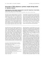
Figure 1 Cloning of a novel gene, LCMR 1. (A) Electrophoresis result of DDRT-PCR in 95C and 95D cells. (B) Nucleotide and amino acid
sequences of LCMR1 cDNA. LCMR1 contains a 74-bp 5’- UTR, a 949-bp ORF, and a 341-bp 3’-UTR. Inframe termination (TER) codons are located
at nt positions 606-608. LCMR1 encodes a 177 aa protein. (C) LCMR1 mRNA expressions in 95C and 95D cells were examined by real-time
quantitative RT-PCR. LCMR1 gene expression level in 95D cells was significantly higher than in 95C cells. (*, P < 0.01) (D) LCMR1 protein
expression in 95D cells was significantly higher than in 95 C cells, examined by western blot. (E) LCMR1 was differentially expressed in the
various human tissue distributions by multiple tissue northern blot (MTN). Numbers indicate tissue types in columns. 1: Brain, 2: Heart, 3: Skeletal
muscle, 4: Colon, 5: Thymus, 6: Spleen, 7: Kidney, 8: Liver, 9: Small intestine, 10: Placental, 11: Lung, 12: Leukocyte.
Chen et al. Journal of Experimental & Clinical Cancer Research 2011, 30:18
/>Page 4 of 8
Figure 1C, LCMR1 gene expression level in 95D cells
was signif icantly higher than in 95C cells. Western blot
analysis with LCMR1 antibody generated as followed
procedure also showed the consistent result (Figure 1D).
Expression of LCMR1 in Various Human Tissues by
Northern blot
Multiple tissue northern blot (MTN) was adopted to
determine the various tissue distribution of human
LCMR1 in RNA level. As shown in Figure 1E, LCMR1
was differentially expressed in all the tissues investi-
gated, with high expression detected in the heart, skele-
tal muscle, kidney, liver, and placental tissue, while low
or hardly detected in others.
Expression and polyclonal antibodies preparation of
recombinant LCMR1 protein
The full length of human LCMR1 CDS region was
cloned into pGEX-5T. Under optimized induction con-
dition, GST-LCMR1 fusion protein was highly expressed
after induction at 20°C with 0.6 mM IPTG for 4 hours
in E.coli. With purification using glutathione-agarose
resin, the fusion protein was separated from those
unwant ed prote ins (Figure 2, lane 5). The GST-LCMR1
fusion protein and GST was recognized clearly by speci-
fic GST antibody (Figure 2, lane 6 and 7). Then the pur-
ified fusion protein was excised and used to immunize
New Zealand rabbits. ELISA was used to determine the
titers of the obtained antibody and the antibody at dif-
ferent dilutions (1000 to 100,000) was reacted with an
equal amount of the recombinant protein (data not
shown). The antibody specificity was examined b y wes-
tern blot (Figure 2, lane 8).
Overexpression of LCMR1 protein in human NSCLC by
immunohistochemistry analysis
There existed various degrees of background staining
that may be caused by tissue processing, such as fixation
and embedding. Because such background staining is
almost nonspecific, occurring in the stromal tissue
(including lymphocytes), we avoided it by counting only
positive epithelial cells. Also, the edge effect was regarded
as negative. Immunohistochemistry analysis results
showed that the expression of LCMR1 was significantly
higher in primary tumor tissue s (84 cases) and metastatic
lymph nodes (51 cases) of NSCLC patients, compared
with its weak expression in adjacent benign tissues
respectively (P < 0.001) (Figure 3, Table 1). There is no
difference in t he expression of LCMR1 between primary
tumor tissues and metastatic lymph nodes (data not
shown). Moreover, immuno staining showed LCMR1 was
expressed mostly in the cytoplasm of cells.
Association between LCMR1 expression and clinical stage
and prognosis of human NSCLC
Patient characteristics, including gender, age (range, 32-
77 years; median, 59 years), smoking status, pathol ogical
type, histologic grade, lymph node metastasis, and clini-
cal stage (classifi ed according to the 2003 TNM classifi-
cation of the International Union Against Cancer) are
recorded in Table 2. Statistical analysis results showed
that LCMR1 expression was significantly associated with
clinical stage of these NSCLC patients (P < 0.05), but no
significant association was found between LCMR1
expression and other clinicopathologic parameters such
as gender, age, smoking status, pathological type, and
histologic grade (Table 2). We further used the stepwise
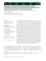
Figure 2 Recombinant LCMR1 protein expression and polyclonal antibody preparation. M, protein marker; lane 1, pGEX-5T-LCMR1 before
induction in E.coli; lane 2, pGEX-5T-LCMR1 after induction in E.coli; lane 3, precipitation after E.coli lysis; lane 4, clear supernatant after E.coli lysis;
lane 5, GST-LCMR1 after purification; lane 6, GST-LCMR1 fusion protein recognized by GST antibody; lane 7, GST protein recognized by GST
antibody; lane 8, GST-LCMR1 fusion protein recognized by LCMR1 polyclonal antibody. (lane 1-5, SDS-PAGE; lane 6-8, western blot).
Chen et al. Journal of Experimental & Clinical Cancer Research 2011, 30:18
/>Page 5 of 8
forward logistic regression analysis to assess the effects
of clinical stages on LCMR1 expression. Logistic regres-
sion analysis revealed that an increased clinical stage
was significantly associated with high LCMR1 expres-
sion (OR = 3.410, P = 0.026) (Table 3). The expression
of LCMR1 protein in metastatic lymph nodes had no
relationship with the clinic features of NSCLC patients
(data not shown).
Survival analysis
Kaplan-Meier analysis of 65 cases of this group, with a
median follow-up of 31 months, showed increased dif-
ference in survival rates between patients with high-level
LCMR1 protein expression and patients with low-level
LCMR1 expression, with overall survival time extension
(Figure 4). But no statistical significance was observed in
overall survival (OS) and progression-free survival (PFS)
of these NSCLC patients using univariate survival analy-
sis and multivariate survival analysis and COX propor-
tional hazard model analysis (data not shown).
Figure 3 LCMR1 expression in human NSCLC. Compared with adjacent normal tissues, LCMR1 was sig nif ica ntly overexpressed in prima ry
tissues and metastatic lymph nodes of patients with NSCLC respectively by immunohistochemistry analysis. (Magnification: ×100).
Table 1 Expression of LCMR1 in primary tumor tissues,
adjacent normal tissues and metastatic lymph nodes
Expression of LCMR1 between two groups P
primary tumor tissues vs paired adjacent normal tissues
(84 cases)
0.000
metastatic lymph nodes vs paired normal tissues (51 cases) 0.000
primary tumor tissues vs paired metastatic lymph nodes
(51 cases)
0.678
Table 2 Correlations between LCMR1 expression and
clinicopathologic characteristics of human NSCLC
n LCMR1 expression P
Negative Positive
Gender
Male 61 12 49 0.147
Female 23 8 15
Age(y)
≥65 22 4 18 0.471
<65 62 16 46
Smoking status
Yes 45 10 35 0.714
No 39 10 29
Pathological type
Adenocarcinoma 41 10 31 0.614
Squamous cell carcinoma 40 10 30
Adenosquamous carcinoma 3 0 3
Histologic grade
PD 28 6 22 0.918
MD 45 11 34
WD 11 3 8
Lymph node metastasis
Yes 62 12 50 0.108
No 22 8 14
Clinical stage
I-II 40 14 26 0.022
III-IV 44 6 38
Abbreviations: WD, well differentiated; MD, moderately differentiated; PD,
poorly differentiated.
Chen et al. Journal of Experimental & Clinical Cancer Research 2011, 30:18
/>Page 6 of 8
Discussion
Tumor development is a complex and multistage process
involving many genetic alterations. It is essential to explore
the molecular mechanisms of tumor formation and pro-
gression to develop rational approaches to the diagnosis
and therapy of cancer, therefore, identifying dysregulated
genes and proteins in neoplasms are critical. 95C and 95D
cells, subcloned from poorly differentiated human large
cell lung carcinoma cell line PLA-801, were of different
metastatic potential, while they came from the same
patient and had similar genetic background [6,7]. We per-
formed DD-PCR between these two cell lines to find some
novel genes involved in lung cancer, and obtained several
cDNA fragments expressed differentially between 95C and
95D cells. All these cDNA fragments were subcloned,
sequenced, searched f or homology with known genes in
the database. Among these, the P9 cDNA fragment did
not reveal homology with any known gene in the database.
Screening the human cDNA library with this specific
cDNA fragment yielded a full-length LCMR1 cDNA, com-
prised of 949 nucleotides , having an ORF encoding for a
177 amino acids peptide. Both nucleotide and amino acid
sequences did not show homology with any gene reported
previously in the database, indicating i t to be a novel
cDNA. It has a 5’-UTR of 74 bp and a 3’-UTR of 341 bp.
The UTRs may be involved in stabilizing mRNA for trans-
lation regulation. Most eukaryotic mRNAs possess
short 5’-UTRs of 20-100 nucleotides that enable efficient
cap-dependent ribosome scanning [9]. We submitted this
result in 2002 and acquired the Genbank accession num-
ber as AY148462. We further confirmed the different
expression of LCMR1 between 95C and 95D cell lines by
real-time quantitative RT-PCR and western blot analysis.
To understand the function of LCMR1, we first investi-
gated LCMR1 mRNA expression in different human nor-
mal tissues by northern blot analysis. The results showed
that LCMR1 was detected in various kinds of human tis-
sues with different expression levels, which suggested th e
functions of LCMR1 might vary in different tissues.
To understand the function of LCMR1,weinvesti-
gated LCMR1 protein expression in 84 cases human
NSCLC tissues by immunohistochemistry analysis. The
results showed that LCMR1 was strongly overexpressed
in NSCLC tissues and metastatic lymph nodes, com-
pared with adjace nt normal tissues. To find out the cor-
relations between LCMR1 expression and the biologic
behavior of NSCLC, we studied clinical data, including
gender, age, smoking status, pathological type, histologic
grade, lymph node metastasis, and clinical stage. Analy-
sis of gender, age, smoking status, pathological type, his-
tologic grade, and lymph node metastasis revealed that
none of them showed a significant correlation with hig h
LCMR1 protein expression. However, high LCMR1
expression was closely associated with clinical stage (P =
0.022). Logistic regression analysis result also showed
that clinical stage was significantly associated with
LCMR1 expression (OR = 3.410, P = 0.026). These
results suggested the critical role of LCMR1 in human
NSCLC development. The Kaplan-Meier analysis of 65
cases of this group showed that LCMR1 expression had
no significance with overall survival, which may be due
to short follow up periods. However, it showed the ten-
dency that positive LCMR1 expression was associated
with poor survival. The results showed that there is no
difference between the levels of LCMR1 expression in
the primary tumors with or without metastasis, neither
between metastatic sites and primary sites. The study on
more pathological specimens would shed light on this
relationship.
LCMR1 was also found to be a member of mamma-
lian Mediator subunits, called MED19 [10,11]. The med-
iator complex is a large collection of DNA binding
transcriptional activators through the action of an inter-
mediary multiprotein coactivator, which controls the
transcription of eukaryotic protein-coding genes with
RNA polymeras e II (pol II) [12]. Specific mediator subu-
nits are dedicated to regulate distinct expression pro-
grams via interactions with relevant gene-specific
transcriptional activators, which lead to activation o f
transcription at the target gene. It has been reported
that normal function of activators, such as VP16 and
p53, interact with different Mediator subunits [ 13].
Table 3 Logistic regression analysis
Wald c
2
P OR
TNM stage 6.995 0.026 3.410
Figure 4 Kaplan-Meier analysis of 65 cases follow-up.The
survival curve showed increased difference in survival rates between
patients with high-level LCMR1 protein expression and patients with
low-level LCMR1 expression, with overall survival time extension.
Chen et al. Journal of Experimental & Clinical Cancer Research 2011, 30:18
/>Page 7 of 8
Recently, it was reported that MED19 (LCMR1) and
MED26 subunits as direct functional targets of the RE1
Silencing Transcription Factor, REST, facilitate d REST-
imposed epigenetic restrictions o n neuronal gene
expression [14]. Mediator serves as a key cofactor and
integrator of signaling in many transcriptional activa-
tions and pathways. Exact temporal and spatial regula-
tion of the transcription of genes is v ital to the
execution of complex gene functions in response to
growth, apoptosis, developmental and homeostatic sig-
nals, etc [15,16]. MED1 has been found to play an
important coregulatory role in the development and
progression of lung adenocarcinoma [17]. Although
Mediator complex has been studied for many years, lim-
ited knowledge was known about MED19/LCMR1. Our
results suggested that LCMR1 has an important clinico-
pathological role in the lung cancer. It will be of consid-
erable interest to further understand these interactions
and elucidate the intrinsic mechanism s, since one of the
most important reasons of cancer development is the
dysfunction of transcriptiona l regulation associated
genes.
In conclusion, we a re the first to identify LCMR1
gene. The present study revealed that the expression of
LCMR1 was significantly up-regulated in primary tissues
and metastatic lymph nodes of patients with NSCLC,
compared with adjacen t normal tissues. Its role in carci-
nogenesis needs to be further investigated. The st rong
correlation between LCMR1 expression and clinical
stage indicates that LCMR1 could serve as a biomarker
for judging the level of malignancy of lung cancer,
which may guide the development of anticancer therapy.
Abbreviations
CDS: coding Sequence; DD: differential display; ELISA: enzyme-linked
immunosorbent assay; ETS: expressed sequence tag; LCMR1: lung cancer
metastasis related protein 1; NSCLC: non-small cell lung cancer; OS: overall
survival; PBS: phosphate-buffered saline; PFS: progression-free survival; RT-
PCR: reverse transcriptase-polymerase chain reaction; UTR: untranslated
Regions.
Acknowledgements
This work was supported by National Natural Science Foundation of China
(30070335, 30370616).
Authors’ contributions
LC and ZL are joint first-authors, and contributed equally to this study. LC
conceived of the work. LC and QT carried out the gene cloning and RNA
expression analysis of LCMR1 in normal human tissues. ZL prepared GST-
LCMR1 protein and antibody. CL participated in the qPCR and drafted the
manuscript. ZL and XM performed immunohistochemistry analysis. CL and
YL carried out qPCR. YZ, ZY, and PW collected the cases and sections. LC
participated in the design and coordination and supervised the whole study.
All authors read and approved the final manuscript. All authors read and
approved the final manuscript.
Competing interests
The authors declare that they have no competing interests.
Received: 13 October 2010 Accepted: 9 February 2011
Published: 9 February 2011
References
1. Santarius T, Shipley J, Brewer D, Stratton MR, Cooper CS: A census of
amplified and overexpressed human cancer genes. Nat Rev Cancer 2010,
10:59-64.
2. Liang P: From differential display to DNA microarrays–a personal
account. J Cell Physiol 2006, 209:653-658.
3. Liang P, Pardee AB: Differential display of eukaryotic messenger rna by
means of the polymerase chain reaction. Science 1992, 257:967-971.
4. Sharma SV, Bell DW, Settleman J, Haber DA: Epidermal growth factor
receptor mutations in lung cancer. Nat Rev Cancer 2007, 7:169-181.
5. Zou X: Epidemiology of lung cancer in china. Chin J Cancer Prev Treat
2007, 14:881-883.
6. Su L, Zhang J, Xu H, Wang Y, Chu Y, Liu R, Xiong S: Differential expression
of cxcr4 is associated with the metastatic potential of human non-small
cell lung cancer cells. Clin Cancer Res 2005, 11:8273-8280.
7. Lu X, Wang J, Li X, Li H, Chen L, Li W: Spontaneous metastasis of clonal
cell subpopulation of human lung large cell carcinoma after
subcutaneous inoculation in nude mice. Chin J Oncol 1989, 11:3-7.
8. Zhang L, Ding F, Cao W, Liu Z, Liu W, Yu Z, Wu Y, Li W, Li Y: Stomatin-like
protein 2 is overexpressed in cancer and involved in regulating cell
growth and cell adhesion in human esophageal squamous cell
carcinoma. Clin Cancer Res 2006, 12:1639-1646.
9. Kozak M: Do the 5’ untranslated domains of human cdnas challenge the
rules for initiation of translation (or is it vice versa)? Genomics 2000,
70:396-406.
10. Guglielmi B, van Berkum NL, Klapholz B, Bijma T, Boube M, Boschiero C,
Bourbon HM, Holstege FC, Werner M: A high resolution protein
interaction map of the yeast Mediator complex. Nucleic Acids Res 2004,
32:5379-5391.
11. Sato S, Tomomori-Sato C, Parmely TJ, Florens L, Zybailov B, Swanson SK,
Banks CA, Jin J, Cai Y, Washburn MP, Conaway JW: A Set of Consensus
Mammalian Mediator Subunits Identified by Multidimensional Protein
Identification Technology. Mol Cell 2004, 14:685-691.
12. Sato S, Tomomori-Sato C, Banks CA, Sorokina I, Parmely TJ, Kong SE, Jin J,
Cai Y, Lane WS, Brower CS, Conaway RC, Conaway JW: Identification of
mammalian mediator subunits with similarities to yeast mediator
subunits srb5, srb6, med11, and rox3. J Biol Chem 2003, 278:15123-15127.
13. Malik S, Roeder RG: Dynamic regulation of pol II transcription by the
mammalian mediator complex. Trends Biochem Sci 2005, 30:256-263.
14. Ding N, Tomomori-Sato C, Sato S, Conaway RC, Conaway JW, Boyer TG:
Med19 and med26 are synergistic functional targets of the re1 silencing
transcription factor in epigenetic silencing of neuronal gene expression.
J Biol Chem 2009,
284:2648-2656.
15. Lewis BA, Reinberg D: The mediator coactivator complex: functional and
physical roles in transcriptional regulation. J Cell Sci 2003, 116:3667-3675.
16. Kornberg RD: Mediator and the mechanism of transcriptional activation.
Trends Biochem Sci 2005, 30:235-239.
17. Yun J, Son C, Um S, Kwon H, Lee K, Choi PJ, Roh M: A different TRAP220
expression in distinct histologic subtypes of lung adenocarcinoma and
the prognostic significance. Lung Cancer 2010.
doi:10.1186/1756-9966-30-18
Cite this article as: Chen et al.: Overexpression of LCMR1 is significantly
associated with clinical stage in human NSCLC. Journal of Experimental &
Clinical Cancer Research 2011 30:18.
Submit your next manuscript to BioMed Central
and take full advantage of:
• Convenient online submission
• Thorough peer review
• No space constraints or color figure charges
• Immediate publication on acceptance
• Inclusion in PubMed, CAS, Scopus and Google Scholar
• Research which is freely available for redistribution
Submit your manuscript at
www.biomedcentral.com/submit
Chen et al. Journal of Experimental & Clinical Cancer Research 2011, 30:18
/>Page 8 of 8