báo cáo khoa học: " Small interference RNA targeting tissue factor inhibits human lung adenocarcinoma growth in vitro and in vivo" ppsx
Bạn đang xem bản rút gọn của tài liệu. Xem và tải ngay bản đầy đủ của tài liệu tại đây (5.17 MB, 11 trang )
RESEARC H Open Access
Small interference RNA targeting tissue factor
inhibits human lung adenocarcinoma growth
in vitro and in vivo
Chengcheng Xu
1
, Qi Gui
2
, Wenshu Chen
1
, Leiming Wu
1
, Wei Sun
1
, Ni Zhang
1
, Qinzi Xu
1
, Jianing Wang
1
and
Xiangning Fu
1*
Abstract
Background: The human coagulation trigger tissue factor (TF) is overexpressed in several types of cancer and
involved in tumor growth, vascularization, and metastasis. To explore the role of TF in biological processes of lung
adenocarcinoma, we used RNA interference (RNAi) technology to silence TF in a lung adenocarcinoma cell line
A549 with high-level expression of TF and evaluate its antitumor effects in vitro and in vivo.
Methods: The specific small interfering RNA (siRNA) designed for targeting human TF was transfected into A549
cells. The expression of TF was detected by reverse transcription-PCR and Western blot. Cell proliferation was
measured by MTT and clonogenic assays. Cell apoptosis was assessed by flow cytometry. The metastatic potential
of A549 cells was determined by wound heal ing, the mobility and Matrigel invasion assays. Expressions of PI3K/Akt,
Erk1/2, VEGF and MMP-2/-9 in transfected cells were detected by Western blot. In vivo, the effect of TF-siRNA on
the growth of A549 lung adenocarcinoma xenografts in nude mice was investigated.
Results: TF -siRNA significantly reduced the expression of TF in the mRNA and protein levels. The down-regulation
of TF in A549 cells resulted in the suppression of cell proliferation, invasion and metastasis and induced cell
apoptosis in dose-dependent manner. Erk MAPK, PI3K/Akt pathways as well as VEGF and MMP-2/-9 expressions
were inhibited in TF-siRNA transfected cells. Moreover, intratumoral injection of siRNA targeting TF suppressed the
tumor growth of A549 cells in vivo model of lung adenocarcinoma.
Conclusions: Down-regulation of TF using siRNA could provide a potential approach for gene therapy against lung
adenocarcinoma, and the antitumor effects may be associated with inhibition of Erk MAPK, PI3K/Akt pathways.
Keywords: Lung adenocarcinoma A549, Tissue factor, RNA interference, Gene therapy
Background
Lung cancer is the leading cause of cancer-related death
worldwide [1,2]. Lung adenocarcinoma, accounted for
approximately 40% of all lung cancers, is currently one
of the most common histological types and its inci-
dence has gradually increased in recent years in many
countries [3].
Tissue factor (TF), a 47-kDa transmemb rane glycopro-
tein, primarily initiates the coagulation cascade by binding
to activated factor VII (FVIIa) [4,5]. Under normal condi-
tions, TF is highly expressed by cells which are not in con-
tact with the blood, such as smooth muscle cells,
mesenchymal and epithelial cells. In addition, numerous
studies have reported that T F is aberrantly expressed in
solid tumors, including cancers of the pancreas, prostate,
breast, colon and lung [6,7], and TF can be detected on
the surface of tumor cells and TF-bearing microparticles
in the blood circulation shed from the cell surface [8,9].
The r ole of TF in coagulation has been much more
focused on, and the association between tumor and coagu-
lation was first revealed by Trousseau as long ago as 1865
[10]. Recently, the roles of TF in tumor growth, angiogen-
esis, and metastasis have become popular fields of
* Correspondence:
1
Department of General Thoracic Surgery, Tongji Hospital, Tongji Medical
College, Huazhong University of Science and Technology, Wuhan, People’s
Republic of China
Full list of author information is available at the end of the article
Xu et al. Journal of Experimental & Clinical Cancer Research 2011, 30:63
/>© 2011 Xu et al; lic ensee BioMed Central Ltd. This is an Open Access article distributed under the terms of the Creative Commons
Attribution Lic ense (http://creativecommons .org/licenses/by/2.0), which permits unrestricted use, distribution, and reproduction in
any medium, provided the original work is properly cited.
research. Precious studies have been implicated that TF
plays an important role in melanoma and pulmonary
metastasis [11,12]. However, no study so far has demon-
strated the antitumor effects and its antitumor mechanism
via inhibition of TF expression by small interfering RNA
(siRNA) in Lung adenocarcinoma. RNA interference
(RNAi) is sequence-specific post-tr anscriptional gene-
silencing process, which is initiated by double-stranded
RNA (e.g. chemically synthetic small interfering RNAs)
and then the RNA-induce d silencing complex (RISC)
degrades targeted mRNA and inhibits the protein expres-
sion [13]. Because of the effective, stable gene suppression
by siRNAs, currently, RNAi technologies are widely used
as knocking down genes in functional genomics [14].
In this study, we successfully used the RNA interfer-
ence (RNAi) technology to silence the expression of TF
in lung adenocarcinoma cell lines A54 9. In vitro and in
vivo experiments described here in, we demonstrate that
the capability of tumor growth and metastasis is reduced,
and apoptosis is induced in TF-siRNA transfected A549
cells. In addition, Molecular mechanisms of the ant itu-
mor effects of TF knockdown are initially revealed, which
could lay a foundation for genetic therapy for lung
adenocarcinoma.
Materials and methods
Cell lines and cell culture
The human lung adenocarcinoma cell lines A549 was pur-
chased from the Institute of Biochemistry and Cell Biol-
ogy, Shanghai Institute for Biological Sciences, Chinese
Academy of Sciences. Cells were grown in RPMI 1640
(Gibco) medium, suppleme nted with 10% fetal bovine
serum (FBS), 100 U/ml penicillin and 100 ug/ml strepto-
mycin in a humidified atmosphere of 5% CO2 at 37 °C.
The cells in the logarithmic phase of growth were used in
all experiments described below.
Specific siRNAs and transfection
One siRNA oligonucleotides targeting human tissue fac-
tor (SiTF) [15] (accession no.M16553, the target mRNA
sequences:5’ -GCGCUUCAGGCACUACAAA-3’ ), one
scrambled non-targeting siRNA (used for a negative con-
trol, Mock) and one fluorescent siRNA were designed
and synthesized by Genepharma Co., Ltd (Shanghai,
China). The sequences were as follows: SiTF, 5’ -
GCGCUUCAGGCACUACAAAtt-3’ (sense) and 5’ -
UUUGUAGUGCCUGAAGCGCtt-3’ (antisense); Mock,
5’ -UUCUCCGAACGUGUCACGUtt-3’ (sense) and 5’-
ACGUGACACGUUCGGAGAAtt-3’ (antisense). The 25
nM, 50 nM and 100 nM siRNAs were transfected into
culture cells with Lipofectamine 2000 reagent (Invitro-
gen, Carlsbad, USA), according to the manufacturer ’ s
protocol. The cells were h arvested 24, 48, or 72 h after
transfection for analyse s. Also as controls, A549 cells
were eithe r untreated or treated only with Lipofectamine
2000 reagent.
Western blotting analysis
Cellular protein were extracted with RIPA lysis buffer and
the concentrations were measured by the Bradford
method using BCA Protein Assay Reagent [16]. Protein
samples (20 ug/well) were separated by 10% SDS-PAGE,
electrophoretically transferred to PVDF membranes, and
the membranes were blocked, and then incubated with
primary antibodies (1:2000) overnight at 4°C, followed by
secondary anti bodies against rabb it or mouse IgG conju-
gated to horseradish peroxidase (1:3000) for 2 hours at
room temperature. Finally, after developed with ECL
detection reagents, the protein bands of membranes were
visualized by exposure to x-ray film. Protein expression
was quantified by densitometry and normalized to b-actin
exp ression. Anti-TF(sc-80952), an ti-PI3K(sc-7174), anti-
Akt(sc-9312)/phosphorylated Akt(sc-16646R), anti-Erk1/2
(sc-93)/phosphorylated Erk1/2(sc-7383), anti-MMP-2(sc-
10736)/-9(sc-12759), anti-VEGF(sc-507), and anti-b-actin
(sc-130300) antibodies were obtained from Santa Cruz
Biotechnology, Inc. (Santa Cruz, CA).
Reverse Transcription-PCR
Total RNA was isolated from transfected cells with TRIzol
reagent (Invitrogen, Carlsbad, CA) according to the manu-
facturer’s protocol. Briefly, 1 ug total cellular RNA was
reverse-transcribed by a Fi rst Strand cDNA Synthesis Kit
(Amersham, Buckinghamshire, UK). Primers used for PCR
amplification of TF were 5’-TGGAGACAAACCTCGGA-
CAG-3’ as the forward primer and 5’-ACGACCTGGT-
TACTCCTTGA-3’ as the reverse primer, amplifying a
626bp fragment; and of GAPDH, the forward primer 5’-
CCACCCATGGCAAATTCCATGGCA-3’ and the reverse
primer 5’-TCTAGACGGCAGGTCAGGTCCACC-3,
amplifying a 600bp fragment. The following conditions
were used for PCR: 94°C for 30s, 58°C for 30s, 72°C for
40s; 30 cycles and 72°C for 5 min for final extension. The
PCR products were separated on 1% agarose gel, visualized
under UV and photographed. The result was analyzed by
Quantity One 4.6.2 software for the optical density.
Cell proliferation assay
Cell proliferation was detected by MTT assay. A549
cells were seeded in 96-well plates at a density of 1 ×
10
4
cells/well. After 24 h, the cells were transfected with
siRNAsandculturedfor0-96h.Cellproliferationwas
determined by adding MTT (5 mg/ml) and incubating
the cells at 37°C further for 4 h, then the precipitate
was solubilized by the addition of 150 ul/well DMSO
(Sigma) and shaken for 10 min. Absorbance at a wave-
length of 490 nm in each well was measured with a
microplate reader (Bio-Tek ELX800, USA).
Xu et al. Journal of Experimental & Clinical Cancer Research 2011, 30:63
/>Page 2 of 11
Clonogenic assay
Cells transfect ed with siRNAs after 48 h were seeded in
6-well plates at a density of 600 cells/well and incubated
for 2 weeks at 37°C in a humidified atmosphere of 5%
CO2. The colonies were fixed with in 4% paraformalde-
hyde at room temperature for 20 min, st ained with 0.1%
crystal violet for 10 min, and finally, positive colony for-
mation (more than 50 cells /colony) was counted and
colony formation rate was calculated.
Wound healing assay
A549 cells were transfected with siRNAs in 6-well plate.
After 48 h, the cells were grown to confluence, and
scratched with sterile P20 pipette tips. Plates were
washed twice with PBS to remove detached cells and
incubated with the com plete growth medium without
FBS. Cells migrated into the wounded area, and photo-
graphs were taken immediately (0 h) and 24 h, respec-
tively. The result was expressed as a migration index:
theareacoveredbythemigratingcells(24h)/the
wound area (0 h)
Invasion and motility assay
Matrigel invasion assay was performed using Transwell
chambers. Briefly, the 8-μm pore size filters were coated
with 100 μl of 1 mg/ml Matrigel ((BD Biosciences, Bed-
ford, MA). 500 ul RPMI1640 medium containing 10%
FBS was added to the lower chambers. After transfection
with siRNA for 48 h, Cells were harvested and homoge-
neous single cell suspensions (2 × 10
5
cells/ well) were
added to the upper chambers. The invasion l asted for 24
h at 37°C in a CO2 incubator. After that, noninvasive
Cells on the upper surface of the filters were carefully
scraped off w ith a cotton swab, and cel ls migrat ed
through the filters were fixed and stained with 0.1% crys-
tal violet for 10 min at room temperature, and finally,
examined and photographed by micr oscopy(×200).
Quantification of migrated cells was performed. The pro -
cedure of motility assay was same to invasion assay as
described above but filters without coating Matrigel.
Flow cytometric analysis of apoptosis
After transfection for 48 h, cells in 6 well plates were har-
vested in 500 ul of binding buffer, stained with 5 ul Annex-
inV-FITC and 5 ul propidium iodide for 10 min using a
apoptosis Kit(keyGen, Nanjing, China), and subjected to
flow cytometric analysis by a CycleTEST™ PLUS (Becton
Dickinson,SanJose,CA)within1h.Theresultswere
quantitated u sing CellQuest and ModFit analysis software.
Nude mouse xenograft model
Female BALB/c nu/nu mice ( 4-5 weeks old) were pur-
chased from Nanjing Qingzilan Technology Co., Ltd
(Nanjing, China). Animal treatment and care were in
accordance with institutional guidelines. A549 cells(1 ×
10
7
) were suspended in 100 ul PBS and injected subcu-
taneously in the right flank region of nude mice. After
2 weeks, when the tumor volume reached 50-100 mm
3
,
mice were randomly divided into three groups (5 mice
per group): (1) control group, untreated; (2) mock
group, intratumoral injection of 50 ug scramble siRNA
every 5 days; (3) SiTF group, intratumoral injection of
50 ug TF-siRNA every 5 days [17-19]. The tumor dia-
meters were measured 2 times a week with a caliper.
The tumor volume (mm
3
) was calculated according to
the following formula: length × width
2
/2 [17,18]. All
mice were sacrificed humanely after 5 times of treat-
ment, and the resected tumors were weighed.
Statistical analysis
All data were shown as mean ± standard deviation (SD).
Statistical significance was determined by analysis of
variance (ANOVA) u sing SPSS 12.0 software package.
The level for statistical differences was set at P < 0.05.
Results
Knockdown of TF expression by TF-siRNA in NSCLC cell
lines A549
To make sure the transfection efficiency of siRNA in
A549 cells, uptake of fluorescently labeled scrambled
siRNAs (25 nM, 50 nM and 100 nM) was detected by
flow cytometry and fluorescence microscopy after 6 h
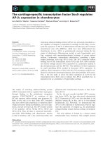
and 48 h post-transfection. It showed a h igh-efficiency
transfection that mor e than 85% cells displayed green
fluorescence with 100 nM fluorescent siRNA (Figure 1).
Figure 1 Efficient delivery of siRNA into lung adenocarcinoma cells.
(A): Detection of transfection efficiency by flow cytometry. Transfect ion
efficiency was maintained at over 85% for 6 h post-transfection. (B):
Detection of transfection efficiency by fluorescence microscopy. High
efficiency of transfection with fluorescent s iRNA (green) in A549 cells
were easily id entified for 4 8 h post-transfection ( ×100).
Xu et al. Journal of Experimental & Clinical Cancer Research 2011, 30:63
/>Page 3 of 11
When cells were treated with TF-targeting siRNA
(25 nM, 50 nM and 100 nM SiTF) and the scramble
siRNA (Mock, 100 nM) for 48 h, the mRNA and protein
expressions of TF were examined by RT- PCR and Wes-
tern blot. As shown in Figure 2 and Figure 3, the Mock
did not affect the expression levels of TF, but in 25 nM,
50 nM and 100 nM SiTF groups, compared with mock,
the TF expression decreased at both protein and mRNA
levels. Specially, 100 nM SiTF indicated a 80-85% reduc-
tion of TF expression in A549 cells. These results
demonstrated that the TF-targeting siRNA was efficient
to knock down the expression of TF in A549 cells.
Inhibition of cell proliferation and colony formation by
TF-siRNA
Since previous studies have shown that the expression of
TF associated with tumor growth [20-22], the effect of
TF siRNA on lung adenocarcinoma cell proliferation
was determined by MTT assay. As shown in Figure 4,
after 24 h-96 h transfection of TF siRNA into A549
cells, cell proliferation was remarkably inhibited in a
time- and dose-dependent manner, when compared
with control and mock groups. Inhibition of cell prolif-
eration at 50 nM and100 nM began at 48 h post-trans-
fection,butat25nMwasobservedat72hpost-
transfection, and higher concentrations of TF siRNA
had greater effects. In addition, the colony formation
assay further revealed effects of TF knockdown on
growth properties of A549 cells. 50 nM and100 nM
SiTF groups, but not 25 nM SiTF group had lower posi-
tive colony formation than control and mock groups,
and it also seemed to depend on doses (Figure 5 and
Figure 6). Overall, down-regulation of TF by siRNA
resulted in a negative effect on growth of lung adenocar-
cinoma cells.
Attenuation of the migration/invasion ability by TF-siRNA
Tumor cell migration and invasi on are two critical steps
in cancer metastatic process [23]. To verify the effect of
TF-siRNA on the migration ability, A549 cells were
tested by wound healing assay and the mobility assay.
Figure 7 and Figure 8 show that the cells in 50 nM and
100 nM SiTF groups demonstrated an attenuated capa-
city of impaired migration, when compared to control
and mock groups. Moreover, untreated and transfected
cells were seeded on transwell chambers with uncoated
filters. After incubation for 24 h, the motility potential
of transfected cells at 50 nM and 100 nM TF-siRNA
was significantly suppressed (Figure 9 and Figure 10). In
addition, the invasion assay using Matrigel-coated
Transwell chambers showed that 50 nM and 100 nM
TF-siRNA transfected cells that passed through the
Matrigel-coated membranes were much more than par-
ental cells and the ce lls transfected with scrambled
siRNA, and it indicated that the invasive capacity was
markedly decreased (Figure 11 and Figure 12). These
results suggested that TF-siRNA attenuated the meta-
static potential of lung adenocarcinoma cells in vitro.
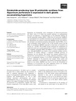
Promoted apoptosis in A549 cells by TF-siRNA
To evaluate further whether knockdown of TF induces
A549 cells apoptosis, at 48 h after transfectio n, the cells
were harvested and analyzed by flow cytometry. As
shown in Figure 13, the apoptosis rates of 25 nM, 50
nM and 100 nM SiTF groups were 7.0%, 9.0% and
16.0%, respectively, which were higher than 4.0% in con-
trol and 4.8% in mock groups, and indicated a dose-
dependent increase.
Molecular mechanisms of the antitumor effects by
TF-siRNA
The protein from transfected cells was extracted to
examine the effects of TF-siRNA on some important
cytokines and signaling m olecules. After 48 h of trans-
fection, the protein relative expression levels of phos-
phorylated Erk1/2 and PI3K in 100 nM SiTF group and
phosphorylated Akt in 25 nM, 50 nM and 100 nM SiTF
groups were decreased, while that in control and mock
groups had no differences (Figure 14 and Figure 15).
Furthermore, compared to control and mock groups,
Figure 2 TF-siRNA suppressed the TF protein express ion in lung adeno carcinoma cells. 48 h after transfecti on, the concentration of 100
nM TF-siRNA (100 nM SiTF group) was identified as the most efficient to knock down the expression of TF by Western blot. *P < 0.05, **P < 0.01
versus mock.
Xu et al. Journal of Experimental & Clinical Cancer Research 2011, 30:63
/>Page 4 of 11
transfection with high concentrations of 50 nM and 100
nM TF-siRNA suppressed the MMP-9/-2 expression
(Figure 16), and the protein expressio n of VEGF of 100
nM SiTF group was decreased (Figure 17). These data
demonstrated that knockdown of TF by siRNA may
inhibit Erk1/2 MAPK, PI3K/Akt signaling pathway,
MMP-9/-2 and VEGF, which all play an important role
in tumor progress.
Inhibition of tumor growth of lung adenocarcinoma cells
in nude mice by TF-siRNA
Intratum oral injection with TF- siRNA was performed to
investigate whether TF-siRNA had the effect of inhibi-
tion on tumor growth in vivo. A nude-mouse model of
human lung adenocarcinoma xenograft was estab lished,
and when the tumor volume reached 50-100 mm
3
,
intratumoral treatment with TF-siRNAs was started and
repeated every 5 days for a total of 5 times. As shown in
Figure 18A, the tumor volume of SiTF group from days
22 to the end was significantly smalle r than control and
mock groups, whereas there was no statistical difference
between control group and mo ck group during the
experiment. All mice were sacrificed on the 42nd day,
and the final tumor volume and weight in SiTF group
(209.6 ± 97.6 mm
3
and 0.21 ± 0.10 g, n = 5) were mark-
edly smaller than that i n control group (600.8 ± 182.0
mm
3
and 0.59 ± 0.18 g, n = 5) and mock group (513.8
± 112.6 mm
3
and 0.52 ± 0.12 g, n = 5) (Figure 18 and
Figure 19). In addition, the relative protein expression of
TF in SiTF group was decreased significantly, but there
was no statistical significance between control group
and mock group (Figure 20). After all, these results
Figure 4 Knockdown of TF with TF-siRNA inhibited cell
proliferation of lung adenocarcinoma cells in vitro. TF-siRNAs
transfected A549 cell growth was significantly attenuated in a time-
and dose-dependent manner compared with mock. *P < 0.05, **P <
0.01 versus mock.
Figure 3 TF-siRNA suppre ssed the mRNA expression in lung adenocarcinoma cells. The concentration of 100 nM TF-siRNA (100 nM SiTF
group) was identified as the most efficient to knock down the expression of TF by RT-PCR. *P < 0.05, **P < 0.01 versus mock.
Figure 5 Knockdown of TF with TF-siRNA inhibited colony
formation of lung adenocarcinoma cells in vitro. Representative
images of the colony formation assay were shown.
Xu et al. Journal of Experimental & Clinical Cancer Research 2011, 30:63
/>Page 5 of 11
indicated that intratumoral injection with TF-siRNA
suppressed the tumor gro wth of lung adenocarcinoma
cells in vivo.
Discussion
Despite advances in the medical and surgical treatments,
lung cancer is the leading cause of cancer deaths [1]and
because of intrinsic properties of lung adenocarcinoma
which cells show a high ability to rapid progress, it has a
poor prognosis in main histological type s of lung cancer
[24,25]. Tumor progression includes tumor cell prolifera-
tion, invasion (loss of cell to cell adhesion, increased cell
motility and basement membrane degradation), vascular
intravasation and extravasation, estab lishment of a meta-
static niche, and angiogenesis [23,26,27]. Therefore, how
to effectively inhibit the proliferative and metastatic bio-
logical behavior of Lung adenocarcinoma cells is a key
problem to improve the outcome.
Recent studies have implicated that TF plays an
important role in biological processes of many ca ncers,
and the main mechanism is mediated via angiogenesis
[28,29]. In non-small-cell lung carcinomas, the increased
TF expression associated with high VEGF levels and
microvessel density has gained widespread acceptance
[6,30]. However, A definite conclusion that silencing the
expression of TF in lung adenocarcinoma affects the
tumor cell proliferation, apoptosis and prometastatic
processes such as migration and matrix degradation
have not yet been established.
In this study, we have shown that chemically synthe-
sized siRNAs specifically targeting TF successfully
knocked down the expression of TF in both protein and
mRNA levels by 80% to 85% in human lung adenocarci-
noma cells A549. Then the assays as described above
detected the effects on biological behavior of A549 cells
in vitro. By the MTT and clonogenic assays, we were able
to first show that the proliferation of the TF-siRNA
transfected lung adenocarcinoma cells is significantly
inhibited in vitro, but previous studies have failed to
show that in colorectal cancer cells and B16F10 mela-
noma cells [11,12,31]. Using wound healing and transwell
assays, TF-siRNA attenuated the potential of invasion
and metastasis in lung adenocarcinoma cells. Further-
more, flow cytometric analysis revealed that knockdown
of TF expression induced apoptosis in A549 cells.
According to these results, we believed that besides parti-
cipating in angiogenesis, TF also plays a key role in cell
proliferation and metastasis of lung adenocarcinoma.
After binding of FVIIa, the TF forms a high affinity
complex with FVIIa or FVIIa-FXa, and other than
Figure 7 Knockdown of TF with TF-siRNA attenuated the
migration ability of lung adenocarcinoma cells in vitro.
Representative images of the wound healing assay were shown
(×40).
Figure 8 Bar graph of the wound healing assay. Bar shows the
means percentage of wound area covered by migrating A549 cells.
A549 cells treated with 50 nM and 100 nM TF-siRNA remarkably
decreased the cell motility. **P < 0.01 versus mock.
Figure 6 Bar graph of the colony formation assay.Theresult
demonstrated that high concentrations of 50 nM and 100 nM TF-
siRNA significantly attenuated the colony formation rate of lung
adenocarcinoma cells. **P < 0.01 versus mock.
Figure 9 Knockdown of TF with TF-siRNA attenuated the
migration ability of lung adenocarcinoma cells in vitro.
Representative images of the mobility assay were shown (×200).
Xu et al. Journal of Experimental & Clinical Cancer Research 2011, 30:63
/>Page 6 of 11
initiating the c oagulation cascade, the complex induce
signal transduction by binding to a family of trans-
membrane domain G protein-coupled cell surface
receptors called protease-activated receptors (PARs),
specially, PAR-1/-2 [32], which are expressed by
numerous tumor cells and tissues [33,34]. In the
tumor, it has recently emerged as important players in
growth and metastasis, butpreviousstudieshave
lacked information about the downstream signal path-
ways induced by the inhibition of the TF expression
via TF-siRNA in lung cancer cells. In the current
study, we established that down-regulation of TF
expression in lung adenocarcinoma cells suppressed
the Erk1/2 MAPK and PI3K/Akt signal pathways,
which are well recognized for mediating cell prolifera-
tion and apoptosis [35,36]. Therefore, the result
explains, at least in part, why TF-siRNA inhibited the
cell proliferation and induced the apoptosis in A549
cells. Furthermore, the expressions of MMP-2/-9 also
were down-regulated in TF-siRNA tran sfected cells,
and it may suggest that MMP-2/-9 are the downstream
products of the T F complex induced cell signaling.
MMPsareafamilyofenzymesthatdegradeproteins
in tissue extracellular matrices, which are clearly
involved in cancer progression, including tumor cell
degradation of basement membranes and stroma and
blood vessel penetration [27]. Consequently, the reduc-
tion of MMP-2/-9 by TF-siRNA exactly results in
attenuating the metastatic potency of lung adenocarci-
noma cells.
Besides experiments in vitro that give new insights
into the antitum or effects of TF-siRNA in lung adeno-
carcinoma, we used a nude m ouse xenograft model of
lung adenocarcinoma to better evaluate the TF-siRNA
effects in vivo. Since in vitro results indicated that
knockdown of TF by chemically synthesized siRNA
lasted for about 5 days, the mice received intratumoral
injectionofTF-siRNAevery5daysoftotal5timesto
down-regulate the expression of TF. Through monitor-
ing the tumor volume for about 4 weeks after injection,
we found that the tumor growth in the treated mice
with TF-siRNA was strongly suppressed. The results
were in agreement with the nude mice bearing tumors
of human breast cancer (MDA-MB-231) treated with
EF24 conjugated to FVIIa [37]. Combined these findings
in vitro and vivo, we confirmed the close relationship
between TF and tumor growth, vascularization, and
metastasis in lung adenocarcinoma.
Conclusions
In summary, our findings clearly demonstrate that TF
plays a crucial role in lung adenocarcinoma tumor
growth and metastasis. This shows the first study in
which silence of TF expression in lung adenocarcinoma
cells by TF-siRNA could inhibit tumor growth and
metastasis in vitro and in vivo, and the antitumor effects
may be associated with inhibition of Erk MAPK, PI3K/
Akt signal pathways in lung cancer. Therefore, RNA
interference targeting TF may be a useful potential tool
Figure 10 Bar graph of the mobility assay. Bar represents the
mean number of the cells per field. Silencing TF by 50 nM and 100
nM TF-siRNA inhibited cell migration in lung adenocarcinoma cells.
**P < 0.01 versus mock.
Figure 11 Knockdown of TF with TF-siRNA attenuated the
invasion ability of lung adenocarcinoma cells in vitro.
Representative microscopy images of the invasion assay are shown
(×200).
Figure 12 Bar graph of the i nvasion assay. Bar represents the
mean number of the cells per field. The invasion assay was
consistent with the migration assay and showed that the high
concentration of 50 nM and 100 nM TF-siRNA attenuated the
invasion ability of lung adenocarcinoma cells. **P < 0.01 versus
mock.
Xu et al. Journal of Experimental & Clinical Cancer Research 2011, 30:63
/>Page 7 of 11
Figure 13 Knockdown of TF with TF-siRNA induced apoptosis of lung adenocarcinoma cells. The transfected cells, labeled with AnnexinV-
FITC and propidium iodide, were subjected to flow cytometric analysis. Two parameter histogram Dot Plot displayed FL1-FITC on the x axis and
FL2-PI on the y axis. The result showed that TF-siRNA increased the apoptotic rate in A549 cells in a dose-dependent manner.
Figure 14 Western blot analysis of Erk1/2 by silencing TF by siRNA in lung adenocacinoma cells in vitro. Representa tive images were
shown and bar represented that the protein relative expression levels of phosphorylated Erk1/2 (P-Erk1/2) in 100 nM SiTF group were decreased.
**P < 0.01 versus mock.
Figure 15 Western blot analysis of PI3K/Akt by silencing TF by siRNA in lung adenocacinoma cells in vitro. Representative images were
shown and bar represented that the protein relative expression levels of PI3K in 100 nM SiTF group and phosphorylated Akt (P-AKT) in 25 nM,
50 nM and 100 nM SiTF groups were decreased. *P < 0.05, **P < 0.01 versus mock.
Xu et al. Journal of Experimental & Clinical Cancer Research 2011, 30:63
/>Page 8 of 11
Figure 16 Western blot analysis of MMP-9/-2 by silencing TF by siRNA in lung adenocacinoma cells in vitro. Representative images were
shown and bar represented that transfection with 50 nM and 100 nM TF-siRNA suppressed the MMP-9/-2 expression. *P < 0.05, **P < 0.01 versus
mock.
Figure 17 Western blot analysis of VEGF by silencing TF by siRNA in lung adenocacinoma cells in vitro. Representat ive images were
shown and bar represented that the protein expression of VEGF of 100 nM SiTF group was decreased. *P < 0.05, **P < 0.01 versus mock.
Figure 18 Tumor volume curve and bar graph of tumor weight
on the 42nd day when mice were killed. (A): The curve showed
that the tumor growth of SiTF group from days 22 to the end was
significantly inhibited compared to that of control and mock
groups. (B): Bar represented that the tumor weight of SiTF group
was decreased than that of control and mock group. **P < 0.01
versus mock.
Figure 19 Knockdown of TF by siRNA inhibited the tumor
growth of lung adenocarcinoma cells in nude mice. (A and B):
Representative images showed that the tumor size of SiTF group
was markedly smaller on the 42nd day after tumor cells inoculation
than that of control and mock group.
Xu et al. Journal of Experimental & Clinical Cancer Research 2011, 30:63
/>Page 9 of 11
for the gene therapy of lung adenocarcinoma, and even
other cancers at high level of TF expression.
Abbreviations
ERK: extracellular signal-regulated kinase; MAPK: mitogen-activated protein
kinase; MMP: matrix metalloproteinase; PARs: protease-activated receptors;
PI3K: phosphoinositide 3-kinase; RNAi: RNA interference; siRNA: small
interfering RNA; TF: tissue factor; VEGF: vascular endothelial growth factor.
Acknowledgements
The work was partially supported by the scientific and technological project
of Hubei Province, China (2008CDB142).
Author details
1
Department of General Thoracic Surgery, Tongji Hospital, Tongji Medical
College, Huazhong University of Science and Technology, Wuhan, People’s
Republic of China.
2
Department of Oncology, Tongji Hospital, Tongji Medical
College, Huazhong University of Science and Technology, Wuhan, People’s
Republic of China.
Authors’ contributions
XC and GQ have contributed to the research design, the data collection and
manuscript writing. CW, WL, SW, ZN, XQ and WJ have contributed to
manuscript writing. FN has contributed to the research design and
manuscript writing. All authors read and approved the final manuscript.
Competing interests
The authors declare that they have no competing interests.
Received: 24 March 2011 Accepted: 28 May 2011
Published: 28 May 2011
References
1. Jemal A, Siegel R, Xu J, Ward E: Cancer statistics, 2010. CA Cancer J Clin
2010, 60:277-300.
2. Parkin DM, Bray F, Ferlay J, Pisani P: Global cancer statistics, 2002. CA
Cancer J Clin 2005, 55 :74-108.
3. Hanagiri T, Baba T, So T, Yasuda M, Sugaya M, Ono K, Uramoto H,
Takenoyama M, Yasumoto K: Time trends of surgical outcome in patients
with non-small cell lung cancer. J Thorac Oncol 2010, 5:825-829.
4. Edgington TS, Mackman N, Brand K, Ruf W: The structural biology of
expression and function of tissue factor. Thromb Haemost 1991, 66:67-79.
5. Rao LV, Pendurthi UR: Tissue factor-factor VIIa signaling. Arterioscler
Thromb Vasc Biol 2005, 25:47-56.
6. Regina S, Rollin J, Blechet C, Iochmann S, Reverdiau P, Gruel Y: Tissue
factor expression in non-small cell lung cancer: Relationship with
vascular endothelial growth factor expression, microvascular density,
and K-ras mutation. Journal of Thoracic Oncology 2008, 3:689-697.
7. Callander NS, Varki N, Rao LV: Immunohistochemical identification of
tissue factor in solid tumors. Cancer 1992, 70:1194-1201.
8. Zwicker JI: Predictive value of tissue factor bearing microparticles in
cancer associated thrombosis. Thromb Res 2010, 125(Suppl 2):S89-91.
9. Aharon A, Brenner B: Microparticles, thrombosis and cancer. Best Pract Res
Clin Haematol 2009, 22:61-69.
10. Rickles FR, Edwards RL: Activation of blood coagulation in cancer:
Trousseau’s syndrome revisited. Blood 1983, 62:14-31.
11. Amarzguioui M, Peng Q, Wiiger MT, Vasovic V, Babaie E, Holen T,
Nesland JM, Prydz H: Ex vivo and in vivo delivery of anti-tissue factor
short interfering RNA inhibits mouse pulmonary metastasis of B16
melanoma cells. Clin Cancer Res 2006, 12:4055-4061.
12. Wang X, Wang M, Amarzguioui M, Liu F, Fodstad O, Prydz H:
Downregulation of tissue factor by RNA interference in human
melanoma LOX-L cells reduces pulmonary metastasis in nude mice. Int J
Cancer 2004, 112:994-1002.
13. Kim DH, Rossi JJ: Strategies for silencing human disease using RNA
interference. Nat Rev Genet 2007, 8:173-184.
14. Lu PY, Xie F, Woodle MC: In vivo application of RNA interference: from
functional genomics to therapeutics. Adv Genet
2005, 54:117-142.
15.
Holen T, Amarzguioui M, Wiiger MT, Babaie E, Prydz H: Positional effects of
short interfering RNAs targeting the human coagulation trigger Tissue
Factor. Nucleic Acids Res 2002, 30:1757-1766.
16. Kruger NJ: The Bradford method for protein quantitation. Methods Mol
Biol 1994, 32:9-15.
17. Fu WJ, Li JC, Wu XY, Yang ZB, Mo ZN, Huang JW, Xia GW, Ding Q, Liu KD,
Zhu HG: Small interference RNA targeting Kruppel-like factor 8 inhibits
the renal carcinoma 786-0 cells growth in vitro and in vivo. J Cancer Res
Clin Oncol 2010, 136:1255-1265.
18. Hou JQ, He J, Wang XL, Wen DG, Chen ZX: Effect of small interfering RNA
targeting survivin gene on biological behaviour of bladder cancer. Chin
Med J (Engl) 2006, 119:1734-1739.
19. Bradley SP, Kowalik TF, Rastellini C, DaCosta MA, Bloomenthal AB, Cicalese L,
Basadonna GP, Uknis ME: Successful incorporation of short-interfering
RNA into islet cells by in situ perfusion. Transplant P 2005, 37:233-236.
20. Toomey JR, Kratzer KE, Lasky NM, Broze GJ Jr: Effect of tissue factor
deficiency on mouse and tumor development. Proc Natl Acad Sci USA
1997, 94:6922-6926.
21. Versteeg HH, Schaffner F, Kerver M, Petersen HH, Ahamed J, Felding-
Habermann B, Takada Y, Mueller BM, Ruf W: Inhibition of tissue factor
signaling suppresses tumor growth. Blood 2008, 111:190-199.
22. Rickles FR, Shoji M, Abe K: The role of the hemostatic system in tumor
growth, metastasis, and angiogenesis: Tissue factor is a bifunctional
molecule capable of inducing both fibrin deposition and angiogenesis
in cancer. Int J Hematol 2001, 73:145-150.
23. Chambers AF, Groom AC, MacDonald IC: Dissemination and growth of
cancer cells in metastatic sites. Nature Reviews Cancer 2002, 2:563-572.
24. Janssen-Heijnen ML, Coebergh JW: The changing epidemiology of lung
cancer in Europe. Lung Cancer 2003, 41:245-258.
25. Devesa SS, Bray F, Vizcaino AP, Parkin DM: International lung cancer
trends by histologic type: male:female differences diminishing and
adenocarcinoma rates rising. Int J Cancer 2005, 117:294-299.
26. Duffy MJ, McGowan PM, Gallagher WM: Cancer invasion and metastasis:
changing views. J Pathol 2008, 214:283-293.
Figure 20 TF-siRNA inhibited the protein expression of TF in vivo as determined by Wester n blot. Representative images were shown
and bar represented that the relative expression of TF in SiTF group was significantly inhibited compared to that in control and mock groups.
**P < 0.01 versus mock.
Xu et al. Journal of Experimental & Clinical Cancer Research 2011, 30:63
/>Page 10 of 11
27. Deryugina EI, Quigley JP: Matrix metalloproteinases and tumor
metastasis. Cancer Metastasis Rev 2006, 25:9-34.
28. Folkman J: Tumor angiogenesis and tissue factor. Nat Med 1996,
2:167-168.
29. Hembrough TA, Swartz GM, Papathanassiu A, Vlasuk GP, Rote WE, Green SJ,
Pribluda VS: Tissue factor/factor VIIa inhibitors block angiogenesis and
tumor growth through a nonhemostatic mechanism. Cancer Res 2003,
63:2997-3000.
30. Koomagi R, Volm M: Tissue-factor expression in human non-small-cell
lung carcinoma measured by immunohistochemistry: correlation
between tissue factor and angiogenesis. Int J Cancer 1998, 79:19-22.
31. Yu JL, May L, Lhotak V, Shahrzad S, Shirasawa S, Weitz JI, Coomber BL,
Mackman N, Rak JW: Oncogenic events regulate tissue factor expression
in colorectal cancer cells: implications for tumor progression and
angiogenesis. Blood 2005, 105:1734-1741.
32. Versteeg HH, Spek CA, Peppelenbosch MP, Richel DJ: Tissue factor and
cancer metastasis: the role of intracellular and extracellular signaling
pathways. Mol Med 2004, 10 :6-11.
33. D’Andrea MR, Derian CK, Santulli RJ, Andrade-Gordon P: Differential
expression of protease-activated receptors-1 and -2 in stromal
fibroblasts of normal, benign, and malignant human tissues. Am J Pathol
2001, 158:2031-2041.
34. Dorsam RT, Gutkind JS: G-protein-coupled receptors and cancer. Nat Rev
Cancer 2007, 7:79-94.
35. Widmann C, Gibson S, Jarpe MB, Johnson GL: Mitogen-activated protein
kinase: conservation of a three-kinase module from yeast to human.
Physiol Rev 1999, 79:143-180.
36. Dudek H, Datta SR, Franke TF, Birnbaum MJ, Yao R, Cooper GM, Segal RA,
Kaplan DR, Greenberg ME: Regulation of neuronal survival by the serine-
threonine protein kinase AKT. Science 1997, 275:661-665.
37. Shoji M, Sun A, Kisiel W, Lu YJ, Shim H, McCarey BE, Nichols C, Parker ET,
Pohl J, Mosley CA, Alizadeh AR, Liotta DC, Snyder JP: Targeting tissue
factor-expressing tumor angiogenesis and tumors with EF24 conjugated
to factor VIIa. J Drug Target 2008, 16:185-197.
doi:10.1186/1756-9966-30-63
Cite this article as: Xu et al.: Small interference RNA targeting tissue
factor inhibits human lung adenocarcinoma growth in vitro and in vivo.
Journal of Experimental & Clinical Cancer Research 2011 30:63.
Submit your next manuscript to BioMed Central
and take full advantage of:
• Convenient online submission
• Thorough peer review
• No space constraints or color figure charges
• Immediate publication on acceptance
• Inclusion in PubMed, CAS, Scopus and Google Scholar
• Research which is freely available for redistribution
Submit your manuscript at
www.biomedcentral.com/submit
Xu et al. Journal of Experimental & Clinical Cancer Research 2011, 30:63
/>Page 11 of 11