báo cáo khoa học: "Recombinant immunotoxin anti-c-Met/PE38KDEL inhibits proliferation and promotes apoptosis of gastric cancer cells" docx
Bạn đang xem bản rút gọn của tài liệu. Xem và tải ngay bản đầy đủ của tài liệu tại đây (2.21 MB, 7 trang )
RESEARC H Open Access
Recombinant immunotoxin anti-c-Met/PE38KDEL
inhibits proliferation and promotes apoptosis of
gastric cancer cells
Xu Wei
1
, Zhu Xiao Juan
1
, Feng Xiao Min
2
, Cai Nan
1
, Zhang Xiu Hua
1
, Feng Zheng Qing
2
and Liu Zheng
1*
Abstract
Background: Our study aims to evaluate the anti-growth effects of recombinant immunotoxin (IT ) anti-c-Met/
PE38KDEL on gastric cancer cells, and its mechnisms.
Methods: Gastric cancer cells were treated with increasing doses of IT and c-Met protein was quantified by
Western blotting. Cell proliferation was determined by Cell Counting Kit-8 assay (CCK). [
3
H]-leucine incorporation
assay was used to evaluate IT inhibition of protein synthesis. Cell apoptosis was quantified by flow cytometry.
Caspase activities were measured using colorimetric prot ease assays.
Results: Cell growth and protein synthesis of the gastric cancer cell lines were suppressed by IT in a dose- and
time-dependent manner. IT also induced apoptosis in a dose-dependent manner. The apoptosis rates of gastric
cancer cell lines MKN-45 and SGC7901 were 19.19% and 27.37%, respectively when treated with 50 ng/ml of IT.
There were significant increase ofcaspase-3 activity at 24 hr of IT treatment (100 ng/ml) (P < 0.01) in these gastric
cancer cell lines.
Conclusions: IT anti-c-Met/PE38KDEL has anti-growth effects on the gastric cancer cell lines in vitro, and it provides
an experimental basis for c-Met-targeted therapy towards in vivo testing.
Introduction
Gastric carcinoma (GC) is one of the most common and
lethal malignant cancers [1]. Despite the improving sur-
gical techniques and new chemotherapeutic treatment
regimens, the patient survival rate remains dismal [2],
and effective alternative treatment approach is in vital
need. GC has been shown to harbor multiple somatic
mutations as well as over-expressions of oncoproteins.
Identification of these GC-associated biomarkers may
entail possible discovery of new therapeutic targets [3].
Among various GC-associated biomarkers, c-MET gene
is frequently found gnomically-amplified and over-
expressed in GC cell lines [4 ]. The proto-oncogene c-
MET, a receptor of hepatocyte growth factor (HGF, also
known as scatter factor), encodes a 190 kDa heterodi-
meric transmembrane tyrosine kinase. HGF binding to
c-Met triggers tyrosine kinase domain auto-
phosphorylation and induces pleio tropic respons es such
as proliferation, motility, morphogenesis and angiogen-
esis in many cell types including normal and tumor cells
[5]. c-MET amplification has been identified in nearly
74% of human GC specimens [6]. HGF and c-MET both
play important roles in the progression and metastasis
of GC [7]. Thus, c-Met has been considered as a pro-
mising therapeutic target for various cancers.
Immunotoxins (ITs) are fusion proteins composed of a
toxin fused to an antibody or growth factor with distinct
target specificity [8]. IT exerts its anti-growth effect by
inhibiting protein synthesis and promoting apoptosis [9].
IT anti-c-Met/PE38KDEL (anti-c-Met Fab, which
resulted from screening and characterization from a nat-
ural human Fab phage antibody library; PE38KDEL,
which is a modi fied structure of PE38, lost the function
of combining with non-mammalian cells specifically, but
retained a complete cytotoxicity after internalization)
has shown specific cytotoxic effects against c-Met-posi-
tive cancer cells [10]. In this study, we investigated the
effects of IT anti-c-Met/PE38KDEL on proliferation and
* Correspondence:
1
Department of Gastroenterology, The Second Affiliated Hospital of Nanjing
Medical University, Nanjing, 210029, PR China
Full list of author information is available at the end of the article
Wei et al. Journal of Experimental & Clinical Cancer Research 2011, 30:67
/>© 2011 Wei et al; licensee BioMed Central Ltd. This is an Open Access article distributed under the terms of the Crea tive Commons
Attribution License ( which p ermits unrestricted use, distribution, and reproduction in
any medium, provided the original work is properly cited.
apoptosis of two different c-Met-positive malignant gas-
tric cell lines, MKN-45 and SGC7901 [11,12], and a nor-
mal gastric mucosa cell GES-1 [13]. We found that IT
anti-c-Met/PE38KDEL exerts its anti-growth effect pri-
marily through rapid inhibition of protein synthesis.
Materials and Me thods
Immunotoxin
IT anti-c-Met/PE38KDEL was described previously [9]. It
induces apoptosis in hepatic carcinoma cells SMMC7721.
Cell Counting Kit 8 (C CK8) was purchased from Sigma
Chemical. Caspase colorimetric assay kit and anti-caspase-
3 antibody were from Biovision. Antibodies against c-Met
and b-actin were purchased from Santa Cruz. Protein lysis
buffer was from TaKaRa Biotechnology.
Cell culture
GC cells lines, MKN-45 and SGC7901, and normal gas-
tric mucosa cells GES-1 were obtained from the Cell
Bank of Type Culture Collection of the Chinese Acad-
emy of Sciences (Shanghai, China), and were grown in
DMEM (Invitrogen) supplemented with 10% fetal calf
serum (FCS) and incubated at 37°C with 5% CO
2
.All
cell lines were routinely tested and found to be free
from mycoplasma contamination.
Western Blotting
GES-1, MKN-45 and SGC7901cellsgrownin6-well
plates were collected in lysis buffer for total cellular pro-
tein. Protein concentrations were measured using a
Bradford reagent (Bio-Rad). Equal amounts of protein
(80 μg/lane) from each cell line were boiled for 5 min,
separated by SDS-PAGE, and then transferred on to a
nitrocellulose membrane before blocking in 5% non-fat
dried milk in Tris-buffered saline (TBS) for 120 min at
room temperature. The membranes were then incubated
with a primary anti-human c-Met polyclonal antibody
(diluted 1:150 in a new batch of the blocking buffer) or
a goat polyclonal primary anti-b-actin (diluted 1:1000,
Santa Cruz, CA, USA) for 2 hr and followed by incuba-
tion with peroxidase-labelled anti-IgG secondary anti-
body for 1 hr. Aft er washing with TBST for 3 times, the
films were developed and the protein bands were quan-
tified by densitometr y using ImageJ software (NIH,
Bethesda, MD, USA).
To detect the caspase-3 activity, both floating and
adherent cells were collected 24 hr following IT treat-
ment. Total cellular protein was prepared as described
above. All the experiments were performed at least
twice with similar results.
Cell proliferation assay
Cell gro wth inhibition rate (IR) was determined using a
CCK- 8 assay following the manufacturer instruc tions
(Sigma). GES-1, MKN-45 and SGC7901 cells were
seeded at a concentration of 1 × 10
5
cells/90 μl/well in
96-well culture plate s. After incubation of cells with the
indicated concentrationsofITfor24hrand48hr,10
μl/well of cell Counting Kit-8 solution was added to the
medium and the cells were incubated for an additional
4 hr. The absorbance at 450 nm was then measured in a
Microplate Reader. IR was calculated using the following
equation: IR = [1-(A value in the treated samples-A
value in the blank samples) / (A value in the control
samples-A value in the blank sam ples)] *100%. The
assays were performed in triplicates and repeated at
least twice [14].
Protein synthesis inhibition assay
IT-induced inhibition of protein synthesis in GES-1,
MKN-45 and SGC7901 ce lls were evaluated using the
[
3
H]-leucine incorporation assay [15]. Cells were seeded
in 48-well plates (1 × 10
4
per well) and allowed to grow
overnight before the addition of IT at different concen-
trations. After 5 or 24 hr incubation, cells were washed
twice with cold phosphate-buffered saline (PBS) contain-
ing 0.1% FCS, and then incubated with [
3
H]-leucine (2
μCi ml
-1
) in leucine-free medium at 37°C for 45 min.
Cells were then washed with 5% trichloroacetic acid
(TCA) for 5 and 10 min, respectively, and dissolved in
0.1M KOH for 10-15 m in. The resultant solution was
transferred to the liquid scintillator. Sample counts were
determined in a liquid scintillation counter. Assays were
performed in duplicates and repeated at least three
times. Counts per minute (cpm) for treated cells were
compared to cpm for untreated cells and reported as a
percentage of leucine incorporation with the control
value set to 100%[16]. The experiment was completed in
the isotope laboratory of Nanjing Medical University.
Flow cytometric analysis of cell apoptosis
Apoptosis were determined by flow cytometric analysis.
Briefly, cells in triplicates, were incubated with or with-
out various concentrations of IT f or 24 hr. Cells were
then harvested, washed in cold PBS, and fixed with 1 ml
75% ice-cold ethanol at -20°C until processing. An ali-
quot (1 ml) of fixed cell suspension containing 1 × 10
6
cells was washed twice in cold PBS and then treated
with fluorochrome DNA staining solution (1 ml) con-
taining 40 μg of propidium iodide and 0.1 mg of RNase
A in the dark at room temperature for 0.5 hr. Flow
cytometric analysis were performed three times [17].
Caspase activity assay
Caspase activity was determined in 96-well plates using
cell lysates from 1 × 10
6
cells for each measurement.
Caspase-3 and caspase-8 activities were dete rmined
using colorimetric assay kits according to the
Wei et al. Journal of Experimental & Clinical Cancer Research 2011, 30:67
/>Page 2 of 7
manufacturer’ s protocol (BioVision). GES-1, MKN-45
and SGC7901 cells were treated with anti-c-Met/PE38K-
DEL (100 ng/ml) for 24 hr prior to the assay. Cell
extracts were incubated with 5 μl of 4 mM tetrapeptide
substrates (DEVD, caspase-3; IETD, a nd caspase-8) at
37°C for 1-2 hr. The reaction was measured at 405 nm
in a Microplate Reader. Background readings from cell
lysat es and buffers were subtracted from the readings of
both IT-induced and control samples before calculating
the relative change increase in caspase activity in the
IT-i nduced samples compared to that of the control. IT
treated samples were normalized to the caspase activity
of the untreated sample, which was set to 1.0. Fold of
increases in caspase activities were presented.
Statistical analysis
Statistical analysis was performed with SPSS 13.0 soft-
ware. Data were presented as mean ± standard devia-
tion. Student’s t-test was used to compare two samples,
and the single -factor analysis of variance (One-way
ANOVA) was used to compare mult iple samples. A p-
value less than 0.05 is considered statistically significant
(*, p < 0.05; **, p < 0.01).
Results
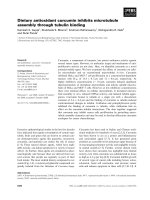
Increased c-Met expression in MKN-45 and SGC7901 cells
To determine the c-Met protein expression levels in GC,
we used western blotting to examine c-Met protein in
two GC cells (MKN-45 and SGC7901) and one normal
gastricmucosacellsGES-1(Figure1A).c-Metproteins
is 3-4 fold higher in MKN-45 and SGC7901cells than
GES-1 cells. SGC7901 cells express slightly more c-Met
than MKN-45 cells (Figure 1B). The optical densities
(OD’s) of the Western blot bands were measured using
ImageJ. The OD for each band was normalized to b-
actin. MKN-45 and SGC7901 had a 0.94 and 1.27 fold
increase in the expression of c-Met over the control, but
only 0.34 fold increased in GES-1.
IT anti-c-Met/PE38KDEL inhibited cell proliferation and
protein synthesis
GC cells hav e significantly higher c-Met protein levels
than normal gastric mucosa cells, therefore we tried to
determine if IT anti-c-Met/PE38KDEL has GC-specific
effects. The anti-proliferative effect of IT anti-c-Met/
PE38KDEL on GES-1, MKN-45 and SGC7901 cells
was measured using CCK8 kit. Cells were harvested at
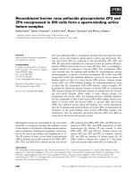
24 or 48 hr after IT treatment. As shown in Figure 2,
IT inhibited GC cell growth in a time- and dose-
dependent manner. 1, 10 and 100 ng/ml of IT caused
a dramatic growth inhibition in MKN-45 and
SGC7901 cells (P< 0.01). 48 hr of IT treatment (100
ng/ml) resulted in a growth inhibition of 30% in GES-
1 cells (Figure 2A). However, inhibitions of 75% and
95%wereobservedinMKN-45andSGC7901cells
(Figure 2B and 2C), respectively. Further, we found
that there is a strong correlation between c-Met
expression and in vitro immunotoxin efficacy.
Given the high c-MET levels in MKN-45 and
SGC7910 cell lines, we hypothesize that anti-c-Met/
PE38KDEL can attenuate cancer cell growth through
inhibition of protein synthesis via c-Met inhibition.
The effects of anti-c-Met/PE38KDEL on protein
synthesis in GES-1, MKN-45 and SGC7901 cells are
showninFigure3.TheIT’ sIC
50
value on GES-1 cells
was approximately 120 ng/ml. However, IT induced
more potent inhibitions of protein synthesis in MKN-
45 and SGC7901 cells, with IC
50
values of 5.34 ng/ml
and 0.83 ng/ml, respectively. Nearly 80% and 100% of
inhibitions were observed with 100 ng/ml of IT treat-
ment in these two GC cells (Figure 3B and 3C). In
contrast, 100 ng/ml of IT only caused a 35% decrease
in protein synthesis in GES-1 cells (Figure 3A). These
results suggested that anti-c-Met/PE38KDEL can
attenuate cell growth through the inhibition of protein
synthesis.
Figure 1 Overexpression of c-Met in castric carcinoma cell
lines. Lysates (80 μg/lane) from normal gastric mucosa cells GES-1
and GC cell lines MKN-45 and SGC7901 were analyzed for c-Met
protein level by western blot using an anti-c-Met antibody and an
anti- b-actin antibody (loading control) (Figure 1A). The optical
densities (OD’s) of the Western blot bands were measured using
Image J (Figure 1B).
Wei et al. Journal of Experimental & Clinical Cancer Research 2011, 30:67
/>Page 3 of 7
IT anti-c-Met/PE38KDEL inhibits tumor cell growth
through induction of apoptosis
To determine whether the anti-proliferative effect of IT
was due to cell apoptosis, we used flow cytometric
(FCM)) to further determine if IT induces cell apoptosis.
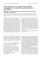
As shown in Figure 4A and 4B, apoptotic rates in MKN-
45 and SGC7901 cells were increased from 1.89% and
2.4% (0 ng/ml), to 19.19% (P < 0.01) and 27.37% (P <
0.01) (50 ng/ml), respectively. The apoptosis rate of
GES-1 cells is significantly lower than two GC cells
(5.98%, P < 0.01) at the IT dose of 50 ng/ml. These data
indicate that anti-c-Met/PE38KDEL induced apoptosis
in GC cells.
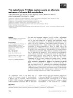
IT anti-c-Met/PE38KDEL activates caspase-3
To determine whether apoptotic pathway is activated by
IT in GC cells, we measured caspase-3 and caspase-8
activities following IT treatment. As shown in Figure 5B
and 5C, MKN-45 and SGC7901 cells showed 3.70 and
5.02 fold of increases in caspase-3 enzyme activity as
compared to untreated controls after 24 hr IT treatment
(P < 0.01). GES-1 exhibited a 2.03-f old increase in cas-
pase-3 enzyme activity (P < 0.05) (Figure 5A). Caspase-8
enzyme activity in two GC cell lines also increased (P <
0.05), suggesting caspase-3 activation mediates IT anti-
c-Met/PE38KDEL-induced biological effects.
The caspases are synthesized as inactive precursors
(zymogens) that are proteolytically processed to generate
active subunits by cleaving specific aspartic acid residues
[18], and are essential for the execution process of apopto-
sis as effector proteases [19]. In the process of IT-inducd
apoptosis, caspase-3 appeared to play a role. We investi-
gated whether caspase-3 is regulated in anti-c-Met/
PE38KDEL-induced cell death. As shown in Figure 6,
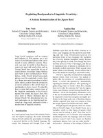
Figure 3 Anti-c-Met/PE38 KDEL induced inhibition of protein synthesis. The ability of IT to inhibit protein synthesis in GES-1, MKN-45 and
SGC7901 cells were evaluated by using the [
3
H]-leucine incorporation assay. [
3
H]-leucine incorporation for protein synthesis as a function of
varying concentration of IT (expressed as a percentage of untreated cells), Normal cell GES-1 (A), GC cells MKN-45 (B) and SGC7901 (C) were
treated with varying concentration of IT for 24 hr and 48 hr.
Figure 2 IT anti-c-Met/PE38KDEL induced inhibition of cell proliferation. Cell growth inhibition as a function of varying concentrations of IT
(expressed as a percentage of untreated cells), Normal cell GES-1 (A), GC cells MKN-45 (B) and SGC7901 (C) were treated with various
concentrations of IT for 24 hr and 48 hr.
Wei et al. Journal of Experimental & Clinical Cancer Research 2011, 30:67
/>Page 4 of 7
Figure 4 IT anti-c-Met/PE38KDEL inhibited tumor cell growth through induction of apoptosis. To measure the dose response effect of IT
on cell apoptosis rate of GES-1, MKN-45 and SGC7901, cells were treated with different concentrations of anti-c-Met/PE38KDEL. Cells were
incubated with IT at 0, 10 and 50 ng/ml for 24 hr, and the percentage of cell apoptosis was determined by flow cytometry. IT induced apoptosis
for its anticancer effect.
Figure 5 IT anti-c-Met/PE38KDEL mainly activates caspase-3. Caspase-3 and caspase-8 activities in GES-1 (A), MKN-45 (B) and SGC7901 (C)
cells were measured in control or IT-treated cells (immunotoxin) (24 hr) using the Caspase colorimetric assay kit. * P < 0.05, **P < 0.01.
Wei et al. Journal of Experimental & Clinical Cancer Research 2011, 30:67
/>Page 5 of 7
procaspase-3 was proteolytically cleaved in a dose-depen-
dent manner after 24 hr of IT treatment, resulting in the
production of the active caspase-3 fragment (17 kDa). In
untreated control cells (0 ng/ml), no caspase-3 was
detected. All these results suggested that IT anti-c-Met/
PE38KDEL causes apoptosis at least partially via activation
of caspase-3.
Discussion
GC is the second leading cause of cancer mortality in
the world [20]. The receptor tyrosine kinase c-Met is
constitutively activated in many GCs [2]. Amplifica-
tions of c-Met have been associated with human GC
progression [21] C-Met i s also related to lymph node
metastasis in GC [22]. Therefore, c-Met is considered
a promsing therapeutic target for this type of cancer
[3]. The aim of t his study w as to evaluate the effects of
recombinant immunotoxin anti-c-Met/PE38KDEL on
proliferation and apoptosis of GC cells and explore the
mechanism underlying the action of anti-c-Met/
PE38KDEL.
SGC7901 was derived from moderately differentiated
GC, with a high metastatic potential [23]. MKN-45 was
derived from poorly differentiated GC with low meta-
static potential [24]. We found that SGC7901 cells
expressed high level of c-Met than MKN-45 cells. Nor-
mal gastric muco sa cells GES-1 expressed a minimum
level of c-Met. Studies have shown that c-Met overex-
pression in carcinoma cells is associated with liver
metastasis of GC [25]. Moreover; c-Met expression can
be used as an indicator of liver metastasis for GC
patients. It has also been reported that HGF is a lym-
phangiogenic factor, which can directly or indirectly sti-
mulate lymphangiogenesis and contribute to lymphatic
metastasis in GC [26]. Therefore, we hypothesized that
IT anti-c-Met/PE38KDEL may be effective in preventing
GC’s metastasis.
Our data showed that IT decreased GC cell prolifera-
tion in a time- and dose-dependent manner. After 48 hr
of IT treatment (100 ng/ml), cell inhibition rate in
MKN-45 and SGC7901 cells was about 75% and 95%,
but only 30% in GES-1 cells, presumably due to low c-
Met expression on GES-1 than the two GC cells. IT
attenuates cancer cell growth not only by inhibiting pro-
tein synthesis but also by inducing apoptosis [27]. We
found that IT anti-c-Met/P E38KDEL induced a rapid
inhibition of protein synthesis with simultaneous induc-
tion of apoptosis in GC cells. Nearly 80% and 100%
inhibitions of protein synthesis were observed after 24
hr treatment with IT (100 ng/ml) in the MKN-45 and
SGC7901 cells, respectively. The inhibition was much
less pronounced in GES-1 cells (35%), suggesting that
IT anti-c-Met/PE38KDEL is selective against GC. In
addition, IT exerts its anticancer effect mostly via induc-
tion of cells apoptos is. The apoptosis rates in three cells
were all increased after treatment with IT, more promi-
nent in the two GC cell lines.
Caspases are classified into two functional sub-
groups-initiator caspases and effector caspases. The
initiator caspases are caspase 2, 8, 9 and 10, and the
effector caspases are ca spase 3, 6 and 7 [28]. Caspases
are critical mediators of apoptosis [29]. Activation of
caspase is responsible for multiple molecular and
structural changes in apoptosis [30]. Caspase-3 is a
potent effector of apoptosis in a variety of cells [31]
and plays a central role in both death-receptor and
mitochondria-mediated apoptosis. Caspase-8 is the
prototypical apoptosis initiator downstream of TNF
super-family death receptors. Our data showed that
caspase-3 enzyme activity exhibited 3.70, and 5.02 fold
increases in IT-treated MKN-45 and SGC7901 cells as
compared to the activity of untreated con trols (P <
0.01). The increase in caspase-8 enzyme activity was
less significant.
Conclusions
Our results demonstrate the time- and dose-dependent
anti-growth effects of IT anti-c-Met/PE38KDEL against
GC cell lines. The anti-cancer effect of IT occurred pri-
marily through inhibition of protein synthesis, and cas-
pase-3-mediated apoptosis, suggesting the potential
value of IT as an anti-c-MET therapeutics for GC.
Abbreviations
IT: Immunotoxins; GC: Gastric carcinoma; HGF: hepatocyte growth factor;
CCK8: Cell Counting Kit 8; FCS: fetal calf serum; TBS: Tris-buffered saline; IR:
inhibition rate; PBS: phosphate-buffered saline; SDS: sodium dodecyl
sulphate; PAGE: polyacrylamide gel electrophoresis.
Acknowledgements and Funding
This study was funded by nature science founation of jiangsu province
(BK2008483).
Author details
1
Department of Gastroenterology, The Second Affiliated Hospital of Nanjing
Medical University, Nanjing, 210029, PR China.
2
Department of Pathology,
Nanjing Medical University, Nanjing, 210029, PR China.
Figure 6 IT-induced caspase 3 cleavage. Lysates from normal
gastric mucosa cells GES-1 and GC cell lines MKN-45 and SGC7901
with or without IT treatment were analyzed for procasoase-3
protein levels and activated caspase protein levels by western blot
using an anti- procaspase-3, anti-activated caspase-3 and anti- b-
actin antibodies (loading control).
Wei et al. Journal of Experimental & Clinical Cancer Research 2011, 30:67
/>Page 6 of 7
Authors’ contributions
LZ AND XW: Conceived, designed, and coordinated the study and acquired
the necessary funding; and carried out the majority of the in vitro studies.
drafted the manuscript. CN and ZXJ: carried out all subsequent analyses;
FXM: carried out some of the in vitro experiments; ZXH and FZQ:
Contributed to the design and coordination of the study and aided with
manuscript preparation. All authors read and approved the final manuscript.
Competing interests
The authors declare that they have no competing interests.
Received: 16 May 2011 Accepted: 7 July 2011 Published: 7 July 2011
References
1. Tepes B: Can gastric cancer be prevented? J Physiol Pharmacol 2009,
60:71-77.
2. Gubanski M, Johnsson A, Fernebro E, Kadar L, Karlberg I, Flygare P,
Berglund A, Glimelius B, Lind PA: Randomized phase II study of sequential
docetaxel and irinotecan with 5-fluorouracil/folinic acid (leucovorin) in
patients with advanced gastric cancer: the GATAC trial. Gastric Cancer
2010, 13:155-161.
3. Corso S, Ghiso E, Cepero V, Sierra JR, Migliore C, Bertotti A, Trusolino L,
Comoglio PM, Giordano S: Activation of HER family members in gastric
carcinoma cells mediates resistance to MET inhibition. Mol Cancer 2010,
9:121.
4. Tahara E: Cancer-stromal interaction through growth factor/cytokine
networks implicated in growth of stomach cancer. Princess Takamatsu
Symp 1994, 24:187-194.
5. Bottaro DP, Rubin JS, Faletto DL, Chan AM, Kmiecik TE, Vande Woude GF,
Aaronson SA: Identification of the hepatocyte growth factor receptor as
the c-met proto-oncogene product. Science 1991, 251:802-804.
6. Drebber U, Baldus SE, Nolden B, Grass G, Bollschweiler E, Dienes HP,
Hölscher AH, Mönig SP: The overexpression of c-met as a prognostic
indicator for gastric carcinoma compared to p53 and p21 nuclear
accumulation. Oncol Rep 2008, 19:1477-1483.
7. Liu SI, Chi CW, Lui WY, Mok KT, Wu CW, Wu SN: Correlation of hepatocyte
growth factor-induced proliferation and calcium-activated potassium
current in human gastric cancer cells. Biochim Biophys Acta 1998,
1368:256-266.
8. Kreitman RJ: Recombinant immunotoxins containing truncated bacterial
toxins for the treatment of hematologic malignancies. BioDrugs 2009,
23:1-13.
9. Martínez-Torrecuadrada JL, Cheung LH, López-Serra P, Barderas R,
Cañamero M, Ferreiro S, Rosenblum MG, Casal JI: Antitumor activity of
fibroblast growth factor receptor 3-specific immunotoxins in a xenograft
mouse model of bladder carcinoma is mediated by apoptosis. Mol
Cancer Ther 2008, 7:862-873.
10. Zhu XJ, Feng ZQ, Zhu J, Tang qi, Liu zheng: Construction, expression and
purification of an immunotoxin containing a human anti-c-Met single-
chain antibody fused to PE38KDEL. Acta Univ Med Nanjing 2009,
29:920-924.
11. Kitamura S, Miyazaki Y, Hiraoka S, Toyota M, Nagasawa Y, Kondo S,
Kiyohara T, Shinomura Y, Matsuzawa Y: PPARgamma inhibits the
expression of c-MET in human gastric cancer cells through the
suppression of Ets. Biochem Biophys Res Commun 1999, 265:453-456.
12. Kaji M, Yonemura Y, Harada S, Liu X, Terada I, Yamamoto H: Participation
of c-met in the progression of human gastric cancers: anti-c-met
oligonucleotides inhibit proliferation or invasiveness of gastric cancer
cells. Cancer Gene Ther 1996, 3:393-404.
13. Zheng S, Ke Y: Study of APC, Rb, c-met gene copy numbers of human
gastric mucosa epithelial cell line GES-1. Zhonghua Zhong Liu Za Zhi
1999, 21:409-411.
14. Koyama M, Izutani Y, Goda AE, Matsui TA, Horinaka M, Tomosugi M,
Fujiwara J, Nakamura Y, Wakada M, Yogosawa S, Sowa Y, Sakai T: Histone
deacetylase inhibitors and 15-deoxy-Delta12,14-prostaglandin J2
synergistically induce apoptosis. Clin Cancer Res 2010, 16:2320-2332.
15. Risberg K, Fodstad Ø, Andersson Y: The melanoma specific 9.2.27PE
immunotoxin efficiently kills melanoma cells in vitro. Int J Cancer 2009,
125:23-33.
16. Andersson Y, Juell S, Fodstad Ø: Downregulation of the antiapoptotic
MCL-1 protein and apoptosis in MA-11 breast cancer cells induced by
an anti-epidermal growth factor receptor-Pseudomonas exotoxin a
immunotoxin. Int J Cancer 2004, 112:475-483.
17. Li Z, Li J, Mo B, Hu C, Liu H, Qi H, Wang X, Xu J: Genistein induces G2/M
cell cycle arrest via stable activation of ERK1/2 pathway in MDA-MB-231
breast cancer cells. Cell Biol Toxicol 2008, 24:401-409.
18. Yamashima T: Implication of cysteine proteases calpain, cathepsin and
caspase in ischemic neuronal death of primates. Prog Neurobiol 2000,
62:273-295.
19. Cohen GM: Caspases: the executioners of apoptosis. Biochem J 1997,
326:1-16.
20. Wagner AD, Wedding U: Advances in the pharmacological treatment of
gastro-oesophageal cancer. Drugs Aging 2009, 26:627-646.
21. Asaoka Y, Tada M, Ikenoue T, Seto M, Imai M, Miyabayashi K, Yamamoto K,
Yamamoto S, Kudo Y, Mohri D, Isomura Y, Ijichi H, Tateishi K, Kanai F,
Ogawa S, Omata M, Koike K: Gastric cancer cell line Hs746T harbors a
splice site mutation of c-Met causing juxtamembrane domain deletion.
Biochem Biophys Res Commun 2010, 394:1042-1046.
22. Lee SW, Kang SB, Kim YS, Nam SW, Lee DS, Lee HK, Han SW: Expression of
c-erbB-2 and c-met proteins in gastric adenoma and adenocarcinoma.
Korean J Gastroenterol 2007, 49:152-157.
23. Pan Y, Zhao L, Liang J, Liu J, Shi Y, Liu N, Zhang G, Jin H, Gao J, Xie H,
Wang J, Liu Z, Fan D: Cellular prion protein promotes invasion and
metastasis of gastric cancer. FASEB J 2006, 20:1886-1888.
24. Rege-Cambrin G, Scaravaglio P, Carozzi F, Giordano S, Ponzetto C,
Comoglio PM, Saglio G: Karyotypic analysis of gastric carcinoma cell lines
carrying an amplified c-met oncogene. Cancer Genet Cytogenet 1992,
64:170-173.
25. Amemiya H, Kono K, Itakura J, Tang RF, Takahashi A, An FQ, Kamei S,
Iizuka H, Fujii H, Matsumoto Y: c-Met expression in gastric cancer with
liver metastasis. Oncology 2002, 63:286-296.
26. Zhang QH, Qian K, Li XJ, Pu J, Wu XT: Experimental study of the
hepatocyte growth factor contributing to lymphangiogenesis and
lymphatic metastasis in gastric cancer. Zhonghua Wei Chang Wai Ke Za
Zhi 2007, 10:212-216.
27. Polito L, Bolognesi A, Tazzari PL, Farini V, Lubelli C, Zinzani PL, Ricci F,
Stirpe F: The conjugate Rituximab/saporin-S6 completely inhibits
clonogenic growth of CD20-expressing cells and produces a synergistic
toxic effect with Fludarabine. Leukemia
2004, 18:1215-1222.
28. Kim MS, Park SW, Kim YR, Lee JY, Lim HW, Song SY, Yoo NJ, Lee SH:
Mutational analysis of caspase genes in prostate carcinomas. APMIS
2010, 118:308-312.
29. Zhou XX, Ji F, Zhao JL, Cheng LF, Xu CF: Anti-cancer activity of anti-
p185HER-2 ricin A chain immunotoxin on gastric cancer cells. J
Gastroenterol Hepatol 2010, 25:1266-1275.
30. Chen L, Zhuang G, Li W, Liu Y, Zhang J, Tian X: RGD-FasL induces
apoptosis of pituitary adenoma cells. Cell Mol Immunol 2008, 5:61-68.
31. Alnemri ES, Livingston DJ, Nicholson DW, Salvesen G, Thornberry NA,
Wong WW, Yuan J: Human ICE/CED-3 protease nomenclature. Cell 1996,
87:171.
doi:10.1186/1756-9966-30-67
Cite this article as: Wei et al.: Recombinant immunotoxin anti-c-Met/
PE38KDEL inhibits proliferation and promotes apoptosis of gastric
cancer cells. Journal of Experimental & Clinical Cancer Research 2011 30:67.
Submit your next manuscript to BioMed Central
and take full advantage of:
• Convenient online submission
• Thorough peer review
• No space constraints or color figure charges
• Immediate publication on acceptance
• Inclusion in PubMed, CAS, Scopus and Google Scholar
• Research which is freely available for redistribution
Submit your manuscript at
www.biomedcentral.com/submit
Wei et al. Journal of Experimental & Clinical Cancer Research 2011, 30:67
/>Page 7 of 7