báo cáo khoa học: " Salivary a-amylase exhibits antiproliferative effects in primary cell cultures of rat mammary epithelial cells and human breast cancer cells" docx
Bạn đang xem bản rút gọn của tài liệu. Xem và tải ngay bản đầy đủ của tài liệu tại đây (698.05 KB, 12 trang )
RESEARCH Open Access
Salivary a-amylase exhibits antiproliferative
effects in primary cell cultures of rat mammary
epithelial cells and human breast cancer cells
Maren Fedrowitz
1*
, Ralf Hass
2
, Catharina Bertram
2
and Wolfgang Löscher
1
Abstract
Background: Breast cancer is one of the most diagnosed cancers in females, frequently with fatal outcome, so
that new strategies for modulating cell proliferation in the mammary tissue are urgently needed. There is some, as
yet inconclusive evidence that a-amylase may constitute a novel candidate for affecting cellular growth.
Methods: The present investigation aimed to examine if salivary a-amylase, an enzyme well known for the
metabolism of starch and recently introduced as a stress marker, is able to exert antiproliferative effects on the
growth of mammary gland epithelial cells.
For this purpose, primary epithelial cultures of breast tissue from two different inbred rat strains, Fischer 344 (F344)
and Lewis, as well as breast tumor cells of human origin were used. Treatment with human salivary a-amylase was
performed once daily for 2 days followed by cell counting (trypan blue assay) to determine alterations in cell
numbers. Cell senescence after a-amylase treatmen t was assessed by b-galactosidase assay. Endogenous a-amylase
was detected in cells from F344 and Lewis by immunofluorescence.
Results: Salivary a-amylase treatment in vitro significantly decreased the proliferation of primary cells from F344
and Lewis rats in a concentration-dependent manner. Noticeably, the sensitivity towards a-amylase was
significantly higher in Lewis cells with stronger impact on cell growth after 5 and 50 U/ml compared to F344 cells.
An antiproliferative effect of a-amylase was also determined in mammary tumor cells of human origin, but this
effect varied depending on the donor, age, and type of the cells.
Conclusions: The results prese nted here indicate for the first time that salivary a-amylase affects cell growth in rat
mammary epithelial cells and in breast tumor cells of human origin. Thus, a-amylase may be considered a novel,
promising target for balancing cellular growth, which may provide an interesting tool for tumor prophylaxis and
treatment.
Keywords: amylase, cell proliferation, breast cancer, primary cell culture, mammary gland
Background
In females, breast cancer still ranks among the primary
reasons of death caused by cancer [1]. Thus, new
approaches for regulating cell prolife ration in the mam-
mary gland are required for the development of improved
therapies. Numerous factors and molecular pathways
have already been reported to influence proliferation and
carcinogenesis in the mammary gland [2,3], and new
findings are constantly prov ided. As shown in this study,
the enzyme a-amylase may join this group of novel
targ ets and ma y become another candidate affecting reg-
ulation of cell growth and providing new insights in pro-
liferation control. In previous investigations of gene
expression in mammary gland tissue from different rat
strains, we unexpectedly discovered that salivary a-amy-
lase might have an impact on cell proliferation [4,5]. This
prompted us to review known facts about this enzyme
and to perform for the first time experiments to elucidate
its effects on proliferation in the breast tissue.
* Correspondence:
1
Department of Pharmacology, Toxicology, and Pharmacy, University of
Veterinary Medicine, Buenteweg 17, Hannover, 30559, Germany
Full list of author information is available at the end of the article
Fedrowitz et al. Journal of Experimental & Clinical Cancer Research 2011, 30:102
/>© 2011 Fedrowitz et al; licensee BioMe d Central Ltd. This is an Open Access article distributed under the terms of the Creative
Commons Attribution License ( s/by/2.0), which permits unrestricted use, distribution, and
reproduction in any medium, provided the original work is properly cited.
a-Amylases, a family of gly coside hydrolases mainly
produced in the saliva ry glands and pancreas, play a well-
known role in the metabolism of starch cleavage by s cis-
sion on 1,4-a-glycosidic bonds [6]. In mammals, there
are mainly two different genes AMY1 and AMY2 includ-
ing occurrence of several haplotypes that encode salivary
(type 1) and pancreat ic (type 2) amylase, respectively [6].
a-Amylases are used as markers for clinical diagnosis o f
diseases, e.g. inflammation and tumors [7-9], exhibit anti-
bacterial effects [10,11], and have been dete cted in the
mammary gland [12], breast milk [13], vaginal secret
[14], and many other tissues [15], but the function there
is mostly un known. a-Amylase has also been determined
in lung tumors [16,17] and in a rare type of breast tumors
[18,19]. The expression of the diff erent a-amylases is tis-
sue-specific; salivary a-amylase is the predominant a-
amylase in the mammary gland [12]. Heitlinger et al. [13]
suggested that a-amylase type 1 in the breast milk com-
pensates for low salivary and pancreatic activity in new-
borns by improving energy utilization of solid nutrition.
Interestingly, there exist some hints for antiproliferative
effects of a-amylase with unknown mechanism. At the
beginning of the last century, Beard [20] used extracts of
a-amylase type 2 and other pancreatic enzymes to treat
patients with tumors in various tissues. Novak and Trnka
[21] reported prolonged survival in amylase-treated mice
after subcutaneous transplantation of melanoma cells. In
comparisons of mouse strains with differing spontaneous
mammary tumor incidence, blood a-amylase was posi-
tively correlated with tumor potential [22]. Malignant
types of breast cysts in human patients contained lower a-
amylase levels than cysts with widely benign behavior [23].
Among several factors, stress is one parameter that
seems to promote breast cancer [24]. Salivary a-amylase
has been recently introduced as an appropriate para-
meter for stress in humans that increases rapidly during
stressful situations [25] reflecting the activity of the sym-
pathoadrenergic system [26,27]. However, to our knowl-
edge, no investigations on a-amylase levels or actions
regarding mammary carcinogenesis have been published.
The objective of the present study was to examine if sali-
vary a-amylase is able to alter growth of mammary epithe-
lial cells by using primary cultures of rat origin. For t his
purpose, we us ed primary mammary epithelial cells from
two inbred r at strains, Fischer 344 (F344) and Lewis,
which originate from the same genetic background, the
Sprague-Dawley outbred rat [28], but differ in their
response to stress and sensitivity to carcinogens [29-31].
Moreover, we performed experiments with primary cul-
tures from human breast tumors in order to compare a-
amylase effects on different mammary cells from various
sources and species. These investigations were expected to
provide evidence if a-amylase serves a s a new candidate
for breast cancer prophylaxis or therapy.
Materials and methods
Animals
Female rats from two inbred rat strains, F344 and Lewis,
were obtained fro m Charles River (Sulzfeld, Germany) at
an age of about six weeks (42-45 days). In total, 18 F344
and 16 Lewis rats were used for five preparations per
strain. Rats were housed in groups of 4-5 animals per cage
with controlled conditions of temperature (23-24°C),
humidity (about 50%), and light (12 h dark/light cycle; light
off 6 p.m.). The experimental protocol was in line with
national and international ethical guidelines, conducted in
compliance with the German Animal Welfare Act, and
approved by the responsible governmental agency, includ-
ing approval by an animal ethics committee. All efforts
were made to minimize pain or discomfort of the animals.
Human cells
Primary human breast cancer-derived epithelial cells
(HBCEC) from ma mmary carcinoma excisions were used
to study the effect of salivary a-amylase in different mam-
mary cells of human origin. Detailed information about
derivation or source of these cells and their maintenance
was described previously [32].
Cell preparation and culture
Rats were killed at an age of 7-9 weeks by CO
2
-anesthesia
and cervical dislocation for dissection of three paired
mammary gland complexes (cranial cervical; abdominal;
cranial inguinal). Cell preparation of the rat mammary
glands was d one according to the protocol of Bissell´s
group for mouse tissue [33] in a modified way. Prior to
dissection of mammary gland complexes, skin and fur
were cleaned with ethanol (70%) or Braunol
®
(Braun,
Melsungen, Germany). Cells from about 20% of the ani-
mals, cleaned with ethanol, turned out to be infected
mostly with fungi. The number of culture infections
decreased from 20% to about 5% by use of the iodine-
based disinfectant Braunol
®
. The mammary gland com-
plexes were taken under sterile conditions and stored in
ice-cold phosphate-buffered saline (PBS). For cell extrac-
tion, tissue was minced by scalpels and incubated in a
pre-warmed enzymatic solution (0.2% trypsin, 0.2% col-
lagenase A, 5% fetal calf serum, and 5 µg/ml gentamicin
in Dulbecco´s Modified Eagle Medium with nutrient
mix ture F12 (DMEM/F12)) on a shaker for 70-90 min at
37°C. After centrifugation (1,500 rpm, 10 min), DNAse
(40-50 U) was used for further cell dissociation (2-5 min,
room temperature, manual shaking). Groups of epithelial
cells were separated by pulse centrifugations from single
cells that were supposed to be mainly fibroblasts. Epithe-
loids were seeded on plates (28 cm
2
, Cellstar, Greiner
BioOne, Frickenhausen, Germany; one plate per animal)
coated with Matrigel
®
(BD Biosciences, Bedford, MA).
Matrigel
®
dilution was ten- or twelvefold in DMEM/F12.
Fedrowitz et al. Journal of Experimental & Clinical Cancer Research 2011, 30:102
/>Page 2 of 12
For cell culture, the Mammary Epithelial Cell Growth
Medium (PromoCell, Heidelberg, Germany) with the
supplement kit (bovine pituitary extract, human epithelial
growth factor, bovine insulin, and hydrocortisone) was
used. The antibiotics penicillin/streptomycin (100 U/ml
and 100 µg/ml, respectively) and gentamicin (50 µg/ml)
were added.
In contrast to the enzymatic digestion of rat mammary
glands, HBCECs were obtained from explant cultures of
human mammary tumor tissue. HBCECs and normal
HMECs, as well as the primary rat mammary cells were
cultured in an incubator at 37°C with 5% CO
2
, 95%
fresh air and saturated humidity as described previously
[32]. Change o f medium was performed the day after
preparation and then every two or three days.
These conditions for preparation and culture were suc-
cessful in predominantly culturing mammary cells with
an epithelial phenotype and to avoid a significant con-
tamination with stromal cells ,e.g.fibroblasts.Moreover,
incubation with trypsin/ethylenediaminetetraacetic acid
(EDTA) for 2-3 minutes at room temperature further
eliminated fibroblasts due to different sensitivities of
epithelial cells and fibroblasts towards trypsin.
For cell counting and passaging, trypsin/EDTA (0.15%)
was used to detach cells, and its reaction was stopped
with fetal calf serum (20%) in DMEM/F12. Remaining
passage 0 (P0)-cells were allowed to proliferate again, so
that a second seeding was possible.
Cell counting was performed within the Fuchs-
Rosenthal-chamber. Cell viability was accessed by trypan
blue exclusion (trypan blue final concentration 0.08%;
Sigma, Schnelldorf, Germany).
Firstly, cells from mammary gland complexes of d iffer-
ent locations were cultured separately. There were no
obvious differences in morphology, behavior in culture,
cell growth, and contamination with stromal cells, so that
cells from all the excised mammary gland complexes per
single animal were cultured together.
Identification of epithelial and mesenchymal cells by
immunocytochemistry
The proportion of epithelial cells in culture was deter-
mined by cytokeratin as epith elial cell marker. Addition-
ally, expression of vimentin was determined, which is
expressed in fibroblasts and mesenchymal precursor cells
[34] but ma y also appear in cultured epithelial cells [35].
To distinguish between different populations of cells, dou-
ble labeling of cellular cytokeratin and vimentin was per-
formed. Cells were seeded on Matrigel
®
-coated cover
slides in 24-well-plates. Fixation with methanol/acetone
(1:1) was followed by washing with PBS, incubation with
blocking solution (PBS with 1% bovine serum albumin
and 0.25% Triton X), incubation w ith the first primary
antibody (1 h, 37°C, monoclonal anti-pan-cytokeratin
(clone PCK-26) from mouse, dilution 1:100; Sigma,
Schnelldorf, Germany), washing, and incubation with Cy2-
fluorescent-marked secon dary antibody (30 min, 37°C,
goat-anti-mouse, dilution 1:100, Jackson Immunoresearch,
Dianova, Hamburg, Germany). After washing, monoclonal
anti-vimentin antibody from mouse was added (1 h, 37°C,
Cy3-labeled, dilution 1:200; Sigma, Schnelldorf, Germany).
Finally, cell nuclei were stained with 4,6-diamidin-2-phe-
nylindol (DAPI). All primary and secondary antibodies
were diluted in blocking solution.
The proporti ons of cytokeratin- and vimentin-positiv e
as a fraction of all DAPI-stained cells were eval uated
microscopically (Zeiss Axioskop; Carl Zeiss Microima-
ging GmbH, Göttingen, Germany). Exclusively vimentin-
positive cells were considered as fibroblasts, cytokeratin-
positive or vimentin- and cytokeratin-positive cells were
counted as epithelial cells.
Detection of cellular a-amylase by immunocytochemistry
Visualization of a-amylase was performed by a primary
anti-antibody against human salivary a-amylase (1 h, 37°C,
fractionated antiserum fro m rabbit; dilution 1:50; Sigma,
Schnelldorf, Germany), the secondary swine-anti-rabbit-
antibody (30 min, 37°C, biotilinated; dilution 1:50; Dako,
Hamburg, Germany), and Cy3-labeled-streptavidin (1 h,
37°C, dilution 1:1,000; Jackson Immunoresearch, Dianova,
Hamburg, Germany). Nuclei were stained by DAPI. Deter-
mination of intracellular localization of a-amylase was
done by confocal microscopy (Leica TCS SP5 II with
AOBS (acousto optical beam splitter), Leica Microsystems,
Wetzlar, Germany).
a-Amylase treatment in rat cells
Salivary a-amylase (a-amylase from human saliva; 300-
1,500 U/mg protein; Sigma, Schnelldorf, Germany) dis-
solved in sterile water was used for treatment in vitro.
The batches of a-amylase used in the experiments con-
tained a specific activity of 66.3 U/mg solid, which was
considered for enzyme solvent preparation. The specif ic
cell s from all animals were merged, seeded onto 12-well-
or 24-well-plates with a seeding density of 15,000 cells/
cm
2
(seeding density in some expe riments 12,000-20,000
cells/cm
2
), and cultured for 2-4 days (in one experiment
7 days) prior to a-amylase treatment. Finally, cells were
detached with trypsin/EDTA, counted in a Fuchs-
Rosenthal-chamber, and viable cells were determined by
tryp an blue exclusion. Evaluated data are shown as cell s/
well or as change in cell number compared to control
treated wells in percentage.
a-Amylase concentrations for treatment of cells were
not available from literature. Novak & Trnka [21] used
a-amylase for in vivo treatment of mice with subcuta-
neous tumors (6-7 U/mouse in 0.1 ml). In order to define
appropriate a-amylase concentrations for cell culture
Fedrowitz et al. Journal of Experimental & Clinical Cancer Research 2011, 30:102
/>Page 3 of 12
treatment, experiments were c onducted with five differ-
ent a-amylase concentrations (0.1 U/ml, 1, 5, 10, and
50 U/ml) applied to F344 and Lewis cells once per d ay
for two days. In another experiment, different durations
of a-amylase treatment (one day, two and four days)
were performed in order to find proper conditions to
examine a-amylase effects. In all following experiments,
a-amylase (5 and 50 U/ml) was added once per day for
two days to the wells after change of medium. Control
cells were treated with vehicle (water). In the majority of
experiments, cells derived from prepared P0-cells were
treated with a-amylase (P1-cells).
As already mentioned, remaining P0-cells were further
cultivated after a first seeding and could be harvested a
second time (second seeding). All these cells were called
P1-cells.
About half of the independently performed experiments
(3 out of 7 for F344; 3 out of 6 for Lewis) were done in a
blind fashion, meaning that the experimenter, who did the
treatment and cell counting, was not aware about the
treatment gro ups. In the first set of experiments, the
experimenter knew about the treatment groups to be able
to notice cellular alterations during a-amylase treatment.
Experiments were evaluated individually and could be ana-
lyzed together because no differences were observed
between blind- and non-blind-performed investigations.
a-Amylase treatment in human mammary epithelial cells
The effect of a-amylase in mammary cells of human origin
was studied in primary HBCEC (mammary carcinoma
excisions). a-Amylase treatment was performed once per
day for 2 days with 0.125 U/ml, 1.25 U/ml, 12.5 U/ml, and
125 U/ml. Control cells were treated with water.
SA-b-galactosidase assay
Expression of senescence-associated- b-galactosidase (SA-
b-gal) is increased in senescent cells [36]. To determine if
a-amylase treatment causes a change in cell senescence,
primary rat mammary cells were cultured on Matrigel
®
-
coated 24-well-plates. Treatment with salivary a-amylase
(5 and 50 U/ml) for 2 days started after 1 (P1) or 4 (P2)
days in culture. The cells were fixed with 1x Fixative Solu-
tion, containing 20% formaldehyde and 2% glutaraldehyde
and stained against SA-b-gal for 24 h/37°C in the dark
according to the manufacturers protocol and reco mmen-
dations (Senescence SA-b-galactosidase Staining Kit, Cell
Signaling Technology, New England Biolabs, Frankfurt,
Germany). The staining was proportional to the amou nt
of substrate (5-bromo-4-chloro-3-indolyl-beta-D-galacto-
pyranoside) enzymatically transformed. Fo llowing two
washes with PBS, the differentially-stained cell cultures
were documented by phase contrast microscopy using
Olympus imaging software cell
®
(Olympus, Hamburg,
Germany) and quantified by counting.
Cells from F344 (P1 and P2) and Lewis (only P2) were
counted in three differ ent wells and portion of SA-b-
gal-positive cells was determined (one well). Positive
and negative cells were counted in 6-9 sections. Data
are shown as percentage SA-b-gal-positive cells. Total
cell numbers per group of 759-963 cells for P1 and 510-
803 cells for P2 were counted. In addition to this, cells
from a human breast tumor (MaCa 700) were also trea-
ted with a-amylase (0.125, 1.25, 12.5, and 125 U/ml)
and used for a SA-b-gal assay (three sections per treat-
ment). Total cell numbers of 266-691 cells were
counted.
Statistical evaluation of data
Data are mainly shown as change in number of cells (a-
amylase-treated) compared to control treated cells in
percent (mean and standard error of the mean (SEM)).
The conversion to percentage was necessary to compare
and merge experiments bec ause absolute n umbers var-
ied nat urally between experiments with different seeding
densities. Statistical analysis was performed by One-way-
ANOVA and the Bonferroni test for selected pairs or
Two-way-ANOVA and Bonferroni test. A p-value of
<0.05 was considered as significant difference.
Results
Primary mammary epithelial cells from female F344 and
Lewis rats
Preparation of the dissected mammary gland complexes
produced comparable amounts of epithelial cells in F344
and Lewis rats. Marked differences between cells from
F344 and Lewis rats could be observed one day after
preparation. Whereas F344 cells attached easily onto the
plates and immediately started to grow (Figure 1a),
attachment and growth of Lewis cells did not show that
progress (Figure 1b). Moreover, cells derived from Lewis
showed signs of senescence (no growth, enlarged cell
body) more quickly during culture than F344 cells.
Immunocytochemical discrimination between epithelial
cells and fibroblasts
As the tissue preparation and culture conditions were
optimized for epithelial cells, the cell cultures predomi-
nantly comprised mammary epitheli al cells. This was
additionally determined by immunofluorescence analysis
using cytokeratin as a marker protein. The mean pro-
portion of cytokeratin-p ositive cells in five different pre-
parations was about 94%, 46% of all c ells were both,
cytokeratin- and vimentin-positive. It is known that
epithelial cells in culture might express vimentin [34],
so that only those cells exclusively stained for vimentin
were considered as mesenchymal cells (about 6%).
There were no obvious differences in the cell fractions
between F344 and Lewis cells (P1).
Fedrowitz et al. Journal of Experimental & Clinical Cancer Research 2011, 30:102
/>Page 4 of 12
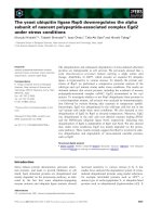
a) F344 cells (P0) b) Lewis cells (P0)
c) F344 cells (P1)
d) Lewis cells (P1)
10 μm
100 μm
100 μm
10 μm
Figure 1 Differ ences in cultures of primary mammary cells from F344 and Lewis rats and cellular localization of a-amylase. One day
after preparation, epitheloids from F344 (a) showed a faster and better attachment and a more effective growth in comparison to those from
Lewis rats (b). Detection of a-amylase (Cy3; red) was performed in mammary gland cells from F344 (c) and Lewis (d) rats (P1). Nuclei were
stained with DAPI (blue). Pictures show cells in xy- and xz-axis by confocal microscopy. a-Amylase was present in F344 and Lewis cells. However,
in Lewis cells, a-amylase was distributed throughout the whole cell, whereas in F344 cells it was found in a more granular manner near the
nuclei (xz-axis).
Fedrowitz et al. Journal of Experimental & Clinical Cancer Research 2011, 30:102
/>Page 5 of 12
Immunocytochemical detection of salivary a-amylase in
F344 and Lewis cells
Salivary a-amylase was similarly expressed in cultured
rat mammary epithelial cells from F344 and Lewis,
showing its localization in the cytoplasm (Figure 1c,d).
In F344 cells, however, a-amylase was a ssociated closer
tothenucleusinamoregranularmanner(Figure1c),
but was spread net-like throughout the whole cell body
in Lewis cells (Figure 1d).
Effects of a-amylase on cell growth in cells from F344
and Lewis rats
It has not yet been described, if a-amylase has effects on
mammary gland cell growth and, if, to what extent.
Experiments with different a-amylase concentrations iden-
tified 5 and 50 U/ml as proper concentrations to reveal
differences in a-amylase efficacy (not illustrated). In order
to find the appropriate treatment duration, experiments
were per formed with a-amylase (5 and 50 U/ml) for one
day, two, and four days (n = 4-14; Figure 2a). Cell numbers
were not altered in F344 and Lewis cells after 5 U/ml for
all treatments. After 50 U/ml, a significant decrease in
number of cells was observed for Lewis cells after 2 days
and also for F344 cells after 2 and 4 days (Figure 2a).
These results were evaluated from the total number of
counted cells including viable as w ell as dead cells after
detachment by trypsin. Comparable results were achieved
when numbers of viable cells were evaluated (Figu re 2b).
In contrast, the number of dead F344 cells varied, depend-
ing on the duration of treatment but not on the a-amylase
concentration (Figure 2c), whereas for Lewis, the amount
of dead cells was not influenced by a-amylase (Figure 2c).
Thus, prolonged a-amylase treatment reduced the number
of non-viable cells in F344 cells, but not in Lewis cells.
Based on these experiments, the cells were treated with
5 and 50 U/ml a-amylase for 2 days (Figure 3). a-Amylase
treatment with 50 U/ml significantly reduced the total cell
number in F344 and Lewis cells indicating an inhibited
cell proliferation. No significant alterations were seen after
5 U/ml compared to water-treated control cells. F344 cells
showed significantly less sensitivity towards a-amylase in
comparison to cells from Lewis rats after both concentra-
tions (5 U /ml: +7.6% and -12.6%; 50 U/ml: -14.7% and -
34.3% for F344 and Lewis, respectively; p < 0.05; Figure 3).
The decrease in total cell number was concentration-
dependent for cells from both rat strains (50 U/ml > 5 U/
ml; p < 0.05).
a-Amylase effects in mammary tumor cells of human
origin
Mammary cells from human breast tumors were also trea-
ted with a-amylase for two days. S imilar to differences
between F344 and Lewis cells, sensitivity towards salivary
a-amylase differed depending on the origin (or source) of
the cells. Cells from two different human breast tumor
patients were treated with four different concentrations of
a-amylase (0.125, 1.25, 12.5, and 125 U/ml). Statistical
analysis revealed that cells cultured from one tumor
(mammary carcinoma (MaCa) 700 II P2; Figure 4a)
showed significant decreases in cell number after 1.25 and
125 U/ml (-76% and -94.6%). Cells from the other tumor
(MaCa 699 II P3; Figure 4b) only significantly responded
to the lowest concentration (0.125 U/ml: -90.5%).
Primary cells from another human breast tumor that
hadbeenculturedfor296daysdidnotrespondwitha
change in cell number. In contrast, a culture of an invasive
ductal human breast tumor showed a concentration-
dependent decrease in number of cells in comparison to
wat er-treated control cells. Results from these cells were
not statistic ally analyzed because only one well per treat-
ment was done.
Cell senescence after a-amylase treatment
A possible influence of a-amylase on cell senescence was
investigated by determination of SA-b-gal-positive cells.
Without treatment, P2-F344 cells showed significantly
increased numbers of SA-b-gal -positive cells compared
to P1-cells (2-3fold). Therewerenosignificantdiffer-
ences in cell growth or SA-b-gal-positive cells after 5 U/
ml. a-Amylase at 50 U/ml significantly decreased num-
ber of cells in P1-F344 cells, but not in P2-F344 or P2-
Lewis, although there was a tendency for P2-F344 (Table
1). Alteration in SA-b-gal-positiv e cells was not strictly
combined with a change in cell number after a-amylase,
because cell counts were decreased in P1-F344 cells, but
SA-b-gal-p ositive cells were not changed. Moreover,
there was a significant increase in SA-b-gal-positive P2-
F344 cells by 50 U/ml, but no significant alteration in
number of cells (Table 1). Lewis cells (P2) did not
respond to a-amylase in this experiment.
In MaCa 700 cells, a primary culture from a human
breast tumor, a-amylase caused a significant decrease in
number of cells after 1.25 and 125 U/ml a-amyla se for 2
days (Figure 4a). The portion of SA-b-gal-posi tive cells
was significantly increased only after 125 U/ml. However,
there was a tendency for a concentration-dependent
increase of SA-b-gal-positive MaCa 700 cells (Figure 4a).
Discussion
The experiments described here revealed for the first time
that salivary a-amylase exhibits in vitro antiproliferative
effects in primary rat mammary epithelial cells and human
breast tumor cells. On the one hand the effects on healthy
rat breast cells indicate that endogenous a-amylase might
be involved in the regulation of mammary cell prolifera-
tion, and on the other hand the results of human breast
tumor cells suggest that it might provide a useful tool for
tumor prophylaxis or therapy. a-Amylase concentrations
Fedrowitz et al. Journal of Experimental & Clinical Cancer Research 2011, 30:102
/>Page 6 of 12
5 U/ml 50 U/ml
a) Total number of cells
b) Viable cells
c) Dead cells
Figure 2 Change in cell number after treatme nt of F344 and Lewis cell s with salivary a-amylase for different incubation times.The
mean a-amylase effect is shown in percent as change compared to control cells treated with water for the total number of cells, exclusively
viable, and for dead cells after 5 and 50 U/ml for 1 day, 2 days, and 4 days (n = 4-14 wells per group). For counting, cells were detached with
trypsin/EDTA, and viable and dead cells could be determined by trypan-blue-exclusion. Results for total cell number and viable cells were
comparable: there were no obvious differences after 5 U/ml a-amylase, but for 50 U/ml, a significant decrease in cell number was apparent after
2 days and more prominent in Lewis cells (a & b). Number of dead cells from Lewis rats was not influenced by amylase treatment (c). In contrast
to this, dead cells from F344 rats markedly changed with duration of treatment in a similar way for 5 and 50 U/ml. After 1 day of a-amylase, the
number was significantly increased, unchanged after 2 days, and significantly decreased after 4 days. Significant differences between controls
and a-amylase are indicated by asterisk (p < 0.05); significant differences between treatment durations and F344 vs. Lewis are indicated by
rhomb (p < 0.05).
Fedrowitz et al. Journal of Experimental & Clinical Cancer Research 2011, 30:102
/>Page 7 of 12
and treatment duration were determined experimentally
because to our knowledge only one previous experimental
study exists that used a-amylase for tumor treatment. In
this study, Novak & Trnka [21] found prolonged survival
in mice with transplanted B16F10 cell melanoma after
subcutaneous application of a-amylase. In the latter study,
pancreatic a-amylase was used to follow the protocol of
Beard [20], who used crude pancreas extract. However,
effects of salivary a-amylase on cell growth in vitro as
described here h ave not been previously reported in the
literature. The present experiments were performed with
salivary a-amylase, because the mammary and the salivary
glands share certain similarities in their embryology [37],
and salivary amylase is the isoenzyme present in the breast
milk [38]. Although it remains unclear if pancreatic a-
amylase exhibits similar effects on cell growth, previous
work has reported that both isoenzymes vary in their
activities on distinct substrates [39,40] suggesting different
properties on mammary cell proliferation.
Interestingly, sensitivity towards a-amylase varied
depending on the cell origin. Mammary cells from Lewis
rats were quite sensitive and showed stronger effects
compared to F344 rats. Cells from human breast tumors
also responded in different ways showing distinct sensi-
tivity. Thus, the impact of a-amylase on cell growth in
vitro depends on cellular conditions, origin, e.g. rat strain,
and distinct cellular characteristics.
The rat primary cells in this study were derived from
F344 and Lewis rats that are histocompatible inbred rat
strains originating from the same background strain [28],
but with differing responses towards stress [30,41], indicat-
ing a stronger stress response of F344 compared to Lewis
rats. Determination of a-amylase was not performed in
these studies.
In line wi th the diverse stress response, F344 rats show
a higher tumor incidence compared to Lewis, particularly
after exposure to man y known carcinogens, which is
attributed to the higher levels of immunosuppressive cor-
tisol in F344 [29]. On the other hand, Lewis appear to be
more susceptible t o autoimmune diseases according to
the low cortisol values, which were observed in this rat
strain [29]. Previous investigations from our group
showed that cell proliferation in mammary gland tissue
was significantly in creased in F344 rats, and not in Lewis,
after magnetic field exposure [42], which is considered to
act as a stressor to sensitive tissues [43-45].
Just a few years ago, salivary a-amylase was discovered
as a stress parameter in humans that, in contrast to corti-
sol, reflects the sympathetic-adrenergic activity [27] and
rapidly increases by stimulation of b-adrenergic receptors
[26]. Due to low a-amylase sensitivity, stress influences
might cause a less regulated cell proliferation in F344
breast tissue. In contrast to this, mammary Lewis cell pro-
liferation was well regulated showing rather soon signs of
senescence. These considerations are supported by the
observation that F344 cells attached easier and grew faster
than Lewis cells (Figure 1a & b). a-Amylase was detected
in both, F344 and Lewis primary mammary epithelial cells
(Figure 1c & d) without obvious differences. Moreover, we
recently determined amylase enzyme activity in the mam-
mary gland tissue of F344 and Lewis rats and observed no
differences in activity between both rat strains (unpub-
lished data). These findings indicate that other factors
than a-amylase protein expression and activ ity must
underlie the observed differences. Thus, the a-amylase
efficacy on its targets is probably altered in F344 cells par-
ticipating in less regulation of cellular proliferation.
Figure 3 a-AmylaseeffectsoncellgrowthinF344andLewis
cells after treatment for 2 days with 5 and 50 U/ml. The mean
a-amylase effect is shown as change in total cell number compared
to the water-treated control cells (percent change; mean and SEM).
Results from four to five different experiments were summarized
and evaluated together for F344 and Lewis cells (n = 29-35 wells
per group). Numbers of cells were significantly decreased after a-
amylase treatment (50 U/ml) indicating antiproliferative effects.
Lewis cells were significantly more sensitive towards a-amylase than
F344 following incubation with both 5 U/ml and 50 U/ml. Statistics:
One-way-ANOVA and Bonferroni for selected pairs: significant
differences between controls and a-amylase are indicated by
asterisk (p < 0.05); Two-way-ANOVA and Bonferroni: significant
differences between F344 vs. Lewis and 5 U/ml vs. 50 U/ml are
indicated by rhomb (p < 0.05).
Fedrowitz et al. Journal of Experimental & Clinical Cancer Research 2011, 30:102
/>Page 8 of 12
However, the enzymatic preparation of mammary gland
tissue might alter cell surface and therefore influence
adhesion properties in vitro. Microenvironmental influ-
ences in the breast tissue , which strongly affect cellular
behavior [46 -48] and which are absent or at least altered
in our primary cultures in vitro, should also be considered.
Currently, the possible mechanisms underlying anti-
proliferative effects of a-amylase remain unclear. How-
ever, some sources in literature can be found that allow
considerations about a possible mechanism and probable
a-amylase targets. a-Amylase might act on mo lecules,
which mediate cell adhesion, and stimulate detachment
and death of cells called anoikis, a type of apoptosis
[49,50]. In our experiments, the proportion of dead cells
reflects the sensitivity to trypsin used for cell detachment
prior to counting. If a-amylase induce s anoikis by action
on cellular adhesion, a more pronounced trypsin effect
would have been expected that is negatively correlated
with number of cells. This was not the case in either,
F344 and Lewis cells.
Furthermore, a-amylase could probably stimulate cel-
lular differentiation or senescence. Investigations of cell
a)
MaCa 700 II P2 (25) 18d
b)
MaCa 699 II P3 (42) 27d
Figure 4 Determinations of a-amylase effects in different cells of human origin. For two HBCEC cult ures, a significantly reduced cell
number after a-amylase treatment was demonstrated (n = 2-6; mean and SEM). MaCa 700 responded in a dose-dependent manner (a).
Additionally, the SA-b-gal assay was performed in MaCa 700 cells, and the proportion of SA-b-gal-positive cells was significantly increased by 125
U/ml a-amylase. The latter parameter showed a tendency for concentration-dependency (Pearson´s correlation coefficient 0.9002; not significant).
In MaCa 699 cells, only the lowest concentration caused a significantly decreased cell number (b). Asteriks indicate significant differences vs.
control cells (One-way-ANOVA and Bonferroni for selected pairs, p < 0.05).
Table 1 SA-b -gal assay and cell number after a-amylase treatment in F344 and Lewis cells
F344, P1 F344, P2 Lewis, P2
SA-b-gal assay SA-b-gal-positive cells (%) SA-b-gal-positive cells (%) SA-b-gal-positive cells (%)
Control (H
2
O) 11.94 ± 1.81 27.35 ± 3.28 33.82 ± 1.48
5 U/ml a-amylase 13.86 ± 1.41 37.15 ± 3.19 34.12 ± 3.20
50 U/ml a-amylase 11.83 ± 2.39 39.48 ± 3.47* 29.81 ± 2.78
n.s. *H
2
O vs. 50 U/ml n.s.
F344, P1 F344, P2 Lewis, P2
Cell counts Number of cells/well Number of cells/well Number of cells/well
Control (H
2
O) 17,250 ± 1,377 4,500 ± 577 4,188 ± 567
5 U/ml a-amylase 17,958 ± 1,514 3,958 ± 240 5,292 ± 163
50 U/ml a-amylase 11,833 ± 870* 2,371 ± 344 4,483 ± 464
*H
2
O vs. 50 U/ml n.s. n.s.
a-Amylase (50 U/ml) decreased the number of cells only in P1-F344-cells, but not in P2-F344- and P2-Lewis-cells. Proportion of SA-b-gal-positive cells did not
correlate with cell number, as this amount of cells was not altered in P1-F344 cells, but significantly increased in P2-F344 cells after 50 U/ml a-amylase. No
difference at all was observed in Lewis-cells (P2) and after 5 U/ml a-amylase. Mean and SEM are shown for three wells per group (cell counts) or 6-9 sections
(SA-b-gal assay). Significant differences (p < 0.05) vs. control cells (One-way-ANOVA and Bonferroni for selected pairs) are indicated by asterisk.
Fedrowitz et al. Journal of Experimental & Clinical Cancer Research 2011, 30:102
/>Page 9 of 12
senescence by SA-b-gal assay presented here did not
show a strong impact of a-amylase on senescence, parti-
cularly not in combination with the effect on cell
growth.
a-Amylase also exerts antibacterial effects, which are
either drawn back to an inhibition of bacteria growth by
dimi nishing nutrients [10] or to a direct interac tion with
a-amylase [11]. Regarding cell culture, known a-amylase-
substrates , like starch, are usually not present in cell cul-
ture media, but an a-amylase effect by metabolism of
nutrients cannot be completely excluded. F344 and Lewis
cells were cultured simultaneously with medium of the
same composition, so that differing dependence on growth
influencing substances could be a possible reason for the
observed differences.
Another explanation for the a-amylase effect on cell
growth might be an interfere nce with growth stimulating
hormones,e.g.estrogens.Hahneletal.[51]showedin
vitro that a-amylase inhibited or diminished binding of
estradiol to its receptor. Previously, a correlation between
a-amylase and hormone lev els was reported in vivo [14],
and hormonal alterations during sexual cycle influenced
a-amylase activity in rat ovaries [52].
In vivo, the sympathetic system and its adrenergic
receptors are activated during stress. a-Amylase is sti-
mulated by adrenergic receptor s [25] and probably
adjusts or counteracts proliferation that h as been eli-
cited by a-andb-adrenergic receptors induced by
stress. It is known that the mammary gland is inner-
vated by sym pathetic fibers. Mammary epithelial cells
express a-andb-receptors, the receptor densities are
hormone-depend ent, and cell proliferation is influenced
by these receptors [53-56], so that there might be a pos-
sible connection or interaction between estrogens, adre-
nergic receptors and a-amylase, which has not yet been
described. In F344 cells, adrenergic receptors might sti-
mulate proliferation in a more pronounced way due to
intensive activation by stress that could not be effec-
tively regulated. According to this hypothesis, cell prolif-
eration in Lewis rats is affected by adrenergic receptors
in a more moderate way and can easily be adjusted by
a-amylase.
In summary, the present results demonstrate antiproli-
ferative properties of salivary a-amylase in mammary
epithelial and breast tumor cells suggesting that a-amylase
might constitute a new strategy to prevent or treat breast
cancer. However, the reasons for the altered cellular sensi-
tivity towards a-amylase should b e identified to allow a
reliable prediction which type of breast cancer cells can be
sufficiently inhibited in proliferation to ensure an appro-
priate efficiency of tumor treatment. The stimulation of
endogenous a-amylase secretion and activity in the vici-
nity of the neoplastic tissue may provide a reasonab le
approach to affect tumor growth. Consequently, a direct
administration of a-amylase into or nearby the tumor
could represent a conceivable opportunity to monitor
both, anti-tumor and potential side effects.
Conclusions
To our knowledge, the findings presented here indicate for
the first time that a-amylase plays a role in the regulation
of mammary cell proliferation. However, the underlying
mechanisms and the influencing factors of a-amylase’s
action must be further elucidated. In view of the potential
impact on regulation of mammary cell proliferation, deter-
mination of a-amylase might be used to disting uish the
risk for cancer development, and a-amylase may provide
an interesting new target for tumor prophylaxis and
treatment.
Abbreviations
ACTH: adrenocorticotropic hormone; BSA: bovine serum albumin; Cy:
cyanine dyes; DAPI: 4,6-diamidino-2-phenylindole; DMBA: 7,12-dimethylbenz
[a]anthracene; DMEM: Dulbecco´s Modified Eagle Medium; EDTA:
ethylenediaminetetraacetic acid; F12: nutrient mixture F12; F344: Fischer 344;
HBCEC: human breast cancer-derived epithelial cells; L/R1: left/right
mammary gland complex at cranial cervical location; MaCa: mammary
carcinoma; P1: cell passage 1; PBS: phosphate-buffered saline; SA-β-gal:
senescence-associated-β-galactosidase; SEM: standard error of the mean
Acknowledgements
The authors would like to acknowledge Britta Sterzik, Jutta Beu, and
Marianne Thren for excellent technical support. This work was funded by a
grant from the German Research Foundation (Lo 274/6-3).
Author details
1
Department of Pharmacology, Toxicology, and Pharmacy, Universi ty of
Veterinary Medicine, Buenteweg 17, Hannover, 30559, Germany.
2
Biochemistry and Tumor Biology Lab, Gynecology Research Unit,
Department of Obstetrics and Gynecology, Carl-Neuberg-Str. 1, Medical
University, Hannover, 30625, Germany.
Authors’ contributions
MF participated in the design of the study, primary rat mammary cell
preparation and culturing, performed the cell counting, immunofluorescence
staining and statistical analysis and drafted the manuscript. RH provided the
human breast tumor cells and expert views in primary cell culture methods,
participated in the SA-β-gal staining and helped draft the manuscript. CB
performed experiments with the human cells and the SA-β-gal staining. WL
participated in the design of the study and helped draft the manuscript. All
authors read and approved the manuscript.
Competing interests
The authors declare that they have no competing interests.
Received: 11 August 2011 Accepted: 25 October 2011
Published: 25 October 2011
References
1. Jemal A, Siegel R, Xu J, Ward E: Cancer Statistics, 2010. CA Cancer J Clin
2010, 60:277-300.
2. Karnoub AE, Dash AB, Vo AP, Sullivan A, Brooks MW, Bell GW,
Richardson AL, Polyak K, Tubo R, Weinberg RA: Mesenchymal stem cells
within tumor stroma promote breast cancer metastasis. Nature 2007,
449:557-563.
3. Finak G, Bertos N, Pepin F, Sadekova S, Souleimanova M, Zhao H, Chen H,
Omeroglu G, Meterissian S, Omeroglu A, Hallett A, Park M: Stromal gene
expression predicts clinical outcome in breast cancer. Nature Med 2008,
14:518-527.
Fedrowitz et al. Journal of Experimental & Clinical Cancer Research 2011, 30:102
/>Page 10 of 12
4. Fedrowitz M, Löscher W: Effects of magnetic field exposure in the
mammary gland tissue of female Fischer 344 rats and the role of
amylase. Eur J Cancer 2007, 5(Suppl):77.
5. Fedrowitz M, Löscher W: Alterations in amylase activity in the mammary
gland of female Fischer 344 rats after exposure to 50 Hertz magnetic
fields. Naunyn Schmiedeberg´s Arch Pharmacol 2008, 377(Suppl 1):83.
6. Zakowski JJ, Bruns DE: Biochemistry of human alpha amylase isoenzymes.
Crit Rev Clin Lab Sci 1985, 21:283-322.
7. Moridani MJ, Bromberg IL: Lipase and pancreatic amylase versus total
amylase as biomarkers of pancreatitis: an analytical investigation. Clin
Biochem 2003, 36:31-33.
8. Brown RC, Chalmers DM, Rowe VL, Kelleher J, Littlewood JM, Losowsky MS:
Comparison of the diagnostic value of serum pancreatic isoamylase and
immunoreactive trypsin measurement in patients with cystic fibrosis. J
Clin Pathol 1982, 35:547-549.
9. Zakowski JJ, Gregory MR, Bruns DE: Amylase from human serous ovarian
tumors: purification and characterization. Clin Chem 1984, 30:62-68.
10. Gregory MR, Gregory WW, Bruns DE, Zakowski JJ: Amylase inhibits
Neisseria gonorrhoeae by degrading starch in the growth medium. J
Clin Microbiol 1983, 18:1366-1369.
11. Chaudhuri B, Rojek J, Vickerman MM, Tanzer JM, Scannapieco FA:
Interaction of salivary alpha-amylase and amylase-binding-protein A
(AbpA) of Streptococcus gordonii with glucosyltransferase of S. gordonii
and Streptococcus mutans. BMC Microbiology 2007, 7:60.
12. Groot PC, Bleeker MJ, Pronk JC, Arwert F, Mager WH, Planta RJ, Eriksson AW,
Frants RR: The human α-amylase multigene family consists of haplotypes
with variable numbers of genes. Genomics 1989, 5:29-42.
13. Heitlinger LA, Lee PC, Dillon WP, Lebenthal E: Mammary amylase: a
possible alternate pathway of carbohydrate digestion in infancy. Pediatr
Res 1983, 17:15-18.
14. Skerlavay M, Epstein JA, Sobrero AJ: Cervical mucus amylase levels in
normal menstrual cycles. Fertil Steril 1968, 19:726-730.
15. Hokari S, Miura K, Koyama I, Kobayashi M, Matsunaga T, Iino N, Komoda T:
Expression of α-amylase isoenzymes in rat tissues. Comp Biochem Physiol
Part B 2003, 135:63-69.
16. Yanagitani N, Kaira K, Sunaga N, Naito Y, Koike Y, Ishihara S, Ishizuka T,
Saito R, Mori M: Serum amylase is a sensitive tumor marker for amylase-
producing small cell lung cancer? Int J Clin Oncol 2007, 12:231-233.
17. Tomita N, Matsuura N, Horii A, Emi M, Nishide T, Ogawa M, Mori T, Doi O,
Matsubara K:
Expression of α-amylase
in human lung cancers. Cancer Res
1988, 48:3288-3291.
18. Coyne JD, Dervan PA: Primary acinic cell carcinoma of the breast. J Clin
Pathol 2005, 55:545-547.
19. Tanahashi C, Yasuki S, Akamine N, Yatabe Y, Ichihara S: Pure acinic cell
carcinoma of the breast in an 80-year-old Japanese woman. Pathol Int
2007, 57:43-46.
20. Beard J: The cancer problem. Lancet 1905, 4:281-283.
21. Novak JF, Trnka F: Proenzyme therapy of cancer. Anticancer Res 2005,
25:1157-1178.
22. Nagasawa H, Kusakawa S: Comparison of plasma component levels in
four strains of female mice with different mammary tumour potentials.
In Vivo 2001, 15:139-144.
23. Simickova M, Pecen L, Eben K, Nekulova M, Vermousek I, Stratil P, Rejthar A,
Cernoch M, Lang B, Sakalova J: Biochemical analysis of breast cyst fluid as
a possible predictor of breast carcinoma development. Neoplasma 1994,
41:245-252.
24. Saez Mdel C, Barriga C, Garcia JJ, Rodriguez AB, Ortega E: Exercise-induced
stress enhances mammary tumor growth in rats: Beneficial effect of the
hormone melatonin. Mol Cell Biochem 2007, 294:19-24.
25. Rohleder N, Nater UM, Wolf JM, Ehlert U, Kirschbaum C: Psychosocial
stress-induced activation of salivary alpha-amylase: An indicator of
sympathetic activity? Ann NY Acad Sci 2004, 1032:258-263.
26. van Stegeren A, Rohleder N, Everaerd W, Wolf OT: Salivary alpha amylase
as marker for adrenergic activity during stress: effect of betablockade.
Psychoendocrinology 2006, 31:137-141.
27. Nater UM, Rohleder N: Salivary alpha-amylase as a non-invasive
biomarker for the sympathetic nervous system: Current state of
research. Psychoendocrinology 2009, 34:486-496.
28. Dhabhar FS, McEwen BS, Spencer RL: Stress response, adrenal steroid
receptor levels and corticosteroid-binding globulin levels - a comparison
between Sprague-Dawley, Fischer 344 and Lewis rats. Brain Res 1993,
616:89-98.
29. Sternberg EM, Hill JM, Chrousos GP, Kamilaris T, Listwak SJ, Gold PW,
Wilder RL: Inflammatory mediator-induced hypothalamic-pituitary-
adrenal axis activation is defective in streptococcal cell wall arthritis-
susceptible Lewis rats. Proc Natl Acad Sci 1989, 86:2374-2378.
30. Dhabhar FS, Miller AH, McEwen BS, Spencer RL: Differential activation of
adrenal steroid receptors in neural and immune tissues of Sprague-
Dawley, Fischer 344, and Lewis rats. J Neuroimmunology 1995, 56:77-90.
31. Haag JD, Newton MA, Gould MN:
Mammary carcinoma suppressor and
susceptibility
genes in the Wistar-Kyoto rat. Carcinogenesis 1992,
13:1933-1935.
32. Hass R, Bertram C: Characterization of human breast cancer epithelial
cells (HBCEC) derived from long term cultured biopsies. J Exp Clin Cancer
Res 2009, 28:127-139.
33. Novaro V, Roskelley CD, Bissell MJ: Collagen-IV and laminin-1 regulate
estrogen receptor α expression and function in mouse mammary
epithelial cells. J Cell Sci 2003, 116:2975-2986.
34. Lavrentieva A, Majore I, Kasper C, Hass R: Effects of hypoxic culture
conditions on umbilical cord-derived human mesenchymal stem cells.
Cell Commun Signal 2010, 8:18.
35. Gilles C, Polette M, Zahm JM, Tournier JM, Volders L, Foidart J, Birembaut P:
Vimentin contributes to human mammary epithelial cell migration. J Cell
Sci 1999, 112:4615-4625.
36. Dimri GP, Lee X, Basile G, Acosta M, Scott G, Roskelley C, Medrano EE,
Linskens M, Rubeli I, Pereira-Smith O, Peacocke M, Campisi J: A biomarker
that identifies senescent human cells in culture and in aging skin in
vivo. Proc Natl Acad Sci 1995, 92:9363-9367.
37. Matoso A, Easley SE, Gnepp DR, Mangray S: Salivary gland acinar-like
differentiation of the breast. Histopathology 2009, 54:262-263.
38. Lindberg T, Skude G: Amylase in human milk. Pediatrics 1982, 70:235-238.
39. Hall FF, Ratliff CR, Hayakawa T, Culp TW, Hightower NC: Substrate
differentiation of human pancreatic and salivary alpha-amylases. Am J
Dig Dis 1970, 15:1031-1038.
40. Stiefel DJ, Keller PJ: Comparison of human pancreatic and parotid
amylase activities on different substrates. Clin Chem 1975, 21:343-346.
41. Dhabhar FS, McEwen BS, Spencer RL: Adaptation to prolonged or
repeated stress - comparison between rat strains showing intrinsic
differences in reactivity to acute stress. Neuroendocrinology 1997,
65:360-368.
42. Fedrowitz M, Löscher W: Power-frequency magnetic fields increase cell
proliferation in the mammary gland of female Fischer 344 rats but not
various other rat strains or substrains. Oncology 2005, 69:486-498.
43. Ossenkopp KP, Kavaliers M, Lipa S: Increased mortality in land snails
(Cepaea nemoralis) exposed to powerline (60-Hz) magnetic fields and
effects of the light-dark cycle. Neurosci Lett 1990, 114:89-94.
44. Pipkin JL, Hinson WG, Young JF, Rowland KL, Shaddock JG, Tolleson WH,
Duffy PH, Casciano DA: Induction of stress proteins by electromagnetic
fields in cultured HL-60 cells. Bioelectromagnetics 1999,
20:347-357.
45.
Yoshikawa T, Tanigawa M, Tanigawa T, Imai A, Hongo H, Kondo M:
Enhancement of nitric oxide generation by low frequency
electromagnetic field. Pathophysiology 2000, 7:131-135.
46. Maffini MV, Soto AM, Calabro JM, Ucci AA, Sonnenschein C: The stroma as
a crucial target in rat mammary gland carcinogenesis. J Cell Sci 2004,
117:1495-1502.
47. Medina D: Stromal fibroblasts influence human mammary epithelial cell
morphogenesis. Proc Natl Acad Sci 2004, 101:4723-4724.
48. Zangani D, Darcy KM, Shoemaker S, Ip MM: Adipocyte-epithelial
interactions regulate the in vitro development of normal mammary
epithelial cells. Exp Cell Res 1999, 247:399-409.
49. Frisch SM, Screaton RA: Anoikis mechanisms. Curr Opin Cell Biol 2001,
13:555-562.
50. Rennebeck G, Martelli M, Kyprianou N: Anoikis and survival connections in
the tumor. Microenvironment: Is there a role in prostate cancer
metastasis? Cancer Res 2005, 65:11230-11235.
51. Hahnel R, Twaddle E, Brindle L: The influence of enzymes on the estrogen
receptors of human uterus and breast carcinoma. Steroids 1974,
24:489-506.
52. Kasperczyk S, Brzoza Z, Kasperczyk A, Beck B, Duliban H, Mertas A: The
changes of alpha-amylase activity in serum and different tissues of
Fedrowitz et al. Journal of Experimental & Clinical Cancer Research 2011, 30:102
/>Page 11 of 12
female rat during sex cycle - isoelectrofocusing studies of alpha-
amylase. Med Sci Monit 2001, 7:49-53.
53. Bruzzone A, Pinero PC, Castillo LF, Sarappa MG, Rojas P, Lanari C, Lüthy IA:
α2-Adrenoceptor action on cell proliferation and mammary tumour
growth in mice. Brit J Pharmacol 2008, 155:494-504.
54. Marchetti B, Spinola PG, Pelletier G, Labrie F: A potential role for
catecholamines in the development and progression of carcinogen-
induced tumors: hormonal control of beta-adrenergic receptors and
correlation with tumor growth. J Steroid Biochem Molec Biol 1991,
38:307-320.
55. Marchetti B, Spinola PG, Plante M, Poyet P, Follea N, Pelletier G, Labrie F:
Beta-adrenergic receptors in DMBA-induced rat mammary tumors:
correlation with progesterone receptor and tumor growth. Breast Cancer
Res Treat 1989, 13:251-263.
56. Lüthy IA, Bruzzone A, Pinero PC, Castillo LF, Chiesa IJ, Vazquez SM,
Sarappa MG: Adrenoceptors: non conventional target for breast cancer?
Curr Med Chemistry 2009, 16:1850-1862.
doi:10.1186/1756-9966-30-102
Cite this article as: Fedrowitz et al.: Salivary a-amylase exhibits
antiproliferative effects in primary cell cultures of rat mammary
epithelial cells and human breast cancer cells. Journal of Experimental &
Clinical Cancer Research 2011 30:102.
Submit your next manuscript to BioMed Central
and take full advantage of:
• Convenient online submission
• Thorough peer review
• No space constraints or color figure charges
• Immediate publication on acceptance
• Inclusion in PubMed, CAS, Scopus and Google Scholar
• Research which is freely available for redistribution
Submit your manuscript at
www.biomedcentral.com/submit
Fedrowitz et al. Journal of Experimental & Clinical Cancer Research 2011, 30:102
/>Page 12 of 12