ABC OF CLINICAL GENETICS - PART 5 pdf
Bạn đang xem bản rút gọn của tài liệu. Xem và tải ngay bản đầy đủ của tài liệu tại đây (246.68 KB, 13 trang )
ABC of Clinical Genetics
44
these disorders. Carrier screening for sickle cell disease has
been less successful. Carrier screening for cystic fibrosis is also
possible, although not all carriers can be identified because of
the diversity of mutations within the cystic fibrosis gene.
Screening programmes instituted in antenatal clinics and in
general practice have reported a substantial uptake for cystic
fibrosis carrier testing when it is offered, but indicate that few
couples actively seek this type of test themselves. It is important
that appropriate information and counselling is available to
individuals being offered screening, as they are likely to have
little prior knowledge of the disorder being screened for and
the implications of a positive test result. Specific training will be
needed by members of primary health care and obstetric teams
before any new screening programmes are instituted, as these
are the settings in which such tests are likely to be offered.
In addition to screening programmes aimed at identifying
carriers, there are well established programmes for screening
all neonates to identify those affected by conditions such as
phenylketonuria and hypothyroidism, where early diagnosis
and treatment is successful in preventing mental retardation.
The value of including other metabolic disorders in screening
programmes depends on the incidence of the disorder and the
prospect of altering the prognosis by its early detection.
Possible candidates include galactosaemia, maple syrup urine
disease and congenital adrenal hyperplasia.
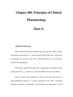
Figure 9.11 Neonatal blood samples used for biochemical screening
Box 9.6 Conditions amenable to population screening
programmes
Antenatal
• Thalassaemia
• Sickle cell disease
• Tay–Sachs disease
• Cystic fibrosis
Neonatal
• Phenylketonuria
• Hypothyroidism
• Galactosaemia
acg-09 11/20/01 7:25 PM Page 44
45
There are thousands of genetic traits and disorders described,
some of which are exceedingly rare. All of the identified
mendelian traits in man have been catalogued by McKusick and
are listed on the Omim (online mendelian inheritance in man)
database described in chapter 16. In this chapter the clinical
and genetic aspects of a few examples of some of the more
common disorders are briefly outlined and examples of genetic
disorders affecting various organ systems are listed. Molecular
analysis of some of these conditions is described in chapter 18.
Central nervous system disorders
Huntington disease
Huntington disease is an autosomal dominant disease
characterised by progressive choreiform movements, rigidity,
and dementia from selective, localised neuronal cell death
associated with atrophy of the caudate nucleus demonstrated
by CNS imaging. The frequency of clinical disease is about
6 per 100 000 with a frequency of heterozygotes of about 1 per
10 000. Development of frank chorea may be preceded by a
prodromal period in which there are mild psychiatric and
behavioural symptoms. The age of onset is often between
30 and 40 years, but can vary from the first to the seventh
decade. The disorder is progressive, with death occurring
about 15 years after onset of symptoms. Surprisingly, affected
homozygotes are not more severely affected than heterozygotes
and new mutations are exceedingly rare. Clinical treatment
trials commenced in 2000 to assess the effect of transplanting
human fetal striatal tissue into the brain of patients affected by
Huntington disease as a potential treatment for
neurodegenerative disease.
The gene (designated IT15) for Huntington disease was
mapped to the short arm of chromosome 4 in 1983, but not
finally cloned until 1993. The mutation underlying Huntington
disease is an expansion of a CAG trinucleotide repeat sequence
(see chapter 7). Normal alleles contain 9–35 copies of the repeat,
whereas pathological alleles usually contain 37–86 repeats, but
sometimes more. Transcription and translation of pathological
alleles results in the incorporation of an expanded polyglutamine
tract in the protein product (huntingtin) leading to
accumulation of intranuclear aggregates and neuronal cell death.
Clinical severity of the disorder correlates with the number of
trinucleotide repeats. Alleles that contain an intermediate
number of repeats do not always cause disease and may not be
fully penetrant. Instability of the repeat region is more marked
on paternal transmission and most cases of juvenile onset
Huntington disease are inherited from an affected father.
Prior to the identification of the mutation, presymptomatic
predictive testing could be achieved by linkage studies if the
family structure was suitable. Prenatal testing could also be
undertaken. In some cases tests were done in such a way as to
identify whether the fetus had inherited an allele from the
clinically affected grandparent without revealing the likely
genetic status of the intervening parent. This enabled adults at
risk to have children predicted to be at very low risk without
having predictive tests themselves. Direct mutation detection
now enables definitive confirmation of the diagnosis in
clinically affected individuals (see chapter 18) as well as
providing presymptomatic predictive tests and prenatal
diagnosis. Considerable experience has been gained with
10 Single gene disorders
Table 10.2 Inheritance pattern and gene product for some
common neurological disorders
Disorder Inheritance Gene product
Childhood onset spinal AR SMN protein
muscular atrophy
Kennedy syndrome XLR androgen
(SBMA) receptor
Myotonia congenita AD muscle chloride
(Thomsen type) channel
Myotonia congenita AD muscle chloride
(Becker type) channel
Friedreich ataxia AR frataxin
Spinocerebellar ataxia type 1 AD ataxin-1
Charcot–Marie–Tooth type 1a AD peripheral
myelin protein
P22
Charcot–Marie–Tooth type 1b AD peripheral
myelin protein
zero
Hereditary spastic paraplegia AD spastin
(SPG4)
Hereditary spastic paraplegia AR paraplegin
(SPG7)
Hereditary spastic paraplegia XLR propeolipid
(SPG2) protein
Table 10.1 Examples of autosomal dominant adult-onset
diseases affecting the central nervous system for which
genes have been cloned
Disease Gene
Familial alzheimer AD1 amyloid precursor
disease gene (APP)
AD2 APOE*4 association
AD3 Presenilin-1 gene (PSEN 1)
AD4 Presenilin-2 gene (PSEN 2)
Familial amyotrophic lateral superoxide dismutase-1
sclerosis ALS1 gene (SOD1)
ALS susceptibility heavy neurofilament subunit
gene (NEFH)
Familial Parkinson disease PARK1 alpha-synuclein gene (SNCA)
+lewy body PARK4
Frontotemporal dementia with microtubule-associated
Parkinsonism protein tau gene (MAPT)
Creutzfeldt-Jakob disease (CJD) prion protein gene (PRNP)
Cerebral autosomal dominant
arteriopathy with subcortical
infarcts and
leucoencephalopathy(CADASIL) NOTCH 3
Familial British dementia (FBD) ITM2B
Box 10.1 Neurological disorders due to trinucleotide
repeat expansion mutations
Huntington disease (HD)
Fragile X syndrome (FRAXA)
Fragile X site E (FRAXE)
Kennedy syndrome (SBMA)
Myotonic dystrophy (DM)
Spinocerebellar ataxias (SCA 1,2,6,7,8,12)
Machado-Joseph disease (SCA3)
Dentatorubral-pallidolysian atrophy (DRPLA)
Friedreich ataxia (FA)
Oculopharyngeal muscular dystrophy (OPMD)
acg-10 11/20/01 7:27 PM Page 45
ABC of Clinical Genetics
46
predictive testing and an agreed protocol has been drawn up
for use in clinical practice that is applicable to other predictive
testing situations (see chapter 3).
Fragile X syndrome
Fragile X syndrome, first described in 1969 and delineated
during the 1970s, is the most common single cause of inherited
mental retardation. The disorder is estimated to affect around
1 in 4000 males, with many more gene carriers. The clinical
phenotype comprises mental retardation of varying degree,
macro-orchidism in post-pubertal males, a characteristic facial
appearance with prominent forehead, large jaw and large ears,
joint laxity and behavioural problems.
Chromosomal analysis performed under special culture
conditions demonstrates a fragile site near the end of the long
arm of the X chromosome in most affected males and some
affected females, from which the disorder derived its name.
The disorder follows X linked inheritance, but is unusual
because of the high number of female carriers who have
mental retardation and because there is transmission of the
gene through apparently unaffected males to their daughters –
a phenomenon not seen in any other X linked disorders. These
observations have been explained by the nature of the
underlying mutation, which is an expansion of a CGG
trinucleotide repeat in the FMR1 gene. Normal alleles contain
up to 45 copies of the repeat. Fragile X mutations can be
divided into premutations (50–199 repeats) that have no
adverse effect on phenotype and full mutations (over 200
repeats) that silence gene expression and cause the clinical
syndrome. Both types of mutations are unstable and tend to
increase in size when transmitted to offspring. Premutations
can therefore expand into full mutations when transmitted by
an unaffected carrier mother. All of the boys and about half of
the girls who inherit full mutations are clinically affected.
Mental retardation is usually moderate to severe in males, but
mild to moderate in females. Males who inherit the
premutation are unaffected and usually transmit the mutation
unchanged to their daughters who are also unaffected, but at
risk of having affected children themselves.
Molecular analysis confirms the diagnosis of fragile X
syndrome in children with learning disability, and enables
detection of premutations and full mutations in female carriers,
premutations in male carriers and prenatal diagnosis (see
chapter 18).
Neuromuscular disorders
Duchenne and Becker muscular dystrophies
Duchenne and Becker muscular dystrophies are due to
mutations in the X linked dystrophin gene. Duchenne
muscular dystrophy (DMD) is one of the most common and
severe neuromuscular disorders of childhood. The incidence of
around 1 in 3500 male births has been reduced to around 1 in
5000 with the advent of prenatal diagnosis for high risk
pregnancies.
Boys with DMD may be late in starting to walk. If serum
creatine kinase estimation is included as part of the
investigations at this stage, very high enzyme levels will indicate
the need for further investigation. In the majority of cases,
onset of symptoms is before the age of four. Affected boys
present with an abnormal gait, frequent falls and difficulty
climbing steps. Toe walking is common, along with
pseudohypertrophy of calf muscles. Pelvic girdle weakness
results in the characteristic waddling gait and the Gower
manoeuvre (a manoeuvre by which affected boys use their
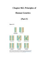
Figure 10.1 Boy with fragile X syndrome showing characteristic facial
features: tall forehead, prominent ears and large jaw
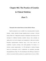
Figure 10.2 Karyotype of a male with fragile X syndrome demonstrating
the fragile site on the X chromosome (courtesy of Dr Lorraine Gaunt
and Helena Elliott, Regional Genetic Service, St Mary’s Hospital,
Manchester)
Figure 10.3 Fragile X pedigree showing transmission of the mutation
through an unaffected male( premutation carrier, ! full mutation)
Figure 10.4 Scapular winging, mild lordosis and enlarged calves in the
early stages of Duchenne muscular dystrophy
acg-10 11/20/01 7:27 PM Page 46
Single gene disorders
47
hands to “climb up” their legs to get into a standing position
when getting up from the floor). Calf pain is a common
symptom at this time. Scapular winging is the first
sign of shoulder girdle involvement and, as the disease
progresses, proximal weakness of the arm muscles becomes
apparent. Most boys are confined to a wheelchair by the age of
12. Flexion contractures and scoliosis are common and require
active management. Cardiomyopathy and respiratory problems
occur and may necessitate nocturnal respiratory support.
Survival beyond the age of 20 is unusual. Intellectual
impairment is associated with DMD, with 30% of boys having
an IQ below 75.
The diagnosis of DMD is confirmed by muscle biopsy with
immunocytochemical staining for the dystrophin protein. Two
thirds of affected boys have deletions or duplications within the
dystrophin gene that are readily detectable by molecular testing
(see chapter 18). The remainder have point mutations that are
difficult to detect. Mutation analysis or linkage studies enable
carrier detection in female relatives and prenatal diagnosis for
pregnancies at risk. However, one third of cases arise by new
mutation. Gonadal mosaicism, with the mutation being
confined to germline cells, occurs in about 20% of mothers of
isolated cases. In these women, the mutation is not detected in
somatic cells when carrier tests are performed, but there is a
risk of having another affected son. Prenatal diagnosis should
therefore be offered to all mothers of isolated cases. Testing for
inherited mutations in other female relatives does give
definitive results and prenatal tests can be avoided in those
relatives shown not to be carriers.
About 5% of female carriers manifest variable signs of
muscle involvement, due to non-random X inactivation that
results in the abnormal gene remaining active in the majority
of cells. There have also been occasional reports of girls being
more severely affected as a result of having Turner syndrome
(resulting in hemizygosity for a dystrophin gene mutation) or
an X:autosome translocation disrupting the gene at Xp21
(causing inactivation of the normal X chromosome and
functional hemizygosity).
Becker muscular dystrophy (BMD) is also due to mutations
within the dystrophin gene. The clinical presentation is similar
to DMD, but the phenotype milder and more variable. The
underlying mutations are commonly also deletions. These
mutations differ from those in DMD by enabling production of
an internally truncated protein that retains some function, in
comparison to DMD where no functional protein is produced.
Myotonic dystrophy
Myotonic dystrophy is an autosomal dominant disorder
affecting around 1 in 3000 people. The disorder is due to
expansion of a trinuceotide repeat sequence in the 3Ј region of
the dystrophia myotonica protein kinase (DMPK ) gene. The
trinucleotide repeat is unstable, causing a tendency for further
expansion as the gene is transmitted from parent to child. The
size of the expansion correlates broadly with the severity of
phenotype, but cannot be used predictively in individual
situations.
Classical myotonic dystrophy is a multisystem disorder that
presents with myotonia (slow relaxation of voluntary muscle
after contraction), and progressive weakness and wasting of
facial, sternomastoid and distal muscles. Other features include
early onset cataracts, cardiac conduction defects, smooth
muscle involvement, testicular atrophy or obstetric
complications, endocrine involvement, frontal balding,
hypersomnia and hypoventilation. Mildly affected late onset
cases may have little obvious muscle involvement and present
with only cataracts. Childhood onset myotonic dystrophy
Table 10.3 Muscular dystrophies with identified genetic
defects
Type of muscular Locus/ Protein Inheritance
dystrophy gene symbol deficiency
Congenital LAMA2 merosin AR
Congenital lTGA7 integrin ␣ 7AR
Duchenne/ DMD/BMD dystrophin XLR
Becker
Emery–Dreifuss EMD emerin XLR
Emery–Dreifuss EDMD-AD lamin A/C AD
Facioscapulo- FSHD (4q34 AD
humeral rearrangement)
Limb girdle LGMDIB lamin A/C AD
with cardiac
involvement
Limb girdle LGMDIC caveolin-3 AD
LGMD2A calpain 3 AR
LGMD2B dysferlin AR
LGMD2C ␥ sarcoglycan AR
LGMD2D ␣ sarcoglycan AR
LGMD2E  sarcoglycan AR
LGMD2F ␦ sarcoglycan AR
LGMD2G telethonin AR
Figure 10.5 Young boy with Duchenne
muscular dystrophy demonstrating the
Gower manoeuvre, rising from the floor by
getting onto his hands and feet, then
pushing up on his knees
a
c
b
Figure 10.6 Marked wasting of the thighs with calf hypertrophy and
scapular winging in young man with Becker muscular dystrophy
acg-10 11/20/01 7:27 PM Page 47
ABC of Clinical Genetics
48
usually presents with less specific symptoms of muscle weakness,
speech delay and mild learning disability, with more classical
clinical features developing later. Congenital onset myotonic
dystrophy can occur in the offspring of affected women. These
babies are profoundly hypotonic at birth and have major
feeding and respiratory problems. Children who survive have
marked facial muscle weakness, delayed motor milestones and
commonly have intellectual disability and speech delay. The age
at onset of symptoms becomes progressively younger as the
condition is transmitted through a family. Progression of the
disorder from late onset to classical, and then to childhood or
congenital onset, is frequently observed over three generations
of a family.
Molecular analysis identifies the expanded CTG repeat,
confirming the clinical diagnosis and enabling presymptomatic
predictive testing in young adults. Prenatal diagnosis is also
possible, but does not, on its own, predict how severe the
condition is going to be in an affected child.
Neurocutaneous disorders
Neurofibromatosis
Neurofibromatosis type 1 (NF1), initially described by
von Recklinghausen, is one of the most common single gene
disorders, with an incidence of around 1 in 3000. The main
diagnostic features of NF1 are café-au-lait patches, peripheral
neurofibromas and lisch nodules. Café-au-lait patches are
sometimes present at birth, but often appear in the first few
years of life, increasing in size and number. A child at risk who
has no café-au-lait patches by the age of five is extremely
unlikely to be affected. Freckling in the axillae, groins or base
of the neck is common and generally only seen in people with
NF1. Peripheral neurofibromas usually start to appear around
puberty and tend to increase in number through adult life.
The number of neurofibromas varies widely between different
subjects from very few to several hundred. Lisch nodules
(iris hamartomas) are not visible to the naked eye but can be
seen using a slit lamp. Minor features of NF1 include short
stature and macrocephaly. Complications of NF1 are listed
in the box and occur in about one third of affected
individuals. Malignancy (mainly embryonal tumours or
neurosarcomas) occur in about 5% of affected
individuals. Learning disability occurs in about one
third of children, but severe mental retardation in
only 1 to 2%.
Clinical management involves physical examination with
measurement of blood pressure, visual field testing, visual
acuity testing and neurological examination on an annual
basis. Children should be seen every six months to monitor
growth and development and to identify symptomatic optic
glioma and the development of plexiform neurofibromas or
scoliosis.
The gene for NF1 was localised to chromosome 17 in 1987
and cloned in 1990. The gene contains 59 exons and encodes
of protein called neurofibromin, which appear to be involved
in the control of cell growth and differentiation. Mutation
analysis is not routine because of the large size of the gene and
the difficulty in identifying mutations. Prenatal diagnosis by
linkage analysis is possible in families with two or more affected
individuals. NF1 has a very variable phenotype and prenatal
testing does not predict the likely severity of the condition. Up
to one third of cases arise by a new mutation. In this situation,
Box 10.2 Diagnostic criteria for NF1
Two or more of the following criteria:
• Six or more café-au-lait macules
Ͼ5 mm diameter before puberty
Ͼ15 mm diameter after puberty
• Two or more neurofibroma of any type or one plexiform
neuroma
• Freckling in the axillary or inguinal regions
• Two or more Lisch nodules
•
Optic glioma
• Bony lesions such as pseudarthrosis, thinning of the long
bone cortex or sphenoid dysplasia
•
First degree relative with NF1 by above criteria
Box 10.3 Complications of NF1
• Plexiform neurofibromas
• Congenital bowing of tibia and fibula due to pseudarthrosis
• Optic glioma
• Scoliosis
• Epilepsy
• Hypertension
• Nerve root compression by spinal neurofibromas
• Malignancy
• Learning disability
Figure 10.7 Ptosis and facial muscle weakness in a woman with myotonic
dystrophy
Figure 10.8 Multiple neurofibromas and scoliosis in NF1
acg-10 11/20/01 7:27 PM Page 48
Single gene disorders
49
the recurrence risk is very low for unaffected parents who have
had one affected child.
Neurofibromatosis type 2 (NF2) is a disorder distinct from
NF1. It is characterised by schwannomas (usually bilateral) and
other cranial and spinal tumours. Café-au-lait patches and
peripheral neurofibromas can also occur, as in NF1. Survival is
reduced in NF2, with the mean age of death being around 32
years. NF2 follows autosomal dominant inheritance with about
50% of cases representing new mutations. The NF2 gene, whose
protein product has been called merlin, is a tumour suppressor
gene located on chromosome 22. Mutation analysis of the NF2
gene contributes to confirmation of diagnosis in clinically
affected individuals and enables presymptomatic testing of
relatives at risk, identifying those who will require annual
clinical and radiological screening.
Tuberous sclerosis complex
Tuberous sclerosis complex (TSC) is an autosomal dominant
disorder with a birth incidence of about 1 in 6000. TSC is very
variable in its clinical presentation. The classical triad of mental
retardation, epilepsy and adenosum sebaceum are present in
only 30% of cases. TSC is characterised by hamartomas in
multiple organ systems, commonly the skin, CNS, kidneys,
heart and eyes. The ectodermal manifestations of the condition
are shown in the table. CNS manifestations include cortical
tumours that are associated with epilepsy and mental
retardation, and subependymal nodules that are found in 95%
of subjects on MRI brain scans. Subependymal giant cell
astrocytomas develop in about 6% of affected individuals. TSC
is associated with both infantile spasms and epilepsy occurring
later in childhood. Learning disability is frequently associated.
Attention deficit hyperactivity disorder is associated with TSC
and severe retardation occurs in about 40% of cases. Renal
angiomyolipomas or renal cysts are usually bilateral and
multiple, but mainly asymptomatic. Their frequency increases
with age. Angiomyolipomas may cause abdominal pain, with or
without haematuria, and multiple cysts can lead to renal failure.
There may be a small increase in the risk of renal carcinoma in
TSC. Cardiac rhabdomyomas are detected by echocardiography
in 50% of children with TSC. These can cause outflow tract
obstruction or arrhythmias, but tend to resolve with age.
Ophthalmic features of TSC include retinal hamartomas,
which are usually asymptomatic.
TSC follows autosomal dominant inheritance but has very
variable expression both within and between families. Fifty
per cent of cases are sporadic. First degree relatives of an
affected individual need careful clinical examination to detect
minor features of the condition. The value of other
investigations in subjects with no clinical features is not of
proven benefit.
Two genes causing TSC have been identified: TSC1 on
chromosome 9 and TSC2 on chromosome 16. The products of
these genes have been called hamartin and tuberin respectively.
Current strategies for mutation analysis do not identify the
underlying mutation in all cases. However, when a mutation is
detected, this aids diagnosis in atypical cases, can be used to
investigate apparently unaffected parents of an affected child,
and enables prenatal diagnosis. Mutations of both TSC1 and
TSC2 are found in familial and sporadic TSC cases. There is no
observable difference in the clinical presentation between TSC1
and TSC2 cases, although it has been suggested that intellectual
disability is more frequent in sporadic cases with TSC2 than
TSC1 mutations.
Table 10.4 Some ectodermal manifestations of tuberous
sclerosis
Feature Frequency (%)
Hypomelanotic macule 80–90
Facial angiofibroma 80–90
(adenosum sebaceum)
Shagreen patch 20–40
Forehead plaque 20–30
Ungual fibroma 5–14 years 20
Ͼ30 years 80
Dental enamel pits 50
Box 10.4 Diagnostic criteria for NF2
•
Bilateral vestibular schwannomas
• First degree relative with NF2 and either
a) unilateral vestibular schwannoma or
b) any two features listed below
• Unilateral vestibular schwannoma and two or more other
features listed below
• Multiple meningiomas with one other feature listed below
meningioma, glioma, schwannoma, posterior subcapsular
lenticular opacities, cerebral calcification
Figure 10.10 Retinal astrocytic hamartoma in tuberous sclerosis
(courtesy of Dr Graeme Black, Regional Genetic Service, St Mary’s
Hospital, Manchester)
a
c
b
Figure 10.9 Facial angiofibroma, periungal fibroma and ash leaf
depigmentation in Tuberous sclerosis
acg-10 11/20/01 7:27 PM Page 49
ABC of Clinical Genetics
50
Connective tissue disorders
Marfan syndrome
Marfan syndrome is an autosomal dominant disorder affecting
connective tissues caused by mutation in the gene encoding
fibrillin 1 (FBN1). The disorder has an incidence of at least 1 in
10 000. It arises by new mutation in 25–30% of cases. In some
familial cases, the diagnosis may have gone unrecognised in
previously affected relatives because of mild presentation and
the absence of complications.
The main features of Marfan syndrome involve the skeletal,
ocular and cardiovascular systems. The various skeletal features
of Marfan syndrome are shown in the box. Up to 80% of
affected individuals have dislocated lenses (usually bilateral)
and there is also a high incidence of myopia. Cardiovascular
manifestations include mitral valve disease and progressive
dilatation of the aortic root and ascending aorta. Aorta
dissection is the commonest cause of premature death in
Marfan syndrome. Regular monitoring of aortic root
dimension by echocardiography, medical therapy
(betablockers) and elective aortic replacement surgery have
contributed to the fall in early mortality from the condition
over the past 30 years.
Clinical diagnosis is based on the Gent criteria, which
require the presence of major diagnostic criteria in two systems,
with involvement of a third system. Major criteria include any
combination of four of the skeletal features, ectopia lentis,
dilatation of the ascending aorta involving at least the sinus of
Valsalva, lumbospinal dural ectasia detected by MRI scan, and a
first degree relative with confirmed Marfan syndrome. Minor
features indicating involvement of other symptoms include
striae, recurrent or incisional herniae, and spontaneous
pneumothorax.
Clinical features of Marfan syndrome evolve with age and
children at risk should be monitored until growth is completed.
More frequent assessment may be needed during the pubertal
growth spurt. Neonatal Marfan syndrome represents a
particularly severe form of the condition presenting in the
newborn period. Early death from cardiac insufficiency is
common. Most cases are due to new mutations, which are
clustered in the same region of the FBN1 gene. Adults with
Marfan syndrome need to be monitored annually with
echocardiography. Pregnancy in women with Marfan syndrome
should be regarded as high risk and carefully monitored by
obstetricians and cardiologists with expertise in management of
the condition.
Marfan syndrome was initially mapped to chromosome 15q
by linkage studies and subsequently shown to be associated with
mutations in the fibrillin 1 gene (FBN1). Fibrillin is the major
constituent of extracellular microfibrils and is widely
distributed in both elastic and non-elastic connective tissue
throughout the body. FBN1 mutations have been found in
patients who do not fulfil the full diagnostic criteria for
Marfan syndrome, including cases with isolated ectopia lentis,
familial aortic aneurysm and patients with only skeletal
manifestations. FBN1 is a large gene containing 65 exons. Most
Marfan syndrome families carry unique mutations and more
than 140 different mutations have been reported. Screening
new cases for mutations is not routinely available, and
diagnosis depends on clinical assessment. Mutations in the
fibrillin 2 gene (FBN2) cause the phenotypically related
disorder of contractural arachnodactyly (Beal syndrome)
characterised by dolichostenomelia (long slim limbs) with
arachnodactyly, joint contractures and a characteristically
crumpled ear.
Box 10.5 Skeletal features of Marfan syndrome
Major features
• Thumb sign (thumb nail protrudes beyond ulnar border of
hand when adducted across palm)
• Wrist sign (thumb and 5th finger overlap when encircling
wrist)
• Reduced upper : lower segment ratio (Ͻ0.85)
• Increased span : height ratio (Ͼ1.05)
• Pectus carinatum
• Pectus excavatum requiring surgery
• Scoliosis Ͼ 20Њ or spondylolisthesis
• Reduced elbow extension
• Pes planus with medical displacement of medial maleolus
• Protrusio acetabulae
Minor features
• Moderate pectus excavatum
• Joint hypermobility
• High arched palate with dental crowding
• Characteristic facial appearance
Figure 10.11 Marked pectus
excavatum in Marfan syndrome
Figure 10.13 Dislocated lenses in Marfan syndrome (courtesy of
Dr Graeme Black, Regional Genetic Service, St Mary’s Hospital,
Manchester)
Figure 10.12 Multiple striae in
Marfan syndrome
acg-10 11/20/01 7:27 PM Page 50
Single gene disorders
51
Cardiac and respiratory disorders
Cystic fibrosis
Cystic fibrosis (CF) is the most common lethal autosomal
recessive disorder of childhood in Northern Europeans. The
incidence of cystic fibrosis is approximately 1 in 2000, with 1 in
22 people in the population being carriers. Clinical
manifestations are due to disruption of exocrine pancreatic
function (malabsorption), intestinal glands (meconium ileus),
bile ducts (biliary cirrhosis), bronchial glands (chronic
bronchopulmonary infection with emphysema), sweat glands
(abnormal sweat electrolytes), and gonadal function (infertility).
Clinical presentation is very variable and can include any
combination of the above features. Some cases present in the
neonatal period with meconium ileus, others may not be
diagnosed until middle age. Presentation in childhood is usually
with failure to thrive, malabsorption and recurrent pneumonia.
Approximately 15% of patients do not have pancreatic
insufficiency. Congenital bilateral absence of the vas deferens is
the usual cause of infertility in males with CF and can occur in
heterozygotes, associated with a particular mutation in intron 8
of the gene.
Cystic fibrosis is due to mutations in the cystic fibrosis
conductance regulator (CFTR) gene which is a chloride ion
channel disease affecting conductance pathways for salt and
water in epithelial cells. Decreased fluid and salt secretion is
responsible for the blockage of exocrine outflow from the
pancreas, accumulation of mucus in the airways and defective
reabsorption of salt in the sweat glands. Family studies localised
the gene causing cystic fibrosis to chromosome 7q31 in 1985
and the use of linked markers in affected families enabled
carrier detection and prenatal diagnosis. Prior to this, carrier
detection tests were not available and prenatal diagnosis, only
possible for couples who already had an affected child, relied
on measurement of microvillar enzymes in amniotic fluid – a
test that was associated with both false positive and false
negative results. Direct mutation analysis now forms the
basis of both carrier detection and prenatal tests (see
chapter 18).
Newborn screening programmes to detect babies affected
by CF have been based on detecting abnormally high levels of
immune reactive trypsin in the serum. Diagnosis is confirmed
by a positive sweat test and CFTR mutation analysis. Within
affected families, mutation analysis enables carrier detection
and prenatal diagnosis. In a few centres, screening tests to
identify the most common CFTR mutations are offered to
pregnant women and their partners. If both partners carry an
identifiable mutation, prenatal diagnosis can be offered prior
to the birth of the first affected child.
Conventional treatment of CF involves pancreatic enzyme
replacement and treatment of pulmonary infections with
antibiotics and physiotherapy. These measures have
dramatically improved survival rates for cystic fibrosis over the
last 20 years. Several gene therapy trials have been undertaken
in CF patients aimed at delivering the normal CFTR gene to
the airway epithelium and research into this approach is
continuing.
Cardiomyopathy
Several forms of cardiomyopathy are due to single gene defects,
most being inherited in an autosomal dominant manner. The
term cardiomyopathy was initially used to distinguish cardiac
muscle disease of unknown origin from abnormalities
secondary to hypertension, coronary artery disease and valvular
disease.
Table 10.5 Frequency of cystic fibrosis mutations screened
in the North-West of England
Mutation Frequency (%)
G85E 0.3
R117H 0.7
621 ϩ 1G→T 1.0
1078delT 0.1
⌬I507 0.5
⌬F508 88.0
1717-1G→T 0.3
G542X 1.3
S549N 0.2
G551D 4.2
R553X 0.7
R560T 0.7
1898ϩ1G→A 1.0
3659delC 0.2
W1282X 0.3
N1303K 0.5
(Data provided by Dr M Schwarz M, Dr G M Malone, and Dr M
Super, Central Manchester and Manchester Children’s University
Hospitals from 1254 CF chromosomes screened)
Table 10.6 Genes causing autosomal dominant
hypertrophic obstructive cardiomyopathy
Gene product Locus Gene location
Cardiac myosin FHC1 14q11.2
heavy chain ␣ or 
Cardiac troponin T FHC2 1q32
Cardiac myosin FHC3 11p11.2
binding protein C
␣ Tropomyosin FHC4 15q22
Regulatory myosin light chain MYL2 12q23–q24
Essential myosin light chain MYL3 3p21
Cardiac troponin l TNNI3 19p12–q13
Cardiac alpha actin ACTC 15q14
Box 10.6 Single gene disorders associated with
congenital heart disease
• Holt Oram syndrome Upper limb defects autosomal
atrial septal defect dominant
cardiac conduction
defect
•
Noonan syndrome ‘Turner-like’ autosomal
phenotype, deafness dominant
pulmonary stenosis
cardiomyopathy
• Leopard syndrome multiple lentigenes autosomal
pulmonary stenosis dominant
cardiac conduction
defect
• Ellis-van Creveld skeletal dysplasia autosomal
polydactyly recessive
mid-line cleft lip
• Tuberous sclerosis neurocutaneous autosomal
features, dominant
hamartomas
cardiac leiomyomas
acg-10 11/20/01 7:27 PM Page 51
ABC of Clinical Genetics
52
Hypertrophic cardiomyopathy (HOCM) has an incidence
of about 1 in 1000. Presentation is with hypertrophy of the left
and/or right ventricle without dilatation. Many affected
individuals are asymptomatic and the initial presentation may
be with sudden death. In others, there is slow progression of
symptoms that include dyspnoea, chest pain and syncope.
Myocardial hypertrophy may not be present before the
adolescence growth spurt in children at risk, but a normal
two-dimensional echocardiogram in young adults will virtually
exclude the diagnosis. Many adults are asymptomatic and are
diagnosed during family screening. Atrial or ventricular
arrhythmias may be asymptomatic, but their presence indicates
an increased likelihood of sudden death. Linkage analysis and
positional cloning has identified several loci for HOCM.
The genes known to be involved include those encoding for
beta myosin heavy chain, cardiac troponin T, alpha
tropomyosin and myosin binding protein C. These are
sarcomeric proteins known to be essential for cardiac muscle
contraction. Mutation analysis is not routine, but mutation
detection allows presymptomatic predictive testing in family
members at risk, identifying those relatives who require
follow up.
Dilated cardiomyopathies demonstrate considerable
heterogeneity. Autosomal dominant inheritance may account
for about 25% of cases. Mutations in the cardiac alpha actin
gene have been found in some autosomal dominant families
and an X-linked form (Barth syndrome) is associated with
skeletal myopathy, neutropenia and abnormal mitochondria
due to mutations in the X-linked taffazin gene.
Dystrophinopathy, caused by mutations in the X-linked gene
causing Duchenne and Becker muscular dystrophies can
sometimes present as isolated cardiomyopathy in the absence of
skeletal muscle involvement.
Restrictive cardiomyopathy may be due to autosomal
recessive inborn errors of metabolism that lead to
accumulation of metabolites in the myocardium, to autosomal
dominant familial amyloidosis or to autosomal dominant
familial endocardial fibroelastosis.
Haematological disorders
Haemophilia
The term haemophilia has been used in reference to
haemophilia A, haemophilia B and von Willebrand disease.
Haemophilia A is the most common bleeding disorder
affecting 1 in 5000 to 1 in 10 000 males. It is an X-linked
recessive disorder due to deficiency of coagulation factor VIII.
Clinical severity varies considerably and correlates with residual
factor VIII activity. Activity of 1% leads to severe disease that
occurs in about half of affected males and may present at birth.
Activity of 1–5% leads to moderate disease, and 5–25% to mild
disease that may not require treatment. Affected individuals
have easy bruising, prolonged bleeding from wounds, and
bleeding into muscles and joints after relatively mild trauma.
Repeated bleeding into joints causes a chronic inflammatory
reaction leading to haemophiliac arthropathy with loss of
cartilage and reduced joint mobility. Treatment using human
plasma or recombinant factor VIII controls acute episodes and
is used electively for surgical procedures. Up to 15% of treated
individuals develop neutralising antibodies that reduce the
efficiency of treatment. Prior to 1984, haemophiliacs
treated with blood products were exposed to the human
immunodeficiency virus which resulted in a reduction
in life expectancy to 49 years in 1990, compared to 70 years
in 1980.
Box 10.7 Familial cardiac conduction defects
Long QT (Romano-Ward) syndrome
• autosomal dominant
• episodic dysrhythmias in a quarter of patients
• risk of sudden death
• several loci identified
• mutations found in sodium and potassium channel genes
Long QT (Jervell and Lange-Nielsen) syndrome
• autosomal recessive
• associated with congenital sensorineural deafness
• considerable risk of sudden death
• mutations found in potassium channel genes
Box 10.8 Haemochromatosis (HFE)
Common autosomal recessive disorder
• One in 10 of the population are heterozygotes
• Not all homozygotes are clinically affected
Clinical features
•
Iron deposition can cause cirrhosis of the liver, diabetes,
skin pigmentation and heart failure
• Primary hepatocellular carcinoma is responsible for one
third of deaths in affected individuals
Management
• Early diagnosis and venesection prevents organ damage
• Normal life expectancy if venesection started in precirrhotic
stage
Diagnosis
• Serum ferritin and fasting transferrin saturation levels
• Liver biopsy and hepatic iron index
Genetics
•
Two common mutations in HFE gene: C282Y and H63D
• >80% of affected northern Europeans are homozygous for
the C282Y mutation
•
Role of H63D mutation (found in 20% of the population)
less clear cut
Table 10.7 Genetic disorders with associated
cardiomyopathy
Condition Inheritance
Duchenne and Becker muscular dystrophy XLR
Emery–Dreifuss muscular dystrophy XLR, AD
Mitochondrial myopathy sporadic/maternal
Myotonic dystrophy AD
Friedreich ataxia AR
Noonan syndrome AD
Figure 10.14 Pedigree demonstrating X linked recessive inheritance of
Haemophilia A
acg-10 11/20/01 7:27 PM Page 52
Single gene disorders
53
The factor VIII gene (F8C) is located on the X chromosome
at Xq28. Mutation analysis is used effectively in carrier
detection and prenatal diagnosis. A range of mutations occur in
the factor VIII gene with point mutations and inversion
mutations predominating. The mutation rate in males is much
greater than in females so that most mothers of isolated cases
are carriers. This is because they are more likely to have
inherited a mutation occurring during spermatogenesis
transmitted by their father, than to have transmitted a new
mutation arising during oogenesis to their sons.
Haemophilia B is less common than haemophilia A and
also follows X-linked recessive inheritance, and is due to
mutations in the factor IX gene (F9) located at Xq27.
Mutations in this gene are usually point mutations or small
deletions or duplications.
Renal disease
Adult polycystic kidney disease
Adult polycystic kidney disease (APKD) is typically a late onset,
autosomal dominant disorder characterised by multiple renal
cysts. It is one of the most common genetic diseases in humans
and the incidence may be as high as 1 in 1000. There is
considerable variation in the age at which end stage renal
failure is reached and the frequency of hypertension, urinary
tract infections, and hepatic cysts. Approximately 20% of APKD
patients have end stage renal failure by the age of 50 and 70%
by the age of 70, with 5% of all end stage renal failure being due
to APKD. A high incidence of colonic diverticulae associated
with a risk of colonic perforation is reported in APKD patients
with end stage renal failure. An increased prevalence of 4–5%
for intracranial aneurysms has been suggested, compared to the
prevalence of 1% in the general population. There may also be
an increased prevalence of mitral, aortic and tricuspid
regurgitation, and tricuspid valve prolapse in APKD.
All affected individuals have renal cysts detectable on
ultrasound scan by the age of 30. Screening young adults at risk
will identify those asymptomatic individuals who are affected
and require annual screening for hypertension, urinary tract
infections and decreased renal function. Children diagnosed
under the age of one year may have deterioration of renal
function during childhood, but there is little evidence that
early detection in asymptomatic children affects prognosis.
There is locus heterogeneity in APKD with at least three loci
identified by linkage studies and two genes cloned. The gene
for APKD1 on chromosome 16p encodes a protein called
polycystin-1, which is an integral membrane protein involved in
cell–cell/matrix interactions. The protein encoded by the gene
for APKD2 on chromosome 4 has been called polycystin-2.
Mutation analysis is not routinely undertaken, but linkage
studies may be used in conjunction with ultrasound scanning to
detect asymptomatic gene carriers.
Deafness
Severe congenital deafness
Severe congenital deafness affects approximately 1 in 1000
infants. This may occur as an isolated deafness as or part of a
syndrome. At least half the cases of congenital deafness have a
genetic aetiology. Of genetic cases, approximately 66% are
autosomal recessive, 31% are autosomal dominant, 3% are
X linked recessive. Over 30 autosomal recessive loci have been
identified. This means that two parents with autosomal
recessive congenital deafness will have no deaf children if their
Table 10.8 Examples of single gene disorder with renal
manifestations
Disorder Features Inheritance
Tuberous sclerosis Multiple hamartomas AD
Epilepsy
Intellectual retardation
Renal cysts/angiomyolipomas
von Hippel-Lindau Retinal angiomas AD
disease Cerebellar haemangioblastomas
Renal cell carcinoma
Infantile polycystic Renal and hepatic cysts AR
kidney disease (histological diagnosis
required)
Cystinuria Increased dibasic amino acid AR
excretion
Renal calculi
Cystinosis Cystine storage disorder AR
Progressive renal failure
Jeune syndrome Thoracic dysplasia AR
Renal dysplasia
Meckel syndrome Encephalocele AR
Polydactyly
Renal cysts
Alport syndrome Deafness X-linked/AD
Microscopic haematuria
Renal failure
Fabry disease Skin lesions XLR
Cardiac involvement
Renal failure
Lesch–Nyhan Intellectual retardation XLR
syndrome Athetosis
Self-mutilation
Uric acid stones
Lowe syndrome Intellectual retardation XLR
Cataracts
Renal tubular acidosis
Table 10.9 Examples of syndromes associated with
deafness
Condition Features Inheritance
Pendred syndrome Severe nerve deafness AR
Thyroid goitre
Usher syndrome Nerve deafness AR
Retinitis pigmentosa
Jervell–Lange–Nielson Nerve deafness AR
syndrome Cardiac conduction
defect
Treacher Collins Nerve deafness AD
syndrome Mandibulo-facial
dysostosis
Waardenberg syndrome Nerve deafness AD
Pigmentary
abnormalities
Branchio-otorenal Nerve deafness AD
syndrome Branchial cysts
Renal anomalies
Stickler syndrome Nerve deafness AD
Myopia
Cleft palate
Arthropathy
Alport syndrome Nerve deafness X linked/AD
Microscopic haematuria
Renal failure
acg-10 11/20/01 7:27 PM Page 53
ABC of Clinical Genetics
54
Box 10.9 Examples of autosomal dominant eye
disorders
• Late onset macular dystrophies
•
Best macular degeneration
• Retinitis pigmentosa (some types)
•
Hereditary optic atrophy (some types)
•
Corneal dystrophies (some types)
• Stickler syndrome (retinal detachment)
•
Congenital cataracts (some types)
• Lens dislocation (Marfan syndrome)
• Hereditary ptosis
• Microphthalmia with coloboma
own deafness is due to different genes, but all deaf children if
the same gene is involved.
Connexin 26 mutations
Mutations in the connexin 26 gene (CX26) on chromosome 13
have been found in severe autosomal recessive congenital
deafness and may account for up to 50% of cases. One specific
mutation, 30delG accounts for over half of the mutations
detected. The carrier frequency for CX26 mutations in the
general population is around 1 in 35. Mutation analysis in
affected children enables carrier detection in relatives, early
diagnosis in subsequent siblings and prenatal diagnosis if
requested.
The CX26 gene encodes a gap junction protein that forms
plasma membrane channels that allow small molecules and
ions to move from one cell to another. These channels play a
role in potassium homeostasis in the cochlea which is
important for inner ear function.
Pendred syndrome
Pendred syndrome is an autosomal recessive form of deafness
due to cochlear abnormality that is associated with a thyroid
goitre. It may account for up to 10% of hereditary deafness.
Not all patients have thyroid involvement at the time the
deafness is diagnosed and the perchlorate discharge test has
been used in diagnosis.
The gene for Pendred syndrome, called PDS, was isolated in
1997 and is located on chromosome 7. The protein product
called pendrin, is closely related to a number of sulphate
transporters and is expressed in the thyroid gland. Mutation
detection enables diagnosis and carrier testing within affected
families.
Eye disorders
Both childhood onset severe visual handicap and later onset
progressive blindness commonly have a genetic aetiology.
X linked inheritance is common, but there are also many
autosomal dominant and recessive conditions. Leber hereditary
optic neuropathy is a late onset disorder causing rapid
development of blindness that follows maternal inheritance
from an underlying mitochondrial DNA mutation. Genes for a
considerable number of a mendelian eye disorders have been
identified. Mutation analysis will increasingly contribute to
clinical diagnosis since the mode of inheritance can often not
be determined from clinical presentation in sporadic cases.
Mutation analysis will also be particularly useful for carrier
detection in females with a family history of X linked
blindness.
Retinitis pigmentosa
Retinitis pigmentosa (RP) is the most common type of inherited
retinal degenerative disorder. Like many other eye conditions it
is genetically heterogeneous, with autosomal dominant (25%),
autosomal recessive (50%), and X linked (25%) cases. In
isolated cases the mode of inheritance cannot be determined
from clinical findings, except that X linked inheritance can be
identified if female relatives have pigmentary abnormalities and
an abnormal electroretinogram. Linkage studies have identified
three gene loci for X linked retinitis pigmentosa and mutations
in the rhodopsin and peripherin genes occur in a significant
proportion of dominant cases.
Box 10.10 Examples of autosomal recessive eye
disorders
• Juvenile Stargardt macular dystrophy
• Retinitis pigmentosa (some types)
• Leber congenital amaurosis
•
Hereditary optic atrophy (some types)
• Congenital cataracts (some types)
• Lens dislocation (homocystinuria)
•
Congenital glaucoma (some types)
• Complete bilateral anophthalmia
Box 10.11 Examples of X-linked recessive eye disorders
• Colour blindness
• Ocular albinisim
• Hereditary oculomotor nystagmus
• Choroideraemia
• Retinoschisis
• Lenz microphthalmia syndrome
• Norrie disease (pseudoglioma)
• Lowe oculocerebrorenal syndrome
• X linked retinitis pigmentosa
• X linked congenital cataract
• X linked macular dystrophy
N
C
cell
membrane
intracellular
extracellular
Figure 10.15 Diagramatic representation of the pendrin protein which
has intracellular, extracellular and transmembrane domains. Mutations in
each of these domains have been identified in the pendrin protein gene
in different people with Pendred syndrome
acg-10 11/20/01 7:27 PM Page 54
Single gene disorders
55
Skin diseases
Epidermolysis bullosa
Epidermolysis bullosa (EB) is a clinically and genetically
heterogeneous group of blistering skin diseases. The main types
are designated as simplex, junctional and dystrophic, based on
ultrastructural analysis of skin biopsies. EB simplex causes
recurrent, non-scarring blisters from increased skin fragility.
The majority of cases are due to autosomal dominant mutations
in either the keratin 5 or keratin 14 genes. A rare autosomal
recessive syndrome of EB simplex and muscular dystrophy is
due to a mutation in a gene encoding plectin. Junctional EB is
characterised by extreme fragility of the skin and mucus
membranes with blisters occurring after minor trauma or
friction. Both lethal and non-lethal autosomal recessive forms
occur and mutations have been found in several genes that
encode basal lamina proteins, including laminin 5,
integrin and type XVII collagen. In dystrophic EB the
blisters cause mutilating scars and gastrointestinal strictures,
and there is an increased risk of severe squamous cell
carcinomas in affected individuals. Autosomal recessive and
dominant cases caused by mutations in the collagen
VII gene.
Mutation analysis in specialist centres enables prenatal
diagnosis in families, which is particularly appropriate for the
more severe forms of the disease. Skin disorders such as
epidermolysis bullosa provide potential candidates for gene
therapy, since the affected tissue is easily accessible and
amenable to a variety of potential in vivo and ex vivo gene
therapy approaches.
Table 10.10 Examples of mendelian disorders affecting
the epidermis
Condition Inheritance
Ectodermal dysplasias
Ectrodactyly/ectodermal dysplasia/clefting AD
Rapp–Hodgkin ectodermal dysplasia AD
Hypohydrotic ectodermal dysplasia AR/XLR
Goltz focal dermal hypoplasia XLD
Incontinentia pigmenti XLD
Ichthyoses
Ichthyosis vulgaris AD
Steroid sulphatase deficiency XLR
Lamellar ichthyosis AD/AR
Bullous ichthyosiform erythroderma AD
Non-bullous ichthyosiform erythroderma AR
Sjögren–Larsson syndrome AR
Refsum syndrome AR
Keratodermas
Vohwinkel mutilating AD
Pachyonychia congenita AD
Papillon le Fevre AR
Palmoplantar keratoderma with leucoplakia AD
Follicular hyperkeratoses
Darrier disease AD
acg-10 11/20/01 7:27 PM Page 55
Table 11.1 Cloned genes in dominantly inherited cancers
Gene Gene Chromosomal
Cancers symbol type* localisation
Familial common cancers
Familial adenomatous APC TS 5q21
polyposis
HNPCC hMSH2 Mis 2p16
hMLH1 Mis 3p21.3-23
hPMS1 Mis 2q31-33
hPMS2 Mis 7p22
MSH6 Mis 2p16
Familial breast–ovarian BRAC1 TS 17q21
cancer BRAC2 TS 13q12-13
Li–Fraumeni syndrome TP53 TS 17p13
Familial melanoma MLM TS 9q21
Cancer syndromes
Basal cell naevus syndrome PTCH TS 9q31
Multiple endocrine MEN1 TS 11q13
neoplasia 1
Multiple endocrine RET Onc 10q11
neoplasia 2
Neurofibromatosis type 1 NFI TS 17q11
Neurofibromatosis type 2 NF2 TS 22q12
Retinoblastoma RB1 TS 13q14
Tuberous sclerosis 1 TSC1 TS 9q34
Tuberous sclerosis 2 TSC2 TS 16p13
von Hippel–Lindau disease VHL TS 3p25
Renal cell carcinoma MET Onc 7q31
Wilms tumour WT1 TS 11p13
Tylosis TOC TS 17q24
*TSϭtumour suppressor; Oncϭoncogene; Misϭmismatch repair
56
Cellular proliferation is under genetic control and
development of cancer is related to a combination of
environmental mutagens, somatic mutation and inherited
predisposition. Molecular studies have shown that several
mutational events, that enhance cell proliferation and increase
genome instability, are required for the development of
malignancy. In familial cancers one of these mutations is
inherited and represents a constitutional change in all cells,
increasing the likelihood of further somatic mutations
occurring in the cells that lead to tumour formation.
Chromosomal translocations have been recognised for many
years as being markers for, or the cause of, certain neoplasms,
and various oncogenes have been implicated.
The risk that a common cancer will occur in relatives of an
affected person is generally low, but familial aggregations that
cannot be explained by environmental factors alone exist for
some neoplasms. Up to 5% of cases of breast, ovary, and bowel
cancers are inherited because of mutations in incompletely
penetrant, autosomal dominant genes. There are also several
cancer predisposing syndromes that are inherited in a
mendelian fashion, and the genes responsible for many of
these have been cloned.
Mechanisms of tumorigenesis
The genetic basis of both sporadic and inherited cancers has
been confirmed by molecular studies. The three main classes of
genes known to predispose to malignancy are oncogenes,
tumour suppressor genes and genes involved in DNA mismatch
repair. In addition, specific mutagenic defects from
environmental carcinogens and viral infections (notably
hepatitis B) have been identified.
Oncogenes are genes that can cause malignant
transformation of normal cells. They were first recognised as
viral oncogenes (v-onc) carried by RNA viruses. These
retroviruses incorporate a DNA copy of their genomic RNA
into host DNA and cause neoplasia in animals. Sequences
homologous to those of viral oncogenes were subsequently
detected in the human genome and called cellular oncogenes
(c-onc). Numerous proto-oncogenes have now been identified,
whose normal function is to promote cell growth and
differentiation. Mutation in a proto-oncogene results in altered,
enhanced, or inappropriate expression of the gene product
leading to neoplasia. Oncogenes act in a dominant fashion in
tumour cells, i.e. mutation in one copy of the gene is sufficient
to cause neoplasia. Proto-oncogenes may be activated by point
mutations, but also by mutations that do not alter the coding
sequence, such as gene amplification or chromosomal
translocation. Most proto-oncogene mutations occur at a
somatic level, causing sporadic cancers. Exceptions include the
germline mutation in the RET oncogene responsible for
dominantly inherited multiple endocrine neoplasia type II.
Tumour suppressor genes normally act to inhibit cell
proliferation by stopping cell division, initiating apoptosis (cell
death) or being involved in DNA repair mechanisms. Loss of
function or inactivation of these genes is associated with
tumorigenesis. At the cellular level these genes act in a
recessive fashion, as loss of activity of both copies of the gene is
required for malignancy to develop. Mutations inactivating
various tumour suppressor genes are found in both sporadic
and hereditary cancers.
11 Genetics of cancer
I
V
IV
III
II
?
Affected females
Females at up to 50% risk having
undergone prophylatic oophorectomy
Figure 11.1 Autosomal dominant inheritance of ovarian cancer (courtesy
of Professor Dian Donnai, Regional Genetic Service, St Mary’s Hospital,
Manchester)
acg-11 11/21/01 8:59 AM Page 56