The Gale Genetic Disorders of encyclopedia vol 1 - part 10 ppsx
Bạn đang xem bản rút gọn của tài liệu. Xem và tải ngay bản đầy đủ của tài liệu tại đây (644.93 KB, 69 trang )
longed QT interval. Genetic testing for JLNS is possi-
ble for high-risk individuals.
Individuals with JLNS sometimes have normal or
borderline-normal QT intervals on an ECG/EKG.
Additional ECGs/EKGs performed during exercise may
reveal an abnormal QT interval. ECGs/EKGs of the par-
ents may also reveal a prolonged QT interval.
Treatment and management
Since JLNS can result in sudden death, including
sudden infant death syndrome (SIDS), treatment is essen-
tial. Beta-blockers are the most common treatment for
the ventricular arrhythmia of JLNS. Treatment with these
drugs usually continues for life. Beta-blockers such as
propranolol are considered to be safe medications. Any
side effects from propranolol are usually mild and disap-
pear once the body has adjusted to the drug. However,
beta-blockers can interact dangerously with many other
medications.
GALE ENCYCLOPEDIA OF GENETIC DISORDERS
619
Jervell and Lange-Nielsen syndrome
KEY TERMS
Action potential—The wave-like change in the
electrical properties of a cell membrane, resulting
from the difference in electrical charge between the
inside and outside of the membrane.
Arrhythmia—Abnormal heart rhythm, examples are
a slow, fast, or irregular heart rate.
Autosomal recessive—A pattern of genetic inheri-
tance where two abnormal genes are needed to dis-
play the trait or disease.
Beta-adrenergic blocker—A drug that works by
controlling the nerve impulses along specific nerve
pathways.
Cochlea—A bony structure shaped like a snail shell
located in the inner ear. It is responsible for chang-
ing sound waves from the environment into electri-
cal messages that the brain can understand, so
people can hear.
Congenital—Refers to a disorder which is present at
birth.
Depolarization—The dissipation of an electrical
charge through a membrane.
Electrocardiogram (ECG, EKG)—A test used to
measure electrical impulses coming from the heart
in order to gain information about its structure or
function.
Endolymph—The fluid in the inner ear.
Fibrillation—A rapid, irregular heartbeat.
Heterozygous—Having two different versions of the
same gene.
Homeostasis—A state of physiological balance.
Homozygous—Having two identical copies of a
gene or chromosome.
Ion channel—Cell membrane proteins which con-
trol the movement of ions into and out of a cell.
QT interval—The section on an electrocardiogram
between the start of the QRS complex and the end
of the T wave, representing the firing or depolariza-
tion of the ventricles and the period of recovery
prior to repolarization or recharging for the next
contraction.
Repolarization—Period when the heart cells are at
rest, preparing for the next wave of electrical cur-
rent (depolarization).
Syncope—A brief loss of consciousness caused by
insufficient blood flow to the brain.
Tachycardia—An excessively rapid heartbeat; a
heart rate above 100 beats per minute.
Torsade de pointes—A type of tachycardia of the
ventricles characteristic of Jervell and Lange-
Nielsen syndrome.
Signs and symptoms
The deafness associated with JLNS usually is appar-
ent in infancy or early childhood. Although the severity
of JLNS varies, children with acute JLNS are profoundly
deaf in both ears.
Depending on the severity of the disorder, the car-
diac symptoms of JLNS may be overlooked. Thus, peo-
ple with JLNS can be at serious risk for sudden death. In
addition to a prolonged QT interval on an ECG/EKG,
cardiac arrhythmias, dizziness, periods of unconscious-
ness (syncopic episodes), and seizures are common
symptoms of JLNS. These symptoms most often occur
upon awakening, during strenuous physical activity, or
during moments of excitement or stress.
Diagnosis
Deaf children, particularly those with a family his-
tory of sudden death, syncopic episodes, or LQTS should
be screened for JLNS, using an ECG to detect a pro-
Surgery may reduce cardiac arrhythmias in people
with JLNS. A mechanical device called a pacemaker or
an automatic implanted cardioverter defibrillator (AICD)
may be used to regulate the heartbeat or to detect and cor-
rect abnormal heart rhythms. Sometimes a pacemaker or
AICD is used in combination with beta-blockers.
In 2000, the first cochlear implant in the inner ear of
a child with JLNS was reported. The child gained limited
hearing and improved speech.
Preventative measures
All individuals who have been diagnosed with JLNS
must avoid reductions in blood potassium levels, such as
those that occur with the use of diuretics (drugs that
reduce fluids in the body). People with JLNS must also
avoid a very long list of drugs and medications that can
increase the QT interval or otherwise exacerbate the syn-
drome.
People with JLNS usually are advised to refrain
from competitive sports and to practice a “buddy system”
during moderate exercise. Family members are advised
to learn cardiopulmonary resuscitation (CPR) in case of
cardiac arrest.
Prognosis
Cochlear implants may improve the hearing of peo-
ple with JLNS. The cardiac abnormalities of JLNS usu-
ally can be controlled with beta-blockers. However,
without treatment, there is a high incidence of sudden
death due to cardiac events.
Family members of a JLNS individual should be
screened with ECGs/EKGs for a prolonged QT interval,
since they are at risk of having LQTS. Genetic counsel-
ing is recommended for people with JLNS, since their
children will inherit a gene causing LQTS.
Resources
PERIODICALS
Chen, Q., et al. “Homozygous Deletion in KVLQT1 Associated
with Jervell and Lange-Nielsen Syndrome.” Circulation
99 (1999): 1344-47.
Schmitt, N., et al. “A Recessive C-terminal Jervell and Lange-
Nielsen Mutation of the KCNQ1 Channel Impairs Subunit
Assembly.” The EMBO Journal 19 (2000): 332-40.
Steel, Karen P. “The Benefits of Recycling.” Science 285
(August 27, 1999): 1363-1364.
ORGANIZATIONS
American Heart Association. 7272 Greenville Ave., Dallas, TX
75231-4596. (214) 373-6300 or (800) 242-8721. inquire
@heart.org. ϽϾ.
American Society for Deaf Children. PO Box 3355,
Gettysburg, PA 17325. (800) 942-ASDC or (717) 334-
7922 v/tty. Ͻ />home.shtmlϾ.
Deafness Research Foundation. 575 Fifth Ave., 11th Floor, New
York, NY 10017. (800) 535-3323.
EAR (Education and Auditory Research) Foundation. 1817
Patterson St., Nashville, TN 37203. (800) 545-HEAR.
Ͻearfound
.orgϾ.
European Long QT Syndrome Information Center. Ronnerweg
2, Nidau, 2560. Switzerland 04(132) 331-5835. jmet-
Ͻ />qt/qt.htmlϾ.
Sudden Arrhythmia Death Syndrome Foundation. PO Box
58767, 508 East South Temple, Suite 20, Salt Lake City,
UT 84102. (800) 786-7723. Ͻhttp://www
.sads.orgϾ.
WEBSITES
Contie, Victoria L. “Genetic Findings Help Tame the Runaway
Heart.” NCAA Reporter. Ͻ />newspub/ nov97rpt/heart.htmϾ (November-December
1997).
“Genetics of Long QT Syndrome/Cardiac Arrest.” DNA Sciences.
Ͻ (2001).
Long QT Syndrome European Information Center. Ͻhttp://
www.qtsyndrome.ch/lqts.htmlϾ
Narchi, Hassib, and Walter W. Tunnessen Jr. “Denouement and
Discussion: Jervell and Lange-Nielsen Syndrome (Long
QT Syndrome).” Archives of Pediatrics and Adolescent
Medicine, 153 (4). Ͻ issues/
v153n4/ffull/ppm8451-1b.htmlϾ (April 1999).
Margaret Alic, PhD
I
Joubert syndrome
Definition
Joubert syndrome is a well documented but rare
autosomal recessive disorder. The syndrome is character-
ized by partial or complete absence of the cerebellar ver-
mis (the connective tissue between the two brain
hemisperes), causing irregular breathing and severe mus-
cle weakness. Other features of the syndrome include
jerky eye movements, abnormal balance and walking,
and mental handicap. There may be minor birth defects
of the face, hands, and feet.
Description
Marie Joubert (whose name is given to the condi-
tion) gave a detailed description of the syndrome in 1969.
She wrote about four siblings (three brothers, one sister)
in one family with abnormal breathing, jerky eye move-
ments (nystagmus), poor mental development, and ataxia
620
GALE ENCYCLOPEDIA OF GENETIC DISORDERS
Joubert syndrome
(staggering gait and imbalance). X ray examination
showed that a particular section of the brain, called the
cerebellar vermis, was absent or not fully formed. This
specific brain defect was confirmed on autopsy in one of
these individuals. Her initial report also described a spo-
radic (non-inherited) patient with similar findings, in
addition to polydactyly. Another name for Joubert syn-
drome is Joubert-Bolthauser syndrome.
Genetic profile
There have been numerous instances of siblings
(brothers and sisters), each with Joubert syndrome. The
parents were normal. A few families have also been seen
where the parents were said to be closely related (i.e. may
have shared the same altered gene within the family). For
these reasons, Joubert syndrome is an autosomal recessive
disorder. Autosomal means that both males and females
can have the condition. Recessive means that both parents
would be carriers of a single copy of the responsible gene.
Autosomal recessive disorders occur when a person
inherits a particular pair of genes that do not work cor-
rectly. The chance that this would happen to children of
carrier parents is 25% (1 in 4) for each pregnancy.
It is known that the cerebellum and brain stem begin
to form between the sixth and twelfth week of pregnancy.
The birth defects seen in Joubert syndrome must occur
during this crucial period of development. As of 2001,
the genetic cause remains unknown.
Demographics
Joubert syndrome affects both males and females,
although more males (ratio of 2:1) have been reported
with the condition. The reason why more males have the
condition remains unknown.
Joubert syndrome is found worldwide, with reports
of individuals of French Canadian, Swedish, German,
Swiss, Spanish, Dutch, Italian, Indian, Belgian, Laotian,
Moroccan, Algerian, Turkish, Japanese, and Portuguese
origin. In all, more than 200 individuals have been
described with Joubert syndrome.
Signs and symptoms
The cerebellum is the second largest part of the
brain. It is located just below the cerebrum, and partially
covered by it. The cerebellum consists of two hemi-
spheres, separated by a central section called the vermis.
The cerebellum is connected to the spinal cord, through
the brain stem.
The cerebellum (and vermis) normally works to
monitor and control movement of the limbs, trunk, head,
GALE ENCYCLOPEDIA OF GENETIC DISORDERS
621
Joubert syndrome
KEY TERMS
Apnea—An irregular breathing pattern character-
ized by abnormally long periods of the complete
cessation of breathing.
Ataxia—A deficiency of muscular coordination,
especially when voluntary movements are
attempted, such as grasping or walking.
Cerebellum—A portion of the brain consisting of
two cerebellar hemispheres connected by a nar-
row vermis. The cerebellum is involved in control
of skeletal muscles and plays an important role in
the coordination of voluntary muscle movement. It
interrelates with other areas of the brain to facili-
tate a variety of movements, including maintaining
proper posture and balance, walking, running, and
fine motor skills, such as writing, dressing, and
eating.
Iris—The colored part of the eye, containing pig-
ment and muscle cells that contract and dilate the
pupil.
Nystagmus—Involuntary, rhythmic movement of
the eye.
Polydactyly—The presence of extra fingers or toes.
Retina—The light-sensitive layer of tissue in the
back of the eye that receives and transmits visual
signals to the brain through the optic nerve.
Vermis—The central portion of the cerebellum,
which divides the two hemispheres. It functions to
monitor and control movement of the limbs, trunk,
head, and eyes.
and eyes. Signals are constantly received from the eyes,
ears, muscle, joints, and tendons. Using these signals, the
cerebellum is able to compare what movement is actually
happening in the body, with what is intended to happen.
Then, it sends an appropriate signal back. The effect is to
either increase or decrease the function of different mus-
cle groups, to make movement both accurate and
smooth.
In Joubert syndrome, the cerebellar vermis is either
absent or incompletely formed. The brain stem is some-
times quite small. The absence or abnormal function of
these brain tissues causes problems in breathing and
vision, and severe delays in development.
One characteristic feature of Joubert syndrome is
the pattern of irregular breathing. Their breathing alter-
nates between deep rapid breathing (almost like pant-
ing) with periods of severe apnea (loss of breathing).
This is usually noticeable at birth. The rate of respira-
tion may increase more than three times that of normal
(up to 200 breaths per minute) and the apnea may last up
to 90 seconds. The rapid breathing occurs most often
when the infant is awake, especially when they are
aroused or excited. The apnea happens when the infants
are awake or asleep. Such abnormal breathing can cause
sudden death or coma, and requires that these infants be
under intensive care. For unknown reasons, the breath-
ing tends to improve with age, usually within the first
year of life.
Muscle movement of the eye is also affected in
Joubert syndrome. It is common for the eyes to have a
quick, jerky motion of the pupil, known as nystagmus.
The retina (the tissue in the back of the eye that receives
and transmits visual signals to the brain) may be abnor-
mal. Some individuals (most often the males) may have a
split in the tissue in the iris of the eye. Each of these prob-
lems will affect their vision, and eye surgery may not be
beneficial.
The central nervous system problem affects the
larger muscles of the body as well, such as those for the
arms and legs. Many of the infants will have severe mus-
cle weakness and delays in development. They reach nor-
mal developmental milestones, such as sitting or
walking, much later than normal. For example, some may
learn to sit without support by around 19–20 months of
age (normal is six to eight months). Most individuals are
not able to take their first steps until age four or older.
Their balance and coordination are also affected, which
makes walking difficult. Many will have an unsteady
gait, and find it difficult to climb stairs or run, even as
they get older.
Cognitive (mental) delays are also a part of the syn-
drome, although this can be variable. Most individuals
with Joubert syndrome will have fairly significant learn-
ing impairment. Some individuals will have little or no
speech. Others are able to learn words, and can talk with
the aid of speech therapy. They do tend to have pleasant
and sociable personalities, but problems in behavior can
occur. These problems most often are in temperament,
hyperactivity, and aggressiveness.
Careful examination of the face, especially in
infancy, shows a characteristic appearance. They tend to
have a large head, and a prominent forehead. The eye-
brows look high, and rounded, and the upper eyelids may
be droopy (ptosis). Their mouth many times remains
open, and looks oval shaped in appearance. The tongue
may protrude out of the mouth, and rest on the lower lip.
The tongue may also quiver slightly. These are all signs
of the underlying brain abnormality and muscle weak-
ness. Occasionally, the ears look low set on the face. As
they get older, the features of the face become less
noticeable.
Less common features of the syndrome include
minor birth defects of the hands and feet. Some individ-
uals with Joubert syndrome have extra fingers on each
hand. The extra finger is usually on the pinky finger side
(polydactyly). It may or may not include bone, and could
just be a skin tag. A few of these patients will also have
extra toes on their feet.
Diagnosis
The diagnosis of Joubert syndrome is made on the
following features. First, there must be evidence of the
cerebellar vermis either being absent or incompletely
formed. This can be seen with a CT scan or MRI of the
brain. Second, the physician should recognize the infant
has both muscle weakness and delays in development. In
addition, there may be irregular breathing and abnormal
eye movements. Having four of these five criteria is
enough to make the diagnosis of Joubert syndrome. Most
individuals are diagnosed by one to three years of age.
Treatment and management
During the first year of life, many of these infants
require a respiratory monitor for the irregular breathing.
For the physical and mental delays, it becomes necessary
622
GALE ENCYCLOPEDIA OF GENETIC DISORDERS
Joubert syndrome
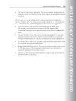
This child is diagnosed with Joubert syndrome. Common
symptoms of this disorder include mental retardation, poor
coordination, pendular eye movement, and abnormal
breathing patterns.
(Photo Researchers, Inc.)
to provide special assistance and anticipatory guidance.
Speech, physical, and occupational therapy are needed
throughout life.
Prognosis
The unusual pattern of breathing as newborns,
especially the episodes of apnea, can lead to sudden
death or coma. A number of individuals with Joubert
syndrome have died in the first three years of life. For
most individuals, the irregular breathing becomes more
normal after the first year. However, many continue to
have apnea, and require medical care throughout their
life. Although the true lifespan remains unknown, there
are some individuals with Joubert syndrome who are in
their 30s.
Resources
ORGANIZATIONS
Joubert Syndrome Foundation Corporation. c/o Stephanie
Frazer, 384 Devon Drive, Mandeville, LA 70448.
OTHER
Alliance of Genetic Support Groups.
ϽϾ.
Joubert Syndrome Foundation Corporation.
ϽϾ.
Kevin M. Sweet, MS, CGC
GALE ENCYCLOPEDIA OF GENETIC DISORDERS
623
Joubert syndrome
I
Kabuki syndrome
Definition
Kabuki syndrome is a rare disorder characterized by
unusual facial features, skeletal abnormalities, and intel-
lectual impairment. Abnormalities in different organ sys-
tems can also be present, but vary from individual to
individual. There is no cure for Kabuki syndrome, and
treatment centers on the specific abnormalities, as well as
on strategies to improve the overall functioning and qual-
ity of life of the affected person.
Description
Kabuki syndrome is a rare disorder characterized by
mental retardation, short stature, unusual facial features,
abnormalities of the skeleton and unusual skin ridge pat-
terns on the fingers, toes, palms of the hands and soles of
the feet. Many other organ systems can be involved in the
syndrome, displaying a wide variety of abnormalities.
Thus, the manifestations of Kabuki syndrome can vary
widely among different individuals.
Kabuki syndrome (also known as Niikawa-Kuroki
syndrome) was first described in 1980 by Dr. N. Niikawa
and Dr. Y. Kuroki of Japan. The disorder gets its name
from the characteristic long eyelid fissures with eversion
of the lower eyelids that is similar to the make-up of
actors of Kabuki, a traditional Japanese theatrical form.
Kabuki syndrome was originally known as Kabuki
Make-up syndrome, but the term “make-up” is now
often dropped as it is considered offensive to some
families.
Scientific research conducted over the past two
decades suggests that Kabuki syndrome may be associ-
ated with a change in the genetic material. However, it is
still not known precisely what this genetic change may
be and how this change in the genetic material alters
growth and development in the womb to cause Kabuki
syndrome.
Genetic profile
As stated above, the etiology of Kabuki syndrome is
not completely understood. While Kabuki syndrome is
thought to be a genetic syndrome, little or no genetic
abnormality has been identified as of yet. Chromosome
abnormalities of the X and Y chromosome or chromo-
some 4 have occurred in only a small number of individ-
uals with Kabuki syndrome, but in most cases,
chromosomes are normal.
In almost all cases of Kabuki syndrome, there is no
family history of the disease. These cases are thought to
represent new genetic changes that occur randomly and
with no apparent cause and are termed sporadic.
However, in several cases the syndrome appears to be
inherited from a parent, supporting a role for genetics in
the cause of Kabuki syndrome. Scientists hypothesize
that an unidentified genetic abnormality that causes
Kabuki syndrome is transmitted as an autosomal domi-
nant trait. With an autosomal dominant trait, only one
abnormal gene in a gene pair is necessary to display the
disease, and an affected individual has a 50% chance of
transmitting the gene and the disease to a child.
Demographics
Kabuki syndrome is a rare disorder with less than
200 known cases worldwide, but the prevalence of the
disease may be underestimated as only a handful of
physicians have first-hand experience diagnosing chil-
dren with Kabuki syndrome. Kabuki syndrome appears
to be found equally in males and females. Earlier cases
were reported in Japanese children but the syndrome is
now known to affect other racial and ethnic groups.
Theoretical mathematical models predict that the
incidence of Kabuki syndrome in the Japanese popula-
tion may be as high as one in 32,000.
Signs and symptoms
The signs and symptoms associated with Kabuki
syndrome are divided into cardinal symptoms (i.e. those
GALE ENCYCLOPEDIA OF GENETIC DISORDERS
625
K
For children with heart defects, surgical repair is
often necessary. This may take place shortly after birth if
the heart abnormality is life threatening, but often physi-
cians will prefer to attempt a repair once the child has
grown older and the heart is more mature. For children
who experience seizures, lifelong treatment with anti-
seizure medications is often necessary.
Children with Kabuki syndrome often have difficul-
ties feeding, either because of mouth abnormalities or
because of poor digestion. In some cases, a tube that
enters into the stomach, is placed surgically in the
abdomen and specially designed nutritional liquids are
administered through the tube directly into the stomach.
People with Kabuki syndrome are at higher risk for
a variety of infections, most often involving the ears and
the lungs. In cases such as these, antibiotics are given to
treat the infection, and occasionally brief hospital stays
are necessary. Most children recover from these infec-
tions with proper treatment.
Nearly half of people affected by Kabuki syndrome
have some degree of hearing loss. In these individuals,
formal hearing testing is recommended to determine if
they might benefit from a hearing-aid device. A hearing
aid is a small mechanical device that sits behind the ear
and amplifies sound into the ear of the affected individ-
ual. Occasionally, hearing loss in individuals with
Kabuki syndrome is severe, approaching total hearing
loss. In these cases, early and formal education using
American Sign Language as well as involvement with the
hearing-impaired community, schools, and enrichment
programs is appropriate.
Children with Kabuki syndrome should be seen reg-
ularly by a team of health care professionals, including a
primary care provider, medical geneticist familiar with
the condition, gastroenterologist, and neurologist. After
growth development is advanced enough (usually late
adolescence or early adulthood), consultation with a
reconstructive surgeon may be of use to repair physical
abnormalities that are particularly debilitating.
During early development and progressing into
young adulthood, children with Kabuki syndrome
should be educated and trained in behavioral and
mechanical methods to adapt to any disabilities. This
program is usually initiated and overseen by a team of
health care professionals including a pediatrician, phys-
ical therapist, and occupational therapist. A counselor
specially trained to deal with issues of disabilities in
children is often helpful is assessing problem areas and
encouraging healthy development of self-esteem.
Support groups and community organizations for people
with disabilities often prove useful to the affected indi-
viduals and their families, and specially equipped
626
GALE ENCYCLOPEDIA OF GENETIC DISORDERS
Kabuki syndrome
KEY TERMS
Autosomal dominant—A pattern of genetic inher-
itance where only one abnormal gene is needed to
display the trait or disease.
Cardinal symptoms—A group of symptoms that
define a disorder or disease.
Gastric tube—A tube that is surgically placed
though the skin of the abdomen to the stomach so
that feeding with nutritional liquid mixtures can be
accomplished.
Gastroenterologist—A physician who specializes
in disorders of the digestive system.
Kabuki—Traditional Japanese popular drama per-
formed with highly stylized singing and dancing
using special makeup and cultural clothing.
Neurologist—A physician who specializes in dis-
orders of the nervous system, including the brain,
spine, and nerves.
that are almost always present) and variable symptoms
(those that may or may not be present). The cardinal and
variable signs and symptoms of Kabuki syndrome are
summarized in the table below.
Diagnosis
The diagnosis of Kabuki syndrome relies on physical
exam by a physician familiar with the condition and by
radiographic evaluation, such as the use of x rays or ultra-
sound to define abnormal or missing structures that are
consistent with the criteria for the condition (as described
above). A person can be diagnosed with Kabuki syn-
drome if they possess characteristics consistent with the
five different groups of cardinal symptoms: typical face,
skin-surface abnormalities, skeletal abnormalities, mild
to moderate mental retardation, and short stature.
Although a diagnosis may be made as a newborn,
most often the features do not become fully evident until
early childhood. There is no laboratory blood or genetic
test that can be used to identify people with Kabuki syn-
drome.
Treatment and management
There is no cure for Kabuki syndrome. Treatment of
the syndrome is variable and centers on correcting the
different manifestations of the condition and on strategies
to improve the overall functioning and quality of life of
the affected individual.
enrichment programs should be sought. Further, because
many children with Kabuki syndrome have poor speech
development, a consultation and regular session with a
speech therapist is appropriate.
Prognosis
The abilities of children with Kabuki syndrome vary
greatly. Most children with the condition have a mild to
moderate intellectual impairment. Some children will be
able to follow a regular education curriculum, while oth-
ers will require adaptations or modifications to their
schoolwork. Many older children may learn to read at a
functional level.
The prognosis of children with Kabuki syndrome
depends on the severity of the symptoms and the extent
to which the appropriate treatments are available. Most
of the medical issues regarding heart, kidney or intes-
tinal abnormalities arise early in the child’s life and are
improved with medical treatment. Since Kabuki syn-
drome was discovered relatively recently, very little is
known regarding the average life span of individuals
affected with the condition, however, present data on
Kabuki syndrome does not point to a shortened life
span.
Resources
BOOKS
Behrman, R.E., ed. Nelson Textbook of Pediatrics. Philadelphia:
W.B. Saunders, 2000.
PERIODICALS
Kawame, H. “Phenotypic Spectrum and Management Issues in
Kabuki Syndrome.” Journal of Pediatrics 134(April
1999): 480-485.
Mhanni, A.A., and A.E. Chudley. “Genetic Landmarks Through
Philately—Kabuki Theater and Kabuki Syndrome.”
Clinical Genetics 56(August 1999): 116-117.
ORGANIZATIONS
CardioFacioCutaneous Support Network. 157 Alder Ave.,
McKee City, NJ 08232. (609) 646-5606.
Kabuki Syndrome Network. 168 Newshaw Lane, Hadfield,
Glossop, SK13 2AY. UK 01457 860110. Ͻhttp://www
.ksn-support.org.ukϾ.
National Organization for Rare Disorders (NORD). PO Box
8923, New Fairfield, CT 06812-8923. (203) 746-6518 or
(800) 999-6673. Fax: (203) 746-6481. Ͻhttp://www
.rarediseases.orgϾ.
WEBSITES
“Entry 147920: Kabuki Syndrome.” OMIM—Online Mendelian
Inheritance in Man. Ͻ />entrez/dispomim.cgi?idϭ147920Ͼ.
Oren Traub, MD, PhD
I
Kallmann syndrome
Definition
Kallmann syndrome is a disorder of hypogo-
nadotropic hypogonadism, delayed puberty, and anosmia.
Description
Hypogonadotropic hypogonadism (HH) occurs
when the body does not produce enough of two important
hormones, luteinizing hormone (LH) and follicle stimu-
lating hormone (FSH). This results in underdeveloped
gonads and often infertility. Anosmia, the inability to
smell, was first described with hypogonadotropic hypog-
onadism in 1856, but it was not until 1944 that Kallmann
reported the inheritance of the two symptoms together
in three separate families. Hence, the syndrome of
hypogonadotropic hypogonadism and anosmia was
named Kallmann syndrome (KS).
Kallmann syndrome (KS) is occasionally called dys-
plasia olfactogenitalis of DeMorsier. Affected people
usually are detected in adolescence when they do not
undergo puberty. The most common features are HH and
anosmia, though a wide range of features can present in
an affected person. Other features of KS may include a
small penis or undescended testicles in males, kidney
abnormalities, cleft lip and/or palate, clubfoot, hearing
problems, and central nervous system problems such as
synkinesia, eye movement abnormalities, and visual and
hearing defects.
Genetic profile
Most cases of Kallmann syndrome are sporadic.
However, some cases are inherited in an autosomal dom-
inant pattern, an autosomal recessive pattern, or an X-
linked recessive pattern. In most cells that make up a
person there are structures called chromosomes.
Chromosomes contain genes, which are instructions for
how a person will grow and develop. There are 46 chro-
mosomes, or 23 pairs of chromosomes, in each cell. The
first 22 chromosomes are the same in men and women
and are called the autosomes. The last pair, the sex chro-
mosomes, are different in men and women. Men have an
X and a Y chromosome (XY). Women have two X-chro-
mosomes (XX). All the genes of the autosomes and the
X-chromosomes in women come in pairs.
Autosomal dominant inheritance occurs when only
one copy of a gene pair is altered or mutated to cause the
condition. In autosomal dominant inheritance, the second
normal gene copy cannot compensate, or make up for, the
altered gene. People with autosomal dominant inheri-
tance have a 50% chance of passing the gene and the con-
dition onto each of their children.
GALE ENCYCLOPEDIA OF GENETIC DISORDERS
627
Kallmann syndrome
mann syndrome. The gene instructs the body to make a
protein called anosmin-1. When this gene is altered in a
male, Kallmann syndrome occurs. Of those families who
have an X-linked recessive form of KS, approximately
1/2 to 1/3 have identifiable alterations in their KAL gene.
Demographics
Kallmann syndrome is the most frequent cause of
hypogonadotropic hypogonadism and affects approxi-
mately 1/10,000 males and 1/50,000 females. Kallmann
syndrome is found in all ethnic backgrounds. Because the
incidence of KS in males is about five times greater than
KS in females, the original belief was that the X-linked
form of Kallmann syndrome was the most common.
However, as of 2001, it is now assumed that the X-linked
recessive form is the least common of all KS. The reason
for Kallmann syndrome being more frequent in males is
not known.
Signs and symptoms
Embryology
Normally, a structure in the brain called the hypothal-
amus makes a hormone called gonadotrophin releasing
hormone (GnRH). This hormone acts on the pituitary
gland, another structure in the brain, to produce the two
hormones: follicle stimulating hormone (FSH) and
luteinizing hormone (LH). Both of these hormones travel
to the gonads where they stimulate the development of
sperm in men and eggs in women. FSH is also involved in
the release of a single egg from the ovary once a month.
Hypogonadotropic hypogonadism results when there is an
alteration in this pathway that results in inadequate pro-
duction of LH or FSH. In Kallmann syndrome, the alter-
ation is that the hypothalamus is unable to produce GnRH.
How hypogonadotropic hypogonadism and the
inability to smell are related can be explained during the
development of an embryo. The cells that eventually
make the GnRH in the hypothalamus are first found in
the nasal placode, part of the developing olfactory system
(for sense of smell). The GnRH cells must migrate, or
move, from the nasal placode up into the brain to the
hypothalamus. These GnRH cells migrate by following
the path of another type of cell called the olfactory neu-
rons. Neurons are specialized cells that are found in the
nervous system and have long tail-like structures called
axons. The axons of the olfactory neurons grow from the
nasal placode up into the developing front of the brain.
Once they reach their final destination in the brain, they
form the olfactory bulb, the structure in the brain that
helps process odors allowing the sense of smell. The
GnRH cells follow the pathway of the olfactory neurons
up into the brain to reach the hypothalamus.
628
GALE ENCYCLOPEDIA OF GENETIC DISORDERS
Kallmann syndrome
KEY TERMS
Hormone—A chemical messenger produced by
the body that is involved in regulating specific
bodily functions such as growth, development,
and reproduction.
Hypothalamus—A part of the forebrain that con-
trols heartbeat, body temperature, thirst, hunger,
body temperature and pressure, blood sugar lev-
els, and other functions.
Neuron—The fundamental nerve cell that con-
ducts impulses across the cell membrane.
Pituitary gland—A small gland at the base of the
brain responsible for releasing many hormones,
including luteinizing hormone (LH) and follicle-
stimulating hormone (FSH).
Puberty—Point in development when the gonads
begin to function and secondary sexual character-
istics begin to appear.
Synkinesia—Occurs when part of the body will
move involuntarily when another part of the body
moves.
Autosomal recessive inheritance occurs when both
copies of a gene are altered or mutated to cause the con-
dition. In autosomal recessive inheritance, the affected
person has inherited one altered gene from their mother
and the other altered gene from their father. Couples who
both have one copy of an altered autosomal recessive
gene have a 25% risk with each pregnancy to have an
affected child.
X-linked recessive inheritance is thought to be the
least common form of inheritance in KS, but is the most
well understood at the genetic level. With X-linked reces-
sive inheritance, the altered gene that causes the condi-
tion is on their X chromosome. Since men have only one
copy of the X chromosome, they have only one copy of
the genes on the X chromosome. If that one copy is
altered, they will have the condition because they do not
have a second copy of the gene to compensate. Women,
however, can have one altered copy of the gene and not
be affected as they have a second copy to compensate. In
X-linked recessive conditions, women are generally not
affected with the condition. Women who are carriers for
an X-linked recessive condition have a 25% chance of
having an affected son with each pregnancy.
Though all three patterns of inheritance have been
suggested for Kallmann syndrome, as of 2001 only one
gene has been found that causes Kallmann syndrome.
The gene, KAL, is located on the X chromosome and is
responsible for most cases of X-linked recessive Kall-
In Kallmann syndrome, the olfactory neurons are
unable to grow into the brain. Hence, the GnRH cells can
not follow their pathway. As a result, the olfactory bulb
does not form, resulting in the inability to smell. The
GnRH cells can not follow the pathway of the axons and
do not reach their final destination in the hypothalamus.
Hence, no GnRH is made to stimulate the pituitary to
make FSH and LH, resulting in hypogonadotropic
hypogonadism.
In X-linked recessive KS, the KAL gene instructs
the body to make the protein anosmin-1. This protein is
involved in providing the pathway in the brain for which
the olfactory axons grow. If it is altered in any way, the
axons will not know where to grow in the brain and the
GnRH cells will be unable to follow. The protein anos-
min-1 is also found in other parts of the body, possibly
explaining some of the other symptoms sometimes seen
in Kallmann syndrome.
Other features
The features of Kallmann syndrome can vary among
affected individuals even within the same family. The
two features most often associated with Kallmann syn-
drome are HH and the inability to smell. Males can also
have a small penis and undescended testicles at birth (tes-
ticles are still in body and have not dropped down into the
scrotal sac). Clubfoot, cleft lip and/or cleft palate can also
be present at birth. Clubfoot occurs when one or both feet
are not properly placed onto the legs and can appear
turned. Cleft lip and/or cleft palate occur when the upper
lip and/or the roof of the mouth fail to come together dur-
ing development. Kidney abnormalities, most often uni-
lateral renal agenesis (one kidney did not form) are
especially common in those males with X-linked reces-
sive KS. Choanal atresia (pathway from the nose is
blocked at birth) and structural heart defects have also
been seen in KS.
Central nervous system problems can also occur in
Kallmann syndrome. These can include nystagmus
(involuntary eye movement), ataxia (involuntary body
movement), hearing loss and problems with vision.
Synkinesia is especially common in men with the X-
linked recessive form of KS. Some people with KS are
also mentally retarded. Holoprosencephaly, when the
brain fails to develop in two halves, can also be seen in
some individuals with KS.
Diagnosis
Individuals with Kallmann syndrome are usually
diagnosed when they do not undergo puberty. Hormone
testing shows that both LH and FSH are decreased.
Affected individuals often do not realize they cannot
smell. MRI can often detect the absence of the olfactory
bulb in the brain. Renal ultrasound can determine if a kid-
ney is missing.
As of 2001, genetic testing for alterations in the
KAL gene is the only genetic testing available. Even with
families with clear X-linked recessive inheritance,
genetic testing does not always detect an alteration in the
KAL gene. Hence, diagnosis is still very dependent upon
clinical features.
Treatment and management
When a child with KS is born with structural abnor-
malities such as cleft lip and/or palate, clubfoot or heart
defects, surgery is often required to fix the defect. Taking
sex hormones treats delayed puberty; women take estro-
gen and men take testosterone. Once puberty is com-
pleted, taking GnRH or both LH and FSH can treat
hypogonadism. For most affected individuals, treatment
is successful and infertility is reversed. However, a small
portion of people will not respond to treatment.
When an isolated case of Kallmann syndrome is
diagnosed, evaluation of first-degree family members,
such as parents and siblings, should be completed. This
should include a detailed family history, measuring hor-
mone levels, assessing sense of smell, and renal ultra-
sound to look for kidney abnormalities. This information
may help to diagnosis previously unrecognized cases of
Kallmann syndrome. Furthermore, this information may
be important for genetic counseling and determining
whom in the family is at risk for also having Kallmann
syndrome.
Prognosis
For individuals with the most common features of
Kallmann syndrome, hypogonadism and the inability to
smell, prognosis is excellent. In most cases, hormone
treatment is able to reverse the delayed puberty and
hypogonadism. For those individuals with other symp-
toms of Kallmann syndrome, prognosis can depend on
how severe the defect is. For example, structural heart
defects can be quite complex and sometimes surgery can
not fix them. Furthermore, no treatment is available for
the mental retardation in the portion of affected individu-
als with this symptom.
Resources
PERIODICALS
Rugarli, Elena, and Andrea Ballabio. “Kallmann Syndrome:
From Genetics to Neurobiology.” JAMA 270, no. 22
(December 8, 1993): 2713–2716.
GALE ENCYCLOPEDIA OF GENETIC DISORDERS
629
Kallmann syndrome
ORGANIZATIONS
American Society for Reproductive Medicine. 1209
Montgomery Highway, Birmingham, AL 35216-2809.
(205) 978-5000. ϽϾ.
RESOLVE, The National Infertility Association. 1310
Broadway, Somerville, MA 02144-1779. (617) 623-0744.
ϽϾ.
WEBSITES
Pediatric Database (PEDBASE) Ͻwww.icondata.com/health/
pedbase/files/KALLMANN.HTMϾ.
Carin Lea Beltz, MS
I
Kartagener syndrome
Definition
Kartagener (pronounced KART-agayner) syndrome
refers to a condition that involves difficulty with clearing
mucus secretions from the respiratory tract, male infertil-
ity, and situs inversus. The defining characteristic of this
syndrome is the situs inversus, which is a reversal of
abdominal and thoracic organs.
Description
This syndrome is named after Kartagener, a physi-
cian from Switzerland. In the 1930’s, Kartagener and a
colleague described a familial form of bronchiectasis
with situs inversus and nasal polyps. This came to be
known as Kartagener syndrome. Kartagener syndrome is
also known as the Siewert syndrome, after another physi-
cian, Siewert, who described the syndrome in the early
1900’s.
Individuals who have Kartagener syndrome form a
subset of the disorder called primary ciliary dyskinesia.
Originally, primary ciliary dyskinesia was known as
immotile cilia syndrome. The name, immotile cilia syn-
drome, is no longer used since the discovery that the
cilia are actually not immotile, but rather, abnormal in
movement. Individuals who have Kartagener syndrome,
basically have primary ciliary dyskinesia, plus partial
or complete situs inversus. The situs inversus is what
sets Kartagener syndrome apart from primary ciliary
dyskinesia.
Kartagener syndrome is caused by abnormalities of
the cilia that line the respiratory tract and also form the
flagella of sperm. Cilia are tiny hair-like structures that
contain a bundle of small parallel tubes that form a cen-
tral core. This core is called the axoneme. Ciliary move-
ment is accomplished by the bending of the axoneme.
One of the most important associated structures that
enable ciliary movement to occur are sets of tiny arms
that project from each tubule. These tiny arms are called
dynein arms.
Cilia line the cells of the lungs, nose and sinuses.
Before reaching the lungs, air travels through the airway
where it is moistened and filtered. The nasal passages and
airway are lined with mucus membranes. The mucus cov-
ering the mucus membrane traps dirt and other foreign
particles that have been breathed in. The cilia, lining the
membranes, beat in a wavelike manner moving the layer
of mucus and carrying away the dirt and debris that has
been trapped. This mucus can then be coughed out or
swallowed into the stomach.
In Kartagener syndrome, the cilia do not move,
move very little, or move abnormally. Because the cilia
do not function properly, the mucus is not cleared from
the respiratory tract, which leads to sinus infection
(sinusitis) and chronic changes of the lung (bronchiecta-
sis), which make it difficult to exhale. Mucus clearance
from the middle ear can also be affected and over time
can lead to hearing loss.
The male infertility in Kartagener syndrome is also
caused by abnormal cilia movement. One spermatozoon
consists of a head, midpiece, and a tail or flagellum. The
tail of a spermatozoon is a long flagellum consisting of a
central axoneme. This axoneme enables the movement of
the flagellum so that the spermatozoon can propel its way
to the fallopian tube and burrow through the egg coat to
fertilize the egg. In Kartagener syndrome, these cilia are
either immotile, or are not able to move normally to com-
plete the journey to the fallopian tubes, nor may they be
able to burrow through the egg coat. This results in male
infertility.
As stated above, situs inversus is what sets
Kartagener syndrome apart from primary ciliary dyski-
nesia. Complete situs inversus involves reversal of both
the abdominal and thoracic organs so that they form a
mirror image of normal. In partial situs inversus, the tho-
racic organs may be reversed, while the abdominal
organs are normally positioned, or vice versa.
Approximately one in 10,000 adults have situs inversus.
Only about 20% of individuals who have complete situs
inversus are diagnosed to have Kartagener syndrome. Of
those with complete situs inversus who are diagnosed to
have Kartagener syndrome, there is only a small risk for
associated cardiac defects. Partial situs inversus may
occur in individuals who have Kartagener syndrome as
well. Partial situs inversus has a higher association with
other abnormalities, including polysplenia or asplenia
(extra or absent spleen) and cardiac defects.
One theory behind the association of situs inversus
with the underlying cause of Kartagener syndrome is that
the lack of ciliary movement in the developing embryo
630
GALE ENCYCLOPEDIA OF GENETIC DISORDERS
Kartagener syndrome
may result in incorrect organ rotation in approximately
50% of affected individuals. In fact, 50% of patients with
PCD will have situs inversus and thus be diagnosed to
have Kartagener syndrome. However, this is a theory
supported only by some researchers.
Genetic profile
Kartagener syndrome is an autosomal recessive con-
dition. This means that in order to have the condition, an
individual needs to inherit two copies of the gene for the
condition, one from each parent. Individuals who carry
only one gene for an autosomal recessive syndrome are
called heterozygotes. Heterozygotes for Kartagener syn-
drome have normal ciliary function and do not have any
clinical features of the condition. If two carriers of
Kartagener syndrome have children, there is a 25%
chance, with each pregnancy, for having a child with
Kartagener syndrome.
The components that form the cilium contain several
hundred different proteins. Each is coded for by different
DNA sequences, potentially on different chromosomes.
A defect in any of these codes could produce an abnor-
mal or missing protein that is a building block for the
cilium and thus could cause abnormal ciliary structure
and movement, resulting in Kartagener syndrome.
When the same condition can be caused by different
genetic abnormalities, this is known as genetic hetero-
geneity. In fact, several different defects in cilia have
been seen in association with Kartagener syndrome,
including; overly long cilia, overly short cilia, absent cilia
and randomly oriented cilia, suggesting genetic hetero-
geneity. Studies have suggested that the most common
defect of cilia in Kartagener syndrome is the lack of
dynein arms. There have been rare cases in which indi-
viduals have Kartagener syndrome, yet have no
detectable abnormality of the cilia, even though the cil-
iary function is abnormal. Results of one study involving
a genome-wide linkage search performed on 31 families,
with multiple individuals affected with either PCD or
Kartagener syndrome, strongly suggested extensive het-
erogeneity. Potential regions involving genes responsible
for PCD or Kartagener syndrome were localized on chro-
mosomes 3, 4, 5, 7, 8, 10, 11, 13, 15, 16, 17 and 19.
Demographics
Kartagener syndrome occurs in approximately one in
32,000 live births, which is half the incidence of primary
ciliary dyskinesia (one in 16,000 live births). Kartagener
syndrome is not found more commonly in any particular
sex, ethnic background or geographic region. Males,
however, may be diagnosed more often than females
because of infertility investigation.
GALE ENCYCLOPEDIA OF GENETIC DISORDERS
631
Kartagener syndrome
KEY TERMS
Bronchiectasis—An abnormal condition of the
bronchial tree, characterized by irreversible
widening and destruction of the bronchial walls of
the lungs.
Cystic fibrosis—A respiratory disease character-
ized by chronic lung disease, pancreatic insuffi-
ciency and an average age of survival of 20 years.
Cystic fibrosis is caused by mutations in a gene on
chromosome 7 that encodes a transmembrane
receptor.
Dyskinesia—Impaired ability to make voluntary
movements.
Tympanoplasty—Any of several operations on the
eardrum or small bones of the middle ear, to
restore or improve hearing in patients with con-
ductive hearing loss.
Signs and symptoms
Newborns who have Kartagener syndrome may
present with neonatal respiratory distress. Often when
individuals are diagnosed to have Kartagener syndrome
in later childhood, problems such as neonatal respiratory
distress may be identified in their history. Symptoms that
may present in childhood include; recurrent ear infec-
tions (otitis media) that can lead to hearing loss, chronic
productive cough, reactive airway disease, pneumonia,
chronic bronchitis, runny nose (rhinitis) with a thin dis-
charge, and sinus infection (sinusitis). Situs inversus usu-
ally does not present symptomatically, unless it is
associated with a congenital heart defect.
The most common clinical expression of
Kartagener syndrome in adults includes chronic upper
and lower airway disease presenting as sinusitis and
bronchiectasis. Clubbing of the digits (fingers) may
occur as the result of chronic hypoxia (lack of oxygen)
from bronchiectasis. In males of reproductive age, male
infertility is almost universal. In females who have
Kartagener syndrome, infertility is not usually a charac-
teristic. This suggests that the egg transport down the
fallopian tube is associated more with muscle contrac-
tions than with ciliary movement.
Several other conditions should be considered when
the aforementioned symptoms present, including; Cystic
fibrosis (CF), immune deficiencies and severe allergies.
Although the causes of Kartagener syndrome and CF are
completely different, the symptoms of these two diseases
are very similar. Often when the symptoms present, chil-
dren with Kartagener syndrome are tested for CF first
because the incidence of CF is much higher (one in
2,400) than the incidence of Kartagener syndrome. CF is
also associated with male infertility.
Diagnosis
Diagnosis of Kartagener syndrome is confirmed by
identifying the ciliary abnormalities of structure and
movement. This is accomplished by biopsy of the mucus
membranes of the respiratory tract and/or by examination
of sperm, looking for ciliary dyskinesia. Situs inversus
can be identified by x ray or ultrasound examination.
Infertility investigation may elicit the possibility of
Kartagener syndrome in a patient previously undiag-
nosed. After a diagnosis is made, genetic counseling
should be provided to discuss the inheritance pattern, to
help identify other possible affected family members and
to discuss reproductive options.
As Kartagener syndrome is an autosomal reces-
sive disorder, individuals who have had a child with
Kartagener syndrome have a 25% chance, with each
future pregnancy, of having another child with
Kartagener syndrome. Prenatal diagnosis may be pos-
sible for a couple with a previously affected child, by
performing ultrasound examination to identify a fetus
who has situs inversus. Although, if the fetus does not
exhibit situs inversus, it is still possible for the fetus to
have PCD. Also, it is important to remember that iden-
tifying a fetus who has situs inversus in a family not
known to be at an increased risk for Kartagener syn-
drome, does not mean that the fetus has Kartagener
syndrome as only 20% of individuals who have situs
inversus have Kartagener syndrome. As of January
2001, DNA testing for Kartagener syndrome is not
possible.
Treatment and management
Treatment for Kartagener syndrome involves treat-
ment of the symptoms. Treatment for sinusitis includes
the use of antibiotics to treat and prevent recurrent infec-
tion. Occasionally, surgery to relieve the sinusitis and
remove nasal polyps that may be present is necessary.
Daily chest physiotherapy to loosen mucus secretions is
a common therapy as well, and if started early in life can
help to prevent or delay development of bronchiectasis.
Tympanoplasty in children with recurrent ear infections
is often necessary.
Advances in reproductive technology allow for men
who have Kartagener syndrome to have the opportunity
to have children. A procedure called intracytoplasmic
sperm injection or ICSI, now allow immotile or dys-
motile sperm to fertilize an egg. ICSI involves injection
of a single sperm into single eggs in order for fertilization
to occur. This procedure first involves ovulation induc-
tion and egg retrieval to obtain eggs for attempt at fertil-
ization by ICSI. In Vitro Fertilization (ICSI) pregnancy
rates vary from center to center. Overall pregnancy rates
of 10%-40% have been quoted worldwide, utilizing these
procedures.
The chance for an affected male and his unaffected
partner to have a child who has Kartagener syndrome is
small. If the disease incidence is one in 32,000, then the
chance for the unaffected woman to be a carrier of
Kartagener syndrome is approximately one in 100 and
the chance for having an affected child would be
expected to be approximately one in 200 (0.5%).
However, all children of affected males or females will be
carriers for Kartagener syndrome.
Prognosis
The severity of Kartagener syndrome is variable.
With the advent of antibiotic use for infection control, the
life expectancy of a patient with Kartagener syndrome is
close to or within the normal range, if there are no imme-
diate problems in the newborn period.
Resources
BOOKS
Jones, Kenneth Lyons. Smith’s Recognizable Patterns of
Human Malformation. Philadelphia: W.B.Saunders
Company, 1997.
PERIODICALS
Guichard, Cècile, et al. “Axonemal Dynein Intermediate-Chain
Gene (DNAI1) Mutations Result in Situs Inversus and
Primary Ciliary Dyskinesia (Kartagener Syndrome).”
American Journal of Human Genetics (April 2001): 1030.
ORGANIZATIONS
American Lung Association. 1740 Broadway, New York, NY
10019-4374. (212) 315-8700 or (800) 586-4872.
Ͻhttp;//www.lungusa.orgϾ.
National Organization for Rare Disorders (NORD). PO Box
8923, New Fairfield, CT 06812-8923. (203) 746-6518 or
(800) 999-6673. Fax: (203) 746-6481. Ͻhttp://www
.rarediseases.orgϾ.
WEBSITES
OMIM Online Mendelian Inheritance in Man. Entries 244400
and 242650. Ͻ />.fcgi?dbϭOMIMϾ.
Tucker, Michael. “Clinical In Vitro Fertilization and Culture”.
IVF.com. Ͻ />Renee A. Laux, MS
632
GALE ENCYCLOPEDIA OF GENETIC DISORDERS
Kartagener syndrome
I
Karyotype
Definition
Karyotype refers to the arrangement of chromo-
somes in their matched (homologous) pairs. For the pur-
poses of this definition, we will be referring to human
chromosomes, although there is a karyotype characteris-
tic for each species. The human chromosomes are
arranged and numbered according to the International
System for Human Cytogenetic Nomenclature (ISCN).
The most recent recommendations of the ISCN are from
1995. Karyotype either refers to the actual composition
of the chromosomes in a body cell of an individual or
species, or to the actual diagram or photograph of those
chromosomes, arranged in their pairs.
Description
The normal human karyotype consists of 23 pairs of
chromosomes. There are 22 pair of autosomes, which are
the chromosomes that are not the sex chromosomes. The
genes on these chromosomes instruct our bodies as to
how they look and function. The 23rd pair of chromo-
somes are the sex chromosomes. Typically, females have
two X sex chromosomes and males have one X sex chro-
mosome and one Y sex chromosome.
Karyotype construction
In the construction of the karyotype, the chromo-
somes are numbered 1 to 22 from longest to shortest. The
last pair are the sex chromosomes and are placed on the
karyotype after the 22nd pair. The chromosomes can be
separated into groups, based on their length and the posi-
tion of the centromere. Group A consists of chromosome
pairs 1, 2 and 3. They are the longest chromosomes and
their centromeres are in the center of the chromosomes
(metacentric). Group B consists of chromosome pairs 4
and 5. They are long; however, their centromeres lie
toward the top of the chromosomes (submetacentric).
Group C consists of chromosome pairs 6, 7, 8, 9, 10, 11
and 12 and also includes the X chromosome. They are
medium-sized and their centromeres either lie in the mid-
dle or toward the top of the chromosomes. Group D con-
sists of chromosome pairs 13,14 and 15. They are
medium-sized and their centromeres lie at the top of the
chromosomes (acrocentric). Additionally, the D group
chromosomes have satellites. Group E consists of chro-
mosome pairs 16, 17 and 18. They are relatively short
chromosomes and their centromeres lie in the center or
towards the top of the chromosomes. Group F consists of
chromosomes 19 and 20. They are short chromosomes
with centromeres that lie in the center of the chromo-
GALE ENCYCLOPEDIA OF GENETIC DISORDERS
633
Karyotype
KEY TERMS
Acrocentric—A chromosome with the centromere
positioned at the top end.
Centromere—The centromere is the constricted
region of a chromosome. It performs certain func-
tions during cell division.
Homologous chromosomes—Homologous chro-
mosomes are two chromosomes of a doublet set
that are identical, particularly for the genes that
are on them.
Metacentric—When a chromosome has the cen-
tromere in the middle of the chromosome it is
called a metacentric chromosome.
Satellites of chromosomes—Small segments of
genetic material at the tips of the short arms of
chromosomes 13, 14, 15, 21, and 22.
Submetacentric—Positioning of the centromere
between the center and the top of the chromo-
some.
some. Lastly, group G consists of chromosome pairs 21,
22 and the Y chromosome. These are short chromosomes
with their centromeres at the top. Chromosome pairs 21
and 22 have satellites. The Y chromosome does not have
satellites.
The actual chromosomes are only individually dis-
tinguishable during a certain stage of cell division. This
stage is called the metaphase stage. Chromosome prepa-
rations are made from pictures of the chromosomes dur-
ing the metaphase stage of division. The metaphase
spread is what the technician sees in one cell under the
microscope and what the photograph of that one cell is
referred to. Usually, the chromosomes in a metaphase
preparation are banded by special staining techniques
used in the laboratory. Each numbered chromosome is
unique in its banding pattern so that all number 1s look
the same and all number 2s look the same, etc. Although,
there can be small normal familial variations in chromo-
somes. Because of banding, the chromosomes are more
easily distinguishable from each other and the banding
makes it is easier to see differences or abnormalities. For
example, if a chromosome is missing a piece, or two
chromosomes are attached to each other (translocation),
it is much easier to see with banded chromosomes than
with unbanded chromosomes.
Chromosome preparations can be made from any
potentially dividing cells, including; blood cells, skin
cells, amniotic fluid cells (the fluid surrounding an
unborn baby), placental tissue or chorionic villi (tissue
that forms the placenta and can be used in prenatal diag-
nosis).
ISCN formulas exist to describe any chromosome
complement. The basic formula for writing a karyotype
is as follows. The first item written is the total number of
chromosomes, followed by a comma. The the second
item written is the sex chromosome complement. The
typical female karyotype is written as 46,XX and the typ-
ical male karyotype is written as 46,XY.
Formulas for abnormal karyotypes
Many formulas for writing abnormal karyotypes
have been determined. Some common examples follow.
A plus or a minus sign before a chromosome number is
used to show that the entire chromosome is extra or miss-
ing. Also, the total number of chromosomes will be dif-
ferent than 46. For example, the condition Down
syndrome occurs when an individual has an extra num-
ber 21 chromosome. For a male, this karyotype is written
as 47,XY,ϩ21. An individual may also have extra or
missing parts of chromosomes. The short arm of a chro-
mosome is called the p arm and the long arm is called the
q arm. For example, the condition Wolf-Hirschhorn
syndrome is caused by a missing part of the top arm of
chromosome 4. For a female, this karyotype would be
written as 46,XX,del(4)(p16). The chromosome that is
involved in the change is specified within the first set of
parentheses and the breakpoint for the missing material is
defined in the second set of parentheses. A final example
is a balanced translocation karyotype. A balanced translo-
cation means that there is no missing or extra genetic
material as the result of the translocation. There are many
types of translocations. One type is called a robertsonian
translocation. A robertsonian translocation occurs when
two acrocentric chromosomes are attached together. One
common example is a translocation involving chromo-
somes 13 and 14. If a male has a balanced robertsonian
translocation of chromosomes 13 and 14, this is written
as 45,XY,der(13;14). The “der” stands for derivative, as
the new 13;14 chromosome is considered a derivative.
There are only 45 separate chromosomes now, which is
why 45 is the number written in the karyotype. There are
many more formulas for the abundant abnormal chromo-
some findings in individuals. For further detailed infor-
mation, please refer to the resource listed below.
Resources
BOOKS
Mitelman, Felix, ed. An International System for Human
Cytogenetic Nomenclature (1995). Farmington, CT: S.
Karger AG, 1995.
Renee A. Laux, MS
Karyotype analysis see Karyotype
Keller syndrome see FG syndrome
I
Kennedy disease
Definition
Kennedy disease (KD) is a disorder characterized by
degradation of the anterior horn cells of the spinal cord
resulting in slow progressive muscle weakness and atro-
phy. Men with Kennedy disease often have breast
enlargement (gynecomastia), testicular atrophy, and may
have infertility.
Description
Kennedy disease, also referred to as spinobulbar
muscular atrophy (SBMA), arises primarily from degra-
dation of the anterior horn cells of the spinal cord, result-
ing in proximal weakness and atrophy of voluntary
skeletal muscle. Anterior horn cells control the voluntary
muscle contractions from large muscle groups such as the
arms and legs. For example, if an individual wants to
move his/her arm, electrical impulses are sent from the
brain to the anterior horn cells to the muscles of the arm,
which then stimulate the arm muscles to contract, allow-
634
GALE ENCYCLOPEDIA OF GENETIC DISORDERS
Kennedy disease
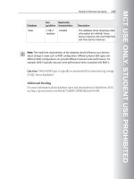
Karyotype showing three copies of chromosome 21.This
indicates Down syndrome.
(Custom Medical Stock Photo, Inc.)
ing the arm to move. Degradation is a rapid loss of func-
tional motor neurons. Loss of motor neurons results in
progressive symmetrical atrophy of the voluntary mus-
cles. Progressive symmetrical atrophy refers to the loss of
function of muscle groups from both sides of the body.
For example, both arms and both legs are equally
affected by similar degrees of muscle loss and the inabil-
ity to be controlled and used properly. Progressive loss
indicates that muscle loss is not instantaneous, rather
muscle loss occurs consistently over a period of time.
These muscle groups include those skeletal muscles that
control large muscle groups such as the arms, legs and
torso. The weakness in the legs is generally greater than
the weakness in the arms.
Proximal weakness is in contrast to distal weakness,
and indicates that muscles such as the arms and the legs
are affected rather than the muscles of the hands, feet,
fingers, and toes. However, the motor neuron of the
brainstem and sensory neurons of the dorsal root ganglia
are also affected in KD. Motor neurons are the neurons
that control large muscle groups (arms, legs, torso) of
which anterior horn cells are a subgroup. Sensory neu-
rons are a distinct class of neurons that control an indi-
vidual’s senses. An example would be pain receptors that
cause an involuntary reaction to a stimuli such as when a
person accidentally grasps a boiling hot kettle and imme-
diately releases the kettle. Dorsal root ganglia are analo-
gous to a headquarters for neurons, through which
essentially all neuronal stimuli are processed.
Diagnosis
Kennedy disease is suspected clinically in a male
with an early adulthood onset of proximal muscle weak-
ness of the limbs, fasticulations (small local contractions
of the musculature that is visible through the skin) of the
tongue, lips or area around the mouth, absence of hyper-
active reflexes and spasticity, and often evidence of
enlarged breasts and/or small testes with few or no sperm.
The diagnosis is made by a specific molecular
genetic test that measures the number of “repeats” in a
particular part of the androgen receptor (AR) gene. The
alteration of the AR gene that causes Kennedy disease is
an expansion of a CAG trinucleotide repeat in the first
PART of the gene. In unaffected individuals, between 11
to 33 copies OF the CAG trinucleotide are present. In
patients with Kennedy disease, this number rises to 40 to
62. The greater the number of expanded repeats, the ear-
lier the age of onset.
Genetic profile
Kennedy disease is an X-linked recessive disease,
meaning the abnormal gene is found on the X chromo-
GALE ENCYCLOPEDIA OF GENETIC DISORDERS
635
Kennedy disease
KEY TERMS
Anterior horn cells—Subset of motor neurons
within the spinal cord.
Atrophy—Wasting away of normal tissue or an
organ due to degeneration of the cells.
Degradation—Loss or diminishing.
Dorsal root ganglia—The subset of neuronal cells
controlling impulses in and out of the brain.
Intragenic—Occuring within a single gene.
Motor neurons—Class of neurons that specifically
control and stimulate voluntary muscles.
Motor units—Functional connection with a single
motor neuron and muscle.
Sensory neurons—Class of neurons that specifi-
cally regulate and control external stimuli (senses:
sight, sound).
Transcription—The process by which genetic
information on a strand of DNA is used to synthe-
size a strand of complementary RNA.
Voluntary muscle—A muscle under conscious
control, such as arm and leg muscles.
some and two copies of the abnormal gene must be pres-
ent for the disorder to occur. Since males only inherit one
X chromosome (the other is the Y chromosome) they will
always express an X-linked disorder if the abnormal gene
is on the X chromosome they receive. Females on the
other hand inherit two X chromosomes. Even if one X
chromosome contains the abnormal gene, the second X
chromosome with a normal functioning gene can usually
compensate for the other. Males lack the second X chro-
mosome that may be able to mask the effect of the abnor-
mal gene.
The disease was first characterized in 1968. The KD-
determining gene, androgen receptor (AR), maps to the
proximal long arm of the X-chromosome.
The AR protein is a member of the steroid-thyroid
hormone receptor family and is involved in transcription
regulation. Transcription regulation is the molecular
process that controls the “reading” of the genetic DNA
information and turning it into RNA which is the mate-
rial which generates proteins.
Demographics
Because of the X-linked inheritance pattern of
Kennedy disease, only males are affected by this disor-
der. Females may be carriers of the disease if they pos-
sess an abnormal gene on one of her X chromosomes.
Due to the rare nature of this disease, and the fact that it
may frequently be misdiagnosed as another form of neu-
romuscular disease, no particular race or ethnicity
appears to be at greater risk than another.
Kennedy disease is primarily an adult disease, with
an onset between the third and fifth decade of life. Once
symptoms present, the disease is slowly progressive. In
addition to neuronal cell loss, breast enlargement
(gynecomatia), reduced fertility and testicular atrophy
have also been reported in affected males.
Treatment and management
To date, there is not treatment for SBMA. However,
there are possible mechanisms through which treatment
could be developed. Gene therapy could be used for
SBMA to replace the abnormal gene associated with
SBMA with a copy carrying fewer CAG repeats.
Currently this is not possible or available.
As the bulbar muscles of the face are affected, eating
and swallowing can become difficult. Due to the weak-
ening of the respiratory muscles, breathing can also be
labored. It is therefore essential for patients to undergo
chest physiotherapy (CPT). CPT is a standard set of pro-
cedures designed to trigger and aid coughing in patients.
Coughing is important as it clears the patient’s lungs and
throat of moisture and prevents secondary problems,
such as pneumonia.
As symptoms progress, patients may require a venti-
lator to aid breathing.
Prognosis
The majority of patients with SBMA have a normal
life span. About 10% of older, severely affected patients
with SBMA may die from pneumonia or asphyxiation
secondary to weakness of the bulbar muscles.
Resources
BOOKS
Zajac, J.D., and H.E. MacLean. “Kennedy’s Disease: Clinical
Aspects.” In Genetic Instabilities and Hereditary
Neurological Diseases, edited by R.D. Wells and S.T.
Warren. New York: Academic Press, 1998, pp. 87-100.
PERIODICALS
Crawford, T.O., and C.A. Pardo. “The Neurobiology of
Childhood Spinal Muscular Atrophy.” Neurobiology of
Disease 3 (1996): 97-110.
Ferlini, A., et al. “Androgen Receptor CAG Repeat Analysis in
the Differential Between Kennedy’s Disease and Other
Motoneuron Disorders.” American Journal of Human
Genetics 55 (1995): 105-111.
ORGANIZATIONS
Kennedy Disease (SBMA) Support Group. 1804 Quivira Road,
Washington, KS 66968. (785) 325-2629. gryphon
@grapevine.net. Ͻ />Villa/1989Ͼ.
National Ataxia Foundation. 2600 Fernbrook Lane, Suite 119,
Minneapolis, MN 55447. (763) 553-0020. Fax: (763) 553-
0167. Ͻ />WEBSITES
Families of Spinal Muscular Atrophy. ϽϾ.
The Andrew’s Buddies web site. FightSMA.com
Ͻ />Muscular Dystrophy Association. ϽϾ.
Philip J. Young
Christian L. Lorson, PhD
Ketotoic hyperglycinemia see Propionic
acidemia
Kinky hair disease see Menkes syndrome
Klein-Waardenburg syndrome, see
Waardenburg syndrome
I
Klinefelter syndrome
Definition
Klinefelter syndrome is a chromosome disorder in
males. People with this condition are born with at least
one extra X chromosome.
Description
Klinefelter syndrome is a condition where one or
more extra X-chromosomes are present in a male. Boys
with this condition appear normal at birth. They enter
puberty normally, but by mid-puberty have low levels of
testosterone causing small testicles and the inability to
make sperm. Affected males may also have learning dis-
abilities and behavior problems such as shyness and
immaturity and are at an increased risk for certain health
problems.
Genetic profile
Chromosomes are found in the cells in the body.
Chromosomes contain genes, structures that tell the body
how to grow and develop. Chromosomes are responsible
for passing on hereditary traits from parents to child.
Chromosomes also determine whether the child will be
636
GALE ENCYCLOPEDIA OF GENETIC DISORDERS
Klinefelter syndrome
male or female. Normally, a person has a total of 46 chro-
mosomes in each cell, two of which are responsible for
determining that individual’s sex. These two sex chromo-
somes are called X and Y. The combination of these two
types of chromosomes determines the sex of a child.
Females have two X chromosomes (the XX combina-
tion); males have one X and one Y chromosome (the XY
combination).
In Klinefelter syndrome, a problem very early in
development results in an abnormal number of chromo-
somes. Most commonly, a male with Klinefelter syn-
drome will be born with 47 chromosomes in each cell,
rather than the normal number of 46. The extra chromo-
some is an X chromosome. This means that rather than
having the normal XY combination, the male has an
XXY combination. Because people with Klinefelter syn-
drome have a Y chromosome, they are all male.
Approximately one-third of all males with Kline-
felter syndrome have other chromosome changes involv-
ing an extra X chromosome. Mosaic Klinefelter syn-
drome occurs when some of the cells in the body have an
extra X chromosome and the other have normal male
chromosomes. These males can have the same or milder
symptoms than non-mosaic Klinefelter syndrome. Males
with more than one additional extra X chromosome, such
as 48,XXXY, are usually more severely affected than
males with 47,XXY.
Klinefelter syndrome is not considered an inherited
condition. The risk of Klinefelter syndrome reoccurring
in another pregnancy is not increased above the general
population risk.
Demographics
Klinefelter syndrome is one of the most common
chromosomal abnormalities. About one in every 500 to
800 males is born with this disorder. Approximately 3%
of the infertile male population have Klinefelter syn-
drome.
Signs and symptoms
The symptoms of Klinefelter syndrome are variable
and not every affected person will have all of the features
of the condition. Males with Klinefelter syndrome appear
normal at birth and have normal male genitalia. From
childhood, males with Klinefelter syndrome are taller
than average with long limbs. Approximately 20–50%
have a mild intention tremor, an uncontrolled shaking.
Many males with Klinefelter syndrome have poor upper
body strength and can be clumsy. Klinefelter syndrome
does not cause homosexuality. Approximately one-third
of males with Klinefelter syndrome have breast growth,
some requiring breast reduction surgery.
GALE ENCYCLOPEDIA OF GENETIC DISORDERS
637
Klinefelter syndrome
KEY TERMS
Chromosome—A microscopic thread-like struc-
ture found within each cell of the body and con-
sists of a complex of proteins and DNA. Humans
have 46 chromosomes arranged into 23 pairs.
Changes in either the total number of chromo-
somes or their shape and size (structure) may lead
to physical or mental abnormalities.
Gonadotrophin—Hormones that stimulate the
ovary and testicles.
Testosterone—Hormone produced in the testicles
that is involved in male secondary sex characteris-
tics.
Most boys enter puberty normally, though some can
be delayed. The Leydig cells in the testicles usually pro-
duce testosterone. With Klinefelter syndrome, the Leydig
cells fail to work properly causing the testosterone pro-
duction to slow. By mid-puberty, testosterone production
is decreased to approximately half of normal. This can
lead to decreased facial and pubic hair growth. The
decreased testosterone also causes an increase in two
other hormones, follicle stimulating hormone (FSH) and
luteinizing hormone (LH). Normally, FSH and LH help
the immature sperm cells grow and develop. In
Klinefelter syndrome, there are few or no sperm cells.
The increased amount of FSH and LH cause hyalinization
and fibrosis, the growth of excess fibrous tissue, in the
seminiferous tubules where the sperm are normally
located. As a result, the testicles appear smaller and firmer
than normal. With rare exception, men with Klinefelter
syndrome are infertile because they can not make sperm.
While it was once believed that all boys with
Klinefelter syndrome were mentally retarded, doctors
now know that the disorder can exist without retardation.
However, children with Klinefelter syndrome frequently
have difficulty with language, including learning to
speak, read, and write. Approximately 50% of males with
Klinefelter syndrome are dyslexic.
Some people with Klinefelter syndrome have diffi-
culty with social skills and tend to be more shy, anxious,
or immature than their peers. They can also have poor
judgement and do not handle stressful situations well. As
a result, they often do not feel comfortable in large social
gatherings. Some people with Klinefelter syndrome can
also have anxiety, nervousness, and/or depression.
The greater the number of X-chromosomes present,
the greater the disability. Boys with several extra X-chro-
mosomes have distinctive facial features, more severe
retardation, deformities of bony structures, and even
more disordered development of male features.
Diagnosis
Diagnosis of Klinefelter syndrome is made by exam-
ining chromosomes for evidence of more than one X
chromosome present in a male. This can be done in preg-
nancy with prenatal testing such as a chorionic villus
sampling or amniocentesis. Chorionic villus sampling is
a procedure done early in pregnancy (approximately
10–12 weeks) to obtain a small sample of the placenta for
testing. An amniocentesis is done further along in preg-
nancy (from approximately 16–18 weeks) to obtain a
sample of fluid surrounding the baby for testing. Both
procedures have a risk of miscarriage. Usually these pro-
cedures are done for a reason other than diagnosing
Klinefelter syndrome. For example, a prenatal diagnostic
procedure may be done on an older woman to determine
if her baby has Down syndrome. If the diagnosis of
Klinefelter syndrome is suspected in a young boy or adult
male, chromosome testing can also be on a small blood
or skin sample after birth.
Treatment and management
There is no treatment available to change chromoso-
mal makeup. Children with Klinefelter syndrome may
benefit from a speech therapist for speech problems or
other educational intervention for learning disabilities.
Testosterone injections started around the time of puberty
may help to produce more normal development including
more muscle mass, hair growth, and increased sex drive.
Testosterone supplementation will not increase testicular
size, decrease breast growth, or correct infertility.
Prognosis
While many men with Klinefelter syndrome go on to
live normal lives, nearly 100% of these men will be ster-
ile (unable to produce a child). However, a few men with
Klinefelter syndrome have been reported who have
fathered a child through the use of assisted fertility serv-
ices. Males with Klinefelter syndrome have an increased
risk of several conditions such as osteoporosis, autoim-
mune disorders such as lupus and arthritis, diabetes, and
both breast and germ cell tumors.
638
GALE ENCYCLOPEDIA OF GENETIC DISORDERS
Klinefelter syndrome
Egg
Sperm
Zygote
47,XXY 47,XXY 47,XY/47,XXY
Mosaic
A
Meiosis I
Meiosis II
B C
Nondisjunction
Nondisjunction
Nondisjunction
Disjunction
Disjunction Disjunction
Disjunction
Disjunction
Fertilization
Mitosls
Nondisjunction, failure of paired chromosomes to separate, can result at different stages of meiosis or mitosis. When
nondisjunction occurs in the first (A) or second (B) phase of meiosis the resulting karyotype will be 47,XXY. If the
chromosomes fail to separate during mitosis (C) a mosaic kayrotype (46,XY/47,XXY) will result.
(Gale Group)
Resources
BOOKS
Bock, R. Understanding Klinefelter’s Syndrome: A Guide for
XXY Males and Their Families. National Institutes of
Health, USA, 1993.
Probasco, Teri, and Gretchen A. Gibbs. Klinefelter Syndrome.
Richmond, IN: Prinit Press, 1999.
PERIODICALS
Smyth, Cynthia M., and W.J. Bremner. “Klinefelter Syndrome.”
Archives of Internal Medicine 158 (1998): 1309–1314.
Smyth, Cynthia M. “Diagnosis and Treatment of Klinefelter
Syndrome.” Hospital Practice (September 15, 1999):
111–120
Staessen, C., et al. “Preimplantation Diagnosis for X and Y
Normality in Embryos from Three Klinefelter Patients.”
Human Reproduction 11, no. 8. (1996): 1650–1653.
ORGANIZATIONS
American Association for Klinefelter Syndrome Information
and Support (AAKSIS) 2945 W. Farwell Ave., Chicago, IL
60645-2925. (773) 761-5298 or (888) 466-5747. Fax:
(773) 761-5298. Ͻ aaksis@aaksis
.orgϾ.
Klinefelter Syndrome and Associates, Inc. PO Box 119,
Roseville, CA 95678-0119. (916) 773-2999 or (888) 999-
9428. Fax: (916) 773-1449.
Ͻ />Klinefelter’s Organization. PO Box 60, Orpington, BR68ZQ.
UK Ͻ />WEBSITES
Klinefelter Syndrome Support Group Home Page.
Ͻ />Carin Lea Beltz, M.S.
I
Klippel-Feil sequence
Definition
Individuals with Klippel-Feil sequence (KFS) were
originally described as having a classic triad of webbed
neck (very short neck), low hairline, and decreased flex-
ibility of the neck. More commonly, abnormal joining or
fusion of two or more vertebrae (bones) of the cervical
spine (neck bones) characterizes Klippel-Feil sequence.
Description
Klippel-Feil sequence is extensive fusion of multiple
cervical vertebrae (the uppermost bones of the spine).
There may be complete fusion or multiple irregular bony
segments in the bones of the upper back (cervical and
often upper thoracic spine). Premature and extensive
arthritis and osseous (bony) spurring affecting the joints
of the spine (facet joints) are common in individuals with
Klippel-Feil sequence.
There are three classifications of Klippel-Feil
sequence.
• Group 1 exhibits fusion of the lower skull (head) and
the first bone of the spine (the first cervical vertebrae
(C1)). The second and third spinal bones (cervical ver-
tebrae C2 and C3) are also usually fused together in
Group 1. The normal cervical spine has seven bones or
vertebrae. Normally half of the ability of humans to
bend their heads forward (flexion) and backwards
(extension) occurs in the joints between the base of the
skull and the uppermost spinal bone. The other half of
the motions of flexion and extension occur in the rest of
the upper spine. Therefore, the danger is due to the
excessive motion of the neck between the joints that are
fused.
• Group 2 has fusion of bones (vertebrae) below the sec-
ond cervical bone (C2). Group 2 also has an abnormal
skull and upper spinal bone connection.
• Group 3 has an open space between two fused segments
of spinal bones.
Genetic profile
Although this is usually a sporadic occurrence, an
abnormal gene responsible for Klippel-Feil sequence has
been found on the q (long) arm of chromosome 8. The
human cell contains 46 chromosomes arranged in 23
pairs. Most of the genes in the two chromosomes of each
pair are identical or almost identical with each other.
However, with KFS individuals, there appears to be a
reversal or inversion on part of chromosome 8.
Demographics
Approximately one out of every 42,000 people has
Klippel-Feil sequence. The classic triad is seen in 52% of
individuals with the syndrome. Men and women are
affected equally, however, some studies have shown
slightly higher numbers for women. There have been
some reports of Klippel-Feil sequence being more com-
mon among infants born with fetal alcohol syndrome
(FAS) because FAS affects bone development of the
fetus. However, there is a genetic component that passes
the syndrome on through the generations in a dominant
inheritance pattern.
Signs and symptoms
The first clinical signs are the classic triad of webbed
neck, low hairline, and decreased flexibility of the neck.
However, the presence of abnormalities of the cervical
GALE ENCYCLOPEDIA OF GENETIC DISORDERS
639
Klippel-Feil sequence
anomalies of the upper neck (cervical vertebrae).
Anomalies of the genital areas and urinary system are
also common.
Individuals diagnosed with Klippel-Feil sequence
frequently have problems with cervical nerves and nerves
that go from the neck to the arms and hands. Individuals
can have pain that starts in their neck and travels into the
arms if the nerve roots coming off of the spinal cord are
irritated or pinched.
Diagnosis
Klippel-Feil sequence is usually diagnosed in early
childhood or adolescence. Observing the clinical signs of
having the classic triad of webbed neck, low hairline, and
limited cervical ranges of motion initiates the diagnosis.
When further testing is done such as x ray, the diagnosis
is confirmed by the fusion of multiple cervical vertebrae.
Treatment and management
If the individual has a very mild case of Klippel-Feil
sequence, then the person can lead a normal life with
only minor restrictions. These restrictions, such as avoid-
ing contact sports that would place the neck at risk, are
necessary because of the instability of the cervical spine.
This is due to the increased motion between the fused
cervical vertebrae.
Symptoms, such as pain, that occur with the arthritis
and degeneration of the joints may also result. The indi-
viduals should be treated with pain medication and pos-
sible cervical traction. If neurological symptoms occur,
the treatment of choice is fusion of the symptomatic area.
However, due to the severe consequences of not having
the preventive surgery, surgery is still the treatment most
performed.
Prognosis
There have been reports of death following minor
trauma because of injuries to the spinal cord in the cervi-
cal spine. Most commonly, individuals with Klippel-Feil
will develop pain. Some diseases are acquired or occur
because of the increased motion of the vertebrae.
Degenerative disc disease, or destruction of the cushion
like disc between the vertebrae is very common. The
most common findings were degenerative disc disease
that affected the entire lower cervical spine. Spondylotic
osteophytes, or bone spurs in the spine, form as a result
of this degeneration. This laying down of new bone may
lead to narrowing of the canal through which the spinal
cord travels (spinal stenosis).
Surgery may prevent a dangerous and fatal accident
because of the instability of the spinal cord. Pain that
640
GALE ENCYCLOPEDIA OF GENETIC DISORDERS
Klippel-Feil sequence
KEY TERMS
Degenerative disc disease—Narrowing of the disc
space between the spinal bones (vertebrae).
Fetal alcohol syndrome—Syndrome characterized
by distinct facial features and varying mental retar-
dation in an infant due to impaired brain develop-
ment resulting from the mother’s consumption of
alcohol during pregnancy.
Hypoplasia—Incomplete or underdevelopment of
a tissue or organ.
Microtia—Small or underdeveloped ears.
Ossicles—Any of the three bones of the middle
ear, including the malleus, incus, and stapes.
Radiculopathy—A bulging of disc material often
irritating nearby nerve structures resulting in pain
and neurologic symptoms. A clinical situation in
which the radicular nerves (nerve roots) are
inflamed or compressed. This compression by the
bulging disc is referred to as a radiculopathy. This
problem tends to occur most commonly in the
neck (cervical spine) and low back (lumbar spine).
Scoliosis—An abnormal, side-to-side curvature of
the spine.
Torticollis—Twisting of the neck to one side that
results in abnormal carriage of the head and is
usually caused by muscle spasms. Also called wry-
neck.
spine found with x rays is the hallmark diagnosis. Other
signs and symptoms may be found, but vary from person
to person.
Some patients may exhibit wryneck or Torticollis,
which is a twisting of the neck to one side that results in
abnormal carriage of the head. The individual may have
differences between the two sides of his face, known as
facial asymmetry. They may also have scoliosis (abnor-
mal curves of the spine).
A variety of miscellaneous abnormalities may clini-
cally manifest themselves in Klippel-Feil sequence.
Deafness occurs in about 30% of the cases. Ear abnor-
malities such as very small ear lobes (microtia), or
deformed bones within the ear (ossicles) may be present.
Patients may even have a small or absent internal ear.
Abnormalities of the blood vessels such as a missing
radial artery in the forearm may decrease the size of the
thumbs (thenar hypoplasia). Anomalies of the right sub-
clavian artery (artery under the clavicle or collar bone)
have been reported as well as higher incidences of artery
originates in the neck and travels into the arms (radicu-
lopathy) is common near the sites of the surgical fusion of
vertebrae. One study found that 25% of the individuals
who had surgery would have had neurological problems
within ten years, therefore requiring additional surgery.
Resources
BOOKS
Guebert, Gary M., et al. “Congenital Anomalies and Normal
Skeletal Variants.” In Essentials of Skeletal Radiology,
edited by Terry Yochum and Lindsay Rowe. 2nd ed.
Baltimore: Williams & Wilkins, 1996.
Juhl J.H., A.B. Crummy, and J.E. Kuhlman, eds. Paul and
Juhl’s Essentials of Radiologic Imaging. 7th ed.
Philadelphia: Lippencot-Raven, 1998.
PERIODICALS
Clarke, Raymond A., et al. “Familial Klippel-Feil Syndrome
and Paracentric Inversion inv(8)(q22.2q23.3).” American
Journal of Human Genetics 57(1995): 1364–1370.
Clarke, Raymond A, et al. “Heterogenectiy in Klippel-Feil
Syndrome: A New Classification.” Pediatric Radiology
28(1998): 967–974.
Hilibrand, A.S., et al. “Radiculopathy and Myelopathy at
Segments Adjacent to the Site of a Previous Anterior
Cervical Arthrodesis.” Journal of Bone and Joint Surgery
81-A, no. 4 (1999): 519–528.
Nagashima, Hideki. “No Neurological Involvement for More
Than 40 Years in Klippel-Feil Syndrome with
Hypermobility of the Upper Cervical Spine.” Archives of
Orthopedic Trauma and Surgery 121(2001): 99–101.
Thomsen, M.N., et al. “Scoliosis and Congenital Anomalies
Associated with Klippel-Feil Syndrome Types I-Ill.” Spine
22, no. 4 (1997): 396–401.
ORGANIZATIONS
National Institutes of Health (NIH). PO Box 5801, Bethesda,
MD 20824. (800) 352-9424.
Ͻ />National Organization for Rare Disorders (NORD). PO Box
8923, New Fairfield, CT 06812-8923. (203) 746-6518 or
(800) 999-6673. Fax: (203) 746-6481. Ͻhttp://www
.rarediseases.orgϾ.
WEBSITES
KFS Circle of Friends support group. Ͻtunecity
.com/millenium/bigears/99/kfs.htmlϾ.
KFS Connection Online, An online Klippel-Feil Support group.
Ͻ />Jason S. Schliesser, D.C.
Knobloch syndrome see Encephalocele
Konigsmark syndrome see Hereditary
hearing loss and deafness
Kowarski syndrome see Pituitary dwarfism
syndrome
I
Krabbe disease
Definition
Krabbe disease is an inherited enzyme deficiency
that leads to the loss of myelin, the substance that wraps
nerve cells and speeds cell communication. Most affected
individuals start to show symptoms before six months of
age and have progressive loss of mental and motor func-
tion. Death occurs at an average age of 13 months. Other
less common forms exist with onset in later childhood or
adulthood.
Description
Myelin insulates and protects the nerves in the cen-
tral and peripheral nervous system. It is essential for effi-
cient nerve cell communication (signals) and body
functions such as walking, talking, coordination, and
thinking. As nerves grow, myelin is constantly being
built, broken down, recycled, and rebuilt. Enzymes break
down, or metabolize, fats, carbohydrates, and proteins in
the body including the components of myelin.
Individuals with Krabbe disease are lacking the
enzyme galactosylceramidase (GALC), which metabo-
lizes a myelin fat component called galactosylceramide
and its by-product, psychosine. Without GALC, these
substances are not metabolized and accumulate in large
globoid cells. For this reason, Krabbe disease is also
called globoid cell leukodystrophy. Accumulation of
galactosylceramide and psychosine is toxic and leads to
the loss of myelin-producing cells and myelin itself. This
results in impaired nerve function and the gradual loss of
developmental skills such as walking and talking.
Genetic profile
Krabbe disease is an autosomal recessive disorder.
Affected individuals have two nonfunctional copies of
the GALC gene. Parents of an affected child are healthy
carriers and therefore have one normal GALC gene and
one nonfunctional GALC gene. When both parents are
carriers, each child has a 25% chance to inherit Krabbe
disease, a 50% chance to be a carrier, and a 25% chance
to have two normal GALC genes. The risk is the same for
males and females. Brothers and sisters of an affected
child with Krabbe disease have a 66% chance of being a
carrier.
GALE ENCYCLOPEDIA OF GENETIC DISORDERS
641
Krabbe disease
Symptoms are more general including weakness, diffi-
culty walking, vision loss, and diminished mental
abilities.
Diagnosis
There are many tests that can be performed on an
individual with symptoms of Krabbe disease. The most
specific test is done by measuring the level of GALC
enzyme activity in blood cells or skin cells. A person with
Krabbe disease has GALC activity levels that are zero to
five percent of the normal amount. Individuals with later
onset Krabbe disease may have more variable GALC
activity levels. This testing is done in specialized labora-
tories that have experience with this disease.
The fluid of the brain and spinal cord (cerebrospinal
fluid) can also be tested to measure the amount of pro-
tein. This fluid usually contains very little protein but the
protein level is elevated in infantile Krabbe disease.
Nerve-conduction velocity tests can be performed to
measure the speed at which the nerve cells transmit their
signals. Individuals with Krabbe disease will have
slowed nerve conduction. Brain imaging studies such as
computerized tomography (CT scan) and magnetic reso-
nance imaging (MRI) are used to get pictures from inside
the brain. These pictures will show loss of myelin in indi-
viduals with Krabbe disease.
DNA testing for GALC mutations is not generally
used to make a diagnosis in someone with symptoms but
it can be performed after diagnosis. If an affected person
has identifiable known mutations, other family members
can be offered DNA testing to find out if they are carri-
ers. This is helpful since the GALC enzyme test is not
always accurate in identifying healthy carriers of Krabbe
disease.
If an unborn baby is at risk to inherit Krabbe disease,
prenatal diagnosis is available. Fetal tissue can be
obtained through chorionic villus sampling (CVS) or
amniocentesis. Cells obtained from either procedure can
be used to measure GALC enzyme activity levels. If both
parents have identified known GALC gene mutations,
DNA testing can also be performed on the fetal cells to
determine if the fetus inherited one, two, or no GALC
gene mutations.
Some centers offer preimplantation diagnosis if both
parents have known GALC gene mutations. In-vitro fer-
tilization (IVF) is used to create embryos in the labora-
tory. DNA testing is performed on one or two cells taken
from the early embryo. Only embryos that did not inherit
Krabbe disease are implanted into the mother’s womb.
This is an option for parents who want a biological child
but do not wish to face the possibility of abortion of an
affected pregnancy.
642
GALE ENCYCLOPEDIA OF GENETIC DISORDERS
Krabbe disease
KEY TERMS
Globoid cells—Large cells containing excess toxic
metabolic “waste” of galactosylceramide and psy-
chosine.
Motor function—The ability to produce body
movement by complex interaction of the brain,
nerves, and muscles.
Mutation—A permanent change in the genetic
material that may alter a trait or characteristic of
an individual, or manifest as disease, and can be
transmitted to offspring.
The GALC gene is located on chromosome 14. Over
70 mutations (gene alterations) known to cause Krabbe
disease have been identified. One specific GALC gene
deletion accounts for 45% of disease-causing mutations
in those with European ancestry and 35% of disease-
causing mutations in those with Mexican ancestry.
Demographics
Approximately one in every 100,000 infants born in
the United States and Europe will develop Krabbe dis-
ease. A person with no family history of the condition has
a one in 150 chance of being a carrier. Krabbe disease
occurs in all countries and ethnic groups but no cases
have been reported in the Ashkenazi Jewish population.
A Druze community in Northern Israel and two Moslem
Arab villages near Jerusalem have an unusually high
incidence of Krabbe disease. In these areas, about one
person in every six is a carrier.
Signs and symptoms
Ninety percent of individuals with Krabbe disease
have the infantile type. These infants usually have normal
development in the first few months of life. Before six
months of age, they become irritable, stiff, and rigid.
They may have trouble eating and may have seizures.
Development regresses leading to loss of mental and
muscle function. They also lose the ability to see and
hear. In the end stages, these children usually cannot
move, talk, or eat without a feeding tube.
Ten percent of individuals with Krabbe disease have
juvenille or adult type. Children with juvenile type begin
having symptoms between three and ten years of age.
They gradually lose the ability to walk and think. They
may also have paralysis and vision loss. Their symptoms
usually progress slower than in the infantile type. Adult
Krabbe disease has onset at any time after age 10.
Treatment and management
Once a child with infantile Krabbe disease starts to
show symptoms, there is little effective treatment.
Supportive care can be given to keep the child as com-
fortable as possible and to counteract the rigid muscle
tone. Medications can be given to control seizures. When
a child can no longer eat normally, feeding tubes can be
placed to provide nourishment.
Affected children who are diagnosed before devel-
oping symptoms (such as through prenatal diagnosis) can
undergo bone marrow transplant or stem cell transplant.
The goal of these procedures is to destroy the bone mar-
row which produces the blood and immune system cells.
After the destruction of the bone marrow, cells from a
healthy donor are injected. If successful, the healthy cells
travel to the bone marrow and reproduce. Some children
have received these transplants and had a slowing of their
symptom’s progression or even improvement of their
symptoms. However, these procedures are not always
successful and research is being done in order to reduce
complications.
Scientists are also researching gene therapy for
Krabbe disease. This involves introducing a normal
GALC gene into the cells of the affected child. The goal
is for the cells to integrate the new GALC gene into its
DNA and copy it, producing functional GALC enzyme.
This is still in research stages and is not being performed
clinically.
Prognosis
Prognosis for infantile and juvenile Krabbe disease
is very poor. Individuals with infantile type usually die at
an average age of 13 months. Death usually occurs within
a year after the child shows symptoms and is diagnosed.
Children with juvenile type may survive longer after
diagnosis but death usually occurs within a few years.
Adult Krabbe disease is more variable and difficult to
predict but death usually occurs two to seven years after
diagnosis.
Resources
BOOKS
Wenger, D.A., et al. “Krabbe Disease: Genetic Aspects and
Progress Toward Therapy.” Molecular Genetics and
Metabolism 70(2000):1-9.
ORGANIZATIONS
Hunter’s Hope Foundation. PO Box 643, Orchard Park, NY
14127. (877) 984-HOPE. Fax: (716) 667-1212.
ϽϾ.
United Leukodystrophy Foundation. 2304 Highland Dr.,
Sycamore, IL 60178. (815) 895-3211 or (800) 728-5483.
Fax: (815) 895-2432. Ͻhttp://www. ulf.orgϾ.
WEBSITES
Wenger, David A. “Krabbe Disease.” GeneClinics. Ͻhttp://www
.geneclinics.org/profiles/krabbe/details.htmlϾ.
Amie Stanley, MS
GALE ENCYCLOPEDIA OF GENETIC DISORDERS
643
Krabbe disease
Lamellar ichthyosis see Ichthyosis
I
Langer-Giedion syndrome
Definition
Langer-Giedion syndrome (LGS) is a rare genetic
disorder characterized by skeletal abnormalities and dys-
morphic (distinctive) facial features. Most people with
LGS also have mental retardation.
Description
LGS affects mostly the skeletal system and facial
structure. Since the features include abnormalities in the
hair (tricho), nose shape (rhino), and fingers and toes
(phalangeal), another name for LGS is tricho-rhino-pha-
langeal syndrome, type II.
Genetic profile
LGS is not usually passed through generations in a
family. However, the condition is considered a contigu-
ous-gene syndrome. This means that it is caused by the
loss of functional copies of two genes near each other on
chromosome 8. Research suggests that another gene may
be involved. Genetic counseling is suggested for any-
one considering pregnancy who has a relative with this
condition.
Demographics
About 50 cases of Langer-Giedion syndrome have
been reported in the literature. Males are affected three
times more often than females.
Signs and symptoms
Craniofacial features associated with Langer-
Giedion syndrome include a bulbous, pear-shaped nose;
a small jaw; a thin upper lip; and large ears. The hair is
usually sparse, and the head is small in 60% of individu-
als with LGS. Mild to severe mental retardation is pres-
ent in 70% of people; it often affects speech more than
other skills.
Skeletal features include exostoses—spiny growths
on the bone—which occur before age five and usually
increase in number until the skeleton matures.
Compression of nerves or blood vessels, asymmetric
limb growth, and limitation of movement are problems
that can result from the exostoses. Scoliosis—a curvature
of the spine—is found in some people, as well as thin
ribs. Short stature is often seen as a result of epiphyses—
cone-shaped bone ends. Longitudinal bone growth
appears to be slowed. Short and/or curved fingers are
common. Loose skin often occurs, but that tends to
improve with age.
Features of LGS that are less commonly seen
include loose joints and low muscle tone. Others are wan-
dering eye (exotropia), droopy eyelid, widely spaced
eyes, fractures in the bones, birthmarks that increase with
age, hearing loss, heart or genito-urinary abnormalities,
and webbing of the fingers.
Diagnosis
The criteria for diagnosis of LGS are a bulbous,
pear-shaped nose, and epiphyses and exostoses. These
signs are probably all related to abnormal bone growth,
but researchers do not yet understand the link to mental
retardation and hair abnormalities. The distinctive facial
features may be recognized at birth. Changes in the epi-
physes are recognizable through x ray by age three, and
exostoses are visible by age five. Chromosome analysis
will likely reveal an abnormality in a certain region of
chromosome 8.
There are no reports of prenatal diagnosis of this
condition. To provide accurate genetic counseling regard-
ing prognosis and risk of recurrence, it is important to
distinguish this condition from others that are similar to
it, such as tricho-rhino-phalangeal syndrome, type 1.
GALE ENCYCLOPEDIA OF GENETIC DISORDERS
645
L