Gastroenterology an illustrated colour text - part 6 pps
Bạn đang xem bản rút gọn của tài liệu. Xem và tải ngay bản đầy đủ của tài liệu tại đây (2.79 MB, 13 trang )
(a-CHAIN
DISEASE
(IMMUNOPROLIFERATIVE
SMALL
INTESTINAL
DISEASE
-
IPSID)
This
condition
is
specifically located
in the
Eastern Mediterranean area, particularly
Iran.
The
basic aetiology seems
to be
simi-
lar
to
that
of
MALT tumours
of the
stom-
ach in
that
the
condition
may be
initiated
by
chronic bacterial antigenic stimulation
which
results
in
subsequent malignant
change. Chronic malnourishment
and
unhygienic
environs produce
a
prolifera-
tion
of
immune cells which produce
the
heavy
chain portion
of
IgA. There
is
asso-
ciated suppression
of
normal
IgA
produc-
tion,
which
may
then result
in
small bowel
bacterial overgrowth, which exacerbates
the problem.
There
is a
premalignant stage
during
which prolonged treatment with
antibiotics such
as
tetracycline
may
result
in
cure. This
is
followed, however,
by a
frankly
malignant stage which requires
chemotherapy. Clinical features
are of
abdominal pain, weight loss, diarrhoea
and
finger
clubbing
in a
young adult
from
the
appropriate geographical area.
SMALL
BOWEL
BACTERIAL
OVERGROWTH
The
proximal small bowel
has
relatively
low
concentrations
of
organisms. This sit-
uation
is
maintained
by
rapid transit
of
small bowel content, mucous secretion
and
a
lack
of
stasis. When these mechanisms
are
inadequate (Table
1), a
rise
in
small
intestinal
flora
occurs that
can
result
in
diarrhoea, malabsorption
and
vitamin
defi-
ciency.
The
protective aspects
of
intestinal
motility
and
gastric acid production
are
less
effective
in the
elderly and, conse-
quently,
small bowel bacterial overgrowth
is
more
common
in the
aged
and
probably
under-recognised.
The
diarrhoea seems
to
occur
as a
result
of
deconjugation
of
bile
salts
by
bacteria
and fat
malabsorption.
There
may be a rise in
serum
folic
acid
as
this
may be
produced
by gut
bacteria.
Diagnosis
may be
made
by
document-
ing
an
early
rise in
exhaled hydrogen,
owing
to
small bowel bacterial metabo-
lism, following
an
ingested carbohydrate
load.
This test lacks sensitivity
and
speci-
ficity
but is
easily performed.
Use of
14
C-
xylose
as the
carbohydrate substrate
is
more accurate
as
xylose
is
completely
absorbed
in the
proximal small bowel
and
none reaches
the
colon. Culture
of
jejunal
contents demonstrating
> 10
5
organisms
per ml is the
gold standard test
but is not
routinely
performed.
Treatment
is
aimed
at the
predisposing
condition,
and
antibiotics such
as
tetracy-
cline
and
metronidazole
in
combination
for
14—28
days
may be
necessary. Relapses
are
common.
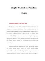
LACTOSE
INTOLERANCE
Lactase
(a
disaccharidase), normally
located
in the
brush
border
of the
small
bowel, hydrolyses
lactose
to
glucose
and
galactose (Fig.
2). In the
period following
weaning,
lactase activity
in
most popula-
tions
of the
world reduces,
such
that adults
tend
to
have
an
acquired lactose intoler-
ance. This tends
not to be the
case amongst
Caucasians
in
whom
the
lactase activity
persists into adulthood
in the
majority.
In
the
10-20%
of
individuals
who are
lactose
intolerant,
the
non-absorbed sugars
are
metabolised
by the
colonic flora, produc-
ing
gas, with distension, borborygmi
and
diarrhoea.
Secondary lactase deficiency
may
develop following small bowel
diseases
such
as
gastroenteritis, malnutrition,
coeliac
disease
and
Crohn's disease.
If
sus-
pected,
a
trial
of
dairy-free products
is
straightforward,
but
more formal testing
may
be
done with
a
lactose
hydrogen
breath test.
DRUGS
The
list
of
drugs that
may
cause diarrhoea
is
impressive (Table
2) and a
very
careful
drug
history
is
essential
in all
patients. This
should include
not
only prescribed med-
ication
but
also over-the-counter prepara-
tions
and
herbal remedies.
The
only
way to
be
sure that
a
drug
is not
playing
a
part
is to
discontinue
it.
Occasionally patients with
psychological problems deliberately abuse
laxatives, which
may
make diagnosis
diffi-
cult.
Phenolphthalein-containing laxatives
can be
detected
by
alkalinising stool water,
which
goes
red in the
presence
of
phe-
nolphthalein.
Anthraquinone laxatives
can
be
detected
by
chromatography
in
urine
or
stool.
Enterocyte
Fig.
2
Carbohydrate
digestion
and
absorption.
Table
1
Conditions
that
may
result
in
small
bowel
bacterial
overgrowth
Table
2
Common
drugs
that
may
cause
diarrhoea
Reduced
gastric
acid
production
Ulcer
surgery
Acid
suppression
therapy
Atrophic
gastritis
Stagnation
and
reduced
transit
Small
bowel
diverticula
Surgical
blind
loops
Obstruction
(strictures,
adhesions)
Motility
disorders
(diabetes,
scleroderma)
Fistulas
between
colon
and
small
bowel
•
Antibiotics
•
Promotility
agents
-
metoelopramide
•
Proton
pump
inhibitors
-
omeprazole,
lansoprazole
•
Non-steroidal
anti-inflammatory
drugs
•
Colchicine
•
Biguanides
-
metformin
•
Misoprostol
•
Cytotoxics
•
5-HT
reuptake
inhibitors
(SSRIs)
•ASA
compounds
Miscellaneous colitides
and
other causes
of
diarrhoea
•
Consider microscopic colitis
in a
middle-aged woman with watery diarrhoea.
•
Colonic biopsy should
be
performed
in all
patients with chronic diarrhoea.
•
Diarrhoea
in a
middle-aged
man
with
an
extra-intestinal phenomenon such
as
arthralgia
should lead
one to
consider Whipple's disease.
•
Intestinal lymphoma
is
often
a
difficult diagnosis
to
make
and may
require open surgical
biopsy
to
confirm.
•
Small bowel bacterial overgrowth
is
underdiagnosed
in the
elderly.
•
Lactose intolerance
may be
detected either following
a
breath test
or by a
trial
of
dairy
product
avoidance.
•
Many drugs have
the
potential
to
cause diarrhoea
and
discontinuation
is the
only
way of
excluding them
as a
cause.
THYROTOXICOSIS
Gut
disturbance
is
common
in
thyrotoxicosis, occurring
in
approximately
25% of
cases. Symptoms
are of
diarrhoea, col-
icky abdominal pain
and
weight loss.
The
diarrhoea
is
probably
due
to a
combination
of
increased small bowel motility
and
increased mucous secretion
via
increased cAMP production.
The
other systemic signs
of
thyrotoxicosis should
be
sought
-
namely tachycardia, tremor,
eye
signs, brisk reflexes
and
signs
of
weight loss.
Gastrinomas
Gastrin-secreting tumours usually occur
in the
pancreas
or
duo-
denum
and are
associated with persistent peptic ulceration
but
frequently
cause diarrhoea
also
(see
p.
27).
VIPoma
A
VIPoma (vasoactive intestinal polypep-
tide-oma)
is a
rare
functional
tumour
of
the
pancreas, producing excess amounts
of
VIP, which results
in
severe
watery
(secretory) diarrhoea, hypokalaemia
and
hypochlorhydria.
The
diarrhoea
is of
large
volume, continues during fasting
and
often
results
in
dehydration. Diagnosis
is
confirmed
by
demonstrating
an
elevated
serum
VIP
concentration
in the
presence
of
diarrhoea
and
frequently
a
mass
in the
tail
of the
pancreas. Functional suppres-
sion
of the
tumour
can be
achieved with
the
somatostatin analogue octreotide
but
surgical excision
is the
treatment
of
choice.
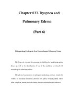
Carcinoid
syndrome
Tumours secreting 5-hydroxytryptamine
(5-HT
or
serotonin) most commonly
occur
in the
terminal ileum
and
appendix,
but
do not
produce
the
syndrome because
5-HT
is
readily metabolised
by the
liver.
Only when there
is
metastatic disease
in
the
liver (Fig.
1) or the
tumour drainage
is
not via the
portal system
(as in
bronchial
or
ovarian carcinoids), does
the
syndrome
occur.
The
clinical features (Fig.
2) are of
diarrhoea,
flushing
affecting
the
chest
and
head (Fig.
3),
bronchospasm, right-sided
heart valve lesions
and
rarely pellagra
(due
to
excessive tryptophan usage, caus-
ing
wasting, dermatitis, dementia
and
diarrhoea). Diagnosis depends
on
demon-
strating
an
elevated 5-HIAA concentra-
tion
in the
urine associated with bulky
hepatic metastatic disease
or a
primary
in
the
lung
or
ovary.
Treatment isdirected
at
controlling,
symptoms
by
debulking
the
tumour
in the
liver (either surgically
or
radiologically),
by
hepatic artery embolisation
or by
sup-
pressing 5-HT secretion with octreotide.
This
often
controls both
the
flushing
and
diarrhoea, whilst cyproheptadine
is
most
useful
in
controlling diarrhoea.
The
tumour
obtains
its
blood supply
from
the
hepatic artery, whereas liver tissue
obtains
the
majority
of its
oxygen supply
from
the
portal vein.
By
selective cannu-
lation
of the
hepatic artery
and
embolisa-
tion
of
radicals supplying
the
tumour,
tumour tissue necrosis
can be
achieved
with
debulking
of the
tumour, whilst leav-
ing
the
liver tissue undamaged. This,
however,
often
produces profound meta-
bolic disturbance
as
there
is a
surge
of 5-
HT
release.
Diabetes
mellitus
Insulin-dependent diabetes
is
compli-
cated
by
diarrhoea
in
about
5% of
patients.
The
stool
is
usually watery, with
occasio-nal
steatorrhoea.
Symptoms
often
occur
at
night
and
tend
to be
refractory
to
therapy. Mechanisms that
may
contribute
include diabetic autonomic neuropathy
(where there
may be
other signs
of
auto-
nomic dysfunction such
as
orthostatic
hypotension, impotence, neurogenic
bladder, pupillary
dysfunction,
and
gusta-
tory sweating), small bowel bacterial
overgrowth
and
abnormal
gut
motility.
Tight diabetic control, antibiotic therapy
for
bacterial overgrowth, opiates
and
cholestyramine
can all be
tried.
Concomitant conditions that occur
more
frequently
in
association with dia-
betes such
as
coeliac
disease
and
hyper-
thyroidism should
be
excluded.
Fig.
2
Clinical features
of the
carcinoid
syndrome.
Fig.
3
Flushing
of the
face
and
neck
in
carcinoid
syndrome.
ENDOCRINE POST-SURGICAL AND LIFESTYLE CAUSES OF DIARRHOEA
The
oral hypoglycaemic metformin
is
a
common cause
of
diarrhoea
in
non-
insulin-dependent
diabetics,
and
sorbitol,
a
sucrose substitute
in
prepared foods,
may
also cause diarrhoea (Fig.
4).
POST-SURGICAL
CAUSES
OF
DIARRHOEA
Bile
salt diarrhoea
The
majority
of
bile acids
are
reabsorbed
by
the
terminal ileum
as
part
of the
enterohepatic circulation. Following
resection
of the
terminal ileum, non-
absorbed bile salts induce
a
watery diar-
rhoea
by
stimulating colonic secretion.
The
same mechanism
may
contribute
to
the
diarrhoea
in
patients with Crohn's dis-
ease
affecting
the
terminal ileum.
Cholestyramine,
an
ion-exchange resin,
is
effective
in
controlling diarrhoea caused
by
this mechanism.
Following
cholecystectomy,
10-20%
of
patients complain
of
mild diarrhoea.
The
mechanism
is not
clear
but
presum-
ably
the
diarrhoea
is a
result
of
disruption
of
the
normal
enterohepatic
circulation
of
bile
salts. Treatment with cholestyramine
or
aluminium hydroxide
may be
helpful.
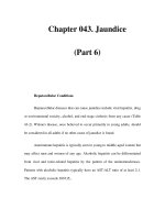
Short
bowel syndrome
The
small bowel absorbs approximately
7.5
litres
of
fluid
per
day.
Following
resection, there
is
considerable capacity
for
compensation
but
when more than
1.5m
is
resected, diarrhoea
usually
ensues
(the normal length
is
estimated
at
between
3 and 8 m). The
diarrhoea
is
most marked
immediately
following
surgery
and may
require intravenous
nutritional
support whilst compensation
occurs. However,
it is
important
to
con-
tinue
enteral feeding during this time
as
this
promotes adaptation. Resection
of
segments
of
small
bowel
can
lead
to
spe-
cific
nutrient deficiencies (Fig.
5).
Resection
is
most usually performed
for
Crohn's disease
and
less
frequently
for
mesenteric infarction
and
radiation enteri-
tis.
The
clinical features
are of
diarrhoea,
steatorrhea
and
macro-
and
micronutrient
deficiency.
Features
are
predictable
depending
on the
amount
and
site
of
bowel
resected.
Moderate resection
may
allow
the
patient
to
remain adequately
nourished
on a
low-fat, high-carbohy-
drate diet with vitamin supplementation.
Calorie
intake
is
often
two to
three
times
that
required preoperatively. More exten-
sive small bowel resection requires long-
term parenteral nutrition. Oral intake
may
promote
a
pronounced secretory phase
which
also
results
in
patients limiting
their
oral
intake
so as to
avoid volume
depletion.
Cholesterol gallstones, liver disease
and
oxalate
kidney stones
are
more com-
mon in
patients with short bowel syn-
drome.
Drugs: metformin
Diet: sorbitol
Associated
conditions
•
Thyrptoxicosis
•
Coeliac disease
_
Autonomic
neuropathy
Small bowel
bacterial overgrowth
Fig.
4
Causes
of
diarrhoea
in
diabetics.
Colon
and
small bowel
Electrolytes
Water
Surgical transplantation
of
small
bowel
may be
possible
in
some patients
although
it has
still been performed
in
only
small numbers
of
patients.
MISCELLANEOUS
CAUSES
OF
DIARRHOEA
Exercise
As
recreational exercise becomes more
widespread, individuals
often
observe
an
urge
to
defaecate, increased bowel fre-
quency
or
episodes
of
watery diarrhoea
before,
during
or
after
exercise.
'Nervous' diarrhoea, just before
a
race,
occurs
in
over
a
third
of
regular runners
and
nearly
a
half experience diarrhoea
during
a
race. Colonic transit times
appear
to
reduce following regular exer-
cise.
Reassurance, reducing workload
and
occasionally prophylactic antidiarrhoeals
can
be
tried.
Alcohol
Alcohol binges
often
lead
to
episodes
of
diarrhoea, possibly owing
to
decreased
gut
transit times
and
inhibition
of gut di-
saccharidases. Chronic alcohol abuse
can
result
in
exocrine pancreatic
insuffi-
ciency, which
may be
reversible,
or
chronic pancreatitis. Some beers have
naturally
occurring high concentrations
of
salts which
act as a
cathartic, inducing
diarrhoea.
Small bowel
Fat
Protein
Carbohydrate
Minerals:
Ca
2+
,
Mg
2+
,
Fe
Vitamins:
B, C,
folate,
A, D, E, K
Trace elements:
Zn, Cu
Terminal ileum
B12
Bile salts
Fig.
5
Potential components malabsorbed following
small
bowel resection.
Endocrine
and
post-surgical
causes
of
diarrhoea
•
Hyperthyroidism
is
common
but
diarrhoea
as a
sole presenting feature
is
unusual.
•
Tumours causing oversecretion
of the gut
hormones gastrin,
VIP and
5-HT
can all
cause
diarrhoea,
but are
rare.
•
Diabetes
can be
complicated
by
diarrhoea
due to the
medication, small bowel bacterial
overgrowth
and gut
dysfunction associated with autonomic neuropathy.
•
Diarrhoea associated with
the
short bowel syndrome
is
accompanied
by
micro-
and
macronutrient deficiency.
62
CONSTIPATION
AND
PERIANAL
PAIN
DEFINITION
OF
CONSTIPATION
The
normal range
of
bowel
frequency
is
between three times
per day and
once
every
3
days. Anything less
frequent
than
this
may be
defined
as
constipation.
Patients
may
also describe straining
at
stool
and
passing pellet-like stools
(often
described
as
being like 'rabbit droppings').
There
may be a
sensation
of
incomplete
evacuation. Symptoms persisting
for
more
than
6
weeks
may be
termed chronic con-
stipation.
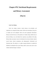
PHYSIOLOGY
OF
DEFAECATION
The
urge
to
defaecate
is
triggered
by
dis-
tension
of the
rectum
by
faeces transported
from
the
sigmoid reservoir
by
mass motor
contractions. Privacy
is
sought
and a
squatting
position adopted.
A
Valsalva
manoeuvre
is
often
used
to
increase intra-
abdominal pressure
in
order
to
promote
faecal
expulsion.
The
pelvic
floor
muscles
relax, allowing
the
pelvic
floor
to
descend.
The
angle between
the
anus
and
rectum
is
straightened, allowing
faecal
passage (Fig.
1).
Defaecation
is a
spinal
reflex
under
sympathetic
control
via the
sympathetic
chain
in
front
of the
aorta
and
parasympa-
thetics
from
S2, 3, and 4 to the
rectum
and
internal anal sphincter.
The
striated muscle
of
the
external anal sphincter
is
controlled
via
the
somatic pudendal nerve (S2,
3 and
4).
When
it is
inappropriate
to
defaecate,
it
is
the
voluntary contraction
of the
external
anal sphincter that prevents defaecation.
PATHOPHYSIOLOGICAL
MECHANISMS
OF
CONSTIPATION
Because there
are so
many varied causes
of
constipation,
it is
necessary
to
have
a
At
rest
Sacrum
Rectum
Internal
anal
sphincter
External
anal
sphincter
Fig.
1 The
pelvis
at
rest
and on
defaecation.
structure
for
investigating
the
causes that
may
be
encountered.
Of the
intestinal
causes,
one
should consider mechanical
obstruction
-
either luminal
or due to
external compression abnormalities
of
muscle
function,
rectal
and
anal disorders
and
functional
constipation. Extraintestinal
causes include drugs, metabolic/endocrine
causes, abnormalities
of the
nervous sys-
tem
(central
or
peripheral)
and
psychologi-
cal
causes (Table
1).
HISTORY
As
always, taking
a
thorough history gives
the
clinician
the
best chance
of
making
a
correct diagnosis
and
investigating
patients appropriately.
The
individuals
most likely
to
suffer
with constipation
are
young
women
who
have
often
had
their
symptoms since their teenage years.
If
sought,
there
may
also
be a
family
history
with
mother
and
sisters being similarly
Table
1
Causes
of
constipation
Idiopathic
Dietary
Inadequate
fibre or
fluid
intake
Intestinal
Luminal
tumours
(also
with
external
compression)
Strictures
(diverticular,
ischaemic,
infective,
inflammatory)
Irritable
bowel
syndrome
Hirschsprung's
disease
Rectocele
Solitary
rectal
ulcer
syndrome/mucosal
prolapse
Anismus
Anal
fissure
Pseudo-obstruction
Extraintestinal
Spinal
cord
damage
Parkinson's
disease
Cerebrovascular
disease
Metabolic/endocrine
(hypothyroidism,
hypercalcaemia,
hypokalaemia)
Drugs
At
defaecation
Loss
of
anorectal
angle
afflicted.
Symptoms
of
abdominal bloat-
ing,
pain relieved
by
defaecation,
and an
alternating diarrhoea
and
constipation sug-
gest
the
irritable bowel syndrome.
In the
older individual
who
suddenly notices
a
change
in
bowel habit associated with
symptoms
of
pain
and
distension, there
may
be a
mechanical obstruction
-
stenos-
ing
carcinomas
of the
colon
not
infre-
quently
cause these symptoms
and
injudicious
use of
purgatives
in
prepara-
tion
for a
barium enema
may tip
patients
into complete obstruction, requiring emer-
gency resection.
In
these circumstances
a
barium enema without colonic preparation
may
give
the
diagnosis without
the
risks.
Particular care should
be
exercised
in
taking
a
thorough drug history
-
patients
often
forget
or
omit
the
non-prescribed
treatments they
are
taking (Table
2).
Careful
dietary assessment
is
important
because
the
poor quality
of
individual diets
is
often
surprising, particularly
in
regard
to
intake
of
dietary
fibre.
It is
worth going
through
each meal
of the day and
enquir-
ing
what would normally
be
eaten.
Endocrine
or
metabolic abnormalities
such
as
hypothyroidism, hypokalaemia
and
hypercalcaemia
may all
present with
constipation
but are
often
associated with
other systemic changes. Neurological
causes would usually have constipation
as
an
associated symptom rather than
as a
presenting feature.
Patients' presenting symptoms
may
often
be
masking underlying worries, par-
ticularly
regarding cancer,
and it is
worth
enquiring
about this specifically,
as
directly addressing
the
issue
and
answer-
ing
the
patients' concerns
will
usually lead
to
resolution
of
their symptoms.
EXAMINATION
If
a
neurological
or
endocrine cause
is
sus-
pected, then abnormal clinical signs
may
be
elicited during
the
general physical
examination.
The
abdominal examination
Table
2
Drugs that
may
cause constipation
•
Anticholinergics
•
Tricyclicantidepressants
(anticholinergic
side-effects)
•
Calcium
channel
blockers
•
Antihistamines
•
Diuretics
•
Antacids
(calcium
and
aluminium
salts)
•Iron
•
Chronic
laxative
abuse
THE CLINICAL APPROACH
THE
CLINICAL
APPROACH
63
Fig.
2
Investigation algorithm
for
constipation.
Fig.
3
Stenosing colon cancer seen
on
barium enema.
may
reveal masses
due to
either tumours
or
distended bowel proximal
to an
obstruc-
tion.
Consideration should
be
given
to the
patient
during rectal examination
as
this
may be
painful
in the
presence
of
anal fis-
sures
or
increased anal tone,
and it may be
kinder
to
perform
rectal
examination under
sedation prior
to
flexible sigmoidoscopy
in
these cases.
In the
elderly,
a
loaded rectum
suggests
faecal
impaction, which
may be
associated with periods
of
spurious diar-
rhoea,
due to
overflow.
Pain
in the
perineum
at the
time
of
defaecation
which begins suddenly, partic-
ularly
when straining
to
pass
a
hard stool,
and is
often
associated with
a few
spots
of
blood suggests
an
anal
fissure.
Intense,
episodic, sharp rectal pain which lasts
a
few
moments
and
then resolves com-
pletely
is
termed proctalgia
fugax
and may
be
associated with symptoms
of
irritable
bowel syndrome.
INVESTIGATIONS (Fig.
2)
Deciding
who and how far to
investigate
is
an
important clinical skill.
In the
younger
age
group where irritable bowel syndrome
is
common, history, examination
and
flexi-
ble
sigmoidoscopy, with
a
full
blood
count, serum biochemistry, thyroid
func-
tion tests
and
measurement
of
serum cal-
cium
concentration
may be all
that
is
necessary. Simple advice regarding diet,
physical
activity
and the
condition itself
may
be
effective
treatment.
It
would
be
inappropriate
to
perform barium examina-
tion
in
individuals
who
respond
to
these
measures.
In an
older
age
group (patients
over
40
years)
or in
younger patients with
a
strong
family
history
of
colon cancer,
particularly
at an
early age, visualisation
by
either
radiology
or
colonoscopy should
be
performed, looking
for
colonic neopla-
sia
(Fig.
3) - the
incidence
of
which
increases with age. Colonic dilatation
is
best demonstrated
by
radiology (Fig.
4).
Colonic transit studies (Fig.
5) and
anorectal physiology measurements
may
be
necessary
in a
small subset
of
patients
such
as
those
with
megacolon
and in
patients
with severe intractable symptoms.
Fig.
5
Pellets
for
transit studies seen
in
right
upper
quadrant
in gut
transit
study.
The
clinical approach
• A
careful history should include both
a
dietary evaluation
and a
drug
history-prescribed
and
over
the
counter.
•
Clearly establish what
the
patient means
by
constipation
and
what symptom
he or she
would
like
to
have solved.
•
Examination
may be
unhelpful. Rectal examination
and,
usually, sigmoidoscopy must
be
performed-if
likely
to be
particularly painful, they
can be
done under sedation.
•
Avoid over-investigation
if the
symptoms
are not
severe
and
there
is no
evidence
of
megacolon.
•
Psychological factors often play
a
part
and
sympathetic management
will
often
be
most
successful.
Fig.
4
Megacolon.
CONSTIPATION
AND
PERIANAL PAIN
SEVERE IDIOPATHIC CONSTIPATION
This condition
usually
afflicts
young
women
who may
have
a
family
history
of
the
condition
and
whose symptoms began
in
their teenage years.
There
is
usually
abdominal pain
and
bloating
and
patients
describe
infrequent
stool passage. Patients
have
often
tried dietary
fibre
supplements
and
are
usually taking stimulant laxatives
at
the
time
of
presentation.
Occasionally, patients
describe
an
incredible bowel habit with defaecation
every
few
weeks.
Colonic
transit time
can
be
established
from
X-ray images taken
at
5-day intervals
of a
patient
who has
swal-
lowed radio-opaque
pellets.
Retention
of
more
than
20% of
pellets suggests slow
transit
constipation.
In
others,
a
more
nor-
mal
bowel habit
is
demonstrated, reflect-
ing
patients' perceptions
of
their bowel
habit.
Anorectal physiology studies
may
show
an
inability
to
relax
the
external anal
sphincter when
the
rectal pressure
is
increased
-
such that
the
rectum
is
pushing
against
a
'closed
door'
(anismus).
The
aetiology
of
this
is
unknown
but is
proba-
bly
an
acquired
condition
following
persis-
tent
suppression
of the
urge
to
defaecate.
Treatment
Mild
to
moderately constipated patients
will
usually
have increased their dietary
fibre
intake, although some
may be
helped
by
formal dietary assessment. Bulking lax-
atives
and
then
a
stimulant suppository
such
as
bisacodyl should
be
used next.
Table
1
Laxatives
and
their mode
of
action
More severe constipation
may
require ene-
mas, oral stimulant laxatives,
or a
non-
absorbed polyethylene glycol preparation
(PEG) (Table
1).
Rarely, surgery
is
considered. Subtotal
colectomy
and
ileorectal anastomosis
has
an
unpredictable outcome with one-third
developing diarrhoea
and 10%
remaining
constipated.
MEGACOLON
If
patients complain
of
constipation since
childhood
and
demonstrate
a
dilated
gut
(diameter
of the
rectum
at the
pelvic brim
exceeds
6.5
cm), adult Hirschsprung's dis-
ease
should
be
considered.
In
this condi-
tion,
a
segment (usually distal)
of
bowel
fails
to
relax, producing
a
functional
obstruction.
Presentation
is
usually
in
childhood
but the
condition
may
appear
in
later
life.
There
is
aganglionosis with loss
of
intramural nerve plexuses, which
can be
demonstrated
at
histology
following
a
full-
thickness mucosal biopsy taken
at
least
2
cm
above
the
dentate line. Alternatively,
rectal physiology studies show
a
failure
of
anal relaxation following rectal distension
(the
recto-anal
inhibitory reflex)
- its
pres-
ence excludes Hirschsprung's disease.
Surgical resection
is
required
for the
rare
cases
of
Hirschsprung's
disease.
Acquired megacolon
can
occur follow-
ing
neurological diseases
such
as
spinal
cord
injury,
Parkinson's disease, diabetic
neuropathy, dystrophia myotonica
and
Chagas'
disease,
or may be
idiopathic.
Action
Bulking agents
Bran
Isphagula husk
Sterculia
Methylcellulose
Faecal
softeners
Docusate
sodium
Paraffin
Arachis
oil
Osmotic
laxatives
Magnesium
salts (e.g.
magnesium
Sodium
sate (e.g.
sodium phosphate)
Lactulose
Polyethylene glycol
Stimulant laxatives
Senna
Bisacodyl
Danthron
Sodium picosulphate
Retain
water
in the
gut,
onset
of
action
12-24
hours, require adequate oral fluid intake
Has
a
detergent
effect
Now
out of
favour
as a
faecal
softener
owing
to
the
possibility
of
aspiration
and
lipoid pneumonia
Given
as an
enema
Stimulate colonic activity
as
well
as
acting
as
osmotic laxative
Should
be
avoided when sodium overload
may
be
harmful (e.g. heart
or
renal failure)
Oral
or
rectal,
can
cause colicky
pain,
induce
hypokalaemia
and
cathartic colon.
Effect
takes
6-12 hours. Often combined with softeners
Treatment should include that
of the
underlying
condition
if
present,
but is
aimed
at
keeping
the
colon empty.
Acute megacolon
can
complicate acute
severe inflammatory bowel disease
and
infectious
colitis. There
is
another group
in
whom megacolon develops acutely, usu-
ally
with
coexisting conditions such
as
trauma
or
orthopaedic events; such
a
development
is
termed pseudo-obstruction
or
'Ogilvie's
syndrome'.
The
clinical fea-
tures
are of
marked gaseous abdominal
distension
developing
in an
elderly,
frail
or
postoperative patient. Abdominal X-ray
shows gaseous distension,
and
mechanical
obstruction
is
excluded
by
water-soluble
contrast enema (Fig.
1).
This
may
also
be
therapeutic
as
treatment
is
aimed
at
decompressing
the
bowel
with
rectal
flatus
tubes
and
enemas. Biochemical abnormal-
ities should
be
corrected
and if
this
fails
decompression
by
colonoscopy
may be
required, which
will
usually
be
effective.
This
can be
repeated
and
neostigmine
added
if
necessary.
SOLITARY RECTAL ULCER SYNDROME
Following
chronic constipation
and
strain-
ing
at
stool, particularly
in
women,
mucosa
from
the
anterior rectal wall
may
prolapse through
the
anal margin. This
results
in
mucosal damage
and
ulceration,
typically
on the
anterior rectal wall.
Straining
at
defaecation
is
accompanied
by
Fig.
1
Intestinal pseudo-obstruction.
RELATED CONDITIONS
RELATED
CONDITIONS
Fig.
2
Rectocele
encroaching
on
posterior
vaginal
wall.
blood
and
pain.
A
defaecating proctogram
may
show
the
mucosa prolapsing through
the
anal margin. Histology
is
characteristic
with
fibrosis in the
lamina propria.
Bulking
agents
and
avoidance
of
straining
at
stool
may
help,
but
surgical
fixation
may
be
required.
RECTOCELE
The
posterior vaginal wall
may
prolapse,
pulling
the
anterior rectal wall with
it,
pro-
ducing
a
rectocele
(Fig.
2). A
rectocele
is
usually
asymptomatic until large, when
the
patient
has a
feeling
of
incomplete evacua-
tion
and may
need
to
place
a
finger
in the
vagina
to
empty
the
rectal
sac of
faeces.
Surgical repair
is
required.
DESCENDING
PERINEUM
SYNDROME
Most commonly
affecting
women follow-
ing
childbirth,
the
anal margin descends
excessively causing closure
of the
anal
canal
and
obstructed
defaecation.
Rectal
prolapse
often
results. Observation
of the
perineum
at the
time
of
straining demon-
strates
the
descent
of the
perineum below
a
line
between
the
ischial tuberosities.
Bulking agents
and
repair
of
rectal pro-
lapse
may be
required.
PERIANAL
PAIN
ANAL
FISSURES
Characteristic intense anal pain,
of
sudden
onset
at the
time
of
passing
a
hard stool,
and
often
associated with
a few
drops
of
blood,
is
characteristic
of an
anal
fissure.
The
vast
majority
occur
in the
posterior
midline
or
anteriorly,
and
deviation
from
these sites raises
the
possibility
of an
alter-
native underlying disease such
as
Crohn's
disease.
At the
upper margin there
may be
a
hypertrophic anal papilla and, distally,
a
sentinel
pile
at the
anal verge
may be
seen.
Anal fissures
are
usually
associated
with
constipation,
and
bulking agents
and
analgesia
may
allow healing. Glyceryl
trinitrate
gel and
lignocaine
gel
applied
topically will help more severe cases.
Lateral sphincterotomy lowers
the
anal
resting pressure
and
allows healing.
PROCTALGIAFUGAX
A
severe pain
in the
rectum which lasts
a
few
moments
and
then resolves sponta-
neously
is
typical
of
proctalgia
fugax.
It is
a
common symptom,
often
experienced
when individuals
are
feeling
under stress.
Reassurance
and
avoidance
of
constipation
are
usually
sufficient.
HAEMORRHOIDS
The
three
major
symptoms caused
by
haemorrhoids
or
'piles'
are
fresh
rectal
bleeding, local pain
and
pruritus.
Of the
mammals
it
would appear that only
man is
afflicted
with haemorrhoids, although
it is
unclear
why
this should
be so. It is
proba-
bly
due to
straining
to
pass
the
low-vol-
ume,
firm
stools that result
from
a
residue-deficient
diet.
The
anal cushions
have
a
rich venous plexus
and it is
these
venous cushions that
become
enlarged
to
form
haemorrhoids. They characteristi-
cally
appear
in the 3, 7, and 11
o'clock
positions (Fig.
3) and may be
internal
or
prolapse
through
the
anal canal (Table
2).
Bleeding
and
prolapse
may be
made
worse when
the
patient attempts
to
pass
Table
2
Classification
of
haemorrhoids
Degree
First
Second
Third
Fourth
Symptoms/findings
Bleeding,
but not
prolapsing
Prolapse
but
reduce spontaneously
Prolapse
but
require manual
reduction
Permanently prolapsed
hard stools
and if
attempts
to
defaecate
are
made before
a
natural call
to
stool.
The
bleeding typically occurs
after
stool
has
been passed
and may be
seen
on the
toilet
paper
or
dripping into
the
pan. Blood
may
appear
on the
surface
of the
stool
but
should
not be
admixed with
it. A
history
of
rectal bleeding warrants some
further
investigation even
in the
young
and
should
include
a
sigmoidoscopy.
An
explanation
and
reassurance
are
necessary
for
minor haemorrhoids
as the
natural
history
of
haemorrhoids
is for
them
to
come
and go, and
treatment
may not be
necessary. Patients should
be
encouraged
to
take more
fibre
in
their diets
in
order
to
produce
softer
stools. Banding
of the
haemorrhoids
is an
outpatient procedure
in
which
a
band
is
placed onto
the
exuberant
venous
plexus.
Care
must
be
taken
to
ensure that
the
band
is
above
the
dentate
line,
otherwise
the
patient experiences
severe pain
and the
band requires removal.
Injection
sclerotherapy
can
also
be
per-
formed,
but
there
are
reports
of
erectile
dysfunction
in men
and,
if
warned
of
this
possibility, most would decline this
form
of
treatment. Surgical excision
is
required
for
irreducible haemorrhoids.
Fig.
3
Haemorrhoid
positions.
Conditions causing constipation and/or
perianal
pain
•
Constipation-predominant irritable bowel syndrome
is a
common problem which
requires reassurance
and
advice rather than extensive investigation.
•
Anismus
is
detected
by
anorectal physiology studies
and is
best treated
by
biofeedback
techniques.
•
Laxatives work
by
bulking
the
stool,
by
acting
as a
faecal softener,
by
creating
an
osmotic gradient
in the
bowel,
or by
stimulating
the
colon.
•
Treatment
for
haemorrhoids includes bulking
the
stool
to
keep
it
soft,
reassurance,
and
therapy
to the
haemorrhoid only
if
necessary.
The
annual incidence
of
acute upper gas-
trointestinal haemorrhage
is
approximately
1 per
1000 adults
per
year with
a
mortality
in
the
region
of
10%,
the
majority
of
deaths occurring
in the
older
age
group.
This mortality rate appears
to
have
fallen
only
slightly despite attempts
at
endo-
scopic therapy
and the
development
of
algorithms attempting
to
identify
high-risk
patients.
Management
of
patients with
an
acute
upper gastrointestinal bleed
is
slightly dif-
ferent
from
the
management
of
many other
emergencies because initial treatment does
not
usually depend
on
establishing
a
diag-
nosis. Patients
may
present with vomiting
of
frank
red
blood
(haematemesis), which
usually
does
not
present
a
diagnostic
conundrum,
although swallowed blood
from
substantial nose bleeds
can be
misin-
terpreted
as
coming
from
the
gastrointesti-
nal
tract (GIT). Estimating
the
volume
of
blood vomited
is
difficult
and
patients
may
often
overestimate
the
amount. Smaller
bleeds
can
present with vomiting altered
blood, which
is
often
described
as
'coffee
grounds'.
The
passing
of
'melaena'
-
black sticky stool with
a
characteristic
odour
-
represents
a
significant upper
GI
bleed
but may or may not be
associated
with
haematemesis.
If the
bleed
is
torren-
tial,
degradation
of the
blood
may not
have
had
time
to
occur
and
partly altered
red
blood
is
passed
per
rectum (haema-
tochezia).
ASSESSMENT
The
first
step
in
management, having been
convinced that there
has
been
an
upper
GI
bleed,
is to
establish
the
severity
and
risk
to
the
patient. This requires ongoing mea-
surement
of
pulse
and
blood pressure
(including
looking
for the
presence
of a
postural drop
in BP,
which should warn
the
clinician that
the
haemorrhage
is
larger
than
may
otherwise have been suspected).
Peripheral venous
access
should
be
gained
in
minor bleeds
or a
central venous line
should
be
placed
to
allow central venous
pressure
monitoring
and
maintain
good
venous
access when
a
larger bleed
is
sus-
pected. This
is
particularly
so in
patients
who
present with
a
systolic
blood
pressure
of
<
l00
mmHg
or who
have significant
comorbidity, particularly liver disease,
in
whom
a
variceal bleed
is a
possibility.
Blood should
be
drawn
for
haemoglobin
estimation, liver
function
tests, coagulation
tests, biochemistry
and
cross-matching.
Age, shock, comorbidity, diagnosis,
major
stigmata
of
recent haemorrhage
at
endoscopy
and
rebleeding have
all
been
shown
to be
independent predictors
of
mortality
and a
scoring system
has
been
developed
in
order
to
identify
these cases
(Table
1). Use of
this scoring system
allows
prediction
of
mortality
and
rebleed-
ing
rates
and
should allow focusing
of
monitoring
and
treatment.
Patients should have their intravascular
volume
restored with colloid
or
blood
when
it
becomes available. This should
be
enough
to
maintain
an
adequate blood
pressure
or
raise
the
haemoglobin above
10
g/dl
in the
less acute situation.
HISTORY
Having stabilised
the
patient, more time
can
be
given
to
taking
a
history.
A
history
of
recurrent epigastric pain
may
point towards peptic ulcer disease,
and
haematemesis
following
a
period
of
vomiting suggests
a
Mallory-Weiss
tear.
Attention should
be
given
to
previous
history
of
haemorrhage, peptic ulcer dis-
ease, liver disease, previous surgery
including
aortic aneurysm repair
and
bleeding disorders. Note should
be
taken
of
current drug therapy, particularly
NSAID usage, remembering that NSAIDs
may
now be
obtained
over
the
counter
without
prescription.
An
attempt
to
quantify
alcohol con-
sumption
should
be
made.
Table
1
Scoring
for
acute
upper
GI
haemorrhage
EXAMINATION
Having
measured
the
vital signs
of
pulse
and
blood pressure, features
of
chronic
liver disease
and
portal hypertension
should
be
sought.
Careful
abdominal
examination should
be
performed
for the
presence
of an
aortic aneurysm
or
previous
surgery
and the
mouth inspected
for
telangiectases. Rectal examination will
determine whether melaena
is
present.
INVESTIGATIONS
The
investigation
of
choice, which also
allows therapy
to be
undertaken,
is
upper
GI
endoscopy. This should
be
undertaken
in
all
patients with
an
upper
GI
bleed
but
the
timing
of its
performance
is a
more
critical question (Fig.
1).
Endoscopy
of an
inadequately resuscitated patient
is
haz-
ardous
and
should
be
avoided; however,
in
the
presence
of
torrential blood loss, such
as
may
occur with oesophageal varices,
resuscitation, diagnosis
and
treatment must
run
concurrently.
The
other patients
who
should
be
endoscoped urgently
are
those
with
a
massive
first
bleed
or a
rebleed,
elderly patients over
the age of 70, and
patients with varices. Otherwise, patients
should
be
endoscoped
on the
next routine
list.
Unfortunately, patients with
the
most
severe disease
who
require urgent
endoscopy
often
have
the
procedure
per-
formed
by the
least experienced endo-
scopists,
out of
hours, with nurses
who
may
not be
highly trained endoscopy
nurses. This
is
unacceptable because
important
therapeutic interventions that
have
an
impact
on
patient outcome
can be
undertaken
during endoscopy.
Following endoscopy,
a
small percent-
Component
Age
Shock
Pulse rate (bpm)
SBP(mmHg)
Comorbidity
Diagnosis
Stigmata
of
recent
haemorrhage
Score
0
<60
No
shock
<100
Normal
None
Mallory-Weiss
tear
No
lesion
None
60-79
Tachycardia
>100
>100
-
All
other
diagnoses
—
2
>80
Hypotension
-
<100
Ischaemic
heart
disease
Malignancy
of
upper
GI
tact
Blood
in
upper
GI
tract,
visible
vessel
spurting
vessel
3
_
-
-
-
Renal
failure
Any
malignancy
-
-
SBP
=
Systolic
blood
pressure
THE CLINICAL APPROACH
age of
patients will have
no
demonstrable
cause
for
their
GI
bleed. This
may
occur
particularly
with
a
Mallory-Weiss
tear
and
much less frequently with
a
Dieulafoy
lesion.
ENDOSCOPIC
STIGMATA
OF
RECENT
HAEMORRHAGE
Certain
stigmata
are
visible endoscopically
which
are
associated with
a
high chance
of
rebleeding
and
usually prompt interven-
tion
with endoscopic therapy. When
oesophageal varices
are
discovered, active
bleeding, adherent clot
or a
cherry
red
spot
on
a
varix indicate active
or
recent bleed-
ing
and
sclerotherapy
or
banding should
be
undertaken.
In
Mallory-Weiss
tears
or
ulcers, active bleeding, adherent clot
or a
visible vessel
-
usually seen
as a
black
dot
in
the
centre
of an
ulcer
-
likewise
signify
a
high risk
of
rebleeding
and
warrant
ther-
apy.
ENDOSCOPIC
THERAPY
Sclerotherapy
and
banding
for
oesophageal
varices
is
dealt with
in the
text
on
portal hypertension
(p.
88).
Sclerotherapy
for
ulcers,
Mallory-Weiss
tears
and
Dieulafoy
lesions
Using
a
similar technique
to
that
of
scle-
rotherapy
for
oesophageal varices, high-
risk
lesions
can be
directly injected
via
the
endoscope, with
a
sclerosant
or an
adrenaline solution.
Up to 10 ml of
1:10000
adrenaline solution
is
injected
around
the
perimeter
of an
ulcer
and
then
directly into
the
visible vessel. This tech-
nique
has
been shown
to
reduce rebleeding
rates.
CAUSES
Peptic ulcer disease,
oesophageal
varices
(see
p.
90),
and
Mallory-Weiss
tears
are
the
commonest causes
of
acute upper
GI
haemorrhage. However, other rarer causes
Table
2
Causes
of
acute
upper
GI
haemorrhage
Common
Less
common
Duodenal
ulcer Duodenitis
Gastric
ulcer Oesophagitis
Gastric
erosions
Tumours
Mallory-Weiss
tear
Hereditary
telangiectasia
Oesophageal
varices
Aortoduodenal
fistula
Clotting
disorder
Portal
hypertensive
gastropathy
Dieulafoy
lesions
should
not be
forgotten, because
no
obvi-
ous
cause
is
found
in up to 20% of
cases
so
the
differential diagnosis
has to be
consid-
ered frequently (Table
2).
Peptic
ulcer
disease
Once diagnosis
has
been established,
patients should
be
started
on a
high-dose
proton pump inhibitor (e.g. omeprazole
40
mg
b.d.)
for 5
days, which reduces
the risk
of
rebleeding. Careful observation should
continue
for
signs
of
rebleeding, which
include
the
development
of a
tachycardia,
a
fall
in BP, or
fall
in the
central venous
pressure. Patients with
a
high-risk lesion
should
be
kept
nil by
mouth
for 48
hours
in
case
surgery
is
required,
and
then food
should
be
reintroduced. Patients with low-
risk
lesions
can
restart food immediately.
Torrential bleeding
at
endoscopy
and
Fig.
1
Investigation
algorithm
for
acute
upper
GI
bleed.
rebleeding following endoscopic therapy
are an
indication
for
surgery.
Mallory-Weiss
tears
The
history
is
characteristic when patients
often
having consumed alcohol begin
to
vomit
and
subsequently have
a
hae-
matemesis. This
is
usually
relatively
mild
and
stops spontaneously. Because
of the
violent
vomiting,
a
tear develops
in the
mucosa
of the
distal oesophagus
or
proxi-
mal
stomach. This
can be
difficult
to see at
endoscopy
but if it
does continue
to
bleed,
injection
therapy
can be
undertaken.
Overnight
observation
in
hospital follow-
ing
the
endoscopy
is all
that
is
required,
and
a
7-day course
of a
proton pump
inhibitor
on
discharge.
Dieulafoy
lesions
These
are
calibre-persistent arteries that
rise to the
surface
of the
gastric mucosa,
erode
through
it and
bleed. They com-
monly
affect
elderly
men and
occur high
in
the
posterior wall
of the
stomach. They
are
easy
to
miss
as
there
is no
surrounding
ulceration
and may
just
be
seen
as a
bleb.
They should
be
considered when
an
elderly patient
has had a
substantial upper
GI
bleed with
an
intial examination that
reveals
no
obvious bleeding source.
To
confirm
small lesions
to be
Dieulafoy,
light
pressure with
an
injection needle that
has
been primed with sclerosant demon-
strates arterial bleeding
and
confirms
the
diagnosis.
It is
then necessary
to
inject
sclerosant into
the
vessel immediately.
If
not
recognised
and
treated, such lesions
result
in a
significant mortality amongst
this
age
group.
The
clinical
approach
•
Assessment
and
resuscitation
should
run
concurrently
to
stabilise
the
patient.
•
Close
questioning
about
drugs,
including
over-the-counter
preparations,
is
essential.
• Age and
comorbidity
increase
the
risk
of a bad
outcome
from
an
upper
GI
haemorrhage.
•
Endoscopy
should
be
carried
out on a
resuscitated
patient,
early
if
high
risk
or on the
next
routine
list
if low
risk.
•
Torrential
bleeding
at
endoscopy,
or
rebleed
following
endoscopic
therapy
for
peptic
ulcers
is an
indication
for
consideration
of
surgery.
68
CHRONIC
GASTROINTESTINAL
BLEEDING
IRON METABOLISM
An
average
diet
provides 10-20
mg of
iron/day
of
which
approximately
1 mg is
absorbed. Sources include
red
meat,
fish,
eggs,
cereals
and
leafy
vegetables.
The
iron
in
vegetable sources
is
usually
present
in the
Fe
3+
state
but it is
best
absorbed
in the
reduced Fe
2+
state. Reduction occurs
in the
stomach with gastric
acid
and
vitamin
C.
Achlorhydria, previous partial gastrectomy,
or a
poor
intake
of
dietary
vitamin
C may
reduce absorption.
Iron
is
actively absorbed across
the
cell wall
of the
intestinal
mucosa, particularly
in the
proximal small bowel. Hence, dam-
age of
this mucosa, which occurs
in
coeliac
disease,
often
leads
to
iron deficiency. Once inside
the
cell, iron
is
either bound
to
ferritin
and
stored within
the
cell
or
passed into
the
circulation
bound
to
transferrin
to be
transported. Storage occurs
in the
liver,
spleen
and
bone marrow
in the
form
of
ferritin
or
haemosiderin.
CLINICAL APPROACH
With
widely available blood testing,
a
full
blood
count revealing
a
microcytic anaemia
is a
very common
finding
in
primary
health
care.
This
may be
simply treated
in the
community
if a
cause such
as
menorrhagia
is
obvious,
or
referred
for
further
investigation
if the
cause
is
obscure.
It is
important, however,
to
confirm
iron deficiency
in the
presence
of a
microcytic anaemia,
and
this
is
best done
by
measuring serum
ferritin
- low
values
confirming
iron deficiency. Secondly, remember that iron
defi-
ciency
has
occurred
for a
reason
and
that
if the
clinician
is
unsure
of
that reason, then
further
investigation
is
necessary.
Fig.
1
Investigation algorithm
for
iron deficiency anaemia.
History
The
history should include
any
symptoms that
may
result
from
anaemia (tiredness,
poor
exercise tolerance, breathlessness,
worsening
angina) although these
may be
absent
in an
otherwise
fit
individual. Teasing
out a
possible cause
is
most logically
done
by
considering that
for
iron
to be
available
for
erythro-
poiesis
it
must
be
ingested, absorbed
and
utilised
and
that
there
should
not be
excessive loss.
So
dietary intake should
be
assessed
and
evidence
for
malabsorption sought. Evidence
for
overt gastrointestinal blood loss,
or GI
symptoms such
as
dys-
pepsia,
or a
change
in
bowel
habit should
be
sought
in all
patients,
and
young women should also
be
asked about their
gynaecological history. Drugs,
as
ever,
are
important because
GI
bleeding
is
commonly caused
by
aspirin
and
NSAIDs.
A
strong
family
history
of
colonic neoplasia should prompt lower
GI
investigation.
Examination
Examination
may
reveal evidence
of
chronic iron deficiency,
such
as
koilonychia, glossitis
and
angular
stomatitis.
Telangiectases under
the
tongue suggest hereditary
haemorrhagic telangiectasia; abdominal masses
may be due to
gastric
or
colonic neoplasia,
and
rectal
examination should
be
performed
in all to
exclude
a
rectal neoplasm.
It is
also
worth
performing
a
urine dipstick test
at
presentation because chronic
blood loss
from
the
bladder
can
result
in
anaemia, particularly
in
elderly men,
and
early discovery
may
prevent
a
sequence
of
unnecessary
GI
investigations.
Investigations
(Fig.
1)
Deciding
who to
investigate
is
difficult
but it is
probably
appro-
priate
to
investigate
the GI
tract
of
postmenopausal women, pre-
menopausal women
who
have light periods,
and all
men.
Investiga-tion
ideally should include
a
serum endomysial anti-
body test
for
coeliac disease, colonoscopy
and
upper
GI
endoscopy with small bowel biopsy (particularly
if the
endomysial antibodies
are
positive). This approach
will
demon-
strate
the
majority
of
lesions
but
approximately
5%
will
remain
obscure
following
these tests. Small bowel examination
is the
next
logical step, using either barium studies, which
are
widely
available,
but
have
the
disadvantage
of
missing small bowel
angiodysplasia (which accounts
for the
majority
of
cases
of
small
bowel blood loss)
or,
preferably, enteroscopy, which
allows direct visualisation
of the
small bowel mucosa.
If
still
no
cause
for the GI
blood loss
is
demonstrated
and the
patient's haemoglobin
can be
maintained
by
oral iron supple-
mentation,
then
it is
reasonable
to do
this.
If
despite iron
the
haemoglobin
falls,
then
further
investigation
can
include
radioisotope
scanning with labelled
red
cells,
but
this requires
5-10
ml of GI
blood loss
per
hour,
or
angiography, which
detects
0.5
ml/min.
Laparoscopy
and
on-table
endoscopy
may
help
in
severe cases.
Faecal
occult
blood
testing
(FOBT)
These
tests
depend
on
pseudoperoxidase activity
in
haemoglo-
IRON DEFICIENCY ANAEMIA
IRON
DEFICIENCY
ANAEMIA
69
bin
reacting
with
substrate
on
guaiac-
impregnated paper
and
producing
a
colour change.
Faeces
is
placed onto
the
test paper,
with
the
paper
dry or
moist-
ened with water. This latter procedure
increases
the
sensitivity
of the
test
but
reduces
its
specificity. Ingested rare
red
meat
and
peroxidase-containing vegeta-
bles such
as
broccoli,
turnip,
cauliflower
and
radish
can
lead
to
false-positive tests,
and
these
foods should
be
avoided
for 3
days prior
to
testing. Widely studied
as a
potential screening mechanism
for
colon
cancer, FOBT
has a
false-negative rate
for
colonic polyps
and
cancer
of
around
40%. This
is
because:
•
tumours bleed intermittently,
and so
FOBT
is
recommended
on 3
consecutive days
•
left-sided colonic lesions tend
to
bleed less than right-sided lesions
and
therefore
can be
missed
•
vitamin
C and
bacterial degradation
of
haemoglobin
by
colonic
bacteria
can
reduce
the
sensitivity
of the
test.
Oral iron therapy
does
not
appear
to
have
an
effect
on
FOBT.
CAUSES
There
are
many lesions within
the GI
tract
that
have
the
potential
to
cause iron
defi-
ciency anaemia (Table
1);
however,
within
the
older
age
group, colonic neo-
plasia (polyps
or
cancer)
and
gastric
ulcers
or
gastric cancer
are
among
the
commonest
and
most
important
causes.
A
frequently
encountered clinical trap
is
that
the
upper
GI
investigations
are
per-
formed
first
and a
benign lesion such
as
oesophagitis, which
is an
unusual cause
of
anaemia,
is
thought
to be the
cause
and
no
lower
GI
investigations
are
under-
taken,
only
to
find
at a
later date that
a
colonic cancer presents.
Gastric antral vascular ectasia
('watermelon stomach')
This
is an
uncommon condition that pre-
dominately
affects
middle-aged
and
elderly women, causing either iron defi-
ciency anaemia
or a
more brisk acute
upper
GI
haemorrhage.
Its
colloquial
name
is
derived from
its
endoscopic
appearance, with
red
streaks radiating
out
from
the
pylorus
of the
stomach (Fig.
2).
Having excluded
a
colonic
cause
for
anaemia, treatment
can be
with
simple
iron replacement therapy
or the
lesions
may be
treated endoscopically with either
laser
or
argon beam photocoagulation.
Hormone replacement therapy
may be
helpful,
as may
transexamic acid.
Angiodysplasia
With
the
widespread
use of
colonoscopy,
this
is
increasingly recognised
as a
cause
of
iron deficiency anaemia affecting
the
middle aged
and
elderly. Small lesions
occur, predominately
in the
caecum
and
right
side
of the
colon,
but
they
may
occur
throughout
the
length
of the GI
tract
and
usually
cause chronic slow blood loss.
The
lesions appear
as red
blushes endo-
scopically
and are due to
fragile ectatic
mucosal vessels (Fig.
3).
Fig.
2
Watermelon stomach.
If
lesions
are
discovered incidentally
and
there
has
been
no
evidence
of GI
bleeding, then they
can be
left
alone.
If
bleeding
has
occurred, then lesions
can be
treated
by
sclerotherapy
or by
ablation
with
either laser therapy
or a
heater probe.
Certainty
that
the
lesions were responsi-
ble for
causing
the
anaemia
is
only possi-
ble
when
the
anaemia
does
not
recur.
Right hemicolectomy
may be
necessary
in
intractable
cases.
Hereditary haemorrhagic telangiectasia
('Osler-Weber-Rendu
disease')
This
is an
autosomal dominant condition
that
may
present
in
childhood with recur-
rent nosebleeds,
but
presents
in
later
adult
life
with recurrent
GI
bleeding. Small
raised vascular blebs occur under
the
tongue (Fig.
3) and
around
the
lips
as
well
as
throughout
the GI
tract. Treatment
of
GI
lesions
is
similar
to
that
for
angiodys-
plasia
and
includes endoscopic ablation,
hormone replacement therapy
and
occa-
sionally surgery.
Table
1
Gastrointestinal
causes
of
iron deficiency
anaemia
Fig.
3
Oral
telangiectases.
Upper
GI
causes
Gastric ulcers
Gastric
erosions/gastritis
Oesophagitis
Gastric
vascular antral ectasia
Angiodysplasia
Previous
partial gastrectomy
Small bowel causes
Coeliac disease
Small
bowel ulcers (NSAIDs)
Crohn's disease
Tumours
Hookworm
Large
bowel causes
Neoplasia (polyps/cancers)
Angiodysplasia
Telangiectasia
Uleerative
colitis
Iron
deficiency
anaemia
•
Anaemia should
be
confirmed
to be due to
iron deficiency
by
demonstrating
a low
serum
ferritin.
•
Unless
it is due to
menorrhagia, iron deficiency requires investigation.
•
Always consider coeliac disease
as a
possible cause,
in
whatever age,
and use
anti-
endomysial
antibodies
as a
screening test.
70
CHRONIC
GASTROINTESTINAL BLEEDING
LOWER
GASTROINTESTINAL
TRACT
BLEEDING
Although
a
rather non-specific term,
lower gastrointestinal (GI) bleeding
is
widely
used
to
describe bleeding that
occurs
from
the
colon.
It can
usefully
be
divided
into overt
or
occult bleeding.
Overt
or
gross bleeding
is
remarkably
common, with
up to 15% of
adults hav-
ing
reported
red
blood
on the
toilet paper
following
defaecation.
The
majority
of
these
will
be due to
bleeding
from
haem-
orrhoids, which
is
dealt with
on
page
65.
Brisk, more continuous
colonic
bleeding
may
be due to
diverticular disease,
or
angiodysplasia (see
p.
69), whilst colonic
neoplasms most
often
present with occult
GI
bleeding. Other causes include colitis,
colonic varices
and
intussusception
of
the
colon, whilst
Meckel's
diverticulum
is the
most common cause
of
lower
GI
bleeding
in
children.
DIVERTICULAR
BLEEDING
The
vasa recta
are
branches
of the
mar-
ginal
artery that supply
the
colon. They
penetrate
the
muscular layer
of the
colon
to
supply
the
mucosa
and it is at
this
point that diverticulae develop.
Consequently,
the
penetrating
vessels
are
only
covered
by
mucosa
and
erosion
at
this
site results
in
haemorrhage. Bleeding
from
diverticulae occurs
in
3-5%
of
patients with diverticulae;
it
usually stops
spontaneously
but may
occasionally
require surgical resection. Diverticulae
cause
an
acute bleed
and are not a
cause
of
chronic iron deficiency anaemia (see
Fig
l.p. 40).
MECKEL'S
DIVERTICULUM
This
is a
congenital anomaly
of the gut
which
occurs
in 3% of the
population
and
arises within
3
feet
(100
cm) of the
ileo-
caecal valve. Ulceration
of
acid-produc-
ing
heterotopic gastric mucosa results
in
haemorrhage,
but
otherwise
the
condi-
tion
often
remains asymptomatic.
A
tech-
netium-99m pertechnetate scan reveals
ectopic gastric mucosa
but the
test
has a
25%
false-negative rate.
PNEUMATOSIS
CYSTOIDES
INTESTINALES
This
is a
rare condition
in
which patients
may
be
symptomatic
or
have colitis-like
symptoms. Air-filled cysts with
a
charac-
teristic appearance occur
in the
colon (Fig.
1).
The
aetiology
is
unclear
and the
condi-
tion
may be
treated
by
oxygen therapy.
COLONIC
POLYPS
The
terminology applied
to
colonic
polyps
can be a
little bewildering
but is
in
fact
quite simple; polyps
may be
described
by
their histological type
including
whether they
are
benign
or
malignant
and by
their morphology.
Great interest
has
been aroused
in
adeno-
matous polyps
because
of
their potential
to
become malignant,
but a
number
of
other polyps exist which have either
no
malignant
potential
or a low
risk
of
becoming malignant.
All
polyps
may be
described
as
sessile
(lacking
a
stalk), pedunculated (with
a
stalk),
or
flat
(Figs
2 and 3).
Hyperplastic
or
metaplastic polyps
Either
term
may be
used
and
describes
small,
less
than
5 mm,
sessile polyps,
which
are
more
frequent
in the
distal
colon
and
appear
to get
more common
with
age. Macroscopically indistinguish-
able
from
small adenomatous polyps,
they
are
thought
to
carry
no
malignant
potential
and do not
appear
to be
associ-
ated
with adenomatous polyps elsewhere
in
the
colon. Histologically, they reveal
a
well-differentiated
epithelium
but the
crypts
are
elongated
and the
epithelial
cells appear papillary. There
is no
cellu-
lar
atypia
and
mitoses occur
as
normal
in
the
base
of the
crypts. They
are
usually
removed
at
endoscopy because they can-
not
be
distinguished
from
small
adeno-
mas
macroscopically,
but if
discovered
at
flexible
sigmoidoscopy they
are not an
indication
for
full
colonoscopy.
Inflammatory
polyps
or
pseudopolyps
As
the
name suggests, these polyps
are
associated with chronic inflammation
and
represent
the
exuberant regeneration
of
mucosa following
injury
and
ulcera-
tion. They
may be
small
and
multiple
or
may
be
singular
and
large
and
macro-
scopically indistinguishable
from
a
large
neoplastic polyp. They
are
often
associ-
ated
with
ulcerative colitis
and may
form
mucosal
bridges across
the
lumen
of the
colon
as
they
heal.
They carry
no
malig-
nant
potential
but
histology
has to
con-
firm
their inflammatory
aetiology.
Histology reveals inflammation
and
granulation
tissue (Fig.
4).
Peutz-Jeghers
polyps
Peutz-Jeghers
syndrome
is
inherited
as
an
autosomal dominant
and is
charac-
terised
by
mucosal pigmentation
and GI
polyps. Polyps
may
occur throughout
the
length
of the GI
tract
and
have
a
charac-
teristic histological appearance. Bands
of
muscle fibres rise
from
the
muscularis
mucosae
and
branch
in a
tree-like fashion
Fig.
1
Pneumatosis coli.
Fig.
2
Sessile colonic polyp.
Fig.
3
Pedunculated colonic polyp.
LOWER
GASTROINTESTINAL
TRACT
BLEEDING
71
to
produce
the
polyp. Originally thought
to
have
no
malignant potential, they
are
now
recognised
as
having
a low
risk
of
malignant
change
and
should
be
removed.
The
polyps
are
often
on a
long
stalk
and may
also induce intussuscep-
tion, particularly when they occur
in the
small bowel.
The
syndrome
is
also asso-
ciated with
an
increased risk
of
develop-
ing
other tumours elsewhere, such
as in
the
pancreas
and
ovary.
The
genetic
defect
has
been located
to the
STK11
gene
on
chromosome
19.
Adenomatous polyps
A
histological spectrum exists
for
adeno-
matous polyps, with
the
following range
of
types:
•
tubular,
in
which more than
80% of
the
glands
are
branching
•
tubulovillous
•
villous,
in
which
at
least
80% of the
glands
are
villiform, i.e. extend
straight
down
from
the
surface
of the
polyp, creating villous-like
projections
to its
surface.
The
interest
in
adenomatous polyps
has
developed because
it was
recognised
that
colorectal cancers
usually
develop
from
pre-existing adenomatous polyps
('adenoma-carcinoma
sequence').
This
was
demonstrated
by
following
up
patients
who had
previously been shown
by
barium enema
to
have
a
polypoid
lesion. Over
a
5-year period,
10% of
these patients developed cancers
at the
site
of the
polyp. Also,
it has
been
demonstrated that resection
of all
adeno-
matous polyps during endoscopy reduces
the
subsequent risk
of
colorectal cancer
by
up to
90%.
Size
and
histological type
influence
the
chance
of
malignant change. Tubular
adenomas, which
are
often
small, have
a
low
risk
of
malignant change; villous
adenomas, which
are
often
larger when
discovered, have
a
higher risk
of
either
being malignant
at the
time
of
discovery
or of
subsequently becoming malignant.
•
Polyps below
1 cm in
size have
a low
risk
of
malignant change
and
grow
in
a
non-linear
fashion;
some
may
regress
and
disappear, whilst others
may
not
grow
at
all.
•
Polyps
of > 1 cm on
average take
5.5
years
to
undergo malignant
transformation,
demonstrating that
the
adenoma-carcinoma
sequence
is
a
slow process.
•
Polyps
of 1-2 cm
often
have
a
higher
villous
component
and up to 10%
may
exhibit malignant change.
•
Polyps larger than
2 cm
have
a 50%
chance
of
being malignant.
Malignant
polyps
are
those that fol-
lowing
resection
are
shown
to
have areas
of
malignancy. They
are
deemed non-
invasive
when
the
malignant cells have
not
crossed
the
muscularis mucosae,
as
the
lymphatic drainage
does
not
extend
up
above this layer
and
therefore
the
chance
of
malignant dissemination
is
very low. Therefore endoscopic resection
can
usually
be
considered
definitive
treatment.
Management
of
adenomatous
polyps
There
is
considerable debate
as to the
most appropriate
way to
follow
up
patients
who
have been shown
to
have
a
colonic polyp. However,
the
recommen-
dations
in
Figure
5
would
be
widely
accepted.
Fig.
5
Follow-up
of
adenomatous
polyps.
Lower
gastrointestinal
tract
bleeding
•
Lower
Gl
bleeding usually stops spontaneously.
• All
lower
Gl
bleeding
requires investigation.
•
Adenomatous polyps have
the
potential
to
grow
and
undergo malignant change, with
tubular adenomas having
the
lowest risk,
and
villous adenomas
the
highest.
•
Adenomatous polyps should
be
completely excised
and the
patients entered into
a
surveillance programme.
•
Hyperplastic/metaplastic polyps
and
inflammatory polyps have
no
risk
of
malignant
change; Peutz-Jeghers polyps have
a low
risk
of
neoplastic transformation.
Fig.
4
Inflammatory
pseudopolyps.