Liver Transplantation - part 2 docx
Bạn đang xem bản rút gọn của tài liệu. Xem và tải ngay bản đầy đủ của tài liệu tại đây (278.83 KB, 13 trang )
6
Liver Transplantation
2
Figure 1. Schematic structure of MHC class I and class II.
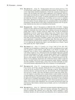
Figure 2. Antigen processing and presentation. The antigen presenting cell pre-
sents exogenous protein bound to class II molecules to CD4+ T-cells and also pro-
cesses intracellular protein and presents it with MHC class I to CD8+ T-cells. The
T-cell receptors recognize peptide bound to MHC.
7
The Allograft Immune Response
2
cell. T-cell recognition is associated with the initation of complex intracellular sig-
naling pathways that result in activation and proliferation of the T-cell (Fig. 3).
There are two classes of T-cells based on the surface expression of CD4 or CD8
molecules. CD4 and CD8 molecules bind to the same MHC on the antigen pre-
senting cell as the T-cell receptor. CD4+ cells are known as helper cells and play an
important role in initiating and directing the immune response (Fig. 4). CD8+ cells
also known as cytotoxic T-cells are responsible for cell directed cytotoxicity.
Whether a T-cell, whose receptor has bound the MHC-peptide displayed on the
antigen presenting cell, becomes activated, depends on a receiving a second set of
signals (co-stimulation) from the antigen presenting cell. These costimulatory inter-
actions act directly through cell surface receptor-ligand interactions and soluble
cytokines that are linked to intracellular signaling pathways (Fig. 4).
CD4+ T-cells are the dominant phenotype initiating acute cellular allograft re-
jection. Once activated the CD4+ T-cells undergo clonal expansion and differentia-
tion. In doing so they secrete cytokines that attract other leukocytes to activate other
T-cells and facilitate the differentiation of B-cells to plasma and memory cells.
The pattern of cytokines elaborated by subsets of CD4+ and CD8+ stimulated
T-cells is important in directing the alloimmune response. CD4 bearing Th lym-
phocytes (T helper) cells have been stratified into two classes of Th cells depending
on the type of cytokines elaborated by the cells in question. The subdivision of Th
cells is called the Th1 and Th2 paradigm. Precursor CD4 cells producing interleukin-12
promote Th 1 cells, whereas precursor CD4 cells producing interleukin-4 (IL-4)
promote Th2 cells.Th1 cells secrete IFN-γ, IL-2, which promote cell-mediated cy-
totoxicity by activating macrophages and cytotoxic T cells. Th2 cells secrete IL-4
and IL-6, cytokines which promote allergic inflammation and stimulate B cells to
produce antibodies. Furthermore, cytokines from Th 1 cells inhibit Th2 cells whereas
cytokines from Th 2 cells inhibit Th1 cells. It appears in certain experimental situa-
tions that a Th 2 predominance is associated with the prolongation of graft survival
or even tolerance (Table 2).
While much attention has been given to T-cell activation, B-cells are also in-
volved in the response. Donor antigen shed from the graft binds to surface Ig and is
then internalized by the B-cell. The antigen is processed and presented on the B-cell
Table 1. Characteristics of MHC molecules
Common features of MHC Class I and II glycoproteins
• allelic diversity
• antigen presentation
Differing characteristics
Class I MHC molecules:
• expressed on all nucleated cells
• activate T-cells bearing the CD8 surface molecule (CD8+)
Class II MHC molecules:
• expressed on dendritic cells, B-cells, and macrophages
• activate T-cells bearing the CD4 antigen (CD4+)
8
Liver Transplantation
2
surface in conjunction with class II to recruit antigen specific T-cell help. The B-cell
undergoes clonal expansion and differentiation becoming a plasma cell capable of
producing soluble Ig (Fig. 5). Other B-cells will become memory cells.
Stages of Allograft Response
The immunologic events surrounding transplantation of an allograft can be
conceptualized as a series of steps starting with changes in the graft prior to
transplantation and extending to the time of rejection.
Antigen Presentation and Allorecognition
When first transplanted, the liver allograft has the immune phenotype of the
donor. Initially therefore, the liver allograft expresses the MHC molecules of the
donor, resulting in two pathways of antigen recognition. Whereas in the non-
transplant setting, T cells recognize foreign (or non-self) peptides bound to native
(or self) MHC molecules, the MHC molecules in the allogeneic liver are non self,
and it is presumed that recipient T cells recognize intact donor MHC molecules as
non-self because their three-dimensional stoichiometry resembles a self MHC bound
to a foreign peptide, a concept referred to as molecular mimicry. This process is
called direct antigen recognition and is thought to be the main mechanism for the
immune response in acute cellular rejection. Later, there is migration of donor
dendritic cells into the host, and migration of recipient APCs into the donor liver.
This leads to a second pathway for alloimmune recognition, in which peptides derived
Figure 3. T-cell receptor: The T-cell receptor (TCR) is composed of two subunits
and is associated with CD3 proteins. Transcriptional activity is initiated in the nucleus
via signaling pathways. (NFAT = nuclear factor of activated T-cells)
9
The Allograft Immune Response
2
from catabolism of the donor MHC molecules are presented by self MHC on re-
cipient APCs.
It is unclear whether the non-immunologic injury incurred by the donor liver in
the process of organ retrieval, preservation and reperfusion contribute to the initiation
or maintenance of the alloimmune response. The period of cold preservation,
ischemia, and reperfusion leads to the differential expression of endothelial cell surface
molecules and cytokines. These include adhesion molecules, interleukins, and
Table 2. Th 1 and Th 2 paradigm
Differentiated by Cytokines Produced Function
Th1 IL-12 IL-2, IFN-γ cell mediated cytotoxicity
suppresses Th2-cell
response
Th2 IL-4 IL-4, Il-5, IL-6 suppresses Th1-cell
response
IL-10 promotes B-cell expansion
Figure 4. Cell surface proteins involved in T-cell activation. The T-cell receptor
complex, including CD3 and CD4 or CD8 bind to an APC displaying MHC and
peptide. Several costimulatory molecules such as CD28 are required for T-cell
activation.
10
Liver Transplantation
2
chemokines which attract inflammatory cells. Oxygen radicals produced during is-
chemia and reperfusion directly harm the graft.
Helper T cells are thought to be the most important cells for initiating allograft
cellular rejection. They are responsible for the production of cytokines, such as
interleukin 2, which are necessary for clonal expansion of activated lymphocytes.
The cytokines act in an autocrine fashion on CD4 expressing surface molecules (Th
cells) and as a paracrine stimulus on other effector cells such as cytotoxic T cells
(CD8 cells), macrophages and B cells.
Leukocyte Migration into the Allograft
As part of the early evolution of the allograft immune response, recipient
leukocytes are recruited to the donor allograft. This involves the elaboration of a
series of soluble molecules as well as cell-to-cell interactions. Three main classes of
receptors are credited with leukocyte migration.
Figure 5. Antibody structure. Heavy chains are in dark, light chains are white.
The antigen binding sites are composed of heavy and light chains.
11
The Allograft Immune Response
2
• Selectins: primarily responsible for allowing the leukocyte to gently ad-
here to the endothelial surface
• Integrins
• Members of the immunoglobulin superfamily: responsible for
extravasation of leukocytes into the allograft.
Graft Destruction
CD8+ T-cells are the main effectors of graft destruction and cause cell death
through direct cell contact. When activated by membrane binding to the allograft
they release cytotoxic molecules termed perforin and granzyme. Perforins create holes
in the target cell membrane and granzymes disrupt intracellular processing. Cytotoxic
T-cells also have a cell surface protein termed Fas ligand which when bound to a
receptor protein called Fas, which is present on target cells, results in death of the
target cell by the process of apoptosis.
Macrophages which have been activated by CD4+ T-cells are capable of causing
tissue destruction through the release of cytotoxic cytokines or through direct cell
lysis. The role of NK cells in organ allograft rejection is unclear. B-cells secrete specific
antibody that binds to the allograft cell surface. The antibody induces tissue damage
through the activation of the complement system (Fig. 6).
Classification of Allograft Rejection
Allograft rejection is classified into three types based on the nature of the immune
response after transplantation:
1. Hyperacute rejection
2. Acute cellular rejection
3. Chronic ductopenic rejection.
Hyperacute Rejection
Hyperacute rejection is characterized by a rapid response of the host immune
system to the allograft. Within minutes to hours of the transplant, preformed
antibodies engage class I MHC or the ABO blood group antigens on the graft. The
antibodies facilitate complement mediated lysis of the endothelium and initiate an
inflammatory cell infiltrate. Hyperacute rejection is rarely observed in liver transplants,
even among those with a positive crossmatch.
Acute Cellular Rejection
Acute cellular rejection is due to an immune reaction mediated by recipient T
lymphocytes’ response to donor MHC antigens. Antibodies and cytokines also
contribute to the immunologic attack. The biliary epithelium and venous
endothelium express MHC class I and II molecules and are the focus of the acute
cellular rejection response. Hepatocytes which express few class I or II MHC antigens
are rarely the target of acute cellular rejection.
The principal clinical features of acute cellular rejection are:
1. Elevated liver transaminases and/or bilirubin
2. Lymphocytic infiltrates in the portal triads seen on biopsy
12
Liver Transplantation
2
3. The development of graft dysfunction manifested by hyperbilirubinemia and
subsequent impaired liver synthetic function.
Acute cellular rejection is usually controlled with additional corticosteroid-based
immunosuppression with no significant impact on graft or patient survival.
Chronic Ductopenic Rejection
Chronic ductopenic rejection occurs months to years after transplantation. The
mechanisms which underlie chronic rejection in any of the solid organs are less well
understood than acute cellular rejection. Both immunologic and non-immunologic
processes are implicated. The impact of chronic rejection on the liver allograft is on
the intralobular bile ducts, a phenomenon termed the “vanishing bile duct syndrome”
and is associated with chronic graft failure. It is believed that many if not all episodes
of chronic ductopenic rejection are preceded by acute cellular rejection.
Mechanisms of Immunosuppressive Drug Action
The rational design and use of drugs is based on an understanding of the immune
response to the donor organ. These agents can be divided by classes based on their
mechanism of action. The antimetabolites include azathioprine and mycophenolate
mofetil. Both interfere with purine synthesis and clonal expansion of T- and B-cells
(Fig. 7).
Cyclosporine and tacrolimus exert their action by inhibiting calcineurin, a pro-
tein responsible for promoting cytokine induced gene activation. By inhibiting IL-
2 production they prevent activation of lymphocytes. Rapamycin (Sirolimus), one
of the most recently approved antirejection drugs, is structurally similar to tacrolimus,
and appears to inhibit the T-cell response to IL-2.
Glucocorticoids bind to cytoplasmic receptors which are translocated to the
nucleus where they regulate gene transcription by binding to specific gene regulatory
regions. They interfere with many aspects of the immune system including the
production of IL-1, IL-2, and IFN-γ.
Antibodies recognize many surface antigen epitopes (polyclonal) or single cell
surface antigen epitopes (monoclonal). Antithymocyte globulin and thymoglobulin
are two polyclonal preparations of immunoglobulin to lymphocytes. OKT-3 is
directed against the CD3 receptor on T-cells. Basiliximab and daclizumab are two
monoclonal antibodies against the α chain of the IL-2 receptor. They are used for
induction therapy and like the other antibodies result in depletion of the cells that
bear the cell surface protein which they bind.
Tolerance
The development of an immunologic state wherein the recipient is unresponsive
to donor alloantigen, but yet the immune system is capable of recognizing and
responding to other foreign proteins such as bacterial or tumor antigens without the
need for immunosuppression is known as tolerance.
Mechanisms of tolerance can be grouped into suppression, anergy, deletion, and
ignorance. Suppression involves the inhibition of donor reactive T and B-cell responses
by a “suppressor” cell population. While functional examples exist, it has been difficult
13
The Allograft Immune Response
2
identifying a suppressor cell. Anergy occurs when T-cells encounter peptide-MHC
complexes that they recognize, but the T-cell does not receive adequate co-stimulatory
signals. Deletion, the destruction of alloreactive T-cells, is likely to occur in the
thymus, and to a lesser extent in the periphery. Ignorance indicates that alloreactive
T-cells are present, but do not respond to stimuli.
Figure 6. Antibody mediated damage to graft endothelium. Recipient antibody binds
to MHC on graft endothelium. Antibody initiates graft damage through antibody
dependent cellular cytotoxicity (ADCC) and activation of the complement system.
Figure 7. Mechanism of action of commonly used immunosuppresants. Cyclosporine
(CSA), tacrolimus (FK506), mycophenolate mofetil (MMF), azathioprine (AZA),
FK506 binding protein (FKBP).
14
Liver Transplantation
2
Glossary
Acquired immunity: All immune processes utilizing immunological memory
(see below). Acquired immunity is the basis of vaccination.
Acute cellular rejection: Inflammation of the allograft elicited by genetic disparity
between the donor and recipient, primarily affecting interlobular bile ducts and
vascular endothelia, including portal veins and hepatic venules, and occasionally the
hepatic artery and its branches.
Allele: Alternative forms of the same gene
Allogeneic: Genetically dissimilar donor and recipient pair of the same species.
The converse is syngeneic.
Allotype: Antigenic determinants that differ among individuals of the same
species. Examples include different epitopes of the HLA system.
Anergy: Immunologic tolerance in which lymphocytes become functionally
unresponsive.
Antigen presenting cell (APC): Functional descriptor of specialized cells bear-
ing MHC cell surface molecules, by which they ‘present’ peptides which are the
product of intracellular degradation of exogenous proteins recognized as non-self.
Activated APCs also express co-stimulatory molecules. Macrophages and dendritic
cells are paradigmatic APCs.
Apoptosis: Also called programmed cell death. A specific form of cell death due
to enzymatic degradation of DNA, without inflammation.
B-cells: Lymphocytes capable of antibody production. Most arise from stem
cells in bone marrow. B lymphocytes produce antibodies as circulating proteins or as
stationary molecules. The latter, which constitute the B cell receptor, contain a
hydrophobic transgenic sequence which tethers the immune recognition segment
of the antibody to the cell surface membrane.
Cell mediated immunity: Immunologic response based on cellular elements of
the immune system.
CD antigen: Cell surface antigens, classified according to ‘cluster of differentiation’
(CD), in which individual molecules are assigned a CD number on the basis of their
reactivity with specific monoclonal antibodies
CD3: A complex of molecules on the cell surface of T cells, that in association
with the T cell receptor (TCR), activate intracellular signal transduction mechanisms
when the TCR binds an antigen. Blockade of CD3 by a monoclonal antibody
(Orthoclone OKT3) depletes the patient of T cells.
CD4: Cell surface molecule expressed by functionally distinct subset of T
lymphocytes. CD4 binds to an invariant part of the MHC class II molecule. CD4
bearing T cells usually act as T helper (Th) cells and recognize antigens processed by
APCs and presented in conjunction with MHC class II molecules.
CD8: Cell surface molecule expressed by functionally distinct subset of T
lymphocytes. CD8 binds to an invariant part of the MHC class I molecule. CD8
bearing T cells usually act as cytotoxic T lymphocytes (CTLs) and recognize antigens
processed by infected or injured nucleated cells and presented in conjunction with
MHC class I molecules.
15
The Allograft Immune Response
2
CD28: The best characterized co-stimulatory molecule. Cell surface molecule
expressed by T lymphocytes, activated by binding of TCR and antigen ligand. CD
28 has two known ligands (variously named B7-1 or CD80 and B7-2 or CD86)
which are expressed on the cell surface of activated APCs.
CTLA-4: Cell surface molecule, structurally similar to CD28, which also binds
B7-1 and B7-2. In contrast to the CD28-B7 interaction, linkage of CTLA-4 and B7
leads to an inhibitory signal that terminates the inflammatory response.
Chemokine: Chemotactic cytokines that regulate leukocyte transit. Each type
of leukocyte bears chemokine receptors on its cell surface that guides it to chemokines
secreted in tissues.
Chronic ductopenic rejection: Defined by two histopathological features:
obliterative vasculopathy and bile duct loss
Clone: Genetically identical cells derived from a common ancestor.
Co-stimulatory signal: Non-antigen specific interaction between lymphocytes
and antigen presenting cells which uses cell surface molecules expressed on APCs to
bind to receptors on lymphocytes (e.g., CD 28 on lymphocytes and B7-1 on APC).
Co-stimulatory signals enhance the immune response by promoting lymphocyte
clonal expansion and cytokine production and are necessary for T cell activation.
Interaction of T lymphocyte and APC in the absence of co-stimulatory signals leads
to anergy or apoptosis of the T cell. A parallel receptor ligand interaction which is
inhibitory of the immune response is described through CTLA-4.
Cytokine: A large family of low molecular weight soluble proteins involved in
regulating cellular activity. Includes the chemokines (see above).
Cytotoxic T lymphocyte (CTL): T lymphocyte that kills its target upon
recognizing complexes of peptides and MHC complexes on the target cell membrane.
Cytotoxic T cells usually express the CD8 cell surface molecule.
Epitope: The structure within an antigen that is recognized by an antigen receptor
(antibody or T cell receptor).
Graft versus host disease (GVHD): Clinical syndrome caused by immune
reaction of allogeneic lymphocytes contained within allograft tissue reacting against
alloantigens in the recipient (usually in skin, liver, and gastrointestinal tract).
Haplotype: Closely linked alleles on the same chromosome, usually inherited as
a group and linked to inheritance of some phenotypic characteristic.
Helper T cell: T lymphocytes that secrete cytokines required for the immune
function of other cells in the immune system. MosT helper T cells express the cell
surface molecule CD 4.
Human leukocyte antigens (HLA): The major histocompatibility complexes
in humans.
Humoral immunity: Immunologic response involving antibodies.
Idiotype: An antigenic determinant within the binding site of an antibody that
is recognised by another antibody.
Immunologic memory: The ability of the immune system to recall an encoun-
ter with a specific antigen, and to generate a greater response in a subsequent expo-
sure to the same alloantigen. Immunologic memory results from the generation of
16
Liver Transplantation
2
memory T and B cells during the initial encounter with an alloantigen, and is the
characteristic feature of ‘acquired immunity’.
Innate immunity: All immunologic defenses that lack immunologic memory.
The characteristic feature is that the response remains unchanged however often a
specific immunogenic moiety (immunogen) is encountered. This contrasts with
‘acquired immunity’ (see above).
Isograft: Transplant between genetically identical members of the same species
such as inborn strain of animals or twins. Also known as a syngeneic transplant.
Major histocompatibility complexes: Histocompatibility antigens expressed
on cell surfaces which are the markers by which the immune system distinguishes
self from non-self. In humans, the MHC molecules are called HLA (see above).
Memory cells: Cells with lasting response to certain immunologic epitopes.
Natural killer (NK) cell: Lymphocytes that have an innate ability to kill infected
or damaged cells, without requiring interaction with MHC surface molecules.
T-cell: Lymphocyte which undergoes selection in the thymus. T cells are the
only cells essential to the acute cellular rejection response. T cells are distinguished
by their cell surface receptor (TCR). T cells are subdivided into categories: T helper
cells and T suppressor cells.
Tolerance: An immunologic state in the absence of immunosuppression wherein
the recipient is unresponsive to donor alloantigens, while retaining the capacity to
recognize and respond to other foreign proteins such as bacteria or tumor antigens.
Vaccination: The exposure of a naïve host to a harmless version of a pathogenic
immunogen (an altered pathogen, or molecular mimic) which in turn generates
memory cells but not the pathologic consequences of the infection itself (the primary
immune response). The immune system is thus primed to deliver an enhanced
secondary immune response in the event that the host is exposed to the infectious
agent in the future.
Xenotransplantation: Transplantation across species. The graft is called a
xenograft.
Based on Delves and Roitt, New Engl J Med 2000; 343:37-49; and Sayegh and
Tu rka, New Engl J Med 1998; 338:1813-1821.
Suggested Reading
1. Delves PJ, Roitt IM. The immune system-the first of two parts. New Engl J Medi-
cine 2000; 343:37-49. The immune system-the second of two parts. New Engl J
Med 2000; 343:108-117.
2. Sayegh MH, Turka LA. The role of T-cell co-stimulatory activation pathways in
transplant rejection. New Eng J Med 1998; 338(25):1813-1821.
3. Wiesner RH, Batts KP, Krom RA. Evolving concepts in the diagnosis, pathogen-
esis, and treatment of chronic hepatic allograft rejection. Liver Trans Surg 1999;
5:388-400.
4. Wiesner RH, Demetris AJ, Belle SH, Seaberg EC, Lake JR, Zetterman RK et al.
Acute hepatic allograft rejection: incidence, risk factors, and impact on outcome.
Hepatology 1998; 28:638-645.
17
Assessment for Liver Transplantation
3
Liver Transplantation, edited by Michael R. Lucey, James Neuberger and Abraham Shaked.
©2003 Landes Bioscience.
CHAPTER 3
Assessment for Liver Transplantation
Michael R. Lucey
Selection for Liver Transplantation
Evaluation of candidates for liver transplantation can be reduced to three core
questions:
• What is the severity and prognosis of the patient’s liver disease?
• Are there confounding medical, surgical or psychological factors which
would reduce the expectation of a successful liver transplant?
• What are the wishes of the patient in regards to liver transplantation?
These questions are best addressed in a multidisciplinary process. The evaluation
may be carried out in an outpatient setting. The prospective candidate is assessed by
transplant surgeons and physicians, social workers, and selected subspecialists
including psychiatrists, cardiologists, pulmonologists and nephrologists. Previous
investigations including radiographs and biopsies are retrieved and new investigations
are ordered where necessary. When the information gathering segment of the
evaluation is complete, the patient is presented to the transplantation evaluation
committee and a decision is made regarding placement on the transplant waiting list.
Liver transplant programs must inform and educate prospective recipients and
their families of the risks and benefits of liver transplantation. It is important to
provide the patient with the opportunity to withdraw from transplant assessment if
they do not wish to proceed. Conversely, whenever the transplant program determines
that the patient is not a suitable candidate, the program should facilitate the patient
in receiving a second opinion regarding their suitability, if they should so wish.
Assessment of Severity and Prognosis
of Chronic Liver Disease
The severity of liver failure in patients with chronic liver disease can be assessed
by several models although the two models currently used are the Child-Pugh
classification and the MELD score (model for end-stage disease).
Child-Turcotte-Pugh Class (Table 1)
This scoring scheme is an empiric compilation of five features of end-stage liver
failure:
• Ascites
• Encephalopathy
18
Liver Transplantation
3
• Prothrombin time
• Serum bilirubin
• Serum albumin
It was developed originally as an instrument to predict outcome after portacaval
shunt surgery. Later Pugh modified it for a study of esophageal transection for bleeding
esophageal varices and modified the score for patients with cholestatic diseases. It
has been adopted as the most easily administered clinical tool to assess severity of
cirrhosis. Survival of cirrhotic patients declines with worsening Child’s class. The
Child’s class is useful for segregation of cirrhotic patients according to risk of dying.
It does not indicate prognosis for an individual patient with cirrhosis. Furthermore,
its origin as an empiric instrument for specific circumstances related to portal
hypertension make it less useful as a prognostic guide in many circumstances in
which liver transplantation is under consideration. These include patients with chronic
cholestatic diseases, liver tumors or fulminant liver failure. The Child-Pugh
classification has not been verified in childhood disorders.
Table 1. Child-Turcotte-Pugh classification
Points
Variable 1 2 3
Encephalopathy None Moderate Severe
Ascites None Slight Moderate
Bilirubin (mg/dl) <2 2-3 >3
Albumin (g/dl) >3.5 2.8-3.5 <2.8
Prothrombin time (sec. prolonged) <4 4-6 >6
(INR) <1.7 1.7-2.3 >2.3
Primary Biliary Cirrhosis/Primary Sclerosing Cholangitis
Bilirubin 1-4 4-10 >10
Scores are summed to determine Child’s class: A = 5-6, B = 7-9 and C = 10-15.
MELD Score
The MELD score is based on the following three variables:
• INR (International Normalized Ratio)
• Serum bilirubin
• Serum creatinine
To obtain the MELD score for any patient, access the Internet at:
www.unos.org
or: www.mayo.edu/int-med/gi/model/mayomodl-5-unos.htm
There are other prognostic scoring schemes:
• Primary biliary cirrhosis: sustained elevation of total bilirubin is the single
most influential factor in predicting outcome. Patient age, serum albumin,
prothrombin time and the presence of edema are minor influential factors.
The presence of cirrhosis is a weak prognostic factor.
• Primary sclerosing cholangitis: patient age, serum bilirubin, albumin and