Cardiology Core Curriculum A problem-based approach - part 3 pdf
Bạn đang xem bản rút gọn của tài liệu. Xem và tải ngay bản đầy đủ của tài liệu tại đây (256.21 KB, 64 trang )
Intracoronary stents
There is a wide variety of intracoronary stents available for
deployment in the coronary arteries after PTCA. These will reduce the
rate of restenosis, as compared with PTCA alone, when introduced
into vessels of sufficiently large caliber. Two randomized trials
compared Palmaz-Schatz stent placement following PTCA versus
PTCA alone for elective procedures in large vessels (≥3·0 mm) and
convincingly demonstrated that angiographic restenosis and need for
repeat interventions were lower in stented patients. This benefit was
derived at the cost of longer hospital stays and a greater incidence of
vascular complications in the stented group because of the need for
aggressive anticoagulation following stent placement. The incidence of
stent thrombosis, for which a patient is at risk for approximately
30 days following the procedure, was less than 4% in those studies.
Because stent deployment strategies now include only aspirin and
clopidogrel for 1 month instead of a combination of aspirin and
coumadin, there are currently no disadvantages to using stents, other
than cost.
Although the incidence of lesion recurrence is decreased with
stenting, in-stent restenosis is a difficult situation to treat because of
a high incidence (up to 70%) of a second restenosis, especially if the
renarrowing involves the majority of the stent length. Intravascular
radiation (brachytherapy) using either β or γ emitters have been
demonstrated to reduce conclusively the incidence of recurrent in-
stent restenosis, although at a greater risk for stent thrombosis.
Consequently, patients receiving coronary brachytherapy are treated
with aspirin and clopridogel for 6–9 months rather than the usual
30 days. A promising alternative is the use of coated stents, which
locally release antiproliferative pharmaceuticals continuously over
several weeks. Initial results using paclitaxel and sirolimus eluting
stents have shown a marked reduction in in-stent restenosis, although
further studies are needed.
Coronary atherectomy
Atherectomy refers to a procedure that employs a class of
percutaneously introduced devices to actually remove or pulverize
plaque within the coronary arteries. Like balloon angioplasty, a guide
catheter and guidewire are used with all of these devices.
Directional coronary atherectomy involves the use of a device with
a windowed cylindrical housing that is compressed against the
stenosis as the attached balloon is inflated against the opposite wall
Cardiology Core Curriculum
116
of the artery. Once positioned, a rotating metal cutting blade inside
the housing is advanced, shaving plaque from the vessel wall and
depositing the shaved debris in the catheter’s nose cone. An increased
luminal area is achieved by the dilating effect of the device itself,
angioplasty effect from balloon inflation, and the removal of
atherosclerotic material. When used for non-calcified stenoses, the
incidence of initial success, abrupt closure, and restenosis with
atherectomy is similar to that with PTCA. At present, atherectomy
offers no obvious benefit as compared with PTCA.
Transluminal extraction atherectomy is a different device that
employs a rotating conical blade that is attached to a hollow shaft
through which suction is applied. The contents of the vessel cut by the
blade are liquefied and removed by the vacuum, which is transmitted
to the tip of the device. It has been less well studied than directional
atherectomy, and appears to have a role primarily in lesions with
intracoronary thrombus or in old, degenerated saphenous vein grafts,
which are usually lined with friable atheromatous material.
Rotational atherectomy utilizes a high speed burr that pulverizes
plaque as it is passed through the diseased vessel over the guidewire.
Its utility is greatest in heavily calcified lesions, diffusely diseased
vessels, and ostial stenoses. As with transluminal atherectomy, most
lesions treated with the device are subjected to adjunctive balloon
angioplasty or stenting to achieve an optimal result. Rotational
atherectomy is utilized primarily in circumstances in which primary
PTCA is an unattractive option because of high-risk lesion
characteristics.
Case studies
Case 3.6
A 58-year-old diabetic hypertensive man with a several-year history
of exertional angina presented to the hospital with a prolonged
episode of pain at rest. He was admitted and was rendered pain-free
with the addition of intravenous nitroglycerin and heparin to his
regimen. A myocardial infarction was excluded with serial cardiac
enzymes and electrocardiograms. He had several recurrent episodes of
angina on hospital day 2 and, because of his refractory ischemia, he
was sent to the cardiac catheterization laboratory. Medications:
aspirin 325 mg/day, nitroglycerin 150 micrograms/min intravenously,
heparin 1200 U/hour intravenously, metoprolol 100 mg twice per day,
diltiazem 60 mg four times per day, and glyburide 5 mg/day.
Cardiac catheterization
117
Examination. Physical examination: the patient appeared normal.
Pulse: 54 beats/min, normal character. Blood pressure: 100/60 mmHg
in right arm. Jugular venous pulse: 6 cm. Cardiac impulse: normal.
First heart sound: normal. Second heart sound: split normally on
inspiration. No added sounds or murmurs. Chest examination:
normal air entry, no rales or rhonchi. Abdominal examination: soft
abdomen, no tenderness, and no masses. Normal liver span. No
peripheral edema. Femoral, popliteal, posterior tibial, and dorsalis
pedis pulses: all normal volume and equal. Carotid pulses: normal, no
bruits. Optic fundi: normal.
Investigations. Hematology and biochemistry: normal. Electrocardio-
gram: sinus bradycardia; PR interval 0·24 s; QRS duration 0·09 s.
Flattened T waves in leads I and aVL. During anginal episodes, 1·5 mm
of horizontal ST depression in these same leads. Chest x ray: normal
cardiac silhouette, no pulmonary venous congestion.
Cardiac catheterization. Aortic pressure: 98/60 mmHg. Left
ventricular pressure: 98/20 mmHg. Normal left ventricular wall
motion, ejection fraction 55%, no mitral regurgitation. Left
ventricular systolic and diastolic volumes were within normal limits.
The left main had a normal contour without stenoses. The left
anterior descending artery had a 30% lesion in its mid-portion, there
was a 90% lesion of the proximal left circumflex artery that measured
2·5 mm, and the right coronary artery had a 50% lesion in its distal
segment.
Questions
1. Is it reasonable to continue medical therapy alone?
2. What revascularization options can be offered to the patient?
3. Should stenting be considered as a primary modality for this
patient?
Answers
Answer to question 1
Although many patients with unstable angina
can successfully be treated with medications, this patient is
experiencing ongoing ischemia despite an aggressive regimen. One of
the objectives of medical therapy is to reduce myocardial oxygen
demand by decreasing heart rate and blood pressure. This patient
already exhibits excellent control of both parameters, and in addition
has first-degree atrioventricular block on the electrocardiogram that
may be drug induced. Increased doses of his calcium antagonist or
β-blocker may result in bradyarrhythmias and/or hypotension. The
Cardiology Core Curriculum
118
presence of electrocardiogram changes with pain also places this
patient in a high-risk subgroup for adverse events, and so an
aggressive approach in this case is most appropriate.
Answer to question 2
Using the normal threshold of a lesion of 70%
or greater as denoting a significant stenosis, this patient is classified as
having single vessel coronary disease of the left circumflex artery.
Because of his medically refractory symptoms, mechanical
revascularization is indicated. Although bypass surgery is likely to
offer symptomatic relief, in the case of single vessel disease the lower
morbidity and mortality of PTCA make it the preferred approach in
these circumstances. In the absence of the adverse lesion characteristics
mentioned above, a success rate in excess of 90% can be expected with
an approximately 30% risk of recurrent symptoms caused by restenosis
over the ensuing 6 months (Figure 3.10).
Answer to question 3
The use of intracoronary stents appears to
diminish restenosis when used in de novo (i.e. not previously dilated)
lesions. Because of the 2·5 mm caliber vessel in this case, stenting may
not be the best option for this patient. Other lesion characteristics
that may dissuade the use of stents (and that were excluded from the
studies comparing stent with PTCA) include excessive length, marked
vessel tortuosity, intracoronary thrombus, or lesion location at a
branch point or in a diffusely diseased vessel. On the other hand there
is growing evidence that in diabetic patients PTCA and stenting is
preferred to PTCA alone because of the high rate of restenosis after
PTCA.
Cardiac catheterization
119
Figure 3.10 Cineangiographic frames revealing (a) a 90% stenosis in the proximal
left circumflex coronary artery (arrow), which was (b) successfully dilated to a
10% residual stenosis. The left anterior descending artery is seen in the upper
portion of this right anterior oblique view
(a) (b)
Further reading
Bittl JA. Advances in coronary angioplasty. N Eng J Med 1996;335:1290–302.
Boehrer JD, Lange RA Willard JE, Grayburn PA, Hillis LD. Advantages and limitations of
methods to detect, localize, and quantitate intracardiac left-to-right shunting.
Am Heart J 1992;124:448–55.
Ellis SG, da Silva ER, Heyndrickx G, et al. Randomised comparison of rescue angioplasty
with conservative management of patients with early failure of thrombolysis for
acute anterior myocardial infarction. Circulation 1994;90:2280–4.
Grines RJ, Browne KF, Marco J, et al. A comparison of primary angioplasty with
thrombolytic therapy for acute myocardial infarction. N Engl J Med 1994;331:673–9.
Grossman WG, Baim DS, eds. Cardiac catheterization, angiography, and intervention,
4th ed. Philadelphia: Lea and Febiger, 1991.
King SB, Lembo NJ, Weintraub WS, et al. A randomised trial comparing coronary
angioplasty with coronary bypass surgery. N Engl J Med 1994;331:1044–50.
Nobuyoshi M, Hamasaki N, Kimura T, et al. Indications, complications, and short term
clinical outcome of percutaneous transvenous mitral commissurotomy. Circulation
1989;80:782–92.
Serruys PW, Jaegere P, Kiemeneij F, et al. A comparision of balloon expandable stent
implantation with balloon angioplasty in patients with coronary artery disease.
N Engl J Med 1994;331:489–95.
Shabetai R, Fowler NO, Guntheroth WG. The hemodynamics of cardiac tamponade and
constrictive pericarditis. Am J Cardiol 1980:46:570–5.
Topol EJ, Leya F, Pinkerton CA, et al. A comparison of directional atherectomy with
coronary angioplasty in patients with coronary artery disease. N Engl J Med 1993;
329:221–7.
Cardiology Core Curriculum
120
4: Hypertension
SHARON C REIMOLD
Hypertension is a major common cardiovascular disease, affecting
up to 75% of the population by the eighth decade of life. Although
hypertension is defined as a blood pressure exceeding 140/90 mmHg,
the cardiovascular risk associated with elevated blood pressure forms
a continuum. The primary reason for treating hypertension is to
reduce the risk for vascular complications, including hemorrhagic and
atherothrombotic stroke, congestive heart failure, coronary artery
disease, aortic dissection, sudden death, nephrosclerosis, and
peripheral vascular disease. The risks for these complications rise with
increasing severity of hypertension.
Definitions
The Joint National Committee on the Detection and Treatment of
Hypertension Guidelines
1
form the basis for diagnosis, classification
(Table 4.1),
2
and treatment of hypertension. Although hypertension is
defined as a blood pressure greater than 140/90 mmHg, the guidelines
draw attention to the population of patients with systolic blood
pressure ranging from 130 to 139 mmHg and diastolic blood pressure
from 85 to 89 mmHg as a “high normal group”. This group is
important because blood pressures in this range may be associated
with increased risk for adverse outcomes.
The most typical form of hypertension involves elevation in both the
systolic and diastolic blood pressures. Systolic hypertension, or isolated
elevation in systolic blood pressure to a level greater than
140 mmHg with normal diastolic pressure, is common in the elderly. It
is now recognized that patients with increased pulse pressure (systolic
blood pressure – diastolic blood pressure) are at higher risk for
cardiovascular complications than are those with lower pulse pressure.
3
For example, a patient with a blood pressure of 180/90 mmHg has
greater cardiovascular risk than does a patient with a blood pressure
of 150/95 mmHg.
Blood pressure is determined as the product of cardiac output and
total peripheral resistance. Early in the course of the hypertensive
disease process, cardiac output is elevated and total peripheral
resistance is essentially normal. As the disease progresses, cardiac
output normalizes but total peripheral resistance becomes elevated.
121
Blood pressure therapies can work by decreasing cardiac output or
peripheral resistance, or both.
In response to the elevated systolic arterial blood pressure, the
myocardium hypertrophies. Several electrocardiographic criteria exist
for the detection of hypertrophy. Voltage criteria are the most helpful
(Table 4.2). Left ventricular hypertrophy may be detected on
echocardiography as increased wall thickness and increased
myocardial mass. Echocardiography is more sensitive for the
detection of hypertrophy than is electrocardiography. Detection of
left ventricular hypertrophy is a marker for end-organ damage from
hypertension. This increased wall thickness may lead to systolic
and/or diastolic dysfunction of the myocardium. Individuals who
develop systolic dysfunction may have had an inadequate
hypertrophic response of the ventricle and develop decreased
myocardial contractile function in response to increased afterload.
Systolic dysfunction in hypertension may be identified as a reduction
in ejection fraction accompanied by a small increase in chamber
volumes. Advanced cases of systolic dysfunction may present with a
low cardiac output state.
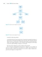
It is more common for patients with hypertension and left
ventricular hypertrophy to develop diastolic dysfunction (Figure 4.1).
4
In these patients, the relaxation or compliance of the left ventricle is
Cardiology Core Curriculum
122
Table 4.1 Classification of adult blood pressure
Category Systolic pressure (mmHg) Diastolic pressure (mmHg)
Optimal <120 <80
Normal <130 <85
High normal 130–139 85–89
Hypertension
Stage I 140–159 90–99
Stage II 160–179 100–109
Stage III ≥180 ≥110
From the Sixth Report of the Joint National Committee on Prevention, Detection,
Evaluation, and Treatment of High Blood Pressure
2
Table 4.2 Voltage criteria for left ventricular hypertrophy
Lead Criteria
Limb lead R amplitude in aVL >11 mm
R wave in aVF >14 mm
R wave in lead I plus S wave in lead III >25 mm
Precordial lead R wave in V
5
–V
6
plus S wave in V
1
>35 mm
R wave in V
5
or V
6
>26 mm
abnormal. The ejection fraction may be normal or increased with
normal chamber volumes. For these normal volumes, however, left
ventricular filling pressures are elevated, leading to pulmonary venous
congestion and symptoms of decreased exercise tolerance or dyspnea.
Patients with systolic dysfunction also have underlying diastolic
dysfunction of the ventricle. The hypertrophy produced by
hypertension predisposes patients to the development of ventricular
arrhythmias and sudden death.
Approximately 95% of all patients with high blood pressure have
essential hypertension. Essential hypertension may also be referred to
as primary or idiopathic. Although no underlying etiology is identified
for patients with essential hypertension, it is likely that multiple
factors play a role in the development of hypertension. Up to 5% of
patients have secondary hypertension. Secondary hypertension
implies that a specific etiology for the elevated blood pressure has
been identified. Treatment of secondary hypertension is based on the
specific underlying etiology (see Secondary hypertension, below).
The frequency of hypertension increases with age and varies by
race. African-American and Hispanic patients are more likely to have
hypertension before age 40 years than are Caucasian patients. Genetic
abnormalities may be responsible for the development of
hypertension in many of these patients. Only a few single gene
mutations capable of producing hypertension have been identified.
Abnormalities in the gene encoding aldosterone synthase/
11β-hydroxylase and 11β-hydroxysteroid dehydrogenase deficiency
Hypertension
123
IBP
LVH
Ventricular
arrhythmias
Diastolic
dysfunction
Systolic
dysfunction
Ejection fraction ↓
End diastolic volume ↑
LV dilation
Low cardiac
output syndrome
Ejection fraction → or ↑
End diastolic volume → or ↓
LV size normal
LV filling pressure ↑
Pulmonary venous
congestion
Dyspnea
Figure 4.1 Impact of elevated blood pressure on systolic and diastolic function of
the left ventricle. IBP, arterial blood pressure; LVH, left ventricular hypertrophy.
From Shepherd
et al
.
4
are examples of monogenic forms of hypertension. The development
of hypertension may also be polygenic, requiring several genetic
abnormalities to be present at one time. Multiple genes may
contribute to the regulation of blood pressure, and the ultimate
development of hypertension may relate to the interaction of a
genetic substrate with environmental and dietary factors.
Pathophysiology
Several mechanisms underlie the development of hypertension.
These mechanisms include sympathetic nervous system overactivity,
renin–angiotensin excess, abnormal nephron number, genetic
abnormalities, obesity, and endothelial abnormalities.
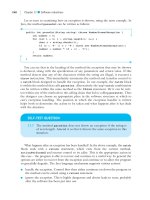
The renin–angiotensin system is important in the development of
hypertension (Figure 4.2).
5
The juxtaglomerular cells of the kidneys
secrete renin. Renin works together with renin substrate to produce
angiotensin I. Angiotensin-converting enzyme converts angiotensin I
to angiotensin II. The biologic effect of angiotensin II is to directly
produce vasoconstriction and to upregulate aldosterone synthesis.
Aldosterone facilitates sodium retention. The combined effects of
angiotensin II and aldosterone production lead to elevated blood
pressure. Elevated blood pressure results in negative feedback on renin
production by the juxtaglomerular cells. On the basis of this negative
feedback loop, it would be expected that patients with essential
hypertension would have low renin states. However, many of these
patients may actually have elevated renin states. Several explanations
for elevated renin have been suggested, including catecholamine
mediated release of renin from the kidney.
Patients with single or multiple genetic mutations may have
abnormal enzymes or proteins, which alter either sodium and volume
regulation in the kidney, sympathetic nervous system activity, or the
vasculature. Reduced nephron number may be present in patients
with low birth weight and prematurity. The reduced renal sodium
excretion and decreased filtration surface of the kidney may
predispose these patients to hypertension.
Extrinsic and intrinsic stressors lead to sympathetic nervous
overactivity. Activation of the sympathetic nervous system produces
increased contractility of the ventricle and may lead to arterial and
venous constriction. Elevated circulating catecholamine levels also
lead to increased renin release.
The association between hypertension and hyperinsulinemia is
most pronounced in patients with truncal obesity. Patients who are
obese or hypertensive may demonstrate augmented sympathetic
Cardiology Core Curriculum
124
pressor activity and/or decreased vasodilatory response to insulin.
These abnormalities may result in elevation of blood pressure in this
patient population. Non-obese hypertensive persons may have
abnormal vascular responses to circulating insulin. Increasing age is
associated with the development of hypertension. This may be related
to increased vascular stiffness that occurs with advancing age.
Other growth factors and endothelial cell dysfunction may play a role
in the control of blood pressure. Patients with high blood pressure may
fail to synthesize sufficient nitric oxide, a substance that is important
to the maintenance of vascular tone and smooth muscle cell relaxation.
In addition, patients with hypertension have abnormal nitric oxide
mediated vasodilatation. The absence of appropriate nitric oxide
mediated vasodilatation may lead to abnormal vascular remodeling and
promote vascular damage, predisposing the patient to atherosclerosis.
Nitric oxide mediated forearm vasodilatation may be normalized by
treatment with antihypertensive therapy. Endothelin and related
factors produce vasoconstriction but have not been shown to have a
role in human hypertension.
Risk factors for the development of hypertension
Genetic predisposition to hypertension is an extremely important
risk factor for the development of hypertension. Although very few
Hypertension
125
Renin
substrate
Renin J–G
Angiotensin I
converting
enzyme
Angiotensin II
Vasoconstriction
FEEDBACK
BLOOD
PRESSURE
Sodium retention
Aldosterone
synthesis
Figure 4.2 Renin–angiotensin system and its role in hypertension. J–G, renal
juxtaglomerular cell. From Kaplan
5
genetic abnormalities capable of producing hypertension have been
found, a detailed family history can often identify a familial
predisposition to hypertension, especially for the development of
high blood pressure at a young age. A detailed history should be taken
from a patient with elevated blood pressure to identify familial
clustering of hypertension.
Diet and physical activity may influence the development of high
blood pressure. Chronic ingestion of alcohol (30 g/day) has a pressor
effect. Excess sodium, low calcium, and low potassium intake are
associated with the development of hypertension. The relationship of
sodium intake to the development of hypertension is not predictable.
The response of blood pressure to sodium intake may vary between
families, suggesting an interaction between genetics and diet. Sodium
retention may be impaired in certain populations (low birth weight)
because of a deficit in nephron development.
Obesity, physical inactivity, and psychologic stress predispose
patients to the development of hypertension. Individuals with truncal
(high waist/hip ratios) obesity have a greater likelihood of developing
hypertension than do those with other distributions of obesity. Truncal
obesity is associated with insulin resistance and hyperlipidemia in the
“metabolic syndrome”. Such patients are at high risk for the subsequent
development of coronary artery disease. There is an inverse relationship
between physical activity and hypertension risk; individuals with
decreased physical activity have a higher risk for the development of
hypertension. Obesity is also associated with decreased physical
activity, and may explain a portion of the hypertensive risk in this
population.
Hypertension is more common in smokers because of enhanced
sympathetic nervous system activity provoked by nicotine. Abnormal
peripheral vascular compliance can be seen in smokers, and this may
contribute to the development of hypertension. Sleep apnea, which
is often related to obesity, is associated with hypertension. The
mechanism of hypertension in patients with sleep apnea is related to
enhanced catecholamine and possibly endothelin release in the
setting of hypoxia.
Laboratory evaluation of the patient with hypertension
Performing diagnostic testing in the patient with hypertension is
aimed at assessing overall cardiovascular risk, identifying end-
organ damage related to hypertension, and highlighting potential
secondary etiologies of hypertension. The laboratory evaluation of a
Cardiology Core Curriculum
126
patient with hypertension should include a variety of serologic tests
and an electrocardiogram (Box 4.1). Identification of end-organ
damage, such as renal dysfunction or left ventricular hypertrophy,
should lead to a decision to institute pharmacologic therapy. The
identification of glucose intolerance or dyslipidemia should prompt
patient education, dietary interventions, and/or drug therapy,
depending on the severity and type of underlying disorder. Baseline
renal dysfunction may also be the etiology rather than the result of
hypertension. Detection of renal dysfunction should lead to further
imaging or diagnostic studies to determine the etiology of the
problem. These studies can include a variety of tests, including
24-hour urine collection for protein and creatinine, renal ultrasound,
and magnetic resonance angiography.
Box 4.1 Basic laboratory evaluation of the patient with new onset
hypertension
Electrolytes
Blood urea nitrogen, creatinine
Calcium
Phosphate
Liver function tests
Glucose
Uric acid
Lipoproteins
Urinalysis
Electrocardiogram
Secondary hypertension
A small proportion of patients with hypertension (approximately
5%) has a secondary, or definable, cause for the hypertension. The
decision to pursue the evaluation of a secondary cause of
hypertension may be based on several clinical features (Box 4.2).
2
The
onset of hypertension at a young or old age is suggestive of a
secondary etiology. Severe hypertension and end-organ damage may
signal a secondary cause of hypertension. Of those patients with
secondary hypertension (Table 4.3),
2
chronic renal disease and
renovascular hypertension account for the vast majority of
underlying causes. Endocrine etiologies such as pheochromocytoma
and Cushing’s syndrome each account for 0·1–0·3% of hypertensive
cases.
Hypertension
127
Box 4.2 Clinical features of “unusual hypertension”
Diagnosis before age 20 years or after age 50 years
Blood pressure greater than 180/110 mmHg
Organ damage: creatinine ≥1·5 mg/dl, cardiomegaly, grade 2 or higher
fundoscopic findings
Features suggestive of secondary causes: unprovoked hypokalemia;
abdominal bruit; syndrome of variable pressure with tachycardia, sweating,
tremor; family history of renal disease; unequal upper/lower extremity
pressures
Poor response to therapy that is usually effective
Cardiology Core Curriculum
128
Table 4.3 Secondary causes of hypertension
Secondary cause Examples
Renal disorders Renal parenchymal disease (diabetic nephropathy,
hydronephrosis, polycystic disease, nephritis,
acute glomerulonephritis), renin producing tumors
Renovascular (renal vascular stenosis, renal
vasculitis)
Primary sodium retention (Liddle syndrome,
Gordon syndrome)
Cardiac Coarctation of the aorta
6
Neurologic disorders Increased intracranial pressure (brain tumor,
encephalitis)
Sleep apnea
Quadriplegia
Miscellaneous (acute porphyria, familial
dysautonomia, lead poisoning, Guillain–Barré
syndrome)
Pregnancy-induced
hypertension
Endocrine disorders Thyroid disorders (hypothyroidism, hyperthyroidism)
Acromegaly, hyperparathyroidism, carcinoid
Adrenal disorders: Cushing syndrome, primary
aldosteronism,
7
congenital adrenal hyperplasia,
pheochromocytoma, apparent mineralocorticoid
excess (licorice intake)
Exogenous hormones
Acute stress Surgery, burns, postresuscitation, sickle cell crisis
Alcohol and drug use
From the Sixth Report of the Joint National Committee on Prevention, Detection,
Evaluation, and Treatment of High Blood Pressure
2
Establishing the diagnosis of hypertension
The physical examination of the patient with suspected
hypertension should focus on appropriate recording of blood
pressure. The patient should be seated in a quiet place for 5 min
without exogenous adrenergic stimulants. Caffeine and smoking
should be avoided for up to 1 hour before measuring the blood
pressure. The blood pressure cuff should cover two-thirds of the
length of the arm. Using a smaller cuff results in inappropriately high
readings. At least two recordings should be made at each visit. At the
initial visit, blood pressure should be taken in both arms. The highest
arm blood pressure should be used. A minimum of three recordings
should be made at least a week apart to establish the diagnosis of
hypertension. To record the blood pressure, the cuff should be
inflated to a pressure 20 mmHg above the systolic pressure. The cuff
should be deflated at a rate of 3 mmHg/second. The disappearance of
the beat (Korotkoff phase V) should be recorded in adults.
A discrepancy between the blood pressures measured in both arms
(of more than 5–10 mmHg) may indicate involvement of the great
arteries leaving the heart in a disease process (arterial occlusive
disease, aortic dissection, or coarctation).
The radial and femoral arteries should be palpated simultaneously
during the cardiovascular examination. A significant radial–femoral
delay (the appearance of the pulses in the lower extremities is delayed
as compared with their appearance in the upper extremities) suggests
coarctation of the aorta and must be excluded. In this instance, an
initial step is to measure blood pressures in the lower extremities and
compare them with the pressures in the upper extremities. Again, if
there is a major discrepancy (i.e. if the blood pressures measured in
the lower extremities are lower than those in the upper extremities),
then coarctation of the aorta must be excluded.
Some patients have a phenomenon known as “white coat
hypertension”; in the presence of healthcare personnel, their blood
pressure is elevated. Measuring home or ambulatory blood pressures
or use of a 24-hour blood pressure monitor may provide a more
accurate assessment of blood pressure levels in these patients.
Recommendations for follow up of blood pressure have been
established (Table 4.4).
2
Patients with normal blood pressure should
have it rechecked approximately every 2 years. Because high normal
blood pressure (130–139/85–89 mmHg) is associated with an
increased risk for adverse events, lifestyle modifications should be
introduced and the blood pressure followed more closely. Patients
with significant elevation in blood pressure should be evaluated as
outlined in Box 4.1. As expected, those patients with more dramatic
elevation in blood pressure should be seen and evaluated more
Hypertension
129
promptly. Hypertensive patients with evidence of acute end-organ
involvement, including pulmonary edema, intracranial bleeding,
encephalopathy, and acute renal dysfunction, should be seen and
treated emergently.
Although the blood pressure criteria involve resting measurements,
some patients may develop a pronounced hypertensive response to
exercise. These patients have an increased likelihood of developing
hypertension subsequently and should be evaluated carefully.
Physical examination of the patient with hypertension
In addition to recording the degree of blood pressure elevation and
heart rate, eye examination with an ophthalmoscope to evaluate
blood vessels is an important aspect of the physical examination in
patients with hypertension. The most common cardiac finding in
patients with hypertension is an S
4
(fourth heart sound) gallop,
caused by the atria contracting against a stiff ventricle. Evidence of
clinical heart failure such as elevated central venous pressure, rales,
cardiac enlargement, peripheral edema, and the presence of a third
heart sound should be sought. A careful vascular examination should
be performed, looking for abdominal/renal bruits or delayed or
reduced lower extremity pulses suggestive of renal vascular disease or
Cardiology Core Curriculum
130
Table 4.4 Follow up of blood pressure based on initial blood pressure
recordings
Systolic blood pressure Diastolic blood pressure
(mmHg) (mmHg) Follow up
<130 <85 Recheck in 2 years
130–139 85–89 Recheck in 1 year;
lifestyle modifications
140–159 90–99 Confirm within
2 months; lifestyle
modifications
160–179 100–109 Evaluate or refer to
source of care within
1 month
≥180 ≥110 Evaluate or refer for
care immediately or
within 1 week,
depending on clinical
presentation
From the Sixth Report of the Joint National Committee on Prevention, Detection,
Evaluation, and Treatment of High Blood Pressure
2
aortic coarctation, respectively. Blood pressure should be recorded
from both arms and legs when assessing for a coarctation.
In patients’ with hypertension the retinal changes are classified
according to the Keith–Wagner–Barker method. These changes reflect
both atherosclerotic and hypertensive retinopathy. Initially arterial
narrowing is seen (grade 1) and subsequently the arteriolar diameters
increase in relationship to the venous diameters, manifested as
arteriovenous nicking (grade 2 changes). As uncontrolled
hypertension progresses small vessels rupture, and exudates and
hemorrhages are seen (grade 3 changes). Eventually, sustained,
accelerated, or extreme hypertension is associated with raised
intracranial pressure seen as papilledema, usually associated with
retinal exudates and hemorrhages (grade 4 changes). Grade 2 changes
correlate with other evidence of clinical cardiovascular disease or end-
organ damage (left ventricular hypertrophy, renal disease, arterial
disease). The overall risk in patients with hypertension correlates with
presence, or absence, of conventional cardiovascular risk factors and
evidence of uncontrolled hypertension or end-organ damage.
Non-pharmacologic treatment of hypertension
Dietary and environmental factors contribute to risk for developing
hypertension. By modifying these factors it is possible to decrease
blood pressure. In addition, these lifestyle modifications may be
important adjuncts to pharmacologic therapy, decreasing overall
medication requirements. Although no trials have been performed to
assess the impact of lifestyle modifications on cardiovascular end-
points or mortality, they are believed to be an important part of
antihypertensive therapy (Table 4.5).
2
Patients in the high normal
blood pressure group are likely to develop hypertension and are at risk
for complications. Lifestyle modifications should be initiated in all
patients. Drug therapy should be initiated in this population if there
is evidence for target organ damage, cardiovascular disease, or
diabetes. Patients with stage 1 hypertension should undergo a trial of
lifestyle modifications depending on risk profile. If lifestyle
modifications are not successful in decreasing blood pressure, then
pharmacologic therapy should be initiated. All patients with stage
2 and 3 hypertension should receive pharmacologic therapy.
Lifestyle modification includes a variety of changes to dietary and
physical activity (Box 4.3). Moderate sodium restriction is associated
with a reduction in blood pressure. It is recommended that sodium
intake be decreased to about 100 mmol/day. This reduction in sodium
intake is associated with a 5·4 mmHg decrease in systolic pressure and
a 6·5 mmHg decrease in diastolic pressure.
3
Individual responses to
Hypertension
131
decreased sodium intake are variable. Weight reduction is associated
with a reduction in blood pressure. Systolic blood pressure drops by
1·6 mmHg and diastolic blood pressure by 1·3 mmHg for each 1 kg
drop in body weight.
3
The diet of patients on weight loss programs
often includes decreased sodium content. This reduction in sodium
content may account for some of the blood pressure reduction in this
population. Reduction in alcohol intake is associated with a reduction
in blood pressure.
Regular moderate exercise is associated with a reduction in blood
pressure. Walking, jogging, running, swimming, and cycling are
examples of isotonic activities that may be effective in decreasing
blood pressure. Regular exercise also promotes weight loss and
increases levels of high-density lipoproteins, thereby further reducing
cardiovascular risk. Combining these modifications has been shown to
have a moderate impact on blood pressure reduction (approximately
9 mmHg fall in systolic and diastolic blood pressures).
Box 4.3 Lifestyle modifications for the treatment of hypertension
Regular moderate exercise
Weight loss
Smoking cessation
Decreased sodium content in diet
Reduced alcohol intake
Increased calcium intake
Cardiology Core Curriculum
132
Table 4.5 Strategy for lifestyle modifications and pharmacologic therapy of
hypertension
Blood pressure
stage Risk group A
*
Risk group B
†
Risk group C
‡
High normal Lifestyle Lifestyle Drug therapy
modifications modifications
Hypertension Lifestyle Lifestyle Drug therapy
stage I modifications modifications
(up to 12 months), (up to 6 months),
then drug therapy then drug therapy
Hypertension Drug therapy Drug therapy Drug therapy
stages II and III
For definition of blood pressure stages, see Table 4.1. *No cardiovascular risk
factors or disease; no target organ disease.
†
At least one risk factor, not including
diabetes; no target organ disease or clinical cardiovascular disease.
‡
Target organ
damage; clinical cardiovascular disease; diabetes with or without other risk
factors. From the Sixth Report of the Joint National Committee on Prevention,
Detection, Evaluation, and Treatment of High Blood Pressure
2
Pharmacologic treatment of hypertension
Pharmacologic therapy for hypertension is indicated for patients in
whom lifestyle modification is inadequate and for those patients with
target organ damage or other cardiovascular risk factors (see Table 4.5).
The goal of pharmacologic therapy is not only to reduce blood
pressure, but also to reduce adverse outcomes, including stroke,
coronary artery disease, congestive heart failure, and mortality. In
a meta-analysis of randomized, placebo controlled trials in
hypertension using diuretics or β-blockers,
8
both agents were effective
in reducing the risks for stroke and heart failure (Figure 4.3). The
influence of those agents on coronary artery disease was less apparent.
There was a trend for a 10% reduction in mortality due to those
agents. As new agents are evaluated for the treatment of
hypertension, the impact of therapy on stroke, heart failure, and
death is extremely important.
The target blood pressure for therapy has been debated.
Cruickshank called attention to the “J curve” associated with
hypertension.
9
This curve reflects the concept that small to moderate
blood pressure lowering is associated with a reduction in adverse
events but further blood pressure lowering is associated with an
increase in adverse outcomes. To investigate this hypothesis, the
Hypertension Optimal Treatment trial was conducted, which
included nearly 19 000 patients.
10
Patients were randomized to
groups with three different target diastolic blood pressures: 90, 85, or
80 mmHg. Felodipine, a long-acting dihydropyridine, was the
primary therapy initiated. Other agents were given as needed. A small
difference in diastolic pressure was achieved in the three treatment
groups. No detrimental effect of lowering diastolic blood pressure to
80 mmHg was noted. Diabetic patients who achieved a diastolic blood
pressure below 80 mmHg had a markedly reduced risk for major
cardiovascular events. The data from that study support lowering of
blood pressure to less than 140/90 mmHg in all persons, and to less
than 130/85 mmHg in high risk patients such as those with diabetes.
The major classes of pharmacologic agents for hypertension are
reviewed below (Table 4.6).
11,12
Diuretics
Several classes of diuretics exist. These agents may be classified
according to their site of action in the kidney. Carbonic anhydrase
inhibitors exert their action on the proximal tubule. Loop diuretics
such as furosemide are most effective for patients with renal insufficiency
or resistant hypertension. Thiazides and potassium-sparing agents
Hypertension
133
work more distally and have a major role in the treatment of
hypertension.
The initial effect of diuretics is to increase sodium excretion and
decrease plasma and extracellular fluid volume. After 6–8 weeks of
therapy, volume returns to normal but blood pressure remains
lowered because of a reduction in peripheral resistance. As blood
pressure falls and blood volume is reduced, renin and aldosterone are
secreted, blocking further excretion of sodium and reduction of blood
volume.
The net effect of diuretic administration is to lower blood pressure
by approximately 10 mmHg. The actual reduction in blood pressure
is variable and may be influenced by underlying renal function,
the response of the renin–aldosterone system to therapy, severity of
blood pressure elevation, and sodium intake. Excess sodium intake
(>8 g/day) can overwhelm the effects of these agents. The most
frequently used diuretic is hydrochlorothiazide, a thiazide diuretic.
Doses as low as 12·5 mg/day may be effective in patients with creatinine
levels of 2·0 mg/dl (177 mmol/l) or less. Patients with renal dysfunction
may benefit from one or two daily doses of a loop diuretic such as
furosemide or torsemide, or an intermittent dose of metolazone.
Diuretics may be effective single agent therapy for patients with
hypertension and are important adjuncts to other agents, increasing
the efficacy of those agents by diminishing intravascular volume.
Cardiology Core Curriculum
134
Outcome Event, active
Drug Number treatment/
Regimen Dose of trials control RR (95% CI)
RR (95% CI)
0·4 0·7 1·0
Stroke
Diuretics High 9 88/232 0·49 (0·39–0·62)
Diuretics Low 4 191/347 0·66 (0·55–0·78)
β-Blockers 4 147/335 0·71 (0·59–0·86)
HDFP High 1 102/158 0·64 (0·50–0·82)
Coronary heart disease
Diuretics High 11 211/331 0·99 (0·83–1·18)
Diuretics Low 4 215/363 0·72 (0·61–0·85)
β-Blockers 4 243/459 0·93 (0·80–1·09)
HDFP High 1 171/189 0·90 (0·73–1·10)
Congestive heart failure
Diuretics High 9 6/35 0·17 (0·07–0·41)
Diuretics Low 3 81/134 0·58 (0·44–0·76)
β-Blockers 2 41/175 0·58 (0·40–0·84)
Total mortality
Diuretics High 11 224/382 0·88 (0·75–1·03)
Diuretics Low 4 514/713 0·90 (0·81–0·99)
β-Blockers 4 383/700 0·95 (0·84–1·07)
HDFP High 1 349/419 0·83 (0·72–0·95)
Treatment Treatment
better worse
Figure 4.3 Influence of diuretics and β-blockers on stroke, coronary heart disease,
congestive heart failure, and total mortality in patients with hypertension. From
Psaty
et al .
8
Table 4.6 Drugs used in the treatment of hypertension
Usual dose range, total
Drug
Trade name
mg/day (frequency/day) Selected side effects and comments
Diuretics (partial list)
Chlorthalidone
Hydrochlorothiazide (G)
Indapamide
Metolazone
Loop diuretics
Bumetanide (G)
Ethacrynic acid
Furosemide (G)
Torsemide
Potassium-sparing agents
Amiloride hydrochloride (G)
Spironolactone (G)
Triamterene (G)
Adrenergic inhibitors
Peripheral agents
Guanadrel
Guanethidine monosulfate
Reserpine (G)
Hygroton
Hydrodiuril, Microzide,
Esidrix
Lozol
Zaroxolyn
Bumex
Edecrin
Lasix
Demadex
Midamor
Aldactone
Dyrenium
Hylorel
Ismelin
Serpasil
12·5–50 (1)
12·5–50 (1)
1·25–5 (1)
2·5–10 (1)
0·5–4 (2–3)
25–100 (2–3)
40–240 (2–3)
5–100 (1–2)
5–10 (1)
25–100 (1)
25–100 (1)
10–75 (2)
10–150 (1)
0·05–0·25 (1)
Short-term: increase cholesterol and glucose
levels; biochemical abnormalities;
decrease potassium, sodium, and
magnesium levels; increase uric and
calcium levels. Rare: blood dyscrasias,
photosensitivity, pancreatitis,
hyponatremia
(Less or no hypercholesterolemia)
(Short duration of action, no hypercalcemia)
(Only non-sulfonamide diuretic, ototoxicity)
(Short duration of action, no hypercalcemia)
(Gynecomastia)
(Postural hypotension, diarrhea)
(Postural hypotension, diarrhea)
(Nasal congestion, sedation, depression,
activation of peptic ulcer)
Continued
Table 4.6 Continued
Usual dose range, total
Drug
Trade name
mg/day (frequency/day) Selected side effects and comments
Central
α-agonists
Clonidine hydrochloride (G)
Guanabenz acetate (G)
Guanfacine hydrochloride (G)
Methyldopa (G)
α-Blockers
Doxazosin mesylate
Prazosin hydrochloride (G)
Terazosin hydrochloride
β-Blockers
Acebutolol
Atenolol (G)
Betaxolol
Bisoprolol fumarate
Carteolol hydrochloride
Metoprolol tartrate (G)
Metoprolol succinate
Nadolol (G)
Penbutolol sulfate
Catapres
Wytensin
Tenex
Aldomet
Cardura
Minipress
Hytrin
Sectral
Tenormin
Kerlone
Zebeta
Cartrol
Lopressor
Toprol-XL
Corgard
Levatol
0·2–1·2 (2–3)
8–32 (2)
1–3 (1)
500–3000 (2)
1–16 (1)
2–30 (2–3)
1–20 (1)
200–800 (1)
25–100 (1–2)
5–20 (1)
2·5–10 (1)
2·5–10 (1)
50–300 (2)
50–300 (1)
40–320 (1)
10–20 (1)
Sedation, dry mouth, bradycardia, withdrawal
hypertension
(More withdrawal)
(Less withdrawal)
(Hepatic and “autoimmune” disorders)
Postural hypotension
Bronchospasm, bradycardia, heart failure;
may mask insulin-induced hypoglycemia.
Less serious: impaired peripheral
circulation, insomnia, fatigue, decreased
exercise tolerance, hypertriglyceridemia
(except agents with intrinsic
sympathomimetic activity)
Continued
Table 4.6 Continued
Usual dose range, total
Drug
Trade name
mg/day (frequency/day) Selected side effects and comments
Pindolol (G)
Propranolol (G)
Timolol maleate (G)
Combined
α- and β-blockers
Carvedilol
Labetalol hydrochloride (G)
Direct vasodilators
Hydralazine hydrochloride (G)
Minoxidil (G)
Calcium channel blockers
Non-dihydropyridine
Diltiazem hydrochloride
Mibefradil dihydrochloride (T-channel
calcium channel blocker)
Verapamil hydrochloride
Dihydropyridine
Amlodipine besylate
Felodipine
Visken
Inderal
Inderal LA
Biocadren
Coreg
Normodyne, Trandate
Apresoline
Loniten
Cardizem SR
Cardizem CD
Dilacor XR, Tiazac
Posicor
Isoptin SR, Calan SR
Verelan, Covera HS
Norvasc
Plendil
10–60 (2)
40–480 (2)
40–480 (1)
20–60 (2)
12·5–50 (2)
200–1200 (2)
50–300 (2)
5–100 (1)
120–360 (2)
120–360 (1)
50–100 (1)
90–480 (2)
120–480 (1)
2·5–10 (1)
2·5–20 (1)
Postural hypotension, bronchospasm
Headaches, fluid retention, tachycardia
(Lupus syndrome)
(Hirsutism)
Conduction defects, worsening in systolic
dysfunction, gingival hyperplasia
(Nausea, headache)
(No worsening of systolic dysfunction;
contraindicated with terfenadine
[Seldane], astemizole [Hismanal], and
cisapride [Propulsid]. Removed from the
market in the USA)
(Constipation)
Edema of the ankle, flushing, headache,
gingival hypertrophy
Continued
Table 4.6 Continued
Usual dose range, total
Drug
Trade name
mg/day (frequency/day) Selected side effects and comments
Isradipine
Nicardipine
Nifedipine
Nisoldipine
Angiotensin-converting enzyme
inhibitors
Benazepril hydrochloride
Captopril (G)
Enalapril maleate
Fosinopril sodium
Lisinopril
Moexipril
Quinapril hydrochloride
Ramipril
Trandolapril
Angiotensin II receptor blockers
Losartan potassium
Valsartan
Irbesartan
DynaCirc
DynaCirc CR
Cardene SR
Procardia XL, Adalat
CC
Sular
Lotensin
Capoten
Vasotec
Monopril
Prinivil, Zestril
Univasc
Accupril
Altace
Mavik
Cozaar
Diovan
Avapro
5–20 (2)
5–20 (1)
60–90 (2)
30–120 (1)
20–60 (1)
5–40 (1–2)
25–150 (2–3)
5–40 (1–2)
10–40 (1–2)
5–40 (1)
7·5–15 (1–2)
5–80 (1–2)
1·25–20 (1–2)
1–4 (1)
25–100 (1–2)
80–320 (1)
150–300 (1)
Common: cough. Rare: angioedema,
hyperkalemia, rash, loss of taste,
leukopenia
Angioedema (very rare), hyperkalemia
These dosages may vary from those listed in the
Physicians’ desk reference 51st ed
.
11
which may be consulted for additional information.
The listing of side effects is not all-inclusive, and side effects ar
e for the class of drugs except where noted for individual drugs (in
parentheses); clinicians are urged to refer to the package insert for a mor
e detailed listing. (G) indicates generic available.
Adverse metabolic effects of diuretics include elevated glucose,
cholesterol, and uric acid levels. In those individuals who develop
gout as a result of diuretic-induced hyperuricemia, long-term therapy
with a uricosuric agent may be beneficial.
Hypokalemia may be seen with loop diuretics or thiazides due to
kaliuresis. The degree of hypokalemia is generally related to the dose of
the agent. Low doses of hydrochlorothiazide (12·5 mg/day) may not be
accompanied by significant potassium losses. Because hypokalemia may
blunt the blood pressure lowering effect of these agents as well as lead
to ventricular ectopy, muscle weakness, and leg cramps, prevention and
treatment of hypokalemia is important. Some of these patients may
develop hypomagnesemia. Repletion of magnesium may be necessary
if hypomagnesemia develops. Strategies for preventing potassium
depletion include using a low dose of diuretic, restricting sodium intake
(100 mmol/day), increasing dietary potassium intake, or use of
additional agents such as a potassium-sparing diuretic, angiotensin-
converting enzyme inhibitor, or angiotensin II receptor blocker.
Potassium-sparing therapy should be used with caution in patients with
underlying renal dysfunction or in those on other therapies associated
with elevated potassium levels such as angiotensin-converting enzyme
inhibitors, angiotensin receptor blocking agents, or non-steroidal anti-
inflammatory drugs. Potassium-sparing diuretics include spironolactone
(an aldosterone antagonist), and triamterene and amiloride (direct
inhibitors of potassium secretion).
Adrenergic inhibitors
Several classes of agents that inhibit the adrenergic nervous system
are available to treat hypertension. Reserpine and guanethidine are
peripheral neuronal inhibitors. Reserpine inhibits the uptake of
norepinephrine (noradrenaline) into storage vesicles within the
ganglion. Guanethidine inhibits the release of norepinephrine from
the adrenergic neurons but is not commonly used because of its
association with orthostatic hypotension.
Central α-agonists work by stimulating central adrenergic receptors,
reducing sympathetic outflow from the central nervous system. This
action results in a decrease in peripheral vascular resistance, thereby
reducing systemic blood pressure. Drugs in this class include
methyldopa, clonidine, and guanabenz. Methyldopa is used
infrequently because of the incidence of abnormal liver function tests;
however, it remains an important and relatively safe form of
antihypertensive therapy during pregnancy.
The mechanism of action of α-adrenergic receptor antagonists is the
blockade of presynaptic or postsynaptic receptors. Phenoxybenzamine
Hypertension
139
and phentolamine block presynaptic and postsynaptic α-receptors.
Blockage of presynaptic receptors leads to stimulation of norephinephrine
release from the neuron. This increased norepinephrine release may
overwhelm the postsynaptic receptors, blunting the effect of the agents.
Phenoxybenzamine and phentolamine are used infrequently because of
side effects and limited efficacy. Phentolamine may be used in
hypertensive crises.
Examples of selective postsynaptic α-receptor blockers include
prazosin, terazosin, and doxazosin. The postsynaptic α-receptor
mediates vasoconstriction. By blocking these receptors, a decrease in
peripheral resistance is seen because of arterial and venous dilatation.
Norepinephrine release from the neuron is inhibited by the intact
α
2
-receptor on the neuron. This inhibition of norepinephrine release
may be responsible for the first-dose hypotensive responses observed
with prazosin. Terazosin and doxazosin have a slower onset of effect
and longer duration of action. These agents are less likely to produce
orthostasis. These agents are effective in lowering blood pressure in
patients on multiple agents and in those with renal dysfunction. They
also reduce smooth muscle tone in the bladder and prostate, and have
been used extensively in patients with benign prostatic hypertrophy.
The most common side effects are weakness, fatigue, headaches,
and dizziness. The ongoing Antihypertensive and Lipid-Lowering
Treatment to Prevent Heart Attack Trial is a double-blind, randomized
trial of antihypertensive therapy in patients older than 55 years.
13
Compared with cholorthalidone which was superior to other agents
in lowering systolic blood pressures, and was cheaper, doxazosin had
25% more cardiovascular events and caused more heart failure and
was not recommended as a first choice agent to lower blood pressure.
β-Adrenergic blocking agents are a popular class of antihypertensive
agent. These agents are associated with a reduction in mortality if
taken at the time of or after a myocardial infarction, and have now
been shown to decrease mortality in patients with congestive heart
failure. In large clinical trials of hypertension, however, diuretics are
better at reducing the likelihood of a primary cardiac event than are
β-blockers.
8
β-Blockers work by reducing cardiac output. These agents
reduce renin levels up to 60% by blocking the sympathetic release of
renin by the juxtaglomerular cells. These agents may also block
central nervous system sympathetic release.
β-Blockers differ in their lipid solubility, intrinsic sympathomimetic
activity, and cardioselectivity. The antihypertensive effect of the
various agents does not depend on lipid solubility. Nadolol and
atenolol have reduced lipid solubility and cross the blood–brain
barrier to a lesser extent than do other agents, thereby reducing
central nervous system side effects. β-Blocking agents with intrinsic
Cardiology Core Curriculum
140
