Hemostasis and Thrombosis - part 8 pps
Bạn đang xem bản rút gọn của tài liệu. Xem và tải ngay bản đầy đủ của tài liệu tại đây (170.06 KB, 23 trang )
147
Antithrombotic Therapy for Cardiac Disease
20
in Table 20.6. It is relatively safe for short periods of time to stop anticoagulation.
One should avoid large doses of vitamin K (greater than 1-2 mg) for reversal of
warfarin anticoagulation. The higher doses of vitamin K are no more effective than
lower doses, and when the patient needs to be restarted on warfarin it will take a
much longer time to reach a therapeutic prothrombin time when high doses of
vitamin K have been used.
Bioprosthetic Heart Valves
Although the risk is lower, bioprosthetic hearts valves still have a definite risk of
associated embolization. This is highest immediately after surgery and in patients
with bioprosthetic valves who have other risk factors such as atrial fibrillation. There-
fore patients with a new bioprosthetic valve should be anticoagulated for 3 months
after surgery with warfarin to an INR of 2.0-3.0. Furthermore, patients with atrial
fibrillation, history of embolism or those with left atrial thrombi should be antico-
agulated indefinitely with warfarin.
Chronic Heart Failure
Patients with left ventricular ejection fractions under 30% associated with global
dysfunction have rates of stroke of 3-4%/year. In addition, these patients are also at
risk for venous thrombosis with as many as 20% of these patients dying of venous
Table 20.5. Risk stratification and therapy of mechanical valve patients
High Risk
Treat to INR 2.5 - 3.5 + ASA 80-100mg/day.
Valve implanted before 1980
Previous embolism
Vascular disease
Risk of stroke >2%/yr on warfarin alone
Medium Risk
INR 2.5 - 3.5
Low Risk
INR 2-3
Bileaflet valve in aortic position
Treatment and Risk Stratification of Bioprosthetic Valves
AVR or MVR: INR 2.5 - 3.5 for 3 months then ASA
+ a fib: INR 2-3
+ hx embolism, vascular disease or LA thrombus:
INR 2.5-3.5 + ASA 80 - 100 mg/day
Table 20.6. Risk(%) for embolic events when off anticoagulation (Pauker, JAMA)
Aortic Position One Day Two Weeks One Year
Ball 0.03 0.44 11
Bjork 0.03 0.38 10
St Jude 0.02 0.21 5
Mitral Position
Ball 0.08 1.13 30
Bjork 0.05 0.74 19
St Jude 0.03 0.48 13
148
Hemostasis and Thrombosis
20
thrombotic disease. Unfortunately this group of patients has not been well-studied,
but patients with global cardiac dysfunction should be considered candidates for
anticoagulation, especially if they have had previous emboli.
Suggested Reading
1. Albers GW, Dalen JE, Laupacis A et al. Antithrombotic therapy in atrial fibrilla-
tion. Chest 2001; 119(1 Suppl):194-206.
2. Antman EM. ‘I can see clearly now’: a new view on the use of IV GP IIb/IIIa
inhibitors in acute coronary syndromes. Eur Heart J 2002; 23(18):1408-11.
3. Bloomfield P. Choice of heart valve prosthesis. Heart 2002; 87(6):583-9.
4. Braunwald E, Antman EM, Beasley JW et al. ACC/AHA 2002 guideline update
for the management of patients with unstable angina and non-ST-segment eleva-
tion myocardial infarction—summary article: J Am Coll Cardiol 2002;
40(7):1366-74.
5. Brogan GX Jr. Bench to bedside: pathophysiology of acute coronary syndromes
and implications for therapy. Acad Emerg Med 2002; 9(10):1029-44.
6. Chan AW, Moliterno DJ. Defining the role of abciximab for acute coronary syn-
dromes: lessons from CADILLAC, ADMIRAL, GUSTO IV, GUSTO V, and
TARGET. Curr Opin Cardiol 2001; 16(6):375-83.
7. Choo JK, Young JJ, Kereiakes DJ. A guide to drug use during percutaneous coro-
nary intervention. Drugs 2002; 62(18):2589-601.
8. Fuster V, Ryden LE, Asinger RW et al. ACC/AHA/ESC guidelines for the man-
agement of patients with atrial fibrillation. Eur Heart J 2001; 22(20):1852-923.
9. Hart RG, Palacio S, Pearce LA. Atrial fibrillation, stroke, and acute antithrombotic
therapy: analysis of randomized clinical trials. Stroke 2002; 33(11):2722-7.
10. Kereiakes DJ, Montalescot G, Antman EM et al. Low-molecular-weight heparin
therapy for non-ST-elevation acute coronary syndromes and during percutaneous
coronary intervention: an expert consensus. Am Heart J 2002; 144(4):615-24.
11. McKay RG, Boden WE. Small peptide GP IIb/IIIa receptor inhibitors as upstream
therapy in non-ST-segment elevation acute coronary syndromes: results of the
PURSUIT, PRISM, PRISM-PLUS, TACTICS, and PARAGON trials. Curr Opin
Cardiol 2001; 16(6):364-9.
12. Mukherjee D, Topol EJ. The role of low-molecular-weight heparin in cardiovascu-
lar diseases. Prog Cardiovasc Dis 2002; 45(2):139-56.
13. Ohman EM, Harrington RA, Cannon CP et al. Intravenous thrombolysis in acute
myocardial infarction. Chest 2001; 119(1 Suppl):253-277.
14. Osula S, Bell GM, Hornung RS. Acute myocardial infarction in young adults:
causes and management. Postgrad Med J 2002; 78(915):27-30.
15. Stein PD, Alpert JS, Bussey HI et al. Antithrombotic therapy in patients with
mechanical and biological prosthetic heart valves. Chest 2001; 119(1
Suppl):220-227.
16. Crowther MA, Ginsberg JS, Julian J et al. A comparison of two intensities of war-
farin for the prevention of recurrent thrombosis in patients with the antiphos-
pholipid antibody syndrome. N Engl J Med 2003; 349(12):1133-8.
17. Hanly JG. Antiphospholipid syndrome: an overview. CMAJ 2003; 16(13):1675-82.
18. Kearon C, Ginsberg JS, Kovacs MJ et al. Extended low-intensity anticoagulation
for thrombo-embolism investigators. Comparison of low-intensity warfarin therapy
with conventional-intensity warfarin therapy for long-term prevention of recur-
rent venous thromboembolism. N Engl J Med 2003; 349(7):631-9.
CHAPTER 21
Hemostasis and Thrombosis, 2nd Edition, by Thomas G. DeLoughery.
©2004 Landes Bioscience.
Stroke and Peripheral Vascular Disease
Stroke
Cerebrovascular disease may be caused by atherosclerosis, embolism, or unusual
causes such as vasculitis. Most strokes are due to either atherosclerosis or embolism
and discussion here will focus on these causes. However, the clinician must be alert
to more unusual causes of stroke in selected cases.
Acute Stroke
The patient with acute neurological defects demands a rapid decision as to whether
the defect is ischemic in nature and, if so, what therapy should be instituted (Table
21.1). Patients with symptoms less than three hours old are candidates for throm-
bolytic therapy. Evaluation of these patients should be rapid. Patients should un-
dergo a CT scan to rule out hemorrhage or mass lesion. Evaluation for an underlying
systemic process should also be undertaken.
Patients who meet rigorous NINDS trial criteria should be considered for throm-
bolytic therapy (Table 21.2). Thrombolytic therapy is believed to be associated with
an improvement in clinical outcome but the rate of intracranial hemorrhage is high
and patient selection is crucial. The rate of bleeding complications is increased in
patients who did not meet the NINDS criteria but still received thrombolytic therapy.
Absolute contraindications to thrombolytic therapy include CT evidence of intrac-
ranial hemorrhage, systolic blood pressure greater than 185 mmHg or diastolic greater
than 110 mm Hg, or a seizure with or before stroke onset. Patients who undergo
thrombolytic therapy should receive tPA 0.9 mg/kg (maximum 90 mg) with ten
percent of the dose given in one minute. The rest should be given over one hour.
Patients who receive thrombolytic therapy should not receive any other form of
anticoagulation including aspirin for 24 hours. These patients should be carefully
monitored for signs of bleeding.
Table 21.1. Antithrombotic therapy of cerebrovascular disease
Transient Ischemic Attack
Ipsilateral carotid stenosis > 69%: endarterectomy
Others: aspirin 75-160 mg/day or clopidogrel in aspirin-intolerant patients
Acute Stroke
Thrombolytic candidates: tPA 0.9 mg/kg (maximum 90 mg) with ten percent of the
dose given in one minute
Embolic stroke: anticoagulation with heparin after 12-24 hours in patients with no
significant hemorrhage
All others: aspirin 160 mg/day or clopidogrel in aspirin-intolerant patients
150
Hemostasis and Thrombosis
21
Patients who do not undergo thrombolytic therapy and do not have an obvious
embolic source for their stroke should be started on aspirin. Two large trials have
shown a small but real benefit of aspirin in preventing death or disability. Since
patients with stroke also have risk factors for ischemic heart disease the aspirin will
be of benefit for this as well. Clopidogrel can be substituted in the aspirin-intolerant
patient. Currently the combination of aspirin and clopidogrel is being examined in
a large trial to see if it has any advantage over single drug therapy.
Recently a trial using a novel sustained-release form of dipyridamole and aspirin
showed greater benefit in secondary stroke prevention than with aspirin alone. How-
ever, the results of this isolated trial have been controversial and in this trial the
combination pill had no effect on cardiovascular events. Until further data is avail-
able from an ongoing trial, the role of dipyridamole in stroke prevention remains
unknown.
Patients who clearly have an embolic source for their stroke require life-long anti-
coagulation. The timing of the initiation of heparin therapy is controversial, but
patients with small strokes without significant hemorrhage should be started on
heparin within 24 - 48 hours of the event and maintained on warfarin at an INR of
2-3.
It is not clear how long the patient who suffers an intracranial hemorrhage on
warfarin should remain off this drug. Recent data suggest that one to two weeks off
warfarin may be appropriate if the patient has a very strong indication for antico-
agulation such as a mechanical valve. One should carefully investigate the circum-
stances around the time of the bleed to see if there are any reversible factors such as
a very high INR. Given the lack of data, treatment should be tailored to the indi-
vidual patient’s circumstances.
Table 21.2. NINDS trial criteria for tPA
Patients Must Have All the Following
An ischemic stroke with a clearly defined time of onset,
A deficit measurable on the NIHSS (National Institutes of Health Stroke Scale), and
Baseline computed tomographic (CT) scan of the brain showing no evidence of
intracranial hemorrhage.
Exclusions (Any of the following)
Stroke or serious head trauma within the preceding 3 months;
Major surgery within the prior 14 days;
History of intracranial hemorrhage;
Systolic blood pressure above 185 mm Hg or diastolic blood pressure above
110 mm Hg;
Rapidly improving or minor symptoms;
Symptoms suggestive of subarachnoid hemorrhage;
Gastrointestinal hemorrhage or urinary tract hemorrhage within the previous 21 days;
Arterial puncture at a noncompressible site within the previous 7 days;
Seizure at the onset of stroke;
Patient on oral anticoagulants with INR >1.7;
Patients on heparin within the previous 48 hours and still with an elevated aPTT;
Platelet count below 100,000;
Prothrombin time higher than 15 seconds;
Glucose concentration below 50 mg/dl or above 400 mg/dl.
N Engl J Med 1995; 333(24):1581-7.
151
Stroke and Peripheral Vascular Disease
21
A frequent cause of morbidity and mortality in the stroke patient is deep venous
thrombosis and pulmonary embolism. Stroke patients should receive prophylaxis
with low molecular weight heparin which has been shown to be safe and effective in
these patients and does not increase the risk of bleeding.
Transient Ischemic Attacks
Patients with a transient neurological syndrome should be evaluated for the pres-
ence of carotid stenosis. Patients with ipsilateral carotid stenosis of over 70% should
receive endarterectomy if they are surgical candidates. All patients with TIA should
be started on aspirin. Clinical trials have shown that 30-75 mg of aspirin is equiva-
lent to higher doses in secondary prevention. For patients who are intolerant of
aspirin or who are aspirin failures clopidogrel is of benefit. Patients with atheroscle-
rosis should also receive aggressive treatment of risk factors such as adverse lipid
profiles and smoking.
Patients with Recurrent Strokes
Except for a small effect of aspirin, there is no good strategy for secondary pre-
vention of nonembolic stroke. Patients who fail antiplatelet therapy are often placed
on warfarin. Warfarin at high INRs (3-4.5) is associated with a high rate of intracra-
nial bleeding and should not be used. A trial looking at lower INRs (1.4- 2.8) also
demonstrated that warfarin fails to offers any benefit over aspirin in the secondary
prevention of non-embolic strokes. Currently a clinical trial is underway to see if
warfarin at an INR 2-3 is effective for stroke prevention. As noted above, trials are
ongoing to study novel approaches to antiplatelet therapy.
Patent Foramen Ovale and Stroke
Patent foramen ovale (PFO) exists in 20% of normal individuals and the inci-
dence in young stroke patients, especially those with idiopathic stroke, may be as
high as 60%. Much controversy exists over the value of diagnosing PFO and ap-
proach to management. In general the PFO is more likely to be a source of embo-
lism if:
• There is no evidence of atherosclerosis;
• There is a source of venous thrombosis;
• MRI shows areas of multiple infarcts;
• The PFO shows significant shunting;
• There is the presence of atrial septal aneurysm.
Management options for PFO are to 1) close the PFO either surgically or with
catheter devices, 2) use aspirin, or 3) use warfarin. Presently there is much interest in
transcatheter closure devices. However, studies demonstrate a residual risk of throm-
bosis and if the patient has an underlying hypercoagulable state, closure alone is not
adequate therapy. Although data is conflicting, a meta-analysis has demonstrated
warfarin still should be considered for patients with presumed embolic stroke and
PFO.
Stroke in Young Patients
Patients under age 50 with a stroke should receive aggressive evaluation (Table
21.3). In younger patients, premature atherosclerosis and embolism are still the two
most common causes of stroke. Patients should undergo a transesophageal
echocardiogram to investigate for any underlying cardiac source of stroke.
152
Hemostasis and Thrombosis
21
Management of patients who have an embolic source of stroke and abnormali-
ties on echocardiography is still controversial. The rate of recurrent stroke in pa-
tients with patent foreman ovale is approximately 1-2% per year. Therapeutic options
include surgical correction or life-long anticoagulation. Patients with large defects,
more than one event, or posterior circulation events appear to be at higher risk of
recurrence.
Patients with premature strokes should receive a limited evaluation for hyperco-
agulable states. There is no convincing evidence that deficiency of protein S, protein
C, antithrombin or the presence of the factor V Leiden mutation increases risk of
stroke. Patients should be evaluated for the presence of antiphospholipid antibod-
ies, obtain a full lipid profile and have a homocysteine level determined. Given the
association with premature atherosclerosis, levels of lipoprotein(a) should also be
determined.
Peripheral Vascular Disease
Acute Ischemia
Patients who present with an acute occlusion of the arterial blood supply of a
limb require rapid intervention to save the limb. The time window is only four to six
hours from onset of ischemia for limb salvage. Patient may suffer either an embo-
lism or sudden thrombosis of a pre-existent atherosclerotic area. Patients with em-
bolic disease often just require removal of the thrombus. Patients with thrombosis
over atherosclerosis often require re-vascularization; therefore, differentiating be-
tween these two entities is important in order to formulate therapy. Patients who
have embolic occlusion have sudden onset of symptoms and rarely have pre-existing
symptoms. Often there will an obvious source of the embolism such as atrial fibril-
lation. Patients with underlying atherosclerosis will have previous symptoms of pe-
ripheral vascular disease.
The presentation of acute ischemia is the classic “Five P’s”: Pain, Pallor, Paralysis
Paresthesia, and Pulselessness. The affected limb should undergo evaluation to de-
termine the degree of ischemia. Patients with mild weakness and sensory loss but
without profound paralysis of the limb need emergent therapy to salvage the limb.
The patient should undergo rapid evaluation for systemic disease. Although embo-
lic occlusion can often be diagnosed on clinical grounds, angiography is indicated in
many cases to determine the underlying cause by either demonstrating diffuse ath-
erosclerotic disease or a discrete embolism.
Patients with acute ischemia require rapid anticoagulation with heparin. Patients
with embolic disease and salvageable limbs require embolectomy via a Fogarty cath-
eter. Catheter- based thrombolytic therapy is required for thrombi that cannot be
removed with the catheter or for multiple small vessel thrombi. Patients with embolic
Table 21.3. Evaluation of the young patient with stroke
• Angiogram
• Antiphospholipid antibodies
• Lipoprotein (a)
• Plasma homocysteine
•Transesophageal echocardiogram
154
Hemostasis and Thrombosis
21
Patients who have thrombosed their bypass grafts are often treated with warfarin
anticoagulation with or without aspirin. Patients whose grafts fail due to thrombosis
and not due to technical reasons or fibrous hyperplasia should be given a trial of
warfarin combined with low-dose aspirin.
Suggested Reading
1. Albers GW, Amarenco P, Easton JD, Sacco RL, Teal P. Antithrombotic and throm-
bolytic therapy for ischemic stroke. Chest 2001; 119(1 Suppl):300-320.
2. Chant H, McCollum C. Stroke in young adults: the role of paradoxical embolism.
Thromb Haemost 2001; 85(1):22-9.
3. Hiatt WR. Medical treatment of peripheral arterial disease and claudication. N
Engl J Med 2001; 344(21):1608-21.
4. Jackson MR, Clagett GP. Antithrombotic therapy in peripheral arterial occlusive
disease. Chest 2001; 119(1 Suppl):283-299.
5. Johnston SC. Clinical practice. Transient ischemic attack. N Engl J Med 2002;
347(21):1687-92.
6. Klausner HA, Lewandowski C. Infrequent causes of stroke. Emerg Med Clin North
Am 2002; 20(3):657-70.
7. MacWalter RS, Shirley CP. A benefit-risk assessment of agents used in the second-
ary prevention of stroke. Drug Saf 2002; 25(13):943-63.
8. Makin AJ, Silverman SH, Lip GY. Antithrombotic therapy in peripheral vascular
disease. BMJ 2002; 325(7372):1101-4.
9. Mohr JP, Thompson JL, Lazar RM et al. A comparison of warfarin and aspirin for
the prevention of recurrent ischemic stroke. N Engl J Med 2001; 345(20):1444-51.
10. O’Keeffe ST, Woods BO, Breslin DJ et al. Blue toe syndrome. Causes and man-
agement. Arch Intern Med 1992; 152(11):2197-202.
11. Schievink WI. Spontaneous dissection of the carotid and vertebral arteries. N Engl
J Med 2001; 344(12):898-906.
12. Straus SE, Majumdar SR, McAlister FA. New evidence for stroke prevention: sci-
entific review. JAMA 2002; 288(11):1388-95.
13. Olsson SB. Stroke prevention with the oral direct thrombin inhibitor ximelagatran
compared with warfarin in patients with non-valvular atrial fibrillation (SPORTIF
III): randomized controlled trial. Lancet 2003; 362(9397):1691-1698.
Table 21.4. Blue toe syndrome (after O’Keefe)
Atheroembolism
Platelet aggregates
Cholesterol crystals
Warfarin-related cholesterol embolism
Cardiac Embolism
Infective endocarditis
Non-thrombotic endocarditis
Cardiac myxoma
Atrial fibrillation
Prosthetic valve embolism
Hyperviscocity Syndromes
Cryoglobulinemia
Cryofibrinogen
Cold agglutinins
Polycythemia rubra vera
Leukemias
Macroglobulinemia
Hypercoagulable States
Malignancy
Diabetes
Antiphospholipid antibodies
Essential thrombocytosis
Erythromelalgia
Disseminated intravascular
coagulation
Deep venous thrombosis
Vasculitis
Microscopic polyarteritis
Classic polyarteritis nodosa
Lupus vasculitis
156
Hemostasis and Thrombosis
22
For acute venous thrombosis the approved doses are either enoxaparin 1 mg/kg
every 12 hours or tinzaparin 175 µ/kg every day (see Table 22.2 for all LMWH
doses). For low-risk patients (calf vein thrombosis, upper extremity thrombosis),
once-a-day therapy with 1.5 mg/kg of enoxaparin can be used, but this may not be
adequate for higher-risk patients and twice a day therapy should be used. One trial
did demonstrate that once daily dalteparin was inferior to twice a day therapy for
venous disease and this dosing should not be used. For short courses of therapy,
most patients do not need to have LMW heparin levels drawn. Patients who are very
obese (>two times ideal body weight), who have severe liver or heart failure, who are
pregnant, or on long-term LMWH therapy should have levels performed.
LMWH are renally cleared and the dose needs to be adjusted for renal function.
For patients with creatinine clearance between 10-30 cc/hr the dose of enoxaparin is
0.65 mg/kg q12 hours. In patients on dialysis or with creatinine clearances less that
30 cc, the dose of enoxaparin should be 1 mg/kg/day. The pharmacokinetics of
LMWH are not affected by weight and there should be no capping or adjusting of
doses for overweight patients. Levels are drawn four hours after injection and the
therapeutic range for enoxaparin is 0.7-1.2 anti-Xa units. Therapeutic ranges for
other low molecular heparins have not been as well established.
The LMW heparins can be used in either inpatient or outpatient settings. Al-
though LMW heparin is more expensive, inpatient savings can be realized since
multiple aPTT’s, daily platelet counts, and continuous intravenous therapy are not
needed. In addition, it was in the general inpatient population that clinical trials
demonstrated LMW heparin’s effectiveness and safety over that of standard heparin.
Table 22.2. Agents and dosing
Heparin
Route of administration: Subcutaneous or intravenous
Prophylactic: 5,000 units tid
Therapeutic: Bolus 5-10,000 units followed by 1-2,000 units/hour to achieve
heparin levels of 0.35-0.7 anti-Xa units
Low Molecular Weight Heparin
Daltaparin
Prophylactic: 2500 units qday (low risk); 5000 units q day (high-risk abdominal
surgery)
Therapy: 100 units/kg every 12 hours
Enoxaparin
Prophylactic: 40 mg/day or 30 mg every 12 hours (orthopedic indications)
Therapy: 1 mg/kg every 12 hours or 1.5 mg/kg in low risk patients
Nadroparin
Prophylactic: 2850 units every 24 hours (38 units/kg in high risk patients)
Therapy: 86 units/kg every 12 hours or 171 units/kg every 24 hours
Tinzaparin
Prophylactic: 3500 units every 24 hours (4500 units in high risk patients)
Therapy: 175 units every 24 hours
Pentasaccharide
Fondaparinux
Prophylaxis: 2.5 mg every 24 hours
Therapy: 7.5 mg every 24 hours (consider 5.0 mg in patients under 50kg and 10 mg
in patients over 100 kg)
157
Heparin and Heparin-Like Drugs
22
The ability to give LMW heparin subcutaneously has opened the door to outpa-
tient therapy. Careful patient selection is crucial. Patients should be considered for
outpatient therapy if the only reason for admission was giving intravenous heparin
therapy. The first dose of LMW heparin is given as soon as possible after diagnosis,
and warfarin is started the first evening after diagnosis. The second dose of LMWH
should be a “transition” to get the patient on an 8am & 8pm schedule. This is
derived by multiplying the patient’s usual dose of 1 mg/kg by the time of the first
dose until the second subtracted from 12. For example, if a 60 kg patient received
his first dose at midnight, at 8am the patient would get 40 mg and from then on
60mg every 12 hours. Patients should be followed every day with a visit or phone
check. One still needs to overlap LMW heparin and warfarin by 24 hours once the
INR is therapeutic.
Antithrombotic Use of Standard Heparin
Standard heparin is fading from use due to its unfavorable and unpredictable
pharmacokinetics. If standard heparin is used, it is critical to give enough. The stron-
gest predictor of repeat thrombosis is failure to achieve adequate anticoagulation at
24 HOURS. The bolus should be 5,000 units (10,000 for larger thrombi or pulmo-
nary embolism). The initial drip should be 1400 units/hr. The traditional regimen
of starting with 1000 units/hour resulted in woefully inadequate anticoagulation in
the vast majority of patients. The aPTT is checked 6 hours after the bolus and the
drip is adjusted accordingly if the aPTT is subtherapeutic. Since a supratherapeutic
aPTT may just reflect the bolus, one should never turn down the drip until two
consecutive aPTT’s are supratherapeutic 6 hours apart. Therapeutic range varies
with different aPTT reagents and must be standardized at each laboratory with
heparin levels. A national survey showed that therapeutic heparin levels can vary
from aPTT ratios of 1.8-2.5 to 2.5-4.8! Some patients may require as much as
2,000 to 2,400 units/hour.
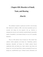
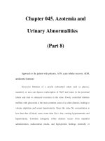
Fig. 22.1. Mechanism of actions of heparin and heparin like anticoagulants.
158
Hemostasis and Thrombosis
22
When using standard heparin, one must be very aggressive in rapidly achieving a
therapeutic aPTT. Unlike the relationship of subtherapeutic heparin levels to recur-
rent thrombosis, there is no association between supratherapeutic aPTT’s and bleed-
ing. Thus, one should not overreact to high aPTT values. Recently, several nomograms
have been published for adjusting heparin to achieve therapeutic anticoagulation. If
a nomogram is used, it must be calibrated for the particular aPTT reagent used in
your lab and not simply copied from a book.
Duration of therapy with any heparin can be as short as five days for patients
with deep venous thrombosis, assuming that the patient has been adequately antico-
agulated with warfarin at that time for at least 24 hours. Patients with a large pul-
monary embolism should have ten days of heparin therapy since it has not yet been
proven that five days is adequate. Heparin should be stopped only after the INR has
been in the therapeutic range for 24 hours.
Antithrombotic Use of Pentasaccharides
One pentasaccharide, fondaparinux, is approved for use in DVT prevention with
hip and knee replacement. The agent is renally cleared and should not be used in
patients with renal insufficiency. Also, given the fixed dose (2.5 mg), it should not
be used in patients weighing under 50 kilograms. Studies for DVT/PE therapy have
been performed using the dose of 7.5 mg daily (5.0 mg in patients under 50 kg and
10 mg in patients over 100 kg). The relative effectiveness of this agent vs LMWH is
still controversial but studies clearly indicated for both treatment and prevention of
DVT/PE these agents are at least equivalent.
Special Problems
Patients with lupus inhibitors who require heparin are difficult to monitor since
their aPTTs are already prolonged. One option is to use LMW heparin due to its
predictable dosing. The other choice is to directly assay for heparin by measuring its
ability to inhibit factor Xa. This assay is insensitive to lupus inhibitors. Therapeutic
range for standard heparin is 0.35 - 0.7 anti-Xa units. Heparin assays are also valu-
able in patients where an acute-phase inflammatory process may lead to nonspecific
heparin binding to inflammatory proteins, resulting in the aPTT not reflecting he-
parin levels. This can be seen in patients on cyclosporin. In pregnant women the
acute rise in factor VIII may also lead to a misleading aPTT; thus, one should use
heparin levels to guide therapy in those patients—even with prophylactic doses of
standard heparin.
The hypercoagulable state associated with malignancy (especially adenocarcino-
mas) may be refractory to warfarin therapy. Long-term LMW heparin is a useful
alternative in these patients. Patients should have LMW heparin levels checked weekly
until the dose is stable.
Pregnant women with prothrombotic states pose a special problem. Pregnancy
adds to the thrombotic risk but is an absolute contraindication to warfarin therapy.
The use of heparin was once also feared, but recent re-analysis of the data shows that
heparin can be safely used in pregnancy. There is abundant experience with LMW
heparin; it is safe and effective in pregnant women for both prophylaxis and therapy.
LMW heparin does not cross the placenta and is associated with less osteoporosis
than standard heparin. For therapy one should follow LMW heparin levels every 4
weeks. Experience shows that levels are more stable than with standard heparin as
the pregnancy progresses.
159
Heparin and Heparin-Like Drugs
22
Since pregnancy is a hypercoagulable state, a patient with additional risk factors
for thrombosis should receive prophylaxis. Prophylactic doses of LMW heparin are
either enoxaparin at 40 mg/day or dalteparin 5000 units q12 hours. Again, the
current recommended approach is to use LMW heparin. To use standard heparin,
start with 5-7,500 units of heparin q12 hours and adjust the dose to keep the hep-
arin assay at 0.1-0.2 anti-Xa units. The plasma protein changes which occur with
pregnancy render the aPTT inadequate even for monitoring prophylactic doses of
heparin. After delivery the patient is on warfarin for six weeks. Warfarin is excreted
in the breast milk in an inactive form and is considered safe for breast feeding.
Young pregnant women with mechanical heart valves present difficult problems.
Thrombosis with both standard heparin and LMWH valve has been reported. How-
ever, the majority of the cases of valve thrombosis have been in patients who were,
for unknown reasons, treated with prophylactic and not therapeutic doses of hep-
arin. At this time the best approach to treatment is unknown. Options include
using standard heparin via continuous infusion pump using heparin levels to moni-
tor therapy, LMWH at doses guided by levels, or use of heparin early, switching to
warfarin mid-pregnancy, and then switching back to heparin at 36 weeks.
Complications of Heparin
Bleeding
Approximately 5% of patients placed on therapeutic heparin will bleed. Some
patients appear to be more at risk than others. Patients without risk factors for bleeding
have a complication rate of 1%, whereas those with risk factors have rates of bleed-
ing of 10-23%. Risk factors include use of aspirin, age greater than 60, liver disease,
and other severe illness (cancer, heart disease). The risk of bleeding is small in pa-
tient receiving prophylactic heparin. Multiple double-blind trials have shown no
increase in major or fatal bleeding with the use of prophylactic heparin.
Protamine is used to reverse heparin and low molecular weight heparin. The
dose for heparin reversal is dependent on timing of the last heparin dose. For imme-
diate reversal (30 minutes or less since the last heparin dose) 1 mg of protamine
should be given for every 100 units of heparin. A suggest nomogram is given in
Table 22.3. One should avoid giving over 50 mg of protamine at one time and
ensure that the infusion does not exceed 5 mg/min.
Protamine does not fully reverse low molecular weight heparin. Due to the longer
half-life of low molecular weight heparin, sometimes a second dose of protamine is
required. The dose is 1mg per 100 units of dalteparin or tinzaparin or 1 mg of
protamine per mg of enoxaparin. If the aPTT is prolonged 2-4 hours later, one-half
of the initial dose of protamine should be given.
Heparin-Induced Thrombocytopenia (HIT)
HIT is caused by the formation of antibodies directed against the complex of
heparin bound to platelet factor 4. These antibodies then bind to the platelet Fc
receptor and activate the platelet. Thus, the thrombocytopenia of HIT is due to
platelet activation and deposition and not immune destruction. The frequency of
HIT is 1-5% when unfractionated heparin is used but less than 1% with low mo-
lecular weight heparin. HIT should be suspected when there is a sudden onset of
thrombocytopenia defined as either 40% or greater reduction in the platelet count,
or the platelet count falling to less than 100,000/uL in a patient receiving heparin in
160
Hemostasis and Thrombosis
22
any form. HIT usually occurs 5-10 days after starting heparin but may occur sud-
denly in patients rechallenged with heparin with recent (less than 3 months prior)
exposure. An often overlooked presentation of HIT is recurrent thrombosis in a
patient receiving heparin despite a normal platelet count. Often when the heparin is
stopped the platelet count rises to above normal levels. Another unique presentation
of HIT is thrombosis occurring up to 2 weeks after heparin exposure. The patients
present with thrombosis, thrombocytopenia, and 25% will also have laboratory evi-
dence of disseminated intravascular coagulation.
The clinical diagnosis of HIT can be challenging, especially in the very sick
patient who has multiple reasons for being thrombocytopenic. In this situation the
laboratory assay for HIT may be helpful. Two general forms of HIT tests exist. One
is a platelet aggregation assay where patient plasma, donor platelets, and heparin are
mixed. If added heparin induces platelet aggregation the test is considered positive.
The test is technically demanding but if performed carefully can be sensitive and
specific. One caveat is that early in the HIT disease process the test can be negative
but then turns positive 24 hours later as the antibody titer increases. There is also an
ELISA assay for the presumed pathogenic anti-platelet factor 4 antibodies. This test
tends to be too sensitive but can also miss up to 20% of HIT due to antibodies other
than anti-platelet factor 4 antibodies; the test is not clinically useful.
Recently Warkentin has developed a scoring system for HIT (Table 22.4). Pa-
tients with high pre-test probabilities should be treated for HIT no matter what the
laboratory testing demonstrated. Lab tests are most helpful for patients with inter-
mediate probability of HIT. Platelet aggregation tests can also be useful in diagnosis
low probability patients with HIT but the ELISA assay may be too sensitive in these
patients.
The first step in therapy of HIT consists of stopping all heparin. Two particular
problems in the critical care setting are that many catheters are kept open with
Table 22.3. Agents for HIT
Argatroban
Therapy: initial dose of 2 µg/kg/min adjusted to an aPTT of 1.5-3.0 times normal.
Bivalirudin (PCI dosing)
Bolus: 1 mg/kg
Infusion: 2.5 mg/kg/hour for 4 hours and then 0.2 mg/kg/hour for 14-20 hrs.
For renal adjustment see Chapter 23
Danaparoid
Therapeutic: bolus of 2250 units (1500 units <60 kg, 3000 units 75-90 kg, 3750
units > 90kg), then a four-hour infusion of 400 units/hour, then 4 hours of 200
units/hour, then a drip at 150-200 u/h to maintain an anti-xa level of 0.5-0.8
anti-xa units.
Fondaparinux
Prophylaxis: 2.5 mg every 24 hours
Therapy: 7.5 mg every 24 hours (consider 5.0 mg in patients under 50 kg and
10 mg in patients over 100 kg)
Use with caution in patients with renal insufficiency
Hirudin
Therapy: bolus of 0.4 mg/kg followed by 0.15 mg/kg/hour to maintain an aPTT of
1.5-3.0 times normal.
For renal adjustment see Chapter 23
161
Heparin and Heparin-Like Drugs
22
heparin and can be a hidden source of heparin for patients with HIT. Also, many
central venous catheters and intra-aortic balloon pumps are coated with heparin.
The presence of heparin-coated catheters is enough to perpetuate the HIT process,
and these must be changed to non-heparin coated devices.
Patients with HIT and active thrombosis are difficult to manage since these pa-
tients cannot receive heparin. Low molecular weight heparins cross-react with the HIT
antibodies and therefore these agents are also contraindicated. Institution of warfarin
therapy alone has been associated with an increased risk of thrombosis. Several
antithrombotic agents are now available for immediate therapy of HIT (see Table 22.3).
Argatroban is a synthetic thrombin inhibitor. It has a short half-life of 40 min-
utes. Dosing is 2 µg/kg/min with the infusion adjusted to keep the aPTT 1.5-3
times normal. Although not widely available, more precise monitoring of argatroban
and other thrombin inhibitors may be obtained by using either the ecarin time or
the quantitative thrombin time. One advantage of argatroban is that it is not renally
excreted and no dose adjustment is necessary in renal failure. These characteristics
make it the most useful agent for patients in the critical care unit. However, argatroban
must be used with caution in patients with severe liver disease (initial dose of 0.5 µg/
kg/min and titrate upward). Argatroban, like all thrombin inhibitors, prolongs the
PT-INR, making initiation of warfarin therapy difficult. If available, the chromoge-
nic X assay may be used to adjust warfarin therapy. Also, if the patient is on a drip of
2 µg/kg/min or less, one can simply aim for a PT-INR of more than 4.0 as thera-
peutic. Once the drip is stopped, one should measure the PT-INR in 4-6 hours to
ensure it is in the therapeutic range. Unfortunately there is no agent that can reverse
argatroban, but given its short half-life this is most often not a problem.
Lepirudin, another direct inhibitor of thrombin, is also monitored by using the
commonly available aPTT. The half-life of lepirudin is short, but the drug accumu-
lates in renal insufficiency with the half-life increasing to more than 50 hours. There
is no antidote for lepirudin. Patients with even slight renal insufficiency (creatinine
greater than 1.5) must have lepirudin doses adjusted to avoid over-anticoagulation.
Table 22.4. Heparin induced thrombocytopenia scoring system
Points 2 1 0
Thrombocytopenia >50% fall or nadir 30-50% fall or nadir Fall < 30% or
20-100,000/ul 10-19,000/ul nadir <10,000/ul
Timing of Onset day 5-10 of consistent but not platelets falls < 5
platelet fall heparin or < 1 day clear records or days and no recent
if patient recently count falls after (100 days) heparin
exposed to heparin day 10
Thrombosis New thrombosis or Progressive or None
skin necrosis or recurrent thrombosis
systemic reaction or suspected but not
with heparin proven thrombosis
OTher cause for No Possible Definite
thrombocytopenia
Pretest Score 6-8=high, 4-5 intermediate, 0-3 low
Warkentin, Heddle Current Hematology Reports 2:148; 2003.
162
Hemostasis and Thrombosis
22
Up to 80% of patients receiving long- term lepirudin therapy will develop antibod-
ies. These antibodies reduce the metabolism of hirudin and increase the therapeutic
effect of lepirudin. In addition, rare cases of anaphylaxis have been reported. Pa-
tients on long-term (> 6 days) lepirudin therapy should still continue to be moni-
tored to avoid over-anticoagulation.
Bivalirudin has been reported in limited studies to also be effective in HIT. How-
ever, like lepirudin, bivalirudin needs dose adjustment in renal disease. There is also
concern about antibody formation since anti-lepirudin antibodies can cross react
with bivalirudin.
Also available is danaparoid, a mixture of various glycosaminoglycans. Unfortu-
nately its half-life is 25 hours, there is no antidote, and monitoring must be done by
specific danapariod levels. These factors greatly limit the use of this agent. One
useful aspect of danapariod is that it can be given as twice a day injections of 2500
units and is useful for the rare patient with HIT who cannot take warfarin but needs
long-term anticoagulation.
The new anti-Xa inhibitor fondaparinux does not cross-react with HIT antibod-
ies. Fondaparinux may be useful for DVT prophylaxis in patients with a history of
HIT and, as clinical experience accumulates, for therapy of these patients. However,
its long half-life and lack of antidote remain a problem. In clinical development is
ximelagatran, an oral thrombin inhibitor which may prove useful for long-term
therapy of HIT patients.
As mentioned above, initiation of warfarin alone has been associated with limb
gangrene, and it should not be started as the sole antithrombotic agent in HIT. In
patients receiving specific antithrombin therapy, warfarin can be started with small
doses (2- 5 mg). These often malnourished patients tend to have a dramatic re-
sponse to warfarin therapy and excessive anticoagulation can easily occur. One should
overlap warfarin and parenteral therapy by 2-3 days as there is evidence that patients
may do worse with shorter durations of specific antithrombin therapy.
Patients with HIT but without evidence of thrombosis are at a high risk of throm-
bosis (53% in one study) and should be considered for antithrombotic therapy.
Patients with HIT should also be carefully screened for any thrombosis, which in-
cludes obtaining lower extremity dopplers. It is unknown whether prophylactic doses
or therapeutic doses of anticoagulants are needed for thrombosis prevention in pa-
tients with HIT but no thrombosis. The duration of such therapy is also controver-
sial. One approach is to give prophylactic doses of antithrombotic agents until the
platelet count has returned to normal. In post-surgical patients prolonged prophy-
laxis for up to six weeks may be of benefit.
Suggested Reading
1. Agnelli G, Sonaglia F. Perspectives on antithrombotic agents: from unfractionated
heparin to new antithrombotics. Haematologica 2002; 87(7):757-70.
2. Hirsh J, Warkentin TE, Shaughnessy SG et al. Heparin and low-molecular-weight
heparin: mechanisms of action, pharmacokinetics, dosing, monitoring, efficacy,
and safety. Chest 2001; 119(1 Suppl):64-94.
3. Keam SJ, Goa KL. Fondaparinux sodium. Drugs 2002; 62(11):1673-85.
4. Ibbotson T, Goa KL. Enoxaparin: an update of its clinical use in the management
of acute coronary syndromes. Drugs 2002; 62(9):1407-30.
5. Laposata M, Green D, Van Cott EM et al. College of American Pathologists Confer-
ence XXXI on laboratory monitoring of anticoagulant therapy: the clinical use and
laboratory monitoring of low-molecular-weight heparin, danaparoid, hirudin and
related compounds, and argatroban. Arch Pathol Lab Med 1998; 122(9):799-807.
163
Heparin and Heparin-Like Drugs
22
6. O’Shea SI, Ortel TL. Issues in the utilization of low molecular weight heparins.
Semin Hematol 2002; 39(3):172-8.
7. Turpie AG. Pentasaccharides. Semin Hematol 2002; 39(3):158-71.
8. Warkentin TE. Platelet count monitoring and laboratory testing for heparin-in-
duced thrombocytopenia. Arch Pathol Lab Med 2002; 126(11):1415-23.
9. Buller HR, Davidson BL, Decousus H et al. Matisse Investigators. Subcutaneous
fondaparinux versus intravenous unfractionated heparin in the initial treatment of
pulmonary embolism. N Engl J Med 2003; 349(18):1695-702.
10. Warkentin TE, Heddle NM. Laboratory diagnosis of immune heparin-induced
thrombocytopenia. Curr Hematol Rep 2003; 2(2):148-57.
11. Warkentin TE. Heparin-induced thrombocytopenia: pathogenesis and manage-
ment. Br J Haematol 2003; 121(4):535-55.
CHAPTER 23
Hemostasis and Thrombosis, 2nd Edition, by Thomas G. DeLoughery.
©2004 Landes Bioscience.
Direct Thrombin Inhibitors
Introduction
Given thrombin’s central role in coagulation, direct inhibition of thrombin is a
potent mechanism for achieving anticoagulation. The first thrombin inhibitor, hiru-
din, was derived from leech saliva and it is clinically available as the recombinant
product lepirudin. Also currently clinically available are argatroban and bivalirudin;
ximelagatran is in clinical trials.
Thrombin inhibitors share certain properties. They raise both the INR and aPTT
since thrombin is part of the common pathway of blood coagulation. Clinically
they are monitored by the aPTT, usually aiming for a goal of 2-2.5 times normal
control. More precise monitoring can be achieved by using the ecarin time. No
effective antidote exists for these agents.
Since all thrombin inhibitors prolong the PT-INR, initiation of warfarin therapy
is difficult. If available, the chromogenic factor X assay can be used to adjust war-
farin therapy. This assay is not affected by the thrombin inhibitor. Warfarin is started
at a low dose of 2.5-5 mg/day and a daily factor X assay is performed. When the
level is down to 15-30% the thrombin inhibitor is stopped. If argatroban is used,
there is a nomogram to guide warfarin therapy if doses of 2
µg/kg/min and under
are used.
Argatroban
Argatroban is a synthetic thrombin inhibitor derived from L-arginine that binds
only to the thrombin active site. It has a short half-life of 40 minutes. The dosing for
anticoagulation is 2
µg/kg/min with the infusion adjusted to keep the aPTT 1.5-2.5
times normal. Argatroban can also be used for percutaneous coronary intervention
with a bolus of 350
µg/kg being given and then a continuous infusion of 25 µg/kg/
min started.
One advantage of argatroban is that it is not renally excreted and therefore no
dosage adjustment is necessary in renal insufficiency or failure. These characteristics
make it the most useful agent for patients who require thrombin inhibitors. How-
ever, argatroban must be used with caution in patients with severe liver disease. An
initial dose of 0.5
µg/kg/min is used and titrated upward guided by the aPTT. To
start warfarin in the patient on a drip of 2
µg/kg/min or less one can simply aim for
a PT-INR of more than 4.0 as therapeutic. Once the drip is stopped, one should
measure the INR in 4-6 hours to ensure it is in the therapeutic range.
Lepirudin
Hirudin is derived from leech saliva. It binds both the active site of thrombin and
the substrate binding site. Currently hirudin is made using recombinant technology.
165
Direct Thrombin Inhibitors
23
Lepirudin, like all thrombin inhibitors, is also monitored by using the com-
monly available aPTT. The half-life of lepirudin is short, but the drug accumulates
in renal insufficiency with the half-life increasing to up to more than 50 hours.
Patients with even mild renal insufficiency (creatinine greater than 1.5) must have
lepirudin doses adjusted to avoid overanticoagulation. Up to 80% of patients re-
ceiving long-term lepirudin therapy will develop antibodies. These antibodies re-
duce the metabolism of hirudin and increase the therapeutic effect of lepirudin.
Patients on long-term (greater than 6 days) lepirudin therapy should still continue
to be monitored to avoid over-anticoagulation. Rare cases of anaphylaxis have been
reported. Lepirudin has also been used for anticoagulation for myocardial infarctions
and as an adjunct to PCI.
Bivalirudin
Bivalirudin is a synthetic thrombin inhibitor that binds to the thrombin active
site and also to the substrate binding site. It also has a short half-life of 30 min-
utes. Its use is best studied for cardiac indications and it is currently approved for
PCI. Bivalirudin is also renally excreted and dosage adjustments should be made
Table 23.1. Argatroban, hirudin and bivalirudin
Argatroban
Therapy: 2 µg/kg/min infusion with dose adjustments to keep aPTT 1.5 - 3 times
normal.
For patients with severe liver disease, start with 0.5 µg/kg/min infusion and follow
aPTT to same goal of aPTT 1.5 - 3 times normal.
For PCI:
Bolus with 350 µg/kg over 3-5 minutes and then infuse with a maintenance drip
of 25 µg/kg/min adjusting to a ACT of 350-400 minutes.
Hirudin
Therapy: bolus of 0.4 mg/kg followed by 0.15 mg/kg/hour to maintain an aPTT of
1.5-3.0 times normal.
Adjustments for Renal Dysfunction:
For creatinine of 1.6-2.0 mg/dl: bolus of 0.2 mg/kg followed by a 50%
reduction in infusion rate.
For creatinine of 2.0-2.5: bolus of 0.2 mg/kg followed by a 75% reduction in
infusion rate.
For creatinine of 2.6-6.0:bolus of 0.2 mg/kg followed by a 90% reduction in
infusion rate.
For creatinine of greater than 6.0 mg/ml: Bolus of 0.1 mg/kg on alternate days
only when the aPTT is less than 1.5 times normal; no continuous infusion.
Bivalirudin
Bolus: 1 mg/kg
Infusion: 2.5 mg/kg/hour for 4 hours and then 0.2 mg/kg/hour for 14-20 hrs.
Renal adjustment:
For creatinine clearance of 30-59 ml/min, decrease dose by 20%
For creatinine clearance of 10-29 ml/min, decrease dose by 60%
For creatinine clearances less than 10 mg/min, decrease dose by 90%
Ximelagatran
DVT prophylaxis: 24 mg po BID
Therapy of DVT: 36 mg po BID
Stroke prevention in atrial fibrillation: 36 mg BID
166
Hemostasis and Thrombosis
23
for patients with renal insufficiency. In theory, bivalirudin should be useful for hep-
arin induced thromocytopenia (HIT) but it has not been clinically studied for this
indication.
Ximelagatran
A novel agent in clinical development is ximelagatran. This agent is a prodrug of
the thrombin inhibitor melagatran. This drug is orally available and has a half-life of
2.5-3.5 hours. The agent is renally metabolized and dosage may need to be adjusted
for renal insufficiency. Unlike warfarin, ximelagatran metabolism is not effected by
food, drugs, or age. Clinical trials have shown that ximelagatran (24 mg bid) is as
effective for prophylaxis in surgery that is high risk for venous thrombosis prophy-
laxis. Data shows that a therapeutic dose of 36 mg bid is just as effective as warfarin
in prevention stroke in patients with atrial fibrillation. Clinical trials have also dem-
onstrated that ximelagatran along is just as effective as enoxaparin followed by war-
farin in the therapy of DVT/PE. The major side effect of ximelagatran is elevations
in liver function tests that occur in 3-6% of patients starting this drug. Most pa-
tients can continue the drug but it is unknown what is the best protocol for moni-
toring patients and if rare patients will develop severe drug induced hepatitis.
Suggested Reading
1. Hopfner R. Ximelagatran (AstraZeneca). Curr Opin Investig Drugs 2002;
3(2):246-51.
2. Kaplan KL, Francis CW. Direct thrombin inhibitors. Semin Hematol 2002;
39(3):187-96.
3. McKeage K, Plosker GL. Argatroban. Drugs 2001; 61(4):515-22
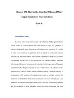
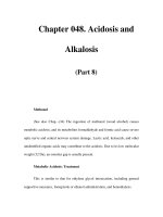
Fig. 23.1. Mechanism of action of thrombin inhibitors.
167
Direct Thrombin Inhibitors
23
4. Nowak G. Clinical monitoring of hirudin and direct thrombin inhibitors. Semin
Thromb Hemost 2001; 27(5):537-41.
5. Robson R, White H, Aylward P et al. Bivalirudin pharmacokinetics and pharma-
codynamics: effect of renal function, dose, and gender. Clin Pharmacol Ther 2002;
71(6):433-9.
6. Weitz JI, Hirsh J. New anticoagulant drugs. Chest 2001; 119(1 Suppl):95-107.
7. Schulman S, Wahlander K, Lundstrom T et al. THRIVE III Investigators. Sec-
ondary prevention of venous thromboembolism with the oral direct thrombin in-
hibitor ximelagatran. N Engl J Med 2003 Oct 30; 349(18):1713-21.
8. Francis CW, Berkowitz SD, Comp PC et al. EXULT A Study Group. Comparison
of ximelagatran with warfarin for the prevention of venous thromboembolism af-
ter total knee replacement. N Engl J Med 2003 Oct 30; 349(18):1703-12.
CHAPTER 24
Hemostasis and Thrombosis, 2nd Edition, by Thomas G. DeLoughery.
©2004 Landes Bioscience.
Warfarin
Warfarin works by interfering with vitamin K-dependent gamma-carboxylation
of coagulation proteins II, VII, IX, and X. As a result of warfarin therapy, these coagu-
lation factors cannot bind calcium. This causes impairment of these factors’ binding
to membranes and to fold into proper configuration. Therapy with warfarin is initi-
ated by giving the patient 5-10 mg in the evening for the first two nights (2.5 mg in
those over 75 years) and adjusting the dose to achieve an adequate prothrombin time.
Although the use of a 10 mg loading dose has been traditional in the past, for most
people this is too much. Multiple trials show that using a 10 mg loading dose causes
one to overshoot and leads to a delay in achieving a stable therapeutic INR. A practi-
cal approach is to use 5mg in loading patients over the age of 50 or in patients with
albumin under three and 10 mg in other patients. The elderly patient (over age 75)
may only need a 2.5 mg loading dose. Nomograms for 5 and 10 mg warfarin loading
doses are given in Table 24.1. The effect of warfarin on the INR takes 36 hours to
occur so the morning INR reflects the effect of the warfarin dose 36 hours before.
Factor VII has the shortest half-life and so it is the first factor reduced as a result
of warfarin therapy. However, the full anticoagulant effect does not occur until there
is a reduction in prothrombin (factor II) and factor X, which may take several days.
Thus, in acute thrombosis heparin needs to be continued for at least 24 hours after
the prothrombin time is therapeutic to allow for factors II and X to fall. For chronic
indications such as atrial fibrillation, warfarin can be started at lower daily doses
(2.5-5.0 mg). This allows for initiation of warfarin therapy without the use of hep-
arin. Table 24.2 gives guidelines for adjusting warfarin doses in patients once thera-
peutic prothrombin times have been reached.
Unfortunately the dose of warfarin required for achieving therapeutic antico-
agulation varies among patients. This is due to a combination of the patient’s ge-
netic ability to metabolize warfarin, concurrent medications and illnesses, and diet.
Patients who are older require less warfarin, with patients over age 65 requiring
one-half to one-third of the warfarin doses of younger patients. Also variations in
cytochrome 2C9 (CYP2C9) can affect warfarin metabolism. The most common
genotype is 2C9*1 with 80-90% of the population carrying this allele. The 2C9*3
allele (found in 6-10%) is much less efficient in metabolism of warfarin. For ex-
ample, studies have shown that 2C9*1/*1 requires an average of 3-4.25 mg/day of
warfarin compared to 1.75-2.5 mg/day for 2C9*1/*3 and 0.4 mg/day for 2C9*3/
*3. Studies are underway to see whether knowing the 2C9 genotype of the patient
can predict warfarin dose or bleeding complications.
Since warfarin is metabolized in the liver by the cytochrome P450 system, the
INR may change with starting or stopping other medications that affect CYP2C9.
Multiple agents can augment or decrease warfarin effect and are listed in Table 24.3.
Unfortunately, many drugs may have an unpredictable effect on the INR. The most
prudent strategy is to check the INR several days after starting a new drug and then
169
Warfarin
24
Table 24.1. Nomograms for warfarin loading
5 Mg Warfarin Nomogram
Day INR Dosage (Mg)
1 5.0
2< 1.5 5.0
1.5-1.9 2.5
2.0-2.5 1.0-2.5
>2.5 0.0
3 <1.5 5.0-10.0
1.5-1.9 2.5-5.0
2.0-2.5 0.0-2.5
2.5-3.0 0.0-2.5
>3.0 0.0
4 <1.5 10.0
1.5-1.9 5.0-7.5
2.0-3.0 0.0-0.5
>3.0 0.0
5 <1.5 10.0
1.5-1.9 7.5-10.0
2.0-3.0 0.0-5.0
>3.0 0.0
6 <1.5 7.5-12.5
1.5-1.9 5.0-10.0
2.0-3.0 0.0-7.5
>3.0 0.0
10 mg Warfarin Nomogram
1 10.0
2< 1.5 7.5-10.0
1.5-1.9 2.5
2.0-2.5 1.0-2.5
>2.5 0.0
3 <1.5 5.0-10.0
1.5-1.9 2.5-5.0
2.0-2.5 0.0-2.5
2.5-3.0 0.0-2.5
>3.0 0.0
4 <1.5 10.0
1.5-1.9 5.0-7.5
2.0-3.0 0.0-0.5
>3.0 0.0
5 <1.5 10.0
1.5-1.9 7.5-10.0
2.0-3.0 0.0-5.0
>3.0 0.0
6 <1.5 7.5-12.5
1.5-1.9 5.0-10.0
2.0-3.0 0.0-7.5
>3.0 0.0
Crowther MA, Harrison L, Hirsh J. Ann Internal Med 1997; 127:333.