Manual of Diagnostic Ultrasound in Infectious Tropical Diseases - part 8 docx
Bạn đang xem bản rút gọn của tài liệu. Xem và tải ngay bản đầy đủ của tài liệu tại đây (681.75 KB, 19 trang )
122 3 Ultrasound Diagnosis of Special Infectious and Parasitic Diseases
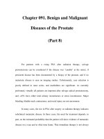
Fig. 3.57. Brazilian25-year-old male. Ul-
trasound of scrotal contents in B-
mode with 3.5-MHz probe s howing ane-
choic cyst-like structures (arrows)corre-
sponding to large lymphangiectasias in
an infected patient with W. bancrof ti.The
FDS (not clear on this image) was seen
inalessdilatedlymphatic(small ar row).
No hydrocele was present
nests and 85% of infected men) and specificity (100%). Detection of the
FDS with the 3.5-MHz transducer was unreliable when the lymphatic vessel
diameter was less than 2.7 mm. Thus, for practical purposes, the limit of
detectionforthe3.5-MHzprobewasreachedatavesseldiameterof2.7mm.
For the 7.5-MHz probe, the limit of detection appears to be at a vessel
diameter of approximately 1 mm. Thus, when maximum resolution of the
ultrasound image is required, such as in studies of drug efficacy, the 7.5-
MHz transducer should continue to be used, in this case, always combined
with physical examination.
3.3.4.8
Alternative and Supplementary Methods
Once living adult worms are identified in an y lymphatic vessel or lymph
node, the diagnosis of active bancroftian filariasis infection is confirmed.
In cases where only lymphangiectasia is found, a search for circulating
an tigen is advised (Og4C3 test or ICT card). These tests are already avail-
able commercially. Also, a “provocative test” with diethylcarbamazine is
an alternate way to reveal the hidden adult worms in lymphatic vessels (es-
pecially in the intrascrotal vessels through the detection of small nodules
perceived by physical examination up to 7 days after treatment). This is
especially useful where vessel dilation is not enough to allow visualization
of living worms by ultrasound.
3.3.4.9
Diagnostic Efficiency
In summary, ultrasound is a very useful tool for complementing the di-
agnosis of bancroftian filariasis and for documenting the extension of the
3.3 Parasitic Diseases 123
lymphatic vessel damage. Its use to monitor absence of lymphangiectasia
in areas where transmission has been interrupted deserves further inves-
tigation.
Acknowledgement. We thank Dr. David Addiss for reviewing the manuscript, and
the NGO (Non-governmental organization) Amaury Coutinho and the World Health
Organization for financial support for the bancroftian ultrasonographic studies in
Brazil.
3.3.5
Liver Trematode Infection (Liver Distosomiasis)
(by Joon-Koo Han)
Distosomiasis isa group of parasitoses due to flat worms that live in contact
with epithelia. Clinical classification depends on the organ infected by
adults: liver, lungs, or intestines.
Liver flukes
– F asciola hepatica is cosmopolitan. It is contracted when eating con-
taminated food (wild watercress, dandelion leaves, or lamb’s lettuce, on
which larvae are encysted).
– Fasciola gigantica or giant fluke is only found in tropical areas.
– Dicrocoeli um dendriticum or small fluke is exceptional in humans; how-
ever, egg s are frequently found in stools.
– Clonor chis sinensis and Opisthorchis viverrini are found in the Orient.
– Opisthorchis felineus canbeseeninEurope.
Pulmonary flukes: Mainly present in the tropics, they are extremely fre-
quent in Far East. Freshwater crustaceans spread the infection:
– Pa ragonimus westermani
– Paragonimus kellicoti
– P aragonimus africanus
Intestinal flukes: Several species are responsible for the disease: Fasci-
olopsis buski is oriental, and can be contracted by eating water chestnuts;
Metago nimus yokoga w ai is also oriental.
– Hetero phyes heteroph yes is more cosmopolitan, and can be contracted
by eating raw fish.
We present as an example clonorchiasis disease.
124 3 Ultrasound Diagnosis of Special Infectious and Parasitic Diseases
3.3.5.1
Clonorchiasis
Epidemiology
Clonor chis s inensis infection is endemic inthe Far East, especially southern
China, Hong Ko ng, and Korea. The custom of eating slices of raw freshwater
fish con tributes to the high incidence of infection in these countries.
Despite a gradual decrease in prevalence over the recent decades, in
1986, it was estimated that about 15 million people were infected in the
world, and a national survey in Korea in 1997 revealed that the prevalence
of clonorchiasis was still 1.4%. C. sinensis is still the most prevalent human
parasitic helminth by stool examination recently in Korea. The difficulty of
eliminating clonorchiasis in the endemic area has been attributed mainly
to the difficulty of detecting infected cases, although other con tributory
factors including re-infection after treatment have been discussed.
Therateofinfectionwithclonorchiasisinendemicareasisgreaterin
older pa tients than in younger ones. Men are more commonly infected
than women. The higher percentage of clonorchiasis in men is probably
related to their dietary habits. In endemic areas, there is a tradition of
eating raw freshwater fish, soaked in vinegar or red-pepper mash, as an
appetizer when drinking liquor at social gatherings.
C. sinensis has a life span of 10–30 years, and this creates a problem
for Asian immigrants who may develop clinical symptoms several years
after leaving the endemic area. Clonorchiasis in North America has been
reported in recent decades, reflecting the immigration of people from
endemic areas.
Pathology
The life cycle of C. sinensis has been well documented. The definite hosts
are humans, dogs, and other mammals. The eggs, shed by the adult worm,
are deposited in the biliary tree of these animals, enter the intestine, and
are passed with the feces. On reaching water, the eggs are ingested by
snails. Within the snail, the eggs undergo metamorphosis, after which
the cercariae erupt. The free-swimming cercariae pass from the snail and
penetratethescales offreshwaterfish. After a developmentperiod ofseveral
weeks, cercariae become encysted inmuscle. Humans ando therfish-eating
animals acquir e the infection by ingesting the infected fish that are raw
or inadequately cooked. W ith digestion, the metacercariae excyst in the
duodenum, migrate into the intrahepatic biliary tree via the common
3.3 Parasitic Diseases 125
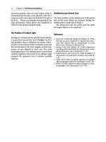
Fig. 3.58. Histopathologic findings of
clonorchiasis. Note the hyperplasia of
biliary epithelial cells and periductal fi-
brosis. Note the flukes within dilated bile
duct (hematoxylin and eosin, original
magnification 12.5x)
duct, and mature into adul t worms (Fig. 3.58). The adult fluke inhabits
the biliary tract, generally localizing within the intrahepatic bile ducts.
The adult worm is a small trematode with an elliptical shape; the average
worm is 10–25 mm in length. Completion of this life cycle is restricted to
endemic areas, reflecting the geographic distribution of the essential snail
species.
C. sinensis causes low-grade inflammatory changesin the biliary tree, se-
vere hyperplasia of epithelial cells and metaplasia of mucopolysac charide-
producing cells in the mucosa, and progressive periductal fibrosis. The
severity of these pathological changes tends to correlate with the duration
of infection, the parasite burden, and the susceptibility of the host.
Thecutsurfaceoftheliverrevealsdilatationofthemedium-sizedbile
ducts, with thickened walls. The histopathological findings of clonorchi-
asis are characterized by bile duct epithelial proliferation followed later
by periductal fibrosis. Biliary hyperplasia is the distinctiv e lesion of early
Clonor chis infection, but the portal tracts do not become so deranged
as to lead to portal venous hypertension or biliary cirrhosis. In addi-
tion to b iliary hyperplasia, the biliary epithelium frequently becomes
edematous, and desquamation may be seen in areas of close proximity
to the flukes. Periductal infiltrates of mononuclear cells are frequently
fo und; however, inflammation of the bile-duct walls is generally slight
in uncomplicat ed cases. Metaplasia of biliary epithelial cells into goblet
cells occurs fairly early in infection, and these may proliferate to pro-
duce many small glandular-like structures in the mucosa, giving the bile
a persistent and excessively high mucus content. Chronic and persistent
infections result in a gradual increase in fibrous tissues, which may even-
tually engulf some of the proliferated glands, giving the appearance of
126 3 Ultrasound Diagnosis of Special Infectious and Parasitic Diseases
cholangiofibrosis. As fibrosis proceeds, the epithelial proliferation sub-
sides.
These histopathological changes are distinctive features of clonor chi-
asis. Therefore, when a variable degree of proliferation of ductal epithe-
lium with metaplastic cells (described as adenomatous hyperplasia) and
periductal fibrosis are observed in an endemic area, it is highly suggestive
of clonorchiasis on histological grounds, even though the parasite is not
included in the section.
The complications of clonorchiasis are the results of obstruction of
the biliary system. Parasite-induced goblet cell metaplasia creates bile
with a high mucin content. This bile, combined with the adult flukes and
ova, serves as a nidus for bacterial superinfection and intrahepatic stone
formation.The ectasia of intrahepatic bile ducts may progress toa pyogenic
cholangitis, liver abscess, cholangiocarcinoma, hepatitis, and cirrhosis.
R etention cysts and dilated v enous radicles in the portal areas are also
observed.
Many studies from endemic areas have documented the high commen-
surate occurrence of cholangiocarcinoma with clonorchiasis. Acause-and-
effect relationship between clonorc hiasis and cholangiocarcinoma is now
generally accepted by most researchers, since epidemiological, experi-
men tal, and pathological data suggesting the relationship have accumu-
lated.
Examination Technique
The patients are recommended to fast for at least four to six hours before
the examination. Although it is now rare in human cases, clonorchiasis
involving the gall bladder or pancreas has been reported.
Examination is in the supine position, if needed with the head elevated
and the patient turned 45 degrees to the left.
The examination should always include the entire biliary system, the
liver, and the pancreas. Because the sonographic diagnosis of clonorchiasis
is generally made by the exclusion of obstruction in the large bile duct, the
common bile duct should be completely evaluated whenever possible.
Sometimes ingestion of water (two or three cups) can improve the
aco ustic window for the distal common bile duct.
Color Doppler is helpful to differentiate the dilated peripheral intrahep-
atic ducts from accompanying portal veins.
3.3 Parasitic Diseases 127
Using a linear array transducer with high frequency (5–12 MHz) some-
times helps the depiction of ductal wall thickening and intraductal flukes.
Pathological Findings
Characteristic ultrasonographic findings of clonorchiasis are summarized
as diffuse, mild, uniform dilatation of the small intrahepatic bile ducts
with no dilatation, or only minimal dilatation, of larger bile ducts without
a focal obstructing lesion.
The ductal wall is often thickened, and its echogenicity is increased.
Occasionally, flukes or aggrega tes of ova can be shown as non-shadowing
echogenic foci or cast within the bile duct (Figs. 3.59a–d and 3.60a–c).
These findings areconsidereda pathognomonicfindingofclonorchiasis.
Differential Diagnosis
Differential diagnosis of clonorchiasis includes cancer along the bile
ducts, choledocholithiasis with recurrent pyogenic cholangitis, sclerosing
cholangitis, Caroli disease, and Fasciola hepatica infection.
Pitfalls
At present, clonorc hiasis is commonly diagnosed incidentally during radi-
ological screening (especially, ultrasonography) of the abdomen for other
purposes, since symptoms of clonorchiasis are vague and nonspecific in
most cases. The biliary dilatation observed in ultrasonography should not
be misinterpreted as being caused by a focal obstructive lesion in the
biliary tree, because this misinterpretation may mislead to unnecessary
diagnostic tests or invasive procedures. Once diagnosed, clonorchiasis is
treated very effectively with praziquantel, with few side-effects.
Efforts should be taken to find an occult cholangiocarcinoma during
the examination.
Alternative and Supplementary Methods
Clonorchiasis should be suspected in a patient who develops manifesta-
tions of hepatic or biliary disease and who has a histo ry of ingesting raw
freshwater fish in an endemic area. The diagnosis of liver fluke infestation
is usually established by microscopic examination of stools for ova and/or
adult parasites. A formalin-ether sedimentation technique is known to be
more reliable than the direct-smear method for detecting eggs in feces.
128 3 Ultrasound Diagnosis of Special Infectious and Parasitic Diseases
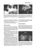
Fig. 3.59a–e.Ultrasonographic findings ofclonorchiasis.Note thediffuse,mild, uniform
dilatation of the small intrahepatic bile d ucts with no dilatation, or only minimal
dilatation, of larger bile ducts. The ductal wall is thickened, and its echogenicity is
increased. Using linear transducer with high frequency helps the depiction of ductal
wall thickening (e). Each figure is from a different patient
3.3 Parasitic Diseases 129
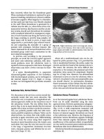
Fig. 3.60a–c. A patient with clonorchiasis-associated cholangiocarcinoma. (a) Trans-
verse contrast-enhanced CT shows diffuse, mild, uniform dilatation of the small intra-
hepatic bile ducts with no dilatation, or only minimal dilatation, of larger bile ducts.
(b) Transverse ultrasonography at epigastrium shows mild dilatation of the small in-
trahepatic bile duct at segment III. The ductal wall is thickened, and its echogenicity
is increased. This finding was observed in diffuse distribution in the entire liver. (c)
Right intercostals scan shows mass in the right liver
Although the diagnosis of clonorchiasis is easily made by the stool ex-
amination, mass screening with fecal examination can be more difficult,
because of poor voluntary cooperation. A number of serologic techniques
have been developed to aid in the diagnosis of clonorchiasis. However, un-
fortunately, the serologic methods currently available exhibit considerable
cross-reactivity. Accordingly, they are not widely accepted as screening
techniques.
Computedtomography,aswellasultrasonography,iswidelyaccepted
as an accurate and feasible diagnostic method for clonorchiasis.
130 3 Ultrasound Diagnosis of Special Infectious and Parasitic Diseases
Although helpful, none of these various ser ologic tests and radiological
examinations has been reported to surpass fecal examination, because of
their limited sensitivity, specificity, or applicability.
Diagnostic Efficiency
Aforementioned ultrasonographic fi ndings are regarded aspathognomonic
for clonorchiasis in endemic areas. However, more recent studies have
shown the low diagnostic accuracy of ultrasonography for clonorchiasis.
Accor ding to a study in an endemic area, the sensitivity was 52% and the
specificity was 51%; the low sensitivity was attributed to false negative
cases with mild infection, and the low specificity was attributed to false
positive cases with residual pathology after cure. This low specificity is
of particular interest, since the number of cases cured has continuo usly
increased in recen t decades, because of na tionwide control and ecologic
changes.
Therefore, ultrasonography is less useful for the differentiation of cured
clono rchiasis and active infection, since it reflects the pathological changes
in the bile ducts, which may persist for years after cure, rather than the
presence of the worm itself.
3.3.6
Schistosomiasis
(by Maria C. Chammas, Ilka R.S. de Oliveira, Giovanni G. Cerri)
Schistosomiasis is a parasitic disease of slow progression caused by trema-
todes of the genus Schistosoma, first described in the mid-19th century by
the German pathologist Theodor Bilharz. It is an important public health
problem in certain regions of the world, including South America, the
Caribbean, Africa, and the Middle East. It is estimated that approximately
250 million individuals are infected in 76 countries, and that 500 to 600
million people are exposed to the infection.
The most prevalent species of the Schistosoma are: Schistosoma man-
soni, Schistosoma japonicum,andSchistosoma haematobium,withthetwo
first species associated with the hepatosplenic form of the illness and the
latter species with the genitourinary form.
The World Health Organization (WHO) recently proposed a standard-
ization of the use of diagnostic ultrasound in schistosomiasis indicated
for field studies. For epidemiological purposes, it is very important that
3.3 Parasitic Diseases 131
ultrasound examinations be carried out and reco rded in a standardized
way, to ensure that results obtained in different places at different times
can be compared. This standardization has been used in several countries
in endemic areas.
The most important clinical signs are related to portal hypertension
in S. mansoni and S. japonicum andtokidneyfunctionimpairmentinS.
haematobium.
3.3.6.1
Schistosoma mansoni
The hepatosplenic form of Manson’s schistosomiasis affects the liver,
spleen, gall bladder, the portal system, and its tributaries.
The advanced hepatosplenic form is identified 5–10 years after the initial
infection, being associated with the development of periportal fibrosis and
portal hypertension. In the absence of other hepatic diseases, such as hep-
atitis caused by the B and C virus, the inflammatory pr ocess resulting from
schistosomiasis usually does not affect the hepatic cell ular parenchyma,
so that no significant alterations in the hepatic function can be observed.
However, portal hypertension leads to repeated bouts of hematemesis sec-
ondary to esophageal and gastric varices, a dr eadful com plication and
acauseofmorbidity.
Other clinical manifestations caused by intestinal schistosome infection
are glomerulonephritis, functional alterations of the exocrine pancreas,
and pulmonary hypertension.
Due to the hemorrhages in the upper digestive tract, repetitive blood
transfusions are carried out. Consequently, the association with viral cir-
rhosis (for virus B or C) is not uncommon, so that the overlapping of the
findings of viral cirrhosis or even hepatocarcinoma with those of schisto-
somiasis should be considered.
Pathophysiology
After penetrating the skin, the parasites, in the form of cercariae, are car-
ried to the lung, and they reach the liver through the systemic circulation,
where they mature and males and females pair off. Subsequently, they
migrate against the blood flow to the mesenteric veins and to the bowel
submucosa, where the egg-laying takes place.
Theevolutionoftheillnessintheliveriscarriedoutthroughthepar-
asite’s egg embolization from the bowel submucosa to the venous portal
132 3 Ultrasound Diagnosis of Special Infectious and Parasitic Diseases
Fig. 3.61. Macrosc opic examination of
the liver, show ing intense fibrous peri-
portal thickening
circulation. The inflammatory reaction to the presence of the eggs (and
sometimes to the dead parasite as well) leads to the formation of chronic
epithelial granulomas and to Symmer’s periportal fibrosis. With the dis-
ease evolution, the periportal fibrosis progresses and establishes a venous
obstruction, contributing to the state of presinusoidal portal hypertension.
Macroscopically, the liver presents an increase of the left lobe, atrophy of
the right lobe, blunt borders, and fibrous thickening of the portal spaces of
up to 3 cm. The fibrous periportal thickening is more intense in the hepatic
hilum, extending in varying degrees to the portal intrahepatic spaces and
to the perivesicular region.
Additionally, the spleen may be enlarged to over 650 g in 50% of the pa-
tients, presenting sinusoidal dilatation, hemorrhages, and later formation
of siderotic nodules (Gamna-Gandi bodies) (Fig. 3.63).
Diagnostic Imaging
The morphologic and biometric aspects of thehepatosplenic form of schis-
tosomiasis can be studied by several diagnostic imaging methods, such as
Doppler ultrasonography (US-Doppler), scintigraphy, compu terized to-
mography (CT scanning), spleno portography, and magnetic resonance
imaging (MRI). The US-Doppler evaluation presents the advantages of
noninvasiveness and of being accessible to the majority of the patients, be-
coming an important step in the practice of modern diagnostic medicine.
Thus, this method has been also applied in a series of epidemiological
studies, in those regions of high prevalence. The morphologic and hemo-
dynamic parameters obtained by this method make both the diagnosis of
portal hypertension and its vascular complications possible.
3.3 Parasitic Diseases 133
US-Doppler Aspects
The anatomical-pathological aspects previously described can be recog-
nized by means of US-Doppler study, preceding, sometimes, the beginning
of the clinical manifestations of the illness. It is important to point o ut that
US-Doppler is useful not only for the diagnosis but for the follow-up of the
clinical treatment. In the initial phases of hepatosplenic schistosomiasis,
discrete and nonspecific alterations are observed, such as hepatomegaly
without periportal thickening, making the characterization of the disease
difficult.
The volumetric alterations of theliver,remarkably the increase of theleft
hepaticlobeandthereductionoftherighthepaticlobe,areobservedinthe
hepatosplenic schistosomiasis (Fig. 3.62). These alterations can easily be
characterized by B-mode ultrasonography, as can the splenomegaly with
the Gamna-Gandibodies. Thesiderotic nodules are evidenced asdispersed
hyperechogenic points in the splenic parenchyma. However, due to their
dimensions (3–15 mm), they are only observed in up to 7% of the patients
(Fig. 3.63).
The fibrous periportal thickening is one of the morphologic features that
allows the recognition of the illness, being characterized on ultrasonog-
raphy as a band of periportal hyperechogenicity in about 73–100% of the
cases. This thickening mainly affects the portal vein at the hepatic hilum,
and also extends to the intrahepatic portal branches and the perivesicular
region (Figs. 3.64, 3.65). However, in the initial phase of the illness, the
periportal fibrosis is difficult to characterize by ultrasound. Periportal fi-
brosis causes an increase in veno us pressure in the splanchnic territory,
constituting the presinusoidal portal hypertension. As a consequence, an
Fig. 3.62. Volumetric alterations of the
liver, remarkable increase of the left hep-
atic lobe, and reduction of the right hep-
atic lobe
134 3 Ultrasound Diagnosis of Special Infectious and Parasitic Diseases
Fig. 3.63a–c. Splenomegaly with Gamna-Gandi bodies (hyperechogenic points in the
splenic parenchyma)
3.3 Parasitic Diseases 135
Fig. 3.64a,b. Fibrous periportal thickening at the hepatic hilum and intrahepatic portal
branches
Fig. 3.65a–c. Fibrous periportal thickening atthehepatic hilumandperivesicular region
136 3 Ultrasound Diagnosis of Special Infectious and Parasitic Diseases
increase in the caliber (diameter) of the portal vein (> 1.2 cm), splenic
vein (> 0.9 cm), and superior mesenteric vein (> 0.9 cm) can be frequently
observed in 73%, 68%, and 42% of the cases, respectively (Fig. 3.66).
An analysis by color duplex-Doppler generally discloses the hepatopetal
flow direction in the portal vein, preserved spectral tracing morphol-
ogy, and blood flow velocity within the normal standards. Some flow
oscillations can be observed, due to cardiac and respiratory movements
(Fig. 3.67).Thrombosis of the portal veinis a rather uncommon finding, be-
ing found in approximately 6% of the patients. However, with the progres-
sive increase of the hepatic resistance to the portal blood flow, thrombosis
may occur in the main portal stem and/or in its branches, filling its lumen
with echogenic material, partially or completely o bstructing the vessel.
Even though the Doppler color mapping does not display the blood flow
Fig. 3.66. Portal hypertension causes in-
crease in the diameter of portal vein
(1.4 cm)
Fig. 3.67. Color duplex-Doppler scan showing hepatopetal flow direction in the portal
vein, preserved spectral tracing morphology, and blood flow velocity (23 cm/sec)
3.3 Parasitic Diseases 137
in the presence of complete thrombosis, in the partial thrombosis events,
flow becomes evident in the vein’s peripheral region (Figs. 3.68–3.70).
In the cases of chronic thrombosis, cavernous transformation of t he
portal vein may occur, being represented by the identification of collateral
circulation in this anatomic region (see Fig. 3.72). US-Doppler demon-
strates an elongated echogenic structure, pervaded by serpiginous images
with low-velocity monophasic flow, without the resulting fluctuating alter-
ations of the cardiac cycle.
The portal thrombosis can also occur as a complication of esophageal
varices sclerotherapy, bypass surgeries (mesenteric-cava, porto-caval, dis-
Fig. 3.68. Partially portal thrombosis,
with echogenic material inside the portal
vein
Fig. 3.69. Complete portal thrombosis, with hypoechogenic ma terial inside the portal
vein (recent thrombosis)
Fig. 3.70. Power Doppler scan showing complete portal thrombosis (chronic) and
identifying collateral circulation in this anatomic region, pervaded by serpiginous
images (cav ernous transformation)
138 3 Ultrasound Diagnosis of Special Infectious and Parasitic Diseases
Fig. 3.71a,b. Spontaneous splenorenal anastomosis, demonstrated by color Doppler
scan
Fig. 3.72a,b. Venous collateral circulation (dilated left gastric vein). B-scan and Doppler
tal splenorenal) (Fig. 3.71), or disconnecting surgeries (azygo-portal and
splenectomy).
US-Doppler studies of the splenic vein and superior mesenteric vein
demonstrate an increase in their caliber/diameter in several degrees,
with hepatopetal flow direction, spectral tracing, and maximum velocities
within the normality standards. However, in the presence of splenorenal
anastomosis, the splenic vein (and eventually the portal vein) can present
flow in the rev erse direction (hepato fugal flow) (Fig. 3.32). Another com-
mon finding is the presence of venous collateral circulation, evidenced in
36–78% of the patients. These v essels can be identified in hepatic hilum,
at the small gastric curve (left or coronary gastric vein) (Fig. 3.72), in the
fundus of the stom ach (short gastric veins) (Fig. 3.73), along the round
ligament (paraumbilical vein) (Fig. 3.74), around the gall bladder (cystic
3.3 Parasitic Diseases 139
Fig. 3.73a,b. Color Doppler scan (a) and power Doppler (b)showingthevenouscollat-
eral circulation (short gastric veins)
Fig. 3.74a,b. Color Doppler scan showing the venous collateral circulation with hep-
atofugal flow in the paraumbilical vein
varices) (Fig. 3.75), and in other retroperitoneal sites (for example, spon-
taneous splenorenal anastomosis) (Fig. 3.71). The collateral circulation
generally presents hepatofugal flow. Finally, the hepatic veins can display
140 3 Ultrasound Diagnosis of Special Infectious and Parasitic Diseases
Fig. 3.75a,b. Perivesicular fibrosis, represented by hyperechogenic tissue (a)andColor
Doppler scan showing the venous collateral circulation (cystic varices) (b)
alterations in the spectral tracing as a result of the reduction of the hep-
atic parenchyma compliance due to the fib rosis, presenting a two-phase or
single-phase flow (“portalized”).
3.3.6.2
Schistosoma japonicum
Many lesions due to S. japonicum are similar to those of S. mansoni.
There are particular fibrous changes in the liver parenchyma differ-
ent from those of S. mansoni,givingthearchitectureamoreprominent
network of fibrosis (fish scales) (Fig. 3.76).
Fig. 3.76a,b. S. japonicum. a Fibr osis, typical aspect of fish scales. b Periportal fibrosis
suggesting the fish scales aspect