Manual of Diagnostic Ultrasound in Infectious Tropical Diseases - part 9 pdf
Bạn đang xem bản rút gọn của tài liệu. Xem và tải ngay bản đầy đủ của tài liệu tại đây (576.98 KB, 19 trang )
3.3 Parasitic Diseases 141
3.3.6.3
Schistosoma haematobium
The alterations in this genito-urinary form are found mainly in the kidneys,
the ureters, and the bladder. The severity and frequency of the lesions are
related to the intensity of the infection.
Obstructive uropathy that varies from mild to severe, with reduction of
the thickness of the renal parenchyma, is a common sign. Bands of fibrosis
(hyperechoic band) are an additional finding in the kidneys. The stage of
hydr onephrosis indicates the severity of the disease.
Dilatation of the ureter could be very important; it is easily identified
with ultrasound (Fig. 3.77).
The most important findings, in the bladder, are as follows: thickening
and irregularity of the wall with development of pseudopolyps and masses
and calcifications (Fig. 3.78).
Liver abnormalities, similar to those related in S. mansoni,mayalsobe
observed.
Ultrasound is very important in the follow-up of portal hypertension,
demonstrating the course of the disease or reduction of periportal fibro-
sis after treatment. Doppler could be used to monitor changes in portal
flow after treatment. In urinary schistosomiasis, the affected bladder and
kidneys could be evaluat ed.
The differential diagnosis includes a chronic liver disease with portal
hypertension for S. mansoni and S. japonicum and inflammatory (partic-
ularly tuberculosis), obstructive and tumoral lesions for S. haematobium.
When thebowel lesion is identified, the differential diagnosis is with granu-
lomatous or ulcerative colitis and tumoral polyps. In the lung, other causes
of interstitial pattern and pulmonary hypertension should be considered.
Usually, the diagnostic presentation of schistosomiasis is very charac-
teristic and specific.
Ultrasound is definitively adopted as an epidemiologic tool in schisto-
somiasis.
142 3 Ultrasound Diagnosis of Special Infectious and Parasitic Diseases
Fig. 3.77a–d. S. haematobium. a Discrete dilatation of the renal cavities. b Important
hydronephrosis. c Dilated pelvis ureter. d IVP same patien t as in Fig. 3.24c
3.3 Parasitic Diseases 143
Fig. 3.78a,b. S. haematobium. a Localized thickening of the bladder wall, pseudo masse
aspect. b Pseudo polyp aspect. c Bladder wall calcification
3.3.7
Echinococcosis
3.3.7.1
Hydatid Disease
(by Ferid Ben Chehida, Heykel Ben Romdhane, Azza Hammou, Ha ssen A.
Gharbi)
Hydatid disease is caused by Echinococcus granulosus; this cestodiasis
of cosmopolitan distribution occurs predominantly in areas of intensive
sheep or cattle farming.
Humans are an accidental host in the animal parasitosis which involves
two hosts:adefinitive, carniv oroushost(usually a dog)andanintermediate
herbivorous animal (sheep, cattle, camel). The adult tapeworm lives in
the jejunum of the dog; the eggs it releases are passed in the excreta;
when ingested by an intermediate host, they reach the stomach, and the
embryos released penetrate the intestinal wall and reach the liver through
the portal system. The larvae are carried to the lungs, and get into the
general circulation; no part of the organism is protected from infestation.
The larvae rapidly become surrounded b y an inflammatory granuloma
and transform into a multinuclear protoplasmic mass that develops over
aweekintoavesicleorhydatid.Overaperiodof16to20weeks,thecyst
doubles in size as it becomes filled with fluid. This period of progressive
development leads to the production of thousands of scolices; at the same
144 3 Ultrasound Diagnosis of Special Infectious and Parasitic Diseases
time, an intense tissue reaction around the vesicle results in the formation
of a tough wall called the pericyst.
I f the intermediate herbivorous host dies and the cyst-containing flesh
is eaten by a dog, each scolex grows to an adult worm in the dog jejunum.
Like intermediate hosts, humans become infected more often during close
co ntact with dogs (hands licked by an infected animal, or hands brought
into contact with the mouth afterhaving touched aninfected dog) thanafter
ingestion of food or water contaminated by the excreta of infected dogs.
The geographic distribution of hyda tid disease is wide and includes
South America, Australia, New Zealand, East and especially North Africa,
and the Mediterranean area. Certain occupations are particularly exposed
to the risk of contracting hydatidosis: these are shepherds and sheep f arm-
ers, veterinarians and laboratory personnel, butchers and meat packers.
All the organs may be affected, but, in adult age, the liver (60%) and the
lungs (20%) are the sites of predilection. The majority of the cases, except
for the liver, are primary, following vascular dissemination. There is no
sex predilection. Infestation may occur at any age, generally from 2 years
onwards. Most cases are seen in young adults.
The clinical manifestations of abdominal hydatidosis are variable, and
depend on the location and the stage of development of the parasite: ab-
dominal mass, abdominal pain, hepatomegaly (with or without jaundice),
and ascites. Sometimes its discovery is fortuitous, during systematic explo-
ration for a lung location or during an epidemiologic study in an endemic
area.
Ultrasound Findings
Several classifications have been proposed to present ultrasound findings,
based on the sonographic analysis of the morphology and structure of
Table 3.1. WHO proposed classification of cystic echinococcosis: Six types
WHO CL CE1 CE2 CE3 CE4 CE5
Gharbi + / – I I III IV IV V
Caremani I-a I-a,b II-a,b III-a,b, IV V-a,b VI-a,b
Perdomoo I I II III IV-a V, VI
CL, cystic lesion, nonspecific CE, cystic echinococc osis
I, II: active
III: transitional
IV,V:inactive
3.3 Parasitic Diseases 145
the hydatid cyst corresponding to its various developmental stages. The
most common worldwide used is the Gharbi classification, with five types
of sonographic patterns, described in 1981, which is simple and can be
adopted for the description of US, CT and MR images. For the future, the
WHO informal working group on cystic Echinococcosis is trying to unify
the most important proposed classifications (Table 3.1).
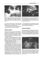
Fig. 3.79a–d. a Liver hydatid cyst type I:
pure fluid collection. b Liver h ydatid cyst
type I: See the localized thickening of the
cyst wall very suggestive of HC. c Orbit US:
retro-ocular hydatid cyst. d Chest hydatid
cyst
146 3 Ultrasound Diagnosis of Special Infectious and Parasitic Diseases
Gharbi Classification
Type I: Pure Fluid Collection (Fig. 3.79).
This type appears as an anechoic space with marked enhancement of back-
wall echoes. The fluid collection is rounded with well-defined borders; its
walls often vary in thickness. This localized thickening should be sought
systematically, and is a very suggestive sign of a hydatid cyst. Only small
cysts appear as anechoic collections; these appear to be ’punched ou t’
and do not show proper walls on echography. Some cysts situated at the
periphery of the liver, or the spleen, in contact with the abdominal wall
or the diaphragm, are no longer rounded but oval-shaped and seem to
follow the parietal contours. The size of the cysts varies greatly, from 1 to
20 cm in diameter. The pure fluid collection is the most notable aspect.
The liquid is clear, and corresponds to cysts that are new, mono vesicular,
and noncomplicated.
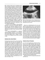
Type II: Fluid Collection with a Split Wall (Fig. 3.80)
The fluid collection retains its well-defined co ntour but it is often less
rounded, and appears to be ‘sagging’ in places. The split wall may be
localized in an area just outside the cyst, or it may become a ‘floating
membrane’ loose inside the cyst. This splitting of the wall, which is often
discreet, must be systematically sought in any intrahepatic liquid collec-
tion, because it is almost pathognomonic for a hydatid cyst. The split wall
may result from a lowering of intracystic pressure, causing the detachment
of the membrane.
Fig. 3.80a,b. a Liver hydatid cyst type II: fluid collection with a split wall.b Thyroid
hydatid cyst; see the membrane
3.3 Parasitic Diseases 147
Type III: Fluid Collection with Septa (Fig. 3.81)
The fluid collection retains its well-defined contour, but it is divided by
septa which are more or less thick and complete, forming oval-shaped
or rounded structures. The enhancement of back-wall echoes is usually
evident. The most typical cases show a ‘honeycomb’ image. The echoes
within the cysts show images of simple or multiple secondary vesicles.
When characteristic, the sonographic appearance of the secondary vesi-
cles allows diagnosis of hydatid cysts. However, this diagnosis is sometimes
difficult to affirm. Intracystic septation may take another aspect: it may
delineate masses of various shapes that are not rounded, but show undu-
lated con tours. This appearance is due to the folding of the detached cystic
membrane.
Type IV: H eterogeneous Echo Patterns (Fig. 3.82).
This type of cyst appears as a roughly rounded mass, with irregular con-
tours and echo pattern. We have found three general pattern types:
IV-1: hypoechoic appearance with a few irregular echoes, always due to
infected multilocular cysts
IV-2: hyperechoic solid pattern without back-wall shadow, and
IV-3: intermediate pattern including both hypoechoic structures and
hyperechoic structures in approximately equal quantity, the latter being
clustered in nodular patterns.
Since it is difficult to make a diagnosis from these structures, it is
necessary to look for other diagnostic signs of hydatid cysts, such as
a membrane seen as a linear ribbon or band pattern, variable appearance
of echographic images from one section to another in the same area,
hyperechoic contour with possible areas of acoustic shadow, presence of
small fluid collections from intra or extracystic secondary vesicles, or the
presence of another cyst at a different stage of development, in the liver or
in another organ.
Type V: Reflecting Thick Walls (Fig. 3.83)
This type appears as a formation with a very hyperechoic contour, and
with a cone-shaped shadow which is usually outlined to some degree.
When this formation is small, we can visualize the whole contour. When it
is bigger, only its immediate front wall is visualized: this appears as a thick
arch-shaped image with a posterior concavity.
148 3 Ultrasound Diagnosis of Special Infectious and Parasitic Diseases
Fig. 3.81a–f. a Liver hydatid cyst type III: Fluid collection with septa. b Renal hydatid
cyst type III. c Renal hydatid cyst, see daugh ter vesicles (same patient as in b). d Chest
X-ray: Left ventricle heart hydatid cyst. e Heart US: hydatid cyst Type III (same patient
as in d). f Macroscopic post mortem study (same patient as in e)
3.3 Parasitic Diseases 149
Fig. 3.82. a Liver hydatid cyst type IV: heterogeneous echo patterns.b Prostate hydatid
cyst, a very rare localization
Fig. 3.83. Liver hydatid cyst type V : re-
flecting thick wall
Complicated Hydatid Cyst, Other Patterns
The natural evolution of a hydatid cyst is difficult to predict. Sometimes
it may develop into a calcified mass or produce compression of adjacent
organs cavities and vessels, e.g., inferior vena cava, portal and hepatic
veins, biliary tract, urinary tract. The cyst may rupture or become infected.
In the liv er, for instance, the most frequent complication is cyst rupture
in to the biliary ducts, through the diaphragm, or into the peritoneum. In
these cases, ultrasound may show a dilated biliary tract with fragments of
membranes in the gall bladder or in the common biliary duct, Budd Chiari
syndr ome with compression of the hepatic veins by hydatid cyst (Fig. 3.84),
multiple perito neal cyst, ascites, and, in some cases, diaphragmatic breach
with a communican t supradiaphragmatic space (Fig. 3.85)
In all these cases, we can see other less freq uent patterns, for example
very hyperechoic masses corresponding to the shell cyst, which is usually
150 3 Ultrasound Diagnosis of Special Infectious and Parasitic Diseases
Fig. 3.84. a Liver hydatid cyst causing compression of the hepatic veins (color Doppler).
b Hydatid cyst ruptured into the biliary ducts. Note the hydatid membranes. c Hydatid
cyst ruptured into the biliary ducts. Note the hydatid membrane inside the main biliary
duct. (courtesy of Dr. Badea, Romania)
calcified to some degree. In liquid collection, some declivitous echoes may
appear and represent hydatid sand (Fig. 3.86).
Other Modalities
In endemic areas, ultrasound plays the main role, in general, among the
imaging modalities. However, conventional X-ray is a very importan t tool
for the chest and bone location; but, for the intracranial and spine location,
CT and MRI, when available, are necessary. These new modalities, with
multislice technique and sophisticated reconstructions, permit a better
overall view of the size, location, and number of cysts within the affected
organs, and vascular relationships.
3.3 Parasitic Diseases 151
Fig. 3.85. Liver hydatid cyst with breach of the diaphragm
Fig. 3.86. Liver hydatid cyst: hydatid sand (very rare)
Differential Diagnosis
The differential diagnosis varies with the type of echographic pattern and
the organ affected. In endemic countries, types II and III are characteristic
of hydatid cyst, and types I and V are suggestive of hydatid cyst. However,
there are alternative diagnoses:
Type I
Cyst (biliary, ovary, mesenteric, pancreatic)
Hematoma
Reduplication cysts
Metastasis, teratoma
Type II
Abscess
Foreign bodies
Type III
Caroli disease
Cystadenoma, cystadeno-carcinoma (Fig. 3.87)
Cystic lymphangioma
R enal cystic mass
152 3 Ultrasound Diagnosis of Special Infectious and Parasitic Diseases
Fig. 3.87. Pancreatic hydatid cyst mim-
icking a cystadenoma
Type IV
Abscess
Solid tumor
Hematoma
Type V
Postoperative calcification
Abscess
Hematoma
Management of Hydatid Cyst
Surgery remains the treatment of choice for hydatidosis. However, in ap-
proximately 16% of patients, surgery may be dangerous or impossible due
to post-surgical recurrence, diffuse disease, or massive peritoneal dissem-
ination.
These situations are frequent in endemic areas and are principally due
to the late discovery of the parasitosis. In addition, the hopes born with
the discovery of benzimidazole compounds as a possible medical cure for
hyda tidosis are fading, as the reported rate of successful treatment is no
more than 9–16%.
3.3 Parasitic Diseases 153
Conseq uently, a new interventional technique was proposed by the
Tunisian Gargouri team, called PAIR:
P = puncture of the cyst under ultrasound guidance, A = aspiration of
the hydatid fluid, I = injection of scolicide, sodium chloride hypertonic
solution, or alcohol into the cystic cavity, and R = reaspiration of the
solution without drainage, under medical treatment protection (Fig. 3.88).
Other scolicide drugs, such as alcohol, have been used with good success,
with or without drainage.
After PAIR, ultrasound controls showed a progressive decrease in the
cyst volume andposterior wall enhancement, anincrease incystechogenic-
ity, and an increase in density at CT.
The involution time is variable, from 2 weeks to 2 years. Repeated PAIR
proced ures may be performed without complication, in order to accelerate
the process of cyst involution. However, the PAIR results also depend on
the cyst location. In the liver, the decrease in size occurs slowly; a minimum
of 6 months is needed.
In the peritoneum, the size reduction is very fast, averaging 2 weeks.
During PAIR, no anaphylactic shock and no secondary dissemination have
been encountered in our experience; however, death may exceptionally
occur.
Fig. 3.88. Liver hydatid
cyst treated by PAIR: a
needle inside the cyst.
b scolicide injection of
hyperosmolar saline so-
lution. c after injection.
d after reaspiration
154 3 Ultrasound Diagnosis of Special Infectious and Parasitic Diseases
A moderate and short allergic reaction, with fever, may occur, but these
can be easily controlled medically.
M orbidity of the PAIR method seems to be acceptable. Its cost is low,
and hospitalization is much shorter than for surgery.
Negative hydatid immunologic tests cannot be considered to be a solid
proofoftheeffectivenessofthePAIRmethod:itneedsatotalevacuation
of hydatid material. There may be recurrence in 0–4% of the cases.
PAIR, today, is accepted and recommended by WHO as a good alterna-
tive for the management of hydatid cysts, but some strict rules must be
respected. Today more than 4000 PAIR procedures have been done around
theworld;onlyonedeathwasreported.
3.3.7.2
Alveolar Echinococcosis of the Liver: Ul trasound Findings
(by Michel Claudon, Alain Gerard, Alix Martin-Bertaux)
Epidemiology
Alveolar echinococcosis (AE) is a rare parasitic disease due to the intra-
hepatic growth of the larva of Echinococcus multilocularis. The disease is
usually found in Central Europe, Turkey, Iran, the Soviet Union, China,
Japan, and North America.
The normal parasitic cycle associates foxes, or sometimes cats or dogs,
in the bowel of which the worm becomes mature, and rodents which
are infested by eating plan ts contaminated by stools containing eggs. The
larvae reach the liver and induce a tumor-like process. The parasitic cycle
starts again when the rodent is caught and eaten by a fox. Up to 75% of
foxes may carry E. mul tilocula ris in endemic countries.
Humans can be accidentally contaminated by ingestion of infected
berries or plants or by direct contact with foxes. However, the hepatic
response in humans is variable, ranging from a prompt healing without
any detectable lesion to the progressive development of a large hepatic
process. Various epidemiological screening studies, using serological tests
based on enzyme-linked immunosorbent assays (ELISAs) and ultrasound
examination, have confirmed that only 0–25% of subjects with positive
serologic tests were found to present abnormal ultrasound images.
Hepatic resections, including rightlobectomy and segmentectomy, were
successfully performed years ago. Transhepatic drainage of dilated biliary
ducts or of large necrotic collections have also been found useful. How-
ever, the prognosis of the disease has changed significantly during the
3.3 Parasitic Diseases 155
last decade, due to the impact of medical treatment based on Mebenda-
zole and Albendazole. Continuous, long-term treatment (even lifelong)
has proven to be well-tolerated and effective, as lesions showed stability
in 97% of the cases. Surgery may still be indicated in the case of a limited
lesion.
Pathology
In contrast to E. gran ulosus, E. mul tilocularis growswithexternalvesicula-
tion, surrounded by a fibroinflammatory reaction (Fig. 3.89). The result is
an infiltrative tumor-like mass that very slowly invades the liver, especially
the portal spaces and hilum, the hepatic veins, and the vena cava. Microcal-
cifications and central necrosis are frequent, due to vascular involvement
and ischemia. Extrahepatic extension through the diaphragm or toward
the duodenum and retroperitoneum is possible, as are metastases to the
lung, brain, or bone.
Fig. 3.89. Pathology specimen. The mass
appears as an infiltrative process, involv-
ing the main portal branches and the bil-
iary tract
Examination Technique
Preparation is not required. The examination is initiated with a longi-
tudinal scan to the left of the midline, demonstrating the left liver lobe,
anterior to the aorta. The transducer is moved to the right i n short steps
to visualize the lobe segments anterior to the vein cava, continuing across
the interlobular area and the right liver lobe to the segments bordering
on the right kidney. The subcostal oblique scan is useful to demonstrate
extension within the right lobe.
Due tothe frequent extension ofthe disease tothe portal veinsandbiliary
tract, particular attention should b e taken in the evaluation of the hilum
156 3 Ultrasound Diagnosis of Special Infectious and Parasitic Diseases
and the main portal branches, using if necessary intercostal or subcostal
oblique scans, and Doppler techniques. In the case of portal hypertension,
the examination should also include the spleen, the splenic and superior
mesen teric veins, and the areas where porto-caval anastomoses could be
demonstrated (pa tent paraumbilical vein, large gastric veins, or spleno-
renal collaterals). Intraperitoneal fluid is nowadays rarely noted.
Pathological Findings
He patomegaly is frequently encountered, mostly due to the parasitic mass
itself, or sometimes to the secondary hypertrophy of normal adjacent
parenchyma.
There is one lesion in most cases, but sometimes multiple lesions or
miliary form can be found. The right hepatic lobe is more often involved
than the left one.
Several patterns of lesions can be described on sonography. Most le-
sions are hyperechoic, with necrotic areas and clusters of small, diffuse
calcifications (Figs. 3.90, 3.91). Other presentations include hypoechoic
masses and massively calcified atrophic lesions. It seems that the older is
the lesion, the more heterogeneous and calcified it is.
Extension to the hilum or hepatic vein convergence is frequently found,
with resulting biliary duct dilation or portal hypertension. Color Doppler
Fig. 3.90. The ultrasound examination reveals a 7-cm-diameter mass located in the
right hepatic lobe, mainly hyperechoic with small clusters of microcalcification
Fig. 3.91. Large necrotic mass of the left hepatic lobe (segment 4), with fluid-debris
level, and a hyperechoic peripheral rim containing calcifications
3.3 Parasitic Diseases 157
Fig. 3.92. The subcostal approach reveals
a large mass, developed from the left
lobe, containing several cystic areas, and
having close contact with the hilum. The
color Dop pler mode sho ws the patency
of the portal vein
may be helpful to demonstrate patency of involvement of the main vessels
(Fig. 3.92). Lymphadenopathy is rare.
Differential Diagnosis
Because of the tumor-like appearance of the parasitic process and the
potential of multiple lesions, the differential diagnosis includes mainly
hepatocarcinoma and metastases. Other entities tobe discussed are hepatic
abscess and benign tumors. However, the frequent presence of clusters of
microcalcifications (90%) is of great value in suggesting the diagnosis,
especially if there are few clinical symptoms and/or suspicious history
(e.g., rural life). The diagnosis can be easily confirmed by serological tests,
which are of high sensitivity and high specificity.
Pitfalls, Alternative and Supplementary Methods
CT scan is very helpful because it can better demonstrate areas of mi-
crocalcification. T2-weighted MR images are more effective than US and
CT in showing cystic or necrotic areas, giving a grape-lik e pattern. Both
imaging methods are required to make a precise evaluation of intra-and
extrahepatic involvement of the disease, which is difficult in the far field
by sonography because of the marked attenuation of the ultrasound beam
throughout the fibrous and calcified parasitic tissue.
Diagnostic Efficiency
Ultrasound is a very useful technique to detect hepatic involv ement by AE,
in large screening studies in endemic areas, or in selected patients. Sero-
logical tests are an easier way to confirm the diagnosis than percutaneous
biopsy. CT scan and/or MRI are required for a better evaluation of the
extension of the disease, and for the follow-u p under medical treatment.
Chapter 4
Ultrasound Features in Childhood Infection
Ibtissem Bellagha · Wi e m D o u i r a · Azza Hammou ·
Hassen A. Gharbi
In medicine, it is obvious that children are not young adults. At this age,
the diseases are mainly different: some diseases are characteristic of the
structure of their growing organs, others are congenital malformations.
In this chapter, we will discuss some diseases in which ultrasound (US)
can play an important role, even in developing countries. Osteomyelitis
is a good example; US is a useful tool to diagnose the early stage of this
affection. Transfontanellar US is also very useful in the study of meningitis
(including early and late complications).
4.1
Ultrasound in Osteomyelitis
Acute hematogenous osteomyelitis still constitutes a diagnostic and ther-
apeutic challenge. Infection almost always occurs by hematogenous colo-
nization of growing bones b y bacteria, usually Staphylococcus aureus.The
metaphysis is usually the site of seeding. The onset of acute osteomyelitis
is typically rapid and progressi ve.
Early clinical consideration, confirmation, and treatment are essential
to prevent or minimize morbidity.
Osteomyelitis usually affects a single bone. Sites of predilection of acute
infection are the fast-growing and large metaphyses around the knee, wrist,
and shoulder. Flat bones are affected in 25% of the cases.
Clinical presentation of pediatric osteomyelitis is a temperature in-
crease, local pain, soft tissue swelling, redness, and tenderness to palpa-
tion. Usually, a history of recent local trauma is found, but infection in
neonates and infants can be clinically silent.
Once clinical suspicion is aroused and the physical examination and
laboratory tests are in keeping with an infectious process, plain film ra-
diographs should be obtained, because they may provide clues for other
160 4 Ultrasound Features in Childhood Infection
pathologic conditions. The earliest sign in osteomyelitis is the deep soft
tissue swelling. Further swelling involves the muscles and the superficial
subcutaneous soft tissues. Bone destruction and periosteal reaction may
be obvious, but only 10 to 21 days after the onset of the disease. At that
stage, antibiotic therapy should be established.
Bone scintigraphy using Technetium 99m methylene diphosphonate
lends itself to the localization of the inflammatory process in osteomyelitis,
but without any specificity. Increased vascularity of hyperemia and initial
bone resorption allow concentration of the isotope at the focus of acute
infection.
Echography must be done as soon as possible, before the antibiotic
therapy is established.
The earliest sign of osteomyelitis is nonspecific soft tissue swelling ad-
jacent to affected bone. The most characteristic ultrasonic feature is a sub-
periosteal fluid collection contiguous with the bone.
The US examination should look for more than 2-mm elevation of the
periosteum by a hypoechoic or anechoic zone tapered in the two ends
located usually in the metaphysis (Figs. 4.1, 4.2). The subperiosteal fluid
Fig. 4.1. Sonography of acute osteomyelitis of the humerus. Coronal scan depicts
echogenic subperiosteal fluid in the metaphysis (white arrows)
Fig. 4.2. Sonography of acute osteomyelitis of the right tibia. Transverse scan depicts
echogenic subperiosteal fluid (white arrows)