Mims pathogenesis of infectious disease - part 7 doc
Bạn đang xem bản rút gọn của tài liệu. Xem và tải ngay bản đầy đủ của tài liệu tại đây (3.34 MB, 48 trang )
8 Mechanisms of Cell and Tissue Damage
283
choroid plexuses, joints and ciliary body of the eye. Factors may include
local high blood pressure and turbulent flow (glomeruli), or the
filtering function of the vessels involved (choroid plexus, ciliary body).
In the glomeruli the complexes pass through the endothelial windows
(Fig. 8.17) and come to lie beneath the basement membrane. The
smallest-sized complexes pass through the basement membrane and
seem to enter the urine. This is probably the normal mechanism of
disposal of such complexes from the body.
Immune complexes are formed in many, perhaps most, acute infec-
tious diseases. Microbial antigens commonly circulate in the blood in
viral, bacterial, fungal, protozoal, rickettsial, etc. infections. When the
immune response has been generated and the first trickle of specific
antibody enters the blood, immune complexes are formed in antigen
excess. This is generally a transitional stage soon giving rise to anti-
body excess, as more and more antibody enters the blood and the
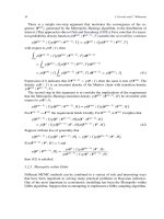
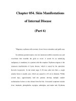
Fig. 8.17 Immune complex glomerulonephritis. Arrows indicate the movement
of immune complex deposits, some moving through to the urine and others
(larger deposits) being retained. M, mesangial cell; U, urinary space; L, lumen
of glomerular capillary; E, endothelial cell (contains 100 nm pores or windows;
see Fig. 3.2b).
284
Mims' Pathogenesis of Infectious Disease
infection is terminated. Sometimes the localisation of immune
complexes and complement in kidney glomeruli* is associated with a
local inflammatory response after complement activation. There is an
infiltration of polymorphs, swelling of the glomerular basement
membrane, loss of albumin, even red blood cells, in the urine and the
patient has acute glomerulonephritis. This is seen following strepto-
coccal infections, mainly in children (see below). As complexes cease to
be formed the changes are reversed, and complete recovery is the rule.
Repeated attacks or persistent deposition of complexes leads to irre-
versible damage, often with proliferation of epithelial cells following
the seepage of fibrin into the urinary space.
Under certain circumstances complexes continue to be formed in the
blood and deposited subendothelially for long periods. This happens in
certain persistent microbial infections in which microbial antigens are
continuously released into the blood but antibody responses are only
minimal or of poor quality (see below). Complexes are deposited in
glomeruli over the course of weeks, months or even years. The normal
mechanisms for removal are inadequate. The deposits, particularly
larger complexes containing high molecular weight antigens or anti-
bodies (IgM) are held up at the basement membrane and accumulate
in the subendothelial space together with the complement components.
As deposition continues, they gradually move through to the mesangial
space (Fig. 8.17) where they form larger aggregates. Mesangial cells,
one of whose functions is to deal with such materials, enlarge, multiply
and extend into the subepithelial space. If these changes are gradual
there are no inflammatory changes, but the structure of the basement
membrane alters, allowing proteins to leak through into the urine.
Later the filtering function of the glomerulus becomes progressively
impaired. In the first place the glomerular capillary is narrowed by the
mesangial cell intrusion. Also, the filtering area is itself blocked by the
mesangial cell intrusion, by the accumulation of complexes (Fig. 8.17),
and by alterations in the structure of the basement membrane. The
foot processes of epithelial cells tend to fuse and further interfere with
filtration. The pathological processes continue, some glomeruli ceasing
to produce urine, and the individual has chronic glomerulonephritis.
Circulating immune complex deposition in joints leads to joint
swelling and inflammation but in choroid plexuses there are no
apparent pathological sequelae. Circulating immune complexes are
also deposited in the walls of small blood vessels in the skin and else-
where, where they may induce inflammatory changes. The prodromal
rashes seen in exanthematous virus infections and in hepatitis B are
probably caused in this way. If the vascular changes are more marked,
they give rise to the condition called erythema nodosum, in which there
* Cells in kidney glomeruli, in joint synovium and in choroid plexuses bear Fc or C3b
receptors. This would favour localisation in these tissues.
8 Mechanisms of Cell and Tissue Damage
285
are tender red nodules in the skin, with deposits of antigen, antibody
and complement in vessel walls. Erythema nodosum is seen following
streptococcal infections and during the treatment of patients with
leprosy. When small arteries are severely affected, for instance in some
patients with hepatitis B, this gives rise to periarteritis nodosa.
Immune complex glomerulonephritis occurs as an indirect immuno-
pathological sequel to a variety of infections. First there are certain
virus infections of animals. The antibodies formed in virus infections
generally neutralise any free virus particles, thus terminating the
infection (see Ch. 6), but the infection must persist if antigen is to
continue to be released into the blood and immune complexes formed
over long periods. Non-neutralising antibodies help promote virus
persistence because they combine specifically with virus particles, fail
to render them noninfectious, and at the same time block the action of
any good neutralising antibodies that may be present. Immune
complexes in antigen excess are formed in the blood when the persis-
tent virus or its antigens circulates in the plasma and reacts with anti-
body which is present in relatively small amounts. Virus infections
with these characteristics are included in Table 8.6. In each instance
complexes are deposited in kidney glomeruli and sometimes in other
blood vessels as described above. In some there are few if any patho-
logical changes (LDV and leukaemia viruses in mice) probably because
there is a slow rate of immune complex deposition, whereas in others
glomerulonephritis (LCM virus in mice, ADV in mink) or vasculitis
(ADV in mink) is severe.
A persistent virus infection that induces a feeble immune response
forms an ideal background for the development of immune complex
glomerulonephritis, but there are no known viral examples in man.
Table 8.6. The deposition of circulating immune complexes in infectious diseases
Kidney Glomerulo- Vascular
Microbe Host deposits nephritis deposits
Leukaemia virus
Lactate dehydrogenase virus (LDV)
Lymphocytic choriomeningitis virus
(LCM)
Aleutian disease virus (ADV)
Equine infectious anaemia virus
Hepatitis B virus
Streptococcus pyogenes
Malaria (nephritic syndrome)
Treponema pallidum
(nephritic
syndrome in secondary syphilis)
Infectious causes of chronic
glomerulonephritis a
Mouse, cat + +_ -
Mouse + _+ -
Mouse ++ + -+
Mink + + ++
Horse + + +
Man + - +
Man + + -
Man + + -
Man + + ?
Man ++ ++
a
Nephrologists and pathologists distinguish ten different types of glomerulonephritis, some of them
infectious in origin, the immune complexes being deposited directly from blood or formed locally in
glomeruli.
286 Mims' Pathogenesis of Infectious Disease
There are one or two other microorganisms that occasionally cause this
type of glomerulonephritis, and it is seen, for instance, in chronic
quartan malaria and sometimes in infective endocarditis. In both these
examples microbial antigens circulate in the blood for long periods.
However, immune complex deposition does not necessarily lead to the
development of glomerulonephritis, and immune complexes are detect-
able in the glomeruli of most normal mice and monkeys. Even in
persistent virus infections the rate of deposition may be too slow to
cause pathological changes as with LDV and leukaemia virus infec-
tions of mice (see Table 8.5). During the acute stage of hepatitis B in
man, when antibodies are first formed against excess circulating viral
antigen (hepatitis B surface antigen), immune complexes are formed
and deposited in glomeruli. However, the deposition is short-lived and
there is no glomerulonephritis. Persistent carriers of the antigen do not
generally develop glomerulonephritis, because their antibody is
usually directed against the 'core' antigen of the virus particle, rather
than against the large amounts of circulating hepatitis B surface
antigen.
Immune complex glomerulonephritis occurs in man as an important
complication of streptococcal infection, but this is usually acute in
nature with complement activation and inflammation of glomeruli, as
referred to above. Antibodies formed against an unknown component
of the streptococcus react with circulating streptococcal antigen,
perhaps also with a circulating host antigen, and immune complexes
are deposited in glomeruli. Streptococcal antibodies cross-reacting
with the glomerular basement membrane or with streptococcal
antigen trapped in the basement membrane may contribute to the
picture. Deposition of complexes continues after the infection is termi-
nated, and glomerulonephritis develops a week or two later. The strep-
tococcal infection may be of the throat or skin, and Streptococcus
pyogenes types 12 and 49 are frequently involved.
Kidney failure in man is commonly due to chronic glomeru-
lonephritis, and this is often of the immune complex type, but the anti-
gens, if they are microbial, have not yet been identified. It is possible
that the process begins when antigen on its own localises in glomeruli,
circulating antibody combining with it at a later stage. The antibody is
often IgA ('IgA nephropathy') which could be explained as follows.
Antigen in intestinal or respiratory tract combines locally with IgA,
and the complex enters the blood. Here, for unknown reasons, it is not
removed in the normal way by the liver, and thus has the opportunity
to localise in glomeruli.
Allergic alveolitis
When certain antigens are inhaled by sensitised individuals and the
antigen reaches the terminal divisions of the lung, there is a local
8 Mechanisms of Cell and Tissue Damage
287
antigen-antibody reaction with formation of immune complexes. The
resulting inflammation and cell infiltration causes wheezing and respi-
ratory distress, and the condition is called allergic alveolitis. Persistent
inhalation of the specific antigen leads to chronic pathological changes
with fibrosis and respiratory disease. Exposure to the antigen must be
by inhalation; when the same antigen is injected intradermally, there
is an Arthus type reaction (see p. 282), and IgG rather than IgE anti-
bodies are involved.
There are a number of microorganisms that cause allergic alveolitis.
Most of these are fungi. A disease called farmer's lung occurs in farm
workers repeatedly exposed to mouldy hay containing the actino-
mycete
Micromonospora faeni.
Cows suffer from the same condition. A
fungus contaminating the bark of the maple tree causes a similar
disease (maple bark stripper's disease) in workers in the USA
employed in the extraction of maple syrup. The mild respiratory symp-
toms occasionally reported after respiratory exposure of sensitised
individuals to tuberculosis doubtless have the same immunopatholog-
ical basis.
Other immune complex effects
In addition to their local effects, antigen-antibody complexes generate
systemic reactions. For instance, the fever that occurs at the end of the
incubation period of many virus infections is probably attributable to a
large-scale interaction of antibodies with viral antigen, although
extensive CMI reactions can also cause fever. The febrile response is
mediated by endogenous pyrogen IL-1 and TNF liberated from poly-
morphs and macrophages, as described on p. 329. Probably the charac-
teristic subjective sensations of illness and some of the 'toxic' features
of virus diseases are also caused by immune reactions and liberation of
cytokines.
Systemic immune complex reactions taking place during infectious
diseases very occasionally give rise to a serious condition known as
disseminated intravascular coagulation. This is seen sometimes in
severe generalised infections such as Gram-negative septicaemia,
meningococcal septicaemia, plague, yellow fever and fevers due to
hantaviruses (see Table A.5). Immune complex reactions activate the
enzymes of the coagulation cascade (Fig. 8.16), leading to histamine
release and increased vascular permeability. Fibrin is formed and is
deposited in blood vessels in the kidneys, lungs, adrenals and pituitary.
This causes multiple thromboses with infarcts, and there are also scat-
tered haemorrhages because of the depletion of platelets, prothrombin,
fibrinogen, etc. Systemic immune complex reactions were once thought
to form the basis for dengue haemorrhagic fever. This disease is seen in
parts of the world where dengue is endemic, individuals immune to one
type of dengue becoming infected with a related strain of virus. They
288 Mims" Pathogenesis of Infectious Disease
are not protected against the second virus, although it shows immuno-
logical cross-reactions with the first one. Indeed the dengue-specific
antibodies enhance infection of susceptible mononuclear cells, so that
larger amounts of viral antigen are produced (see p. 173). It was
thought that after virus replication, viral antigens in the blood reacted
massively with antibody to cause an often lethal disease with haemor-
rhages, shock and vascular collapse. However, it has proved difficult to
demonstrate this pathophysiological sequence, and the role of circu-
lating immune complexes and platelet depletion remains unclear.
Perhaps in this and in some of the other viral haemorrhagic fevers the
virus multiplies in capillary endothelial cells. Disease seems due to
cytokines liberated from infected mononuclear cells.
Immune complex immunopathology is probable in various other
infectious diseases. For instance, the occurrence of fever, polyarthritis,
skin rashes and kidney damage (proteinuria) in meningococcal menin-
gitis and gonococcal septicaemia indicates immune complex deposi-
tion. Circulating immune complexes are present in these conditions.
Certain African arthropod-borne viruses with exotic names
(Chikungunya, O'nyong-nyong) cause illnesses characterised by fever,
arthralgia and itchy rashes, and this too sounds as if it is immune
complex in origin. Immune complexes perhaps play a part in the
oedema and vasculitis of trypanosomiasis and in the rashes of
secondary syphilis.
Sensitive immunological techniques are available for the detection of
circulating complexes and for the identification of the antigens and
antibodies in deposited complexes. The full application of these tech-
niques will perhaps solve the problem of the aetiology of chronic
glomerulonephritis in man.
Type 4: cell-mediated reactions
Although antibodies often protect without causing damage the mere
expression of a CMI response involves inflammation, lymphocyte infil-
tration, macrophage accumulation and macrophage activation as
described in Ch. 6. The CMI response by itself causes pathological
changes, and cytokines such as TNF play an important part. This can
be demonstrated, as a delayed hypersensitivity reaction by injecting
tuberculin into the skin of a sensitised individual. The CMI response to
infection dominates the pathological picture in tuberculosis, with
mononuclear infiltration, degeneration of parasitised macrophages,
and the formation of giant cells as central features. These features of
the tissue response result in the formation of granulomas (see
Glossary) which reflect chronic infection and accompanying inflamma-
tion. There is a ding-dong battle as the host attempts to contain and
control infection with a microorganism that is hard to eliminate. The
8 Mechanisms of Cell and Tissue Damage
289
granulomas represent chronic CMI responses to antigens released
locally. Various other chronic microbial and parasitic diseases have
granulomas as characteristic pathological features. These include
chlamydial (lymphogranuloma inguinale), bacterial (syphilis, leprosy,
actinomycosis), and fungal infections (coccidiomycosis). Antigens that
are disposed of with difficulty in the body are more likely to be impor-
tant inducers of granulomas. Thus, although mannan is the dominant
antigen of
Candida albicans,
glucan is more resistant to breakdown in
macrophages and is responsible for chronic inflammatory responses.
The lymphocytes and macrophages that accumulate in CMI
responses also cause pathological changes by destroying host cells.
Cells infected with viruses and bearing viral antigens on their surface
are targets for CMI responses as described in Chs 6 and 9. Infected
cells, even if they are perfectly healthy, are destroyed by the direct
action of sensitised T lymphocytes, which are demonstrable in many
viral infections. In spite of the fact that the
in vitro
test system so
clearly displays the immunopathological potential of cytotoxic T cells,
this is not easy to evaluate in the infected host. It may contribute to the
tissue damage seen, for instance, in hepatitis B infection and in many
herpes and poxvirus infections. In glandular fever, cytotoxic T cells
react against Epstein-Barr virus-infected B cells to unleash an
immunological civil war that is especially severe in adolescents and
young adults. Antigens from
Trypanosoma cruzi
are known to be
adsorbed to uninfected host cells, raising the possibility of autoimmune
damage in Chagas' disease, caused by this parasite.* It is also
becoming clear that cells infected with certain protozoa (e.g.
Theileria
parva
in bovine lymphocytes) have parasite antigens on their surface
and are susceptible to this type of destruction. Little is known about
intracellular bacteria.
The most clearly worked out example of type 4 (CMI) immuno-
pathology is seen in LCM virus infection of adult mice. When virus is
injected intracerebrally into adult mice, it grows in the meninges,
ependyma and choroid plexus epithelium, but the infected cells do not
show the slightest sign of damage or dysfunction. After 7-10 days,
however, the mouse develops severe meningitis with submeningeal and
subependymal oedema, and dies. The illness can be completely pre-
vented by adequate immunosuppression, and the lesions are attribut-
able to the mouse's own vigorous CD8 § T-cell response to infected cells.
* Chagas' disease, common in Brazil, affects 12 million people, and is transmitted by
blood-sucking bugs. After spreading through the body during the acute infection, the
parasitaemia falls to a low level and there is no clinical disease. Years later a poorly
understood chronic disease appears, involving heart and intestinal tract, which contain
only small numbers of the parasite but show a loss of autonomic ganglion cells. An
autoimmune mechanism is possible (see p. 188), because a monoclonal antibody to T.
cruzi
has been obtained that cross-reacts with mammalian neurons.
290
Mims' Pathogenesis of Infectious Disease
These cells present processed LCM viral peptides on their surface in
conjunction with MHC I proteins, and sensitised CD8§ cells, after
entering the cerebrospinal fluid and encountering the infected cells,
generate the inflammatory response and interference with normal
neural function that cause the disease. The same cells destroy infected
tissue cells
in vitro,
but tissue destruction is not a feature of the neuro-
logical disease. In this disease the CD8 § T cells probably act by liber-
ating inflammatory cytokines. It may be noted that the brain is
uniquely vulnerable to inflammation and oedema, as pointed out
earlier in this chapter. The infected mouse shows the same type of
lesions in scattered foci of infection in the liver and elsewhere, but they
are not a cause of sickness or death. LCM infection of mice is a classical
example of immunopathology in which death itself is entirely due to
the cell-mediated immune response of the infected individual. This
response, although apparently irrelevant and harmful, is nevertheless
an 'attempt' to do the right thing. It has been shown that immune T
cells effectively inhibit LCM viral growth in infected organs. However,
a response that in most extraneural sites would be useful and appro-
priate turns out to be self-destructive when it takes place in the central
nervous system.
Another type of T cell-mediated immune pathology is illustrated by
influenza virus infection of the mouse. When inoculated intranasally,
the virus infects the lungs and causes a fatal pneumonia in which the
airspaces fill up with fluid and cells. The reaction is massive and the
lungs almost double in weight. Effectively the animal drowns. The
cause is an influx of virus-specific CD8 § T cells. Normally when an
appropriate number ofT cells had entered the lungs, the T cells would
issue a feedback response to prevent such overaccumulation, but it is
thought that influenza virus infects the T cells and inhibits this control
process, so that the lungs are eventually overwhelmed. The virus does
not multiply in or kill the infected T cells, and it is presumed that it
undergoes limited gene expression.
One human virus infection in which a strong CMI contribution to
pathology seems probable is measles. Children with thymic aplasia
show a general failure to develop T lymphocytes and cell-mediated
immunity, but have normal antibody responses to most antigens. They
suffer a fatal disease if they are infected with measles virus. Instead of
the limited extent of virus growth and disease seen in the respiratory
tract in normal children, there is inexorable multiplication of virus in
the lung, in spite of antibody formation, giving rise to giant cell pneu-
monia. This indicates that the CMI response is essential for the
control of virus growth. In addition there is a total absence of the
typical measles rash, and this further indicates that the CMI response
is also essential for the production of the skin lesions. Cell-mediated
immune responses also make a contribution to the rashes in poxvirus
infections.
8 Mechanisms of Cell and Tissue Damage
291
Other Indirect Mechanisms of Damage
Stress, haemorrhage, placental infection and tumours
Sometimes in infectious diseases there are prominent pathological
changes which are not attributable to the direct action of microbes or
their toxins, nor to inflammation or immunopathology. The stress
changes mediated by adrenal cortical hormones come into this cate-
gory. Stress is a general term used to describe various noxious influ-
ences, and includes cold, heat, starvation, injury, psychological stress
and infection. An infectious disease is an important stress, and corti-
costeroids are secreted in large amounts in severe infections (see also
Ch. 11). They generally tend to inhibit the development of pathological
changes, but also have pronounced effects on lymphoid tissues, causing
thymic involution and lymphocyte destruction. These can be regarded
as pathological changes caused by stress. It was the very small size of
the thymus gland as seen in children dying with various diseases, espe-
cially infectious diseases, that for many years contributed to the
neglect of this important organ, and delayed appreciation of its vital
role in the development of the immune system.
Appreciation of the effects of stress on infectious diseases and the
immune response in particular has led to the establishment of the sci-
ence of neuroimmunology. Properly controlled experiments are difficult
to mount but it is clear that the nervous system affects the functioning
of the immune system. The pathways of this communication are still
poorly understood, but there is a shared language for immune and
neural cells. For example, neural cells as well as immune cells have
receptors for interleukins, and lymphocytes and macrophages secrete
pituitary growth hormone. Work on
Mycobacterium bovis
grew out of
observations from the turn of the century that stress appears to increase
the death rate in children with tuberculosis (TB). In one type of exper-
iment mice were stressed by being kept in a restraining device where
movement was virtually impossible. This resulted in the reduction of
expression of MHC class II antigens on macrophages, which correlated
with increased susceptibility to infection. Similarly stressing mice
infected with influenza virus caused several immunosuppressive events
including reduction of inflammatory cells in the lung, and decreased
production of IL-2. Suppression of antibody responses is found in people
suffering a type of stress familiar to students - examinations! The best
responses to hepatitis B vaccine in students immunised on the third day
of their examinations were found in those who reported the least stress.
Finally, in a double-blind trial at the Common Cold Research Unit in
England with five different respiratory viruses, it was ascertained in
human volunteers that stress gave a small but statistically significant
increased likelihood of an individual developing clinical disease.
Pathological changes are sometimes caused in an even more indirect
way as in the following example. Yellow fever is a virus infection trans-
292
Mims' Pathogenesis of Infectious Disease
mitted by mosquitoes and in its severest form is characterised by
devastating liver lesions. There is massive mid-zonal liver necrosis
following the extensive growth of virus in liver cells, resulting in the
jaundice that gives the disease its name. Destruction of the liver also
leads to a decrease in the rate of formation of the blood coagulation
factor, prothrombin, and infected human beings or monkeys show
prolonged coagulation and bleeding times. Haemorrhagic phenomena
are therefore characteristic of severe yellow fever, including haemor-
rhage into the stomach and intestine. In the stomach the appearance of
blood is altered by acid, and the vomiting of altered blood gave yellow
fever another of its names, 'black vomit disease'. Haemorrhagic
phenomena in infectious diseases can be due to direct microbial
damage to blood vessels, as in certain rickettsial infections (see p. 140)
or in the virus infection responsible for haemorrhagic disease of deer.
They may also be due to immunological damage to vessels as in the
Arthus response or immune complex vasculitis, to any type of severe
inflammation, and to the indirect mechanism illustrated above. Finally
there are a few infectious diseases in which platelets are depleted,
sometimes as a result of their combination with immune complexes
plus complement, giving thrombocytopenia and a haemorrhagic
tendency (see also disseminated intravascular coagulation, p. 287).
Thrombocytopenic purpura is occasionally seen in congenital rubella
and in certain other severe generalised infections.
Infection during pregnancy can lead to foetal damage or death not
just because the foetus is infected (p. 333), but also because of infection
and damage to the placenta. This is another type of indirect patholog-
ical action. Placental damage may contribute to foetal death during
rubella and cytomegalovirus infections in pregnant women.
Certain viruses undoubtedly cause tumours (leukaemia viruses,
human papillomaviruses, several herpes viruses in animals - see Table
8.1) and this is to be regarded as a late pathological consequence of
infection. As was discussed in Ch. 7 the tumour virus genome can be
integrated into the host cell genome whether a tumour is produced or
not, so that the virus becomes a part of the genetic constitution of the
host. Sometimes the host cell is transformed by the virus and
converted into a tumour cell, the virus either introducing a trans-
forming gene into the cell, activating expression of a pre-existing
cellular gene, or inactivating the cell's own fail-safe tumour suppressor
gene. The transforming genes of DNA tumour viruses generally code
for T antigens which are necessary for transformation, and the trans-
forming genes of RNA tumour viruses are known as
onc
genes.*
* Onc
genes (oncogenes) are also present in host cells, where they play a role in normal
growth and differentiation, often coding for recognised growth factors (e.g. human
platelet-derived growth factor). They can be activated and the cell transformed when
tumour viruses with the necessary 'promoters' are brought into the cell. The
onc
genes of
the RNA tumour viruses themselves originate from cellular oncogenes which were taken
up into the genome of infecting viruses during their evolutionary history.
8 Mechanisms of Cell and Tissue Damage
293
Transformation has been extensively studied
in vitro,
and the features
of the transformed cell described (changed surface and social activity,
freedom from the usual growth restraints).
Dual infections
Simultaneous infection with two different microorganisms would be
expected to occur at times, merely by chance, especially in children. On
the other hand, a given infection generates antimicrobial responses
such as interferon production and macrophage activation which would
make a second infection less likely. Dual infections are commonest
when local defences have been damaged by the first invader. The
pathological results are made much more severe because there is a
second infectious agent present. This can be considered as another
mechanism of pathogenicity. Classical instances involve the respira-
tory tract. The destruction of ciliated epithelium in the lung by viruses
such as influenza or measles allows normally nonpathogenic resident
bacteria of the nose and throat, such as the pneumococcus or
Haemophilus influenzae,
to invade the lung and cause secondary pneu-
monia. If these bacteria enter the lung under normal circumstances,
they are destroyed by alveolar macrophages or removed by the
mucociliary escalator. In at least one instance the initial virus infection
appears to act by interfering with the function of alveolar macro-
phages. Mice infected with parainfluenza 1 (Sendai) virus show greatly
increased susceptibility to infection with
Haemophilus influenzae,
and
this is largely due to the fact that alveolar macrophages infected with
virus show a poor ability to phagocytose and kill the bacteria.
Specialised respiratory pathogens such as influenza, measles, parain-
fluenza or rhinoviruses damage the nasopharyngeal mucosa and can
lead in the same way to secondary bacterial infection, with nasal
catarrh, sinusitis, otitis media or mastoiditis. The normal microbial
flora of the mouth, nasopharynx or intestine are always ready to cause
trouble if host resistance is lowered, but under normal circumstances
they hinder rather than help other infecting microorganisms (see
Ch. 2).
One interesting example of exacerbation of infection occurs in mice
dually infected with influenza virus and microorganisms such as
Streptococcus aureus
or
Serratia marcescens.
Under these conditions
animals suffer a more severe viral infection. This results from the need
to proteolytically cleave the viral haemagglutinin protein which is
done by a cellular enzyme. If the appropriate protease is in short
supply or lacking completely, virions are formed but they are not infec-
tious. Under these circumstances the haemagglutinin can be cleaved
extracellularly by microbial proteases with resulting increased
amounts of infectious virus and disease.
As a final example of dual infections, microorganisms that cause
294 Mires' Pathogenesis of Infectious Disease
immunosuppression can activate certain pre-existing chronic infec-
tions. In measles, for instance, there is a temporary general depres-
sion of CMI; tuberculin-positive individuals become tuberculin
negative, and in patients with tuberculosis the disease is exacerbated.
In the acquired immunodeficiency syndrome (AIDS; see p. 191)
immunosuppression by HIV activates a variety of pre-existing persis-
tent infections.
Diarrhoea
Diarrhoea deserves a separate section, since it is one of the commonest
types of illness in developing countries and a major cause of death in
childhood. Particularly in infants, who have a very high turnover of
water relative to their size, the loss of fluid and salt soon leads to life-
threatening illness. In 1998, diarrhoea was responsible for 2.2 million
deaths world-wide in children under 5 years old. In villages in West
Africa and Guatemala, the average 2-3-year-old child has diarrhoea
for about 2 months in each year.* Diarrhoea also interacts with malnu-
trition and can cause stunted growth, defective immune responses and
susceptibility to other infections (pp. 377-379). Fluid and electrolyte
replacement is a simple, highly effective, life-saving treatment that can
be used without determining the cause of the diarrhoea. Oral rehydra-
tion therapy (ORF) means giving a suitable amount of salt and sugar
in clean water, and this is something that can be done by the mother.
Diarrhoea is also a common affliction of travellers from developed
countries, and business deals, athletic successes and holiday pleasures
can be forfeited on the toilet seats of foreign lands. The most reliable
prophylaxis is to 'cook it, peel it, or forget it'. Most attacks of diarrhoea
are self-limiting. Diarrhoea means the passage of liquid faeces,t or
faeces that take the shape of the receptacle rather than have their own
shape. This could arise because of increased rate of propulsion by
intestinal muscles, giving less time for reabsorption of water in the
large bowel, or because there was an increase in the amount of fluid
held or produced in the intestine. In many types of infectious diarrhoea
the exact mechanism is not known. Diarrhoea, on the one hand, can be
* Diarrhoea on a massive scale is not always confined to developing countries. There was
a major outbreak of Cryptosporidium infection in Milwaukee, USA, in 1993 with more
than 400 000 cases; 285 of these were diagnosed in the laboratory and they suffered
watery diarrhoea (mean 12 stools a day) for a mean of 9 days. The small (4-5 mm)
oocysts, probably from cattle, had entered Lake Michigan, and then reached the commu-
nity water supply because of inadequate filtration and coagulation treatment.
t Liquid faeces are not abnormal in all species. The domestic cow experiences life-long
diarrhoea, but presumably does not suffer from it.
8 Mechanisms of Cell and Tissue Damage
295
regarded as a microbial device for promoting the shedding and
spreading of the infection in the community, or, on the other hand, as a
host device to hasten expulsion of the infectious agent. Diarrhoea is a
superb mechanism for the dissemination of infected faeces (see p. 58)
and there is no doubt that strains of microbes are selected for their
diarrhoea-producing powers. The advantages to the host of prompt
expulsion of the infectious agent was illustrated when volunteers
infected with
Shigella flexneri
were given Lomotil, a drug that inhibits
peristalsis. They were more likely to develop fever and had more diffi-
culty in eliminating the pathogen.
Before attempting to explain the pathophysiology of diarrhoeal
disease, the normal structure and function of gut will be considered.
The main function of the gut is the active inward transport of ions and
nutrient solutes which is followed by the passive movement of water
(Fig. 8.18). The driving force is the Na+/K § ATPase situated in the baso-
lateral membrane of enterocytes on the villus (Fig. 8.18), which main-
tains a low intracellular [Na+], thus creating the electrochemical
gradient favourable for Na § entry and a high regional [Na § in the
intercellular spaces; C1- follows Na § A similar situation exists in crypt
cells: Na§ § ATPase drives secretion. The key difference is the location
of the carrier systems responsible for the facilitated entry of the
actively transported species. In villus cells the carriers are present in
the brush border, whereas in crypt cells they are located in the basal
membrane: this is responsible for the vectorial aspects of ion/fluid
traffic in villus/crypt assemblies. However, it is clear that several
factors in addition to enterocytes are involved in regulating fluid trans-
port in the gut; these include the enteric nervous system and the
anatomy of the microcirculation. The latter plays a profoundly impor-
tant role in the uptake of fluid. This is illustrated in Fig. 8.19, which
shows the existence of zones of graded osmotic potential. At the tips of
villi in adult human gut, osmolalities range from 700 to 800 mOsm kg -1
H20, which would generate huge osmotic forces. Thus, current percep-
tions are that enterocytes are responsible for generating this gradient
and the blood supply acts as a countercurrent multiplier which ampli-
fies the gradient in a manner analogous to the loops of Henle in the
kidney. The hypertonic zone has been demonstrated directly in whole
villi of infant mice in terms of the changing morphology of erythro-
cytes: in the lower regions of villi they show characteristic discoid
morphology, whereas in the upper region they are crenated, indicating
a hyperosmotic environment. The hypertonicity is dissipated if the
blood flow is too slow and washed out if too fast. It is the villus unit
rather than enterocytes by themselves that is responsible for fluid
uptake. Another consequence of the microcirculatory anatomy is that
villus tip regions are relatively hypoxic. In addition, neonatal brush
borders contain disaccharidases (principally lactase) which break
down nonabsorbable disaccharides (e.g. lactose) into constituent
absorbable monosaccharides.
296
Mires' Pathogenesis of Infectious Disease
CrHCCF HtNa + Na+
~t~
~Of('"
Glucose or _H2O Na +
* ~ Tight
,~~/~t junctions
2o
~/r 3. ;," !
~/~-~= ~ IL~a~ ~+ 3IP~ / I [
/, f'/l(', Basement
T JJ ! If-"
lkla+Lme? brahe
_ _
, ~ _ C(L ~ L.GI[Jc o~ ~_~_ Na T. uap_,llary
(a) (b)
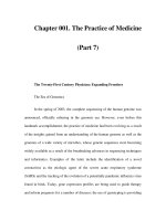
Fig. 8.18 Simplified schematic representation of electrolyte transport by ileal
mucosal tissue and its consequence for (a) absorption and (b) secretion. Active
processes involve the movement of ions and nutrient solutes; water follows
passively.
(a) Two methods of Na § co-transport are shown involving a glucose-linked
symport and two coupled antiports; the latter results in the co-transport of
C1 The coupled antiports are functionally linked via H § and HCO3, the rela-
tive concentrations of which are a reflection of metabolic activity. These
processes occur within the same cells but are shown separately for clarity. The
driving force for Na § uptake is the low Na § concentration maintained by the
Na§ § pump (ATPase) which creates the electrochemical gradient that
promotes the inward movement of Na+; C1- follows Na § by diffusion. Water is
drawn osmotically across the epithelium paracellularly (i.e. across tight junc-
tions) and/or transcellularly, the former pathway accounting for approximately
80% of fluid movement.
(b) Secretion is the result of the coupled entry ofNa § and C1- across the baso-
lateral membrane. Na § is recycled by the Na§ § pump and C1- exits by
diffusing down an electrochemical gradient and across the undifferentiated
crypt cell apical membrane; Na § follows C1- and water follows passively.
Note: (i) The driving force results from the same mechanism that powers
absorption, i.e. the Na§ § pump located in the basolateral membrane; it is the
location of the 'port' 'diffusion' systems that determines the vectorial aspects of
ion movement. (ii) The tight junctions are less tight in the crypts than villi. (iii)
The apical membrane of the crypt cell is undifferentiated and only acquires
microvilli during ascent into villous regions. O, Na § § pump; O, symport,
antiport or diffusion channel.
Villus tips and crypts are regarded as the anatomical sites of physi-
ological absorption and secretion respectively. Fluid transport is a bi-
directional process in the healthy animal with net absorption in health
and net secretion in disease. The balance between absorption and
secretion is poised at different points throughout the intestinal tract
reflecting differences in both structure and function. Proximal small
intestine is relatively leaky; in contrast the colon is a powerfully
absorptive organ.
8 Mechanisms of Cell and Tissue Damage
297
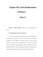
Fig. 8.19 Small intestinal villus: simplified schema of integrated structure and
function. Note the central arterial vessel (AV) which arborizes at the tip into a
capillary bed drained by a subepithelial venous return (VR). Movement of
sodium into VR creates a concentration gradient between VR and AV, causing
absorption of water from AV and surrounding tissue. This results in a progres-
sive increase in the osmolarity of incoming blood moving into the tip region
through to VR. Tip osmolarity is about three times higher than normal.
Hyperosmolarity has been demonstrated in man and can be inferred in mice
from the morphology of erythrocytes which changes during ascent of the same
vessel from base to tip regions of villi. The intensity of shading indicates a
vertical increase in osmolarity. The left crypt represents normal physiological
secretion and the right crypt hypersecretion. ENS, the enteric nervous system,
is depicted schematically and not anatomically.
Finally, crypts are the principal sites of cell regeneration, replacing
cells which migrate up the epithelial escalator. The epithelium is
renewed in approximately 3-5 days. At villus tips senescent cells are
shed.
Diarrhoeal disease can result from interference with almost any one,
or combination of these systems. The range of intestinal pathogens and
the types of disease they cause is illustrated in Tables 8.7 and 8.8. The
pathological/pathophysiological nature of some pathogen/host interac-
tions is illustrated in Fig. 8.20. Noninvasive pathogens like V.
cholerae
and enterotoxigenic
E. coli
(ETEC) secrete toxins which perturb the ion
transport systems. Invasive nonhistotoxic pathogens, such as some
Salmonella
strains (see Ch. 2) and rotavirus, invade villus tip cells
which are then shed into the intestinal lumen. Invasive histotoxic
pathogens, such as some strains of
Salmonella
(see Ch. 2), cause rapid
toxin-mediated detachment of epithelial cells. Experimental rotavirus
infections have been studied in great detail allowing us to delineate
298
Mims' Pathogenesis of Infectious Disease
Table 8.7. Production of diarrhoea by microorganisms shed in faeces
Infectious agent Diarrhoea Site of replication
Rotaviruses +
Parvoviruses (dogs) +
Intestinal adenoviruses (types 40, 41) +
Intestinal coronaviruses a +
Norwalk virus group (caliciviruses) +
Toroviruses (calves, horses, humans) +
Vibrio
cholerae +
Clostridium difficile +
Campylobacter jejuni +
E. coli +
Shigella +
Salmonella
sp. __
Salmonella typhi +
Cryptosporidium +
Giardia lamblia +
Entamoeba histolytica +
Intestinal epithelium
Intestinal epithelium (crypt cells)
Intestinal epithelium
Intestinal epithelium
Intestinal epithelium
Intestinal epithelium and M cells
(see Table A.5)
Intestinal lumen
Intestinal lumen
Intestinal epithelium
Varies b
Intestinal epithelium
Intestinal epithelium (varies)
Intestinal lymphoid tissue, liver,
biliary tract
Intestinal epithelium
Attached to intestinal epithelium
Invasion of intestinal epithelium
a Described for pigs, foals, calves, sheep, dogs, mice, man and turkeys; maximum susceptibility in the
first few weeks of life.
b Strain ETEC remains in the lumen; EIEC is similar to
Shigella,
EHEC reaches subepithelial
tissues.
intermediate stages between initial infection, through clinical diar-
rhoea to recovery from infection. We either do not know or can only
infer what the intermediate stages are for the other examples alluded
to - signified by broken arrows (Fig. 8.20) - leading to a return to
normal in those cases in which disease is self-limiting.
Campylobacter jejuni
does not figure in our treatment so far despite
the fact that
C. jejuni
and related species are the most common bacte-
rial cause of diarrhoea in many industrialised countries. This is
because of a severe lack of relevant 'mechanistic' information due to
the lack of good experimental models; hence we know very little about
the detailed mechanisms of pathogenicity of this hugely important
pathogen. The clinical picture of the pathogenesis of
C. jejuni
infection
may be summarised as follows. In developing countries the most
common clinical presentation is mild watery diarrhoea, whereas in
developed countries disease often manifests as a severe inflammatory
diarrhoea. No evidence has yet been found to suggest that the watery
type and severe bloody type of diarrhoeas can be explained in terms of
a C. jejuni
equivalent of the ETEC and EHEC mechanisms described
above. Current thinking proposes that the different disease patterns
reflect the immunological status of the host. Those with full immunity
experience no clinical disease, whereas those with no pre-immunity
experience the full-blown bloody diarrhoea and those with partial
8 Mechanisms of Cell and Tissue Damage
299
Table 8.8. Types of intestinal infection
Types of infection Microorganism Disease
Microorganism attaches
to epithelium of small
intestine, rarely
penetrates and causes
disease (diarrhoea)
often by forming a
toxin(s) which induces
fluid loss from
epithelial cells
Vibrio cholerae
E. coli (certain
strains)
Giardia lamblia
Cholera
Infantile gastroenteritis
(certain types) or mild
cholera-like disease in
adults (travellers'
diarrhoea)
Calf diarrhoea
Giardiasis
Microorganism attaches
to and penetrates
epithelium of large
intestine (Shigella) or
ileum (Salmonella),
causing disease by
shedding/killing
epithelial cells
(exotoxin?) and
inducing diarrhoea.
Subepithelial
penetration uncommon
Shigella spp.
Salmonella (certain
species) a
E. coli (certain
strains)
Campylobacter
jejuni
Human diarrhoea
viruses
Eimeria spp.
Entamoeba
histolytica
Bacillary dysentery
Salmonellosis
Coliform enteritis or
dysentery
Piglet diarrhoea
Diarrhoea, enteritis in
man b
Gastroenteritis
Coccidiosis in domestic
animals (may cause
diarrhoea and blood
loss)
Amoebic dysentery
Microorganism attaches
to and penetrates
intestinal wall. Also
invades subepithelial
tissues, sometimes
(typhoid, hepatitis A)
spreading systemically
Salmonella typhi Enteric fever (typhoid)
and paratyphi
Salmonella (certain Salmonellosis (severe
species) form)
E. coli (certain Calf enteritis
strains)
Hepatitis A virus Varied
Reoviruses, Hepatitis
enteroviruses
a There are more than 1000 serotypes of Salmonella, distinct from Salmonella typhi and
Salmonella paratyphi. They are primarily parasites of animals, ranging from pythons to
elephants, and their importance for man is their great tendency to colonise domestic
animals. Pigs and poultry are commonly affected, and human disease follows the
consumption of contaminated meat or eggs.
b Other campylobacters cause sepsis, abortion and enteritis in animals.
immunity, watery diarrhoea. The incubation period can range from I to
7 days and acute diarrhoea can last for 1-2 days with abdominal pain
which may persist after diarrhoea has stopped. Diarrhoeal stools often
contain fresh blood, mucus and an inflammatory exudate with leuco-
cytes; bacteremia may also occur though it is rarely reported. Infected
mucosae may be oedematous and hyperaemic with petechial haemor-
rhages. The disease, even its severe form, tends to be self-limiting,
300
Mims' Pathogenesis of Infectious Disease
despite the fact that organisms may be isolated for several weeks after
resolution of the symptoms. We do, however, know that there is a
strong correlation between infection with
C. jejuni
and Guillain-Barr~
syndrome which is the most notable complication of
C. jejuni
infection.
Guillain-Barr~ syndrome is a peripheral neuropathy, and one possible
cause may be an autoimmune phenomenon arising from molecular
8 Mechanisms of Cell and Tissue Damage
301
mimicry between the polysaccharide side chains of
C. jejuni
and neural
gangliosides.*
While there are reasonable models for studying colonisation and
initial invasion, there is a problem regarding experimental animal
models in which to reproduce the extreme form of bloody diarrhoea
seen in humans. However, the situation concerning
C. jejuni
is prob-
ably about to change dramatically. New strategies based on the use of
the new technology of 'microarrays'~ are now being used. By this
means, and by reference to the genomic atlas, it is theoretically poss-
ible to identify which genes are expressed under different sets of
experimental conditions including those which mimic the infection
environment. Doubtless a plethora of new data is about to be generated
from which we hope to learn more of the disease-conferring attributes
of
C. jejuni
and related species.
Rotaviruses are known to invade intestinal epithelial cells and cause
diarrhoea in man, foals, dogs, pigs, mice, etc. Extensive multiplication
takes place and very large amounts of virus (1011 particles g-l) are shed
in faeces. The conventional wisdom is that tips of villi especially are
* Guillain-Barr~ syndrome is also associated with certain virus infections, and 'flu vacci-
nation (see Ch. 12).
t Microarrays: see Ch. 1.
Fig. 8.20 Diarrhoeal mechanisms: initial stages and (for rotavirus) some inter-
mediate stages in disease progression. This represents a schematic summary
of the text on diarrhoeal mechanisms. In all cases, broken arrows indicate
uncertainty about the number and nature of intermediate steps in the return
to normality of affected villi in self-limiting diarrhoeal disease. For clarity, the
blood supply in [2] and both blood supply and enteric nervous system (ENS) in
[3], [4], [5] and [6] have been omitted.
[1] represents a normal villus; the shading intensity (as in Fig. 8.19) repre-
sents the magnitude of osmolarity. [2] Intoxication of villi by noninvasive
pathogens such as V.
cholerae
and ETEC. The main diarrhoeal determinant is
CT in V.
cholerae
and LT and ST in ETEC. However, as discussed in the text,
toxins are not the whole story, hence the broken arrows. [3] Represents disease
caused by invasive pathogens such as nonhistotoxic
S. typhimurium
and
rotavirus. Villi are shortened with presumed loss of absorption and observed
increase in secretion. Again the mechanistic pathway for return to normality is
not known for bacterial infections. [4] Loss of epithelia due to a histotoxin seen
in some strains of
S. typhimurium.
Clearly loss of enterocytes will affect
absorption and open up other routes for progressive invasion. Again note the
broken arrow. [5] A more complete experimentally based understanding of the
pathophysiological mechanisms is possible in rotavirus infection of neonatal
mice ([5], [6] and [7]). The main point is that conventional wisdom is not
sustained: maximum diarrhoea occurred during the resynthesis of truncated
villi and villus shortening was preceded/caused by ischaemia. Prolongation of
diarrhoea coincided with non-hypertonic villi; diarrhoea ceased on reconstitu-
tion of hypertonic villus tip regions. It is possible to infer that some of these
intermediate steps take place in other gut infections.
302 Mires' Pathogenesis of Infectious Disease
affected, leading to reduced absorption of fluid from the lumen. In addi-
tion destruction of enterocytes leads to a loss in lactase resulting in an
accumulation of lactose in the gut causing an osmotic flux of fluid into
the intestine. A major study of rotavirus-induced diarrhoea in neonatal
mice provides a different model of this important disease of children.
The main features of this model are summarised in Fig. 8.20. Oral
infection of the gut induces ischaemia in villi, followed by hypoxia,
enterocyte damage, and shortening of villi. The perception is that it is
the induction of ischaemia and not viral replication per se that results
in these changes. It is during rapid resynthesis of the atrophied villi
that maximum diarrhoea occurs due to the transient accumulation of
excess NaC1 in dividing cells. Prolongation of diarrhoea is seen to be
due to the hyperaemic state of the newly reconstructed villi which
reduces the hypertonicity of villi. Resolution of the diarrhoea occurs
when microcirculation is restored to normal with concomitant restora-
tion of hypertonic tip zones in villi.
The preceding description of the self-limiting diarrhoea induced by
rotavirus in neonatal mice is that of a basic response probably applic-
able to many diarrhoeas since the features of the post-peak phase have
often been reported or can be inferred in other infections. However, the
observed pathology will be different according to age, host species, or
the inducing pathogen. For example, in rotavirus-infected lambs, villus
atrophy and crypt hypertrophy occur (the latter indicative of crypt cell
division) but as in mice, infected lambs are not lactose intolerant. In
rotavirus-infected swine piglets, crypt hypertrophy occurs but villus
atrophy is severe, the animals are lactose intolerant and mortality is
high; a similar situation exists for the coronavirus, transmissible
gastroenteritis (TGE) virus of swine. The latter has often been used as
the model for infantile diarrhoea but the question is whether human
infants are more like piglets or lambs. Clinical studies have shown that
recovery from mild, acute gastroenteritis of rotavirus origin occurs
within 2 weeks irrespective of the carbohydrate ingested. Clearly, the
severity of disease and the clinical outcome will depend on the extent
of 'vertical' villus/crypt involvement and the regions of intestine
infected. When villus erosion is severe, then lactose may cause an
'osmotic' purge or be fermented by intestinal bacteria to short-chain
fatty acids which stimulate secretion in the colon. Astroviruses,
Norwalk virus, caliciviruses and certain adenoviruses all cause
gastroenteritic disease by infecting enterocytes. However, parvoviruses
cause severe intestinal disease in dogs by virtue of their predilection
for the mitotically active crypt cells which is the cause of the near-
complete erosion of villi similar to that seen after exposure to sublethal
doses of irradiation.
Can we be more specific about the viral determinants responsible for
triggering these complex host reactions? It has recently been shown
that a non-structural rotavirus protein, NSP4, induces diarrhoea in
mice when introduced into the ileum, by causing increased C1- secre-
8 Mechanisms of Cell and Tissue Damage
303
tion. An apparent exception to the 'rule' that viruses do not form toxins!
Entamoeba histolytica causes lysis of target cells apparently by
direct contact with the cell membrane. This pathogen produces under
in vitro conditions a spectacular array of potential (but as yet
unproven) virulence determinants including: proteases that round up
cells, pore-forming proteins, collagenases and oligosaccharidases and
neurotransmitter-like compounds; the latter can induce intestinal fluid
secretion. Some of these factors have been implicated as the determi-
nants responsible for liver abscess formation.
Although much research has been focused on toxins, their mode of
action, and their role in disease, it is useful to compare different types
of intestinal infection and to refer to the concept of food poisoning.
Types of intestinal infection are set out in Table 8.8. Food poisoning is
a loosely used term, and usually refers to illnesses caused by
preformed toxins in food, or sometimes to illnesses that come on within
a day or so after eating contaminated food. Food may be contaminated
with plant poisons, fungal poisons (e.g. poisoning due to Amanita phal-
loides), fish poisons,* heavy metals, as well as with bacterial toxins or
bacteria.
References
Alouf, J. E. and Freer, J. (eds) (1999). 'The Comprehensive Sourcebook
of Bacterial Protein Toxins'. Academic Press, London. (This excellent
book contains up to date treatments on many of the protein toxin
topics alluded to in Ch. 8 of this book.)
Borriello, S. P. (1998). Pathogenesis of Clostridium difficile infection. J.
Antimicrob. Chemother. 41, 13-19.
Buchmeier, M. J. et al. (1980). The virology and immunology of lympho-
cytic choriomeningitis virus infection. Adv. Immunol. 30, 275-331.
Burke, B. and Desselberger, U. (1996). Rotavirus pathogenicity.
Virology 218, 299-305.
Casali, P. and Oldstone, M. B. A. (1983). Immune complexes in viral
infection. Curr. Topics Microb. Immunol. 104, 7-48.
Dale, J. B. and Beachey, E. H. (1985). Epitopes of streptococcal M
proteins shared with cardiac myosin. J. Exp. Med. 162, 583-591.
Fitzgerald, T. J. (1981). Pathogenesis and immunology of Treponema
pallidum. Annu. Rev. Microbiol. 35, 29-54.
Fleischer, B. and Hartwig, U. (1992). T-lymphocyte stimulation by
microbial antigens. In 'Biological Significance of Superantigens' (B.
Fleischer, ed.). Chem. Immunol. Vol. 55, pp. 36-64. Karger, Basel.
* Ingestion of scombroid fish (mackerel, etc.) containing large amounts of histamine or
similar substances leads to headache, flushing, nausea and vomiting within an hour.
304 Mims' Pathogenesis of Infectious Disease
Fontaine, A., Arondel, J. and Sansonetti, P. J. (1988). Role of Shiga toxin
in the pathogenesis of bacillary dysentery, studied by using a Tox-
mutant of Shigella dysenteriae-1. Infect. Immun. 56, 3099-3109.
Hamilton, P. J. et al. (1977). Disseminated intravascular coagulation: A
review. J. Clin. Path. 31,609-619.
Hirst, T. R. (1999). Cholera toxin and Escherichia coli heat labile
enterotoxin. In 'The Comprehensive Sourcebook of Bacterial Protein
Toxins' (J. E. Alouf and J. Freer, eds), pp. 104-129. Academic Press,
London.
Hormaeche, C. E., Penn, C. W. and Smyth, C. J. (eds) (1992). 'Molecular
Biology of Bacterial Infection. Current Status and Future
Perspectives'. Soc. Gen. Microbiology Symposium 49, Cambridge
University Press, Cambridge.
Hornick, R. B. et al. (1970). Typhoid fever: pathogenesis and immuno-
logic control. N. Engl. J. Med. 283, 739.
Kaper, J. B., Morris, J. G. and Levine, M. M. (1995) Cholera. Clin.
Microbiol. Rev. 8, 48-86.
Karaolis, D. K. R., Somara, S., Maneval, D. R., Johnson, J. A. and Kaper,
J. B. (1999). A bacteriophage encoding a pathogenicity island, a type-
IV pilus and a phage receptor in cholera bacteria. Nature 399,
375-379.
Ketley, J. M. (1997). Pathogenesis of enteric infection by
Campylobacter. Microbiology 143, 5-21.
Khan, S. A. et al. (1998). A lethal role for lipid A in Salmonella infec-
tions. Molec. Microbiol. 29, 571-579.
Laforce, F. M. (1994). Anthrax. Clin. Infect. Dis. 19, 1009-1114.
Levin, J., van Deventer, S. J. H., van der Poll, T. and Sturk, A. (eds)
(1994). 'Bacterial Endotoxins. Basic Science to Anti-Sepsis
Strategies'. Progress in Clinical and Biological Research. Vol. 388,
John Wiley & Sons Inc., New York.
Levin, J., Alving, C. R., Munford, R. S. and Redl, H. (eds). (1995).
'Bacterial Endotoxins. Lipopolysaccharides From Genes to Therapy'.
Progress in Clinical and Biological Research, Vol. 392, John Wiley &
Sons Inc., New York.
Lencer, W. I., Hirst, T. R. and Holmes, R. K. (1999). Membrane traffic
and the cellular uptake of cholera toxin. Biochim. Biophys. Acta
Molec. Cell Res. 1450, 177-190.
Lodge, J. M., Bolton, A. J., Martin, G. D., Osborne, M. P., Ketley, J. M.
and Stephen, J. (1999). A histotoxin produced by Salmonella. J. Med.
Microbiol. 48, 811-818.
Lundgren, O. and Jodal, M. (1997). The enteric nervous system and
cholera toxin-induced secretion. Comp. Biochem. Physiol. A 118,
319-327.
Mathan, M. M., Chandy, G. and Mathan, V. I. (1995). Ultrastructural
changes in the upper small intestinal mucosa in patients with
cholera. Gastroenterology 109, 422-430.
McGee, Z. A. et al. (1981). Pathogenic mechanism of Neisseria gonor-
8 Mechanisms of Cell and Tissue Damage 305
rhoeae: observations on damage to human fallopian tubes in organ
cultures by gonococci of colony Type I or Type 4. J. Infect. Dis. 143,
413-422; 432-439.
Mims, C. A. (1957). Rift Valley Fever virus in mice VI: Histological
changes in the liver in relation to virus multiplication. Austral. J.
Exp. Biol. Med. Sci. 35, 595.
Mims, C. A. (1985). Viral aetiology of diseases of obscure origin. Brit.
Med. Bull. 41, 63-69.
Nataro, J. P. and Kaper, J. B. (1998). Diarrheagenic Escherichia coli.
Clin. Microbiol. Rev. 11, 142-201.
Olsnes, S., Wesche, J. and Falnes, P. O. (1999). Binding, uptake, routing
and translocation of toxins with intracellular sites of action. In 'The
Comprehensive Sourcebook of Bacterial Protein Toxins' (J. E. Alouf
and J. Freer, eds), pp. 73-93. Academic Press, London.
Osborne, M. P., Haddon, S. J., Worton, K.J., Spencer, A. J., Starkey, W.
G., Thornber, D. and Stephen, J. (1991). Rotavirus-induced changes
in the microcirculation of intestinal villi of neonatal mice in relation
to the induction and persistence of diarrhea. J. Pediatr.
Gastroenterol. Nutr. 12, 111-120.
Poewe, W., Schelosky, L., Kleedorfer, B., Heinen, F., Wagner, M. and
Deuschl, G. (1992). Treatment of spasmodic torticollis with local
injections of botulinum toxin. J. Neurol. 239, 21-25.
Raudin, J. I. (1986). Pathogenesis of diseases caused by Entamoeba
histolytica: studies of adherence, secreted toxins and contact-depen-
dent cytolysis. Rev. Infect. Dis. 8, 247-260.
Rodriguez, M., von Wedel, R. J., Garrett, R. S. et al. (1983). Pituitary
dwarfism in mice persistently infected with lymphocytic chori-
omeningitis virus. Lab. Invest. 49, 48.
Schiavo, G., Benfenati, F., Poulain, B., Rossetto, O., de Laureto, P. P.,
DasGupta, B. R. and Montecucco, C. (1992). Tetanus and botulinum-
B neurotoxins block neurotransmitter release by proteolytic
cleavage of synaptobrevin. Nature 359, 832-833.
Schiavo, G., Poulain, B., Rossetto, O., Benfenati, F., Tauc, L. and
Montecucco, C. (1992). Tetanus toxin is a zinc protein and its inhibi-
tion of neurotransmitter release and protease activity depend on
zinc. EMBO J. 11, 3577-3583.
Silva, T. M. J., Schleupner, M. A., Tacket, C. O., Steiner, T. S., Kaper, J.
B., Edelman, R. and Guerrant, R. L. (1996). New evidence for an
inflammatory component in diarrhea caused by selected new, live
attenuated cholera vaccines and by E1Tor and O 139 Vibrio cholerae.
Infect. Immun. 64, 2362-2364.
Spencer, A. J., Osborne, M. P., Haddon, S. J., Collins, J., Starkey, W. G.,
Candy, D. C. A. and Stephen, J. (1990). X-ray-microanalysis of
rotavirus-infected mouse intestine- a new concept of diarrheal
secretion. J. Pediatr. Gastroenterol. Nutr. 10, 516-529.
Stephen, J. (2000). Pathogenesis of infectious diarrhea: A minireview.
Can. J. Gastroenterol. (in press).
306 Mires' Pathogenesis of Infectious Disease
Svanborg, C., Godaly, G. and Hedlund, M. (1999). Cytokine responses
during mucosal infections: role in disease pathogenesis and host
defence. Curr. Opin. Microbiol. 2, 99-105.
Uchida, H., Kiyokawa, N., Horie, H., Fujimoto, J. and Takeda, T. (1999).
The detection of Shiga toxins in the kidney of a patient with
hemolytic uremic syndrome. Pediatr. Res. 45, 133-137.
VanderSpeck, J. C. and Murphy, J. R. (1999). Diphtheria toxin-based
interlukin-2 fusion proteins. In 'The Comprehensive Sourcebook of
Bacterial Protein Toxins' (J. E. Alouf, and J. Freer, eds), pp. 682-690.
Academic Press, London.
Welliver, R. C. et al. (1981). The development of respiratory syncytial
virus-specific IgE and the release of histamine in naso-pharyngeal
secretions after infection. N. Engl. J. Med. 305, 841-845.
Williams, R. C. (1981). Immune complexes in human diseases. Annu.
Rev. Med. 32, 13-28.
Yuki, N. (1999). Pathogenesis of Guillain-Barre and Miller Fisher
syndromes subsequent to Campylobacter jejuni enteritis. Jap. J.
Infect. Dis. 52, 99-105.
9
Recovery from Infection
Immunological factors in recovery 307
Inflammation 321
Complement 323
Interferons 324
Multimechanistic recovery: an example 327
Temperature 329
Tissue repair 331
Resistance to re-infection 334
References 337
If there is to be recovery from an infection, it is first necessary that the
multiplication of the infectious agent is brought under control. The
microbe must decrease in numbers and cease to spread through the
body or cause progressive damage. This is accomplished by immuno-
logical and other factors whose action is now to be described. The
average multiplication rate of various microorganisms in the infected
host as shown by doubling times (Table 8.2), is nearly always longer
than in artificial culture under optimal conditions. This in itself reflects
the operation of antimicrobial forces. In the process of recovery from an
infectious disease, damaged tissues must of course be repaired and
reconstituted. Sometimes the microorganism is completely destroyed
and tissues sterilised, but often this fails to take place and the micro-
organism persists in the body, in some instances continuing to cause
minor pathological changes. The individual is nevertheless said to have
recovered from the acute infection and is usually resistant to re-
infection with the same microorganism. Persistent infections are dealt
with in Ch. 10.
Immunological Factors in Recovery
The mechanisms of recovery from a primary infection are not neces-
sarily the same as those responsible for resistance to re-infection (see
below). For instance, antibody to measles is of prime importance in
resistance to re-infection and susceptible children can be passively
307