Mollison’s Blood Transfusion in Clinical Medicine - part 4 pot
Bạn đang xem bản rút gọn của tài liệu. Xem và tải ngay bản đầy đủ của tài liệu tại đây (517.71 KB, 92 trang )
antigens, bind to additional as yet unidentified antigens.
These authors draw an analogy between the binding of
Ii carbohydrate structures to the hydrophobic patch
on KAU and the way in which oligosaccharide chains
of antibody molecules bind to a hydrophobic patch on
the Cγ2 domains of IgGFc. This alternative carbo-
hydrate antigen-binding region also provides a possible
explanation for the cold agglutinin activity of other
V4–34 encoded antibodies including monoclonal anti-
Ds (Thorpe et al. 1998).
Of the relatively few examples of anti-Pr, six were IgAκ and
five of these had Pr
1
and one, Pr
a
specificity (Angevine et al.
1966; Garratty et al. 1973; Roelcke 1973; Tonthat et al.
1976; Roelcke et al. 1993); one that was IgMκ was anti-Pr
2
and another (IgMλ), anti-Pr
3
(Roelcke et al. 1974, 1976). An
IgAκcold agglutinin had anti-Sa specificity (Roelcke et al. 1993).
IgM cold agglutinins with λ light chains are rarely directed
against the I antigen. They are frequently cryoprecipitable
and are often found in malignant conditions. Such agglutinins
thus differ markedly from cold agglutinins with κ light chains.
Patients with chronic CHAD synthesize IgM at approx-
imately 10 times the normal rate; treatment with alkylating
agents results in a diminished rate of synthesis (Brown and
Cooper 1970).
Occasionally, cold IgM anti-I is accompanied by a warm
IgG autoantibody of the same or another specificity (see
below). Examples of anti-I cold agglutinins that appeared to
be solely IgG were described by Ambrus and Bajtai (1969)
and Mygind and Ahrons (1973) and two cases in which the
anti-Pr was IgG1κ have been described (Dellagi et al. 1981;
Curtis et al. 1990). The latter case was unusual because the
cold agglutinins failed to activate complement.
The possibility that IgM anti-I is always accompan-
ied by at least traces of IgG and IgA autoantibodies is
raised by the finding of Hsu and co-workers (1974).
Using a PVP-augmented antiglobulin test in the auto-
analyzer they found that, in patients with typical anti-I
cold agglutinins, IgG and IgA could always be detected
on the patient’s red cells in addition to C3 and C4.
Similarly, Ratkin and co-workers (1973) prepared
eluates from 19 sera from patients with cold agglutinin
disease and regularly found an excess of IgG and of
IgA, both having agglutinating activity of relatively
low titre. They interpreted their observations to mean
that in patients in whom IgM autoantibodies predom-
inated, autoantibodies of classes IgG and IgA were also
regularly present, although in lower titre.
In mycoplasma infection, when a patient develops
potent cold autoagglutinins of anti-I specificity as a
transient phenomenon the antibody is made of hetero-
geneous IgM and contains both κ and λ light chains
(Costea et al. 1966), although the heterogeneity is
restricted (see Feizi 1977).
Production of cold autoagglutinins following
repeated blood transfusions
Rous and Robertson (1918) observed that in rabbits trans-
fused almost daily with the blood of other rabbits, cold
autoagglutinins developed in about one-half of the animals.
The animals with the most potent agglutinins developed a
sudden anaemia, due perhaps to immune clearance of trans-
fused cells. The agglutinins persisted in the animal’s serum
long after transfused cells had disappeared. Thus, in one case,
133 days after the last blood transfusion there was still gross
autoagglutination on chilling the animal’s blood.
Ovary and Spiegelman (1965) gave repeated injections of
Hg
A
-positive red cells to an Hg
A
-negative rabbit: the animal
produced not only the expected anti-Hg
A
active at 37°C, but
also a cold agglutinin.
The production of cold autoagglutinins in humans,
following alloimmunization and in association with a
delayed haemolytic transfusion reaction, has been
observed only occasionally (see Chapter 11).
Cold (biphasic) autohaemolysins
In the syndrome of paroxysmal cold haemoglobinuria
(PCH) the patient’s serum contains a cold, complement-
fixing antibody. This antibody, often referred to as
the Donath–Landsteiner antibody after its discoverers,
produces haemolysis both in vitro and in vivo when
the blood is first cooled (to allow the binding of anti-
body) and then warmed (to provide optimal conditions
for complement-mediated haemolysis). Because of this
behaviour, the antibody is described as a ‘biphasic
haemolysin’.
Although biphasic haemolysin was originally des-
cribed in a patient with tertiary syphilis, the majority
of cases seen nowadays are associated with viral
infections, particularly in children. In one series of
11 cases, only three were definitely syphilitic; of five
which were definitely non-syphilitic, one followed
measles and one mumps (Worlledge and Rousso
1965). Biphasic haemolysin may also occur transiently
following chickenpox, influenza-like illness and pro-
phylactic immunization with measles vaccine (Bird
et al. 1976a).
CHAPTER 7
260
Of 19 patients with biphasic haemolysin reported
by Sokol and co-workers (1982, 1984), 17 were
children. All patients were non-syphilitic. In 10 of the
children the biphasic haemolysin developed after an
upper respiratory tract infection. The other patients
had infections with adenovirus type 2, influenza A
virus or Haemophilus influenzae; one had chickenpox.
The authors stressed the fact that in the acute form that
typically occurs in children, the onset of the haemolytic
anaemia is sudden, usually with haemoglobinuria,
prostration and pallor. In the chronic form haemolysis
is only mild; this form occurred in only two patients,
one a child and the other an adult. Biphasic haemolysin
in an adult patient with pneumonia due to Klebsiella
was described by Lau and co-workers (1983).
In one series, all of 22 patients with biphasic
haemolysins were children, who developed the anti-
bodies after infection, usually of the upper respir-
atory tract (Göttsche et al. 1990b).
It has been suggested that for the prevalent non-
syphilitic form of the syndrome the term Donath–
Landsteiner haemolytic anaemia should be used rather
than PCH, as the clinical manifestations are rarely
paroxysmal, seldom precipitated by cold and not
necessarily characterized by haemoglobinuria (Wolach
et al. 1981).
Several estimates of the relative frequency of bipha-
sic haemolysin in AIHA are available. In one series
of 347 cases of AIHA, the antibody was found in six,
i.e. fewer than 2% (Petz and Garratty 1980, p. 54).
Similarly, of red cell autoantibodies from 2000 patients,
48 (2.4%) were biphasic haemolysins (Engelfriet et al.
1982). On the other hand, the antibody was present
in four of 34 (12%) acute cases of AIHA in children
in one series (Habibi et al. 1974) and in 17 out of
42 (40%) cases in another (Sokol et al. 1984). The
22 patients with biphasic haemolysins described by
Göttsche and co-workers (1990b) were among 599
patients with AIHA, 68 of whom were children.
Although maximum haemolysis is observed when
red cells are left with biphasic haemolysin and com-
plement in the cold phase, the requirement for com-
plement in the cold phase is not absolute. Thus when
red cells are first left at 0°C with EDTA-treated serum
containing fairly potent antibody then washed and
incubated at 37°C with fresh normal serum, some
haemolysis occurs (Polley et al. 1962). Similarly, Hinz
and co-workers (1961a) found that if PNH red cells
were used, haemolysis occurred quite readily when
complement was supplied only in the warm phase of
the reaction. An experiment described by Dacie (1962,
p. 553) shows clearly that the reason why much more
haemolysis is found when complement is present in the
cold phase of the reaction is that, on warming, anti-
body very rapidly elutes from the red cells so that at
higher temperatures there is usually too little antibody
on the cells to activate complement. Hinz and co-
workers (1961b) showed that optimal lysis occurred
even when only C1 was present with antibody in the
cold phase of the reaction; C4 could be present either
in the cold or warm phases but C2 and C3 were essen-
tial in the warm phase.
False-negative results may be observed due to hypo-
complementaemia and it may then be necessary to add
fresh normal serum to demonstrate the presence of the
biphasic haemolysin (Wolach et al. 1981).
In cases in which biphasic haemolysin is associated
with syphilis (tertiary or congenital) the antibody is
seldom active above 20°C; that is to say, red cells and
serum must be cooled to a temperature below 20°C
if there is to be haemolysis on subsequent warming.
In cases in which the antibody appears transiently in
children following infections, the thermal range is
greater and the antibody may be active in vitro up to
a temperature as high as 32°C (see Bird et al. 1976a).
A monophasic haemolysin acting in vitro up to 32°C,
in an adult, was described by Ries and co-workers
(1971).
As mentioned above, potent cold autoagglutinins
which are readily lytic may be confused with biphasic
haemolysin, but the latter is usually non-agglutinating,
produces substantially more lysis, is IgG rather than
IgM and has anti-P rather than anti-I specificity. A test
that helps to distinguish unusually lytic anti-I from
biphasic haemolysin is described above.
Specificity. Classically, biphasic haemolysin has the
specificity anti-P (Levine et al. 1963; Worlledge and
Rousso 1965). Very occasionally, the specificity may
be anti-‘p’ (see Chapter 4).
Biphasic haemolysins with anti-P specificity are
inhibited by globoside: some are more strongly inhib-
ited by the Forssman glycolipid, which contains the
globoside structure with an additional terminal GalNAc
residue, suggesting that the antibodies are probably
evoked by Forssman antigens which are widespread in
animal tissues and microorganisms (Schwarting et al.
1979). Chambers and Rauck (1996) described a case
RED CELL ANTIBODIES AGAINST SELF-ANTIGENS, BOUND ANTIGENS AND INDUCED ANTIGENS
261
of childhood acute haemolytic anaemia following
parvovirus infection. In this case the reticulocyte count
was low (1.0%, attributed to parvovirus infection, see
Chapter 4) despite profound anaemia (haematocrit
14.5%). As P antigen is both the receptor for parvovirus
B19 and the target for most biphasic haemolysins the
authors speculate that interaction of the virus with
P antigen may have triggered an auto-anti-P response.
Occasionally, biphasic haemolysins have a specificity
outside the P system: anti-IH (Weiner et al. 1964),
anti-I (Engelfriet et al. 1968a; Bell et al. 1973a), anti-i,
as described above, or anti-Pr like (Judd et al. 1986). In
practice, determination of the specificity of biphasic
haemolysins is not helpful in diagnosis. On the other
hand, in children with antibodies of wide thermal
range and severe red cell destruction, confirmation of
anti-P specificity may be helpful in treatment, as trans-
fusion of pp red cells is sometimes very successful (see
below).
Immunoglobulin class. Biphasic haemolysin (of speci-
ficity anti-P) is composed of IgG (Adinolfi et al. 1962;
Hinz 1963). If red cells are incubated at a temperature
such as 15°C with fresh serum containing biphasic
haemolysin and then washed at room temperature,
they react weakly with anti-IgG but strongly with
anti-C4 and anti-C3, as expected from the fact that the
antibody elutes rapidly as the temperature is raised.
During, and for some time after, an attack of haemo-
globinuria, the red cells of patients with PCH give a
positive direct antiglobulin test (DAT). Only comple-
ment components (presumably C3d and C4d) can be
detected on the red cells.
Red cell transfusion in patients with biphasic
haemolysins
Red cell transfusion is seldom required in PCH. When
the thermal range of the antibody extends only to 20°C
or so in vitro, the patient is not severely anaemic. In
patients in whom the thermal range extends to 30°C
or more, severe anaemia does occur occasionally but
in these patients the disease is usually transient and
recovery has usually begun before the question of
transfusion has to be considered. The successful use of
P-negative red cells (from a bank of frozen blood) has
been reported (Rausen et al. 1975) but unwashed,
unwarmed P-positive blood has also been used suc-
cessfully in three affected children (Wolach et al.
1981). In a child with PCH and severe anaemia, who
did not respond to transfusion of P-positive blood, the
transfusion of P-negative blood resulted in a sustained
rise in Hb level (I Franklin and M Contreras, personal
observation). The use of plasmapheresis to remove the
Donath–Landsteiner antibody and ameliorate severe
autoimmune haemolytic anaemia in a child follow-
ing gastroenteritis is described by Roy-Burman and
Glader (2002). The authors consider that because
production of the Donath–Landsteiner antibody is
transient and relatively brief in post-viral illness,
removal of the antibody by plasmapheresis is less
likely to be followed by significant rebound antibody
production.
Harmless warm autoantibodies
IgG subclass of harmless warm antibodies
The affinity of Fc receptors for IgG4 is very low and
subjects with only IgG4 on their red cells are expected
to have a positive DAT but no signs of red cell destruc-
tion. With IgG2, the situation is more complex because,
as explained in Chapter 3, there are two alleles of the
gene that encodes the FcRIIa receptor on macrophages.
As a result, some subjects have a low-affinity receptor
for IgG2 and, in the presence of an IgG2 autoantibody
have a positive DAT without signs of red cell destruction;
others have a high-affinity receptor and the potential
to destroy IgG2-coated cells. Indeed, some patients
with only IgG2 on their red cells have haemolytic
anaemia (CP Engelfriet, unpublished observations).
However, IgG2-mediated destruction depends upon
antigen specificity; see pp. 227–228 and 426.
Although IgG1 and IgG3 readily adhere to Fc recep-
tors and antibodies of these subclasses are expected
to cause red cell destruction, in the case of IgG1, the
number of molecules bound per cell must exceed a
certain minimum number to bring about attachment
to phagocytes and thus to cause red cell destruction
(see below). Subjects with a relatively small number
of IgG1 molecules per red cell are expected to have a
positive DAT without signs of red cell destruction.
Positive direct antiglobulin test in apparently
normal subjects
The fact that an apparently normal donor has a posit-
ive DAT is often first discovered when the donor’s red
CHAPTER 7
262
cells are used in crossmatching. Sixty-five cases were
found in this way in one region during a period in
which one million donations were collected. Assuming
that for every 10 donors detected one was missed,
the frequency of donors with a positive DAT was
estimated to be one in 14 000 (Gorst et al. 1980).
In another prospective survey donors with a positive
DAT were discovered either by antiglobulin testing or
by noting autoagglutination of a blood sample in an
automated or manual test and then doing an antiglob-
ulin test. The frequency of donors with a positive DAT
was one in 13 000 (Habibi et al. 1980). Although the
results of these two surveys look very similar there
were apparent differences between the two. In the first
there was only C3d (and C4d) on the red cells of 28 of
the 65 donors. All donors with a positive DAT were
haematologically normal; of 32 of the donors followed
for many years, 31 remained well and only one, with a
strongly positive DAT with anti-IgG, developed AIHA
(Gorst et al. 1980).
In the second series, immunoglobulin was detectable
on the red cells in all of 69 cases (IgG in 67, IgM in 2).
Ten per cent of the donors had subnormal Hb values;
a further 29% had reticulocytosis, with or without
hyperbilirubinaemia: 61% appeared to be normal
haematologically but when Cr survival studies were
carried out in a few of these subjects, results were
below normal in about 50% of the cases (Habibi et al.
1980). It should be noted that 25% of the donors with
a positive DAT were receiving methyldopa, a circum-
stance that might have debarred them from donation
in many countries. In any case, it must be said that the
evidence presented for a haemolytic state in many of
the donors was rather slight. No donor had a reticulo-
cyte count higher than about 4.5% or a bilirubin value
higher than 2.2 mg/dl (37 µmol/l). Slightly reduced
Cr survival in haematologically normal subjects is
difficult to interpret. Finally, in many of the donors
who were followed for a period of 1 year or more,
haematological findings became normal.
A very much higher frequency of positive DATs
in normal donors than that found in the two series
mentioned above was reported by Allan and Garratty
(1980), namely one in 1000, but the discrepancy may
be more apparent than real, as over 90% of the reac-
tions were only ‘1+’ or less.
In 22 out of 23 normal donors with IgG on their red
cells from the series of Gorst and co-workers (1980),
the IgG subclass of the antibody was later investigated.
In 20 of the cases it was solely IgG1 and the number of
IgG1 molecules per red cell varied from 110 to 950; in
the remaining two subjects the red cells were coated
only with IgG4 (Stratton et al. 1983). In another series
of 10 subjects, five had only IgG1, three IgG4, one
IgG2 and one both IgG1 and IgG3 (Allan and Garratty
1980).
In normal donors with a positive DAT, the speci-
ficity of the autoantibody, as in patients with AIHA, is
often related to Rh (Issitt et al. 1976a; Habibi et al.
1980) but may be outside the Rh system, for example
anti-Jk
a
(Holmes et al. 1976) and anti-Xg
a
(Yokohama
and McCoy 1967).
In normal subjects with IgG on their red cells, the
red cells may be agglutinated by anti-complement as
well as anti-IgG, although the frequency with which
both IgG and complement have been found has varied
widely in different series, i.e. 15% (Gorst et al. 1980);
44% (Allan and Garratty 1980) and 70% (Issitt et al.
1976).
Positive direct antiglobulin test (IgG) in
hypergammaglobulinaemia
An association has been observed between hypergam-
maglobulinaemia and a positive DAT. Of 50 patients
with an increased concentration of IgG in their serum,
25 had a positive DAT without signs of increased red
cell destruction. The eluates from the red cells were
unreactive (Huh et al. 1988). In another study of
20 patients with an increased serum IgG and a positive
DAT, there were no signs of increased red cell destruc-
tion. These eluates were also unreactive (Heddle et al.
1988). In a prospective study of 44 patients with
increased serum IgG, the DAT was positive in the
three patients with the highest IgG concentrations.
The DAT became positive in two other patients who
were treated with high-dose intravenous immunoglob-
ulin and, again, the eluates were unreactive (Heddle
et al. 1988). In patients with a positive DAT but with
an unreactive eluate, a significant correlation has
been observed between the strength of the DAT reac-
tion and the serum IgG concentration (Clark et al.
1992).
C3d (and C4d) alone on red cells
In 40–47% of normal donors with a positive DAT,
only complement is detected on the red cells (Allan and
RED CELL ANTIBODIES AGAINST SELF-ANTIGENS, BOUND ANTIGENS AND INDUCED ANTIGENS
263
Garratty 1980; Gorst et al. 1980). C3d can be demon-
strated on all normal red cells by using a sufficiently
potent anti-C3d serum (Graham et al. 1976) and both
C3d and C4d can be demonstrated by using the sensit-
ive PVP-augmented antiglobulin test (Rosenfield and
Jagathambal 1978). The presence of these fragments
on red cells is taken as evidence of continuing low-
grade activation of complement (see Chapter 3). There
is no reason to believe that autoantibodies of any kind
are responsible for this activation and it is therefore
not logical to discuss this subject under the general
heading of ‘harmless warm autoantibodies’, but it is
nevertheless convenient.
The amount of C3d on the red cells of normal adults
has been estimated by using rabbit IgG anti-C3d and
125
I-labelled goat anti-rabbit IgG; in 174 normal
adults there were estimated to be between 50 and 200
C3d molecules per red cell, i.e. too few to be detected
in the ordinary DAT. There was no difference between
males and females and no evidence of any change in
the number of molecules per cell over the age range
20–65 years. There was also no evidence that the
number was different in children (Chaplin et al. 1981).
Other estimates of the number of C3d molecules per
cell in normal adults are 207–427 (Freedman and
Barefoot 1982) and 280–560 (Merry et al. 1983).
Weakly positive DATs due to increased amounts
of C3d on the red cells appear to be relatively frequent
in subjects who are ill. Dacie and Worlledge (1969)
found that 40 out of 489 (8%) patients in hospital gave
weakly positive antiglobulin reactions due to com-
plement. Similarly, Freedman (1979) found that of
100 EDTA samples from hospital patients, taken at
random, seven gave positive reactions with anti-C3d
and anti-C4d; all seven patients were seriously ill.
Again, in 8% of random hospital patients values
greater than 230 C3d molecules per cell were found
by Chaplin and co-workers (1981), who also noted
that in random patients in hospital 33% had values for
the numbers of C3d molecules per red cell that were
above the range found in more than 90% of healthy
adults.
In testing red cells with anti-C3d and anti-C4d,
freshly taken EDTA blood should be used whenever
possible, as the amounts of C3d and C4d on red cells in
ACD blood may increase slightly during brief storage
at 4°C (Engelfriet 1976); after 21 days of storage, the
increase of C3d and C4d may be two-fold (H Chaplin,
personal communication).
Positive direct antiglobulin test associated with
various diseases, but without signs of increased
red cell destruction
Malaria. A positive DAT has been found in 40–50%
of West African children with falciparum malaria
(Topley et al. 1973; Facer et al. 1979; Abdalla and
Weatherall 1982). In most cases, only C3d is detected
on the red cells but in some both C3d and IgG are
present and, in a few, IgG alone. Although in one series
there was a relationship between a positive DAT and
anaemia (Facer et al. 1979), in the others there was
not. It was suggested that a positive test might be asso-
ciated with the development of immunity to malaria
(Abdalla and Weatherall 1982).
On the other hand, some patients with falciparum
malaria, with antibodies against triosephosphate,
associated with a positive DAT, have a prolonged
haemolytic anaemia (Ritter et al. 1993).
Kala azar. The presence of complement on the red
cells of patients with this disorder was reported by
Woodruff and co-workers (1972). Of 67 patients with
kala azar, 33% tested prior to antimonial therapy had
a positive DAT (Vilela et al. 2002).
Patients on
α
-methyldopa and other drugs. The devel-
opment of a positive DAT without any evidence of a
haemolytic process is very common in patients taking
α-methyldopa and is found occasionally in patients
taking a variety of other drugs. The subject is con-
sidered in more detail in the section on drug-induced
haemolytic anaemia.
Patients with autoimmune haemolytic disease in
spontaneous remission without signs of red cell
destruction may have a positive DAT (Loutit and
Mollison 1946). In a patient reported by Goldberg
and Fudenberg (1968), the red cells were initially
agglutinated by anti-IgG and anti-C3; the serum con-
tained an IgM antibody reacting with IgG-coated red
cells. After treatment with steroids, the patient went
into complete haematological remission and the IgM
antibody disappeared from the serum; however, the
red cells were still strongly agglutinated by anti-IgG
and anti-C3.
In a patient reported by von dem Borne and co-
workers (1977), who initially suffered from severe
AIHA, a long-lasting remission was induced with
steroid therapy, and it was then found that the antibody
CHAPTER 7
264
on the patient’s cells was predominantly IgG4; the
coated red cells induced only weak rosetting with
monocytes in vitro and it was postulated that there had
been a switch in the subclass of the autoantibody, with
production of a subclass IgG4, which was incapable of
producing destruction in vivo.
Harmful warm autoantibodies
As mentioned above, antibodies reacting as well, or
better, at 37°C than at lower temperatures are found in
about 80% of all cases of AIHA. In the warm antibody
type of AIHA, the DAT is almost always positive but
the indirect test (for antibody in serum) is sometimes
negative. Harmful warm autoantibodies are of two
kinds: incomplete antibodies and haemolysins.
Incomplete warm autoantibodies
IgG alone has been found in 18.3% (Petz and Garratty
1975), 36% (Worlledge 1978) and 64% (Engelfriet
et al. 1982) of cases.
IgG alone was found invariably in patients with a
positive DAT associated with α-methyldopa in two
series (Worlledge 1969; Issitt et al. 1976), although
in a third, IgM and complement (Clq), in addition to
IgG, were found on the red cells of all patients who
developed α-methyldopa-induced haemolytic anaemia
(Lalezari et al. 1982), results that could not be repro-
duced by one previous author (CP Engelfriet) or by
Ben-Izhak and co-workers (1985). The detection of
the IgM antibodies appears to depend on the anti-IgM
serum used. It has been suggested that if the affinity
of the anti-IgM for IgM is much greater than that of
the IgM red cell antibodies for the red cell antigen, the
IgM antibodies are removed from the red cell in the
antiglobulin phase of the test (P Lalezari, personal
communication).
IgG and complement have been found in 64.5%
(Petz and Garratty 1980), 44.4% (Worlledge 1978)
and about 34% (Engelfriet et al. 1982) of cases; in
the latter series, IgG and complement were found
on the red cells of all patients with a combination
of IgG incomplete warm autoantibodies and warm
haemolysins (see below).
When complement and IgG are found on the red
cells of patients with incomplete warm autoantibodies,
it does not follow that complement has been fixed by
autoantibody. Some of the evidence for this assertion
is as follows: (1) neither IgG incomplete warm auto-
antibodies present in the serum nor those detectable in
an eluate from the red cells are capable of fixing comple-
ment in vitro; (2) in at least 50% of patients with IgA
incomplete warm autoantibodies alone, complement
is detectable on the red cells; and (3) the frequency
with which both IgG and complement are found on
the red cells is much higher in patients suffering from
a typical immune complex disease such as systemic
lupus erythematosus (SLE), than in other cases of the
warm type of AIHA. Thus, in SLE, both IgG and com-
plement were found on the red cells in all cases by
Chaplin (1973) and Worlledge (1978), in virtually all
cases by Petz and Garratty (1980) and in 81% of cases
by Engelfriet and colleagues (1982).
IgG subclass of warm incomplete
autoantibodies
IgG warm autoantibodies are IgG1 in the vast majority
of patients (Engelfriet et al. 1982). IgG1 alone was
found in 72% of patients and IgG1 with antibodies
of another subclass in 25%. In only 23 out of 572
patients was no IgG1 detectable. IgG2 and IgG4 anti-
bodies were found the least frequently. Table 7.2
shows the frequency with which IgG autoantibodies of
only one subclass were detected, and the relation of the
subclass of the autoantibodies to increased red cell
destruction. Determination of the IgG subclass of
autoantibodies in eluates can readily be achieved using
commercially available gel tests (Fabijanska-Mitek
et al. 1997).
As mentioned above, subjects whose red cells are
coated with not more than 950 IgG1 molecules per cell
show no signs of red cell destruction. On the other
hand in patients with AIHA with only IgG1 on their
red cells, the number of molecules per cell was found to
be 1200 or more (Stratton et al. 1983). This finding
agrees well with the observation that at least 1180
IgG1 anti-D molecules must be bound per cell for
adherence to monocyte receptors to occur in vitro
(Zupanska et al. 1986). There is a clear relationship
between the number of IgG1 molecules per cell and the
severity of haemolytic anaemia (van der Meulen et al.
1980). The role of IgG2 autoantibodies is uncertain; in
subjects with high-affinity FcRIIa receptors, alloanti-
bodies with A specificity are lytic but those with Rh
specificity are not (Kumpel et al. 1996). IgG3 anti-
bodies mediate lysis by monocytes even when present
RED CELL ANTIBODIES AGAINST SELF-ANTIGENS, BOUND ANTIGENS AND INDUCED ANTIGENS
265
on red cells at too low a concentration to be detected
in the normal DAT, which explains why the test is
negative in some patients with AIHA. IgG4 autoanti-
bodies do not cause red cell destruction.
The ability of different IgG subclasses to effect lysis
of red cells is related to the nature of their interaction
with Fc receptors and their ability to activate comple-
ment. The IgG Fc receptor family consists of several
activating receptors and a single inhibitory receptor.
Two activating receptors (FcγRI, FcγRIIIa) are com-
mon to humans and mice. Two additional receptors
(FcγRIIa, FcγRIIIb) are found in humans but not in
mice. The inhibitory receptor (FcγRIIb) is common to
mice and man. Experiments carried out in mice lacking
different Fc receptors have demonstrated that absence
of activating receptors ablates tissue destruction in
models of autoimmune disease, whereas inactivation
of FcγRIIb exacerbates existing autoimmunity (reviewed
in Hogarth 2002). Studies using transgenic mice ex-
pressing FcγRIIa show that crosslinking this receptor
with antimouse platelet antibody results in a severe
immune-mediated thrombocytopenia not found in
transgene negative mice (McKenzie et al. 1999). Fossati-
Jimack and colleagues (2000) injected different IgG
subclass switch variants of a low-affinity auto-anti-red
cell antibody (4C8) into mice to induce AIHA and com-
pared the pathogenicity of the different antibodies.
They found the highest pathogenicity with IgG2a
(20- to 100-fold more potent than IgG1or IgG2b) and
IgG3 was not pathogenic at all. By comparing the
results with wild-type mice and FcγR-deficient mice
they could show that the differences in pathogenicity
were related to the ability of the switch variants to
react with the low-affinity FcγRIII. In a subsequent
study, Azeredo da Silveira and co-workers (2002) com-
pared subclass switch variants of a high-affinity anti-red
cell autoantibody (34–3C) with those obtained with
the low-affinity antibody. They found that the high-
affinity antibodies (IgG2a = IgG2b > IgG3) activated
complement, whereas the low-affinity antibodies (and
high-affinity IgG1) did not activate complement. The
pathogenicity of high-affinity IgG2b and IgG3 isotypes
was more than 200-fold higher than the corresponding
low-affinity isotypes. This study in the mouse illus-
trates very clearly that a high density of cell-bound IgG
is required for efficient binding and activation of C1,
with complement activation being related to antibody
affinity and the density and distribution of antigen.
Complement alone was found in about 10% of
cases in two series (Worlledge 1978; Petz and Garratty
1980), although no cases of this kind were found
in another series (Issitt et al. 1976). Only comple-
ment was found on the red cells of all patients with
cold autoagglutinins, biphasic haemolysins or warm
haemolysins without the simultaneous presence (see
below) of incomplete warm autoantibodies (von dem
Borne et al. 1969; Engelfriet et al. 1982).
As in all patients on whose red cells complement is
bound in vivo, C3d (actually C3dg, see Chapter 3) is
the subcomponent of C3 present on circulating red
cells, and similarly C4d (possibly C4dg) is the only
subcomponent of C4 present.
IgA. Incomplete warm autoantibodies may be solely
IgA (Engelfriet et al. 1968b). IgA alone was found in
3 out of 291 cases in one series (Worlledge 1978), in
2 out of 102 cases in another series (Petz and Garratty
1980), and in 11 out of 1374 patients in a third series
(Engelfriet et al. 1982). One example of an IgA incom-
plete autoantibody with Rh specificity (anti-e) has
been described (Stratton et al. 1972). An IgA autoanti-
body with specificity for the third extracellular loop of
band 3 has also been described (Janvier et al. 2002).
For optimal conditions for detecting bound IgA in
the antiglobulin test, see Chapter 8. In about 50% of
CHAPTER 7
266
Number of Increased red cell
patients IgG1 IgG2 IgG3 IgG4 destruction
416 + – – – 75%
4–+ – – None
13 – – + – 100%
5 –––+ None
438*
* 438 of 572 patients with IgG incomplete warm autoantibodies had antibody of
only one subclass (CP Engelfriet, unpublished observations).
Table 7.2 Presence or absence of
increased red cell destruction in
patients with IgG incomplete warm
autoantibodies of only one subclass.
patients with IgA autoantibodies, complement as well
as IgA can be detected on the red cells.
The clinical course of patients with IgA incomplete
warm autoantibodies is very similar to that of patients
with IgG antibodies. Destruction of red cells by IgA
antibodies is brought about by adherence to Fc recep-
tors for IgA on monocytes and macrophages. It has
been shown that adherence to this receptor leads
to cytotoxic damage (Clark et al. 1984) or phago-
cytosis (Maliszewski et al. 1985). The FcR for
IgA(FcαRI,CD89) belongs to the immunoglobulin
superfamily and contains an extracellular region of
206 amino acids, a transmembrane domain of 19
amino acids and a cytoplasmic region of 41 amino
acids. The extracellular region consists of two Ig-like
domains, EC1 and EC2, and six potential sites for
N-glycosylation. The receptor binds IgA1 and IgA2
with an equal affinity (Ding et al. 2003).
IgM incomplete warm autoantibodies occur with
about the same frequency as IgA incomplete warm
autoantibodies, i.e. in about 1% of patients with
incomplete warm autoantibodies. For example, in one
series of 1374 patients, 13 had only IgM autoantibody
on their cells (always accompanied by complement),
13 had mixed IgG and IgM incomplete warm auto-
antibodies (and complement), and a single patient had
a mixture of IgA and IgM incomplete warm auto-
antibodies together with complement (Engelfriet et al.
1982). The presence of autoantibodies of more than
one immunoglobulin class on the red cells is associated
with severe haemolytic anaemia (Ben-Izhak et al.
1985). Garratty and co-workers (1997) describe three
severe cases (two fatal) of AIHA associated with warm
IgM autoantibodies and point out that the specificities
of each antibody (En
a
, Wr
b
and Pr) are all associated
with glycophorin A. The severity of AIHA caused by
antibodies of these specificities may be related to the
role of glycophorin A an inhibitor of red cell lysis by
autologous complement (Okada and Tanaka 1983;
Tomita et al. 1993, see Chapter 6).
Brain and co-workers (2002) obtained evidence that
binding of lectins (Maclura pomifera and wheatgerm
agglutinin) and antibodies to glycophorin A make the
red cell membrane leaky to cations.
Warm autohaemolysins and agglutinins
Nearly all warm autohaemolysins react in vitro only
with enzyme-treated red cells, although some examples
weekly sensitize untreated red cells to agglutination by
anti-complement serum. Most warm autohaemolysins
react with antigens susceptible to destruction by phos-
pholipase; the rest react with antigens that are hardly,
if at all, susceptible; warm haemolysins show no
specificity for Ii or Rh antigens (Wolf and Roelcke
1989).
Warm haemolysins, which are nearly always IgM,
were the only autoantibodies found in 165 out of 2000
patients with red cell autoantibodies (Engelfriet et al.
1982). When only IgM warm haemolysins, reacting only
with enzyme-treated cells in vitro, are demonstrable
in a patient’s serum, red cell survival is only slightly
shortened (von dem Borne et al. 1969). IgM warm
haemolysins also frequently occur together with
incomplete warm autoantibodies, for example in 138
of the 2000 patients in one series (Engelfriet et al.
1982). Complement is found on the red cells of all
patients with IgM warm autohaemolysins.
Rarely, warm autoantibodies are capable of agglutin-
ating and haemolysing untreated normal red cells
suspended in saline. Such autoantibodies were described
by Chaufford and Vincent (1909), Dameshek and
Schwartz (1938) and Dacie (1954), but are very rare.
In one series they were found in only three of 2000
patients with red cell autoantibodies; their presence is
associated with very severe intravascular haemolysis,
which may be directly responsible for the death of the
patient (Engelfriet et al. 1982).
Cold and warm autoantibodies occurring
together
Patients with AIHA with both cold and warm autoanti-
bodies in their serum are not as rare as was thought at
one time: in one series the combination was recorded
in 63 out of 865 patients (Sokol et al. 1981). In 25 of
these patients studied in more detail, IgG and comple-
ment were detectable on the red cells in every case and
anti-I or anti-i cold autoagglutinins, reactive at 30°C
or above, were detectable in the serum. All the cases
were severe; 56% were secondary, the commonest
associated diseases being SLE and lymphoma (Sokol
et al. 1983). In another series, a somewhat lower incid-
ence of this kind of AIHA was reported, namely 12
out of 144 patients (Shulman et al. 1985); again, the
haemolytic anaemia was severe in all cases and, again,
many cases were secondary to SLE or lymphoma.
Three of 46 patients with AIHA described by Kajii and
RED CELL ANTIBODIES AGAINST SELF-ANTIGENS, BOUND ANTIGENS AND INDUCED ANTIGENS
267
colleagues (1991) had both IgGκ warm autoantibodies
and IgMκ cold autoagglutinins. One patient had a
lymphoma and the other two idiopathic AIHA. A few
other cases have been described in which a patient with
AIHA has had both IgG and IgM autoantibodies
active at 37°C but in which the features have not been
exactly the same as in the series described above. In
one of these atypical cases both the IgM and the IgG
autoantibodies reacted better in the cold but had a
wide thermal range, the IgG antibody lysing enzyme-
treated cells at 37°C (Moore and Chaplin 1973). In
two other cases both IgM and IgG autoantibodies had
anti-I specificity. There were many features that were
quite atypical of CHAD; thus, the patients had a very
severe haemolytic process unrelated to exposure to
cold and responding well to steroids (Freedman and
Newlands 1977). A case with many similarities was
reported by Dacie (1967, p. 751).
Association of red cell autoantibodies,
autoimmune haemolytic anaemia and carcinoma
Erythrocyte autoantibodies and carcinoma are found
together 12–13 times more often than expected from
their relative frequencies. In patients with carcinoma,
warm autoantibodies were about twice as common as
cold ones; about 50% of carcinoma patients with
autoantibodies had AIHA (Sokol et al. 1994).
Negative direct antiglobulin test in autoimmune
haemolytic anaemia
About 10% of patients with the clinical picture of
AIHA have a negative conventional DAT (Garratty
1994). In many of these cases IgG, IgM or IgA auto-
antibodies can be demonstrated by more sensitive
methods (Petz and Branch 1983; Salama et al. 1985;
Sokol et al. 1987). In five out of seven patients with a
negative DAT on whose red cells an increased amount
of IgG was detected with a more sensitive method, the
anaemia was corrected by steroid therapy (Gutgsell
et al. 1988).
Specificity of warm autoantibodies
Rh related
A few warm autoantibodies are specific for one par-
ticular Rh antigen such as e (Weiner et al. 1953) or D
(Holländer 1954); others react more strongly with
e-positive than with e-negative samples (Dacie and
Cutbush 1954) but the commonest pattern, found by
Weiner and Vos (1963) in two-thirds of cases is to react
well with all cells except for those of the type Rh
null
.
Celano and Levine (1967) concluded that three
specificities could be recognized: (1) anti-LW; (2) an
antibody reacting with all samples except Rh
null
; and
(3) an antibody reacting with all samples including
Rh
null
.
Weiner and Vos (1963) classified their cases accord-
ing to whether they reacted only with normal (nl) D-
positive cells or also with ‘partially deleted’ (pdl)
Rh-positive cells, for example D– –, or with both these
types of cell and also with ‘deleted’ (dl) cells, i.e. Rh
null
;
of 50 cases tested by Marsh and co-workers (1972),
three had specificity involving both Rh and U – about
40% of the antibodies in the series had no recognizable
specificity. Anti-dl specificity, or ‘no recognizable
specificity’ as some would call it, was found in 23 out
of 33 cases associated with α-methyldopa and in 23
out of 30 normal subjects with a positive DAT by Issitt
and co-workers (1976).
Subsequent biochemical studies have confirmed that
many warm autoantibodies precipitate Rh polypep-
tides and RhAG from normal red cells, whereas others
immunoprecipitate band 3, or band 3 and glycophorin
A (Leddy et al. 1993). Iwamoto and co-workers
(2001) expressed band 3, Rh polypeptides D, cE, ce,
CE and chimeric antigens CE-D and D-CE in the
eythroleukaemic line KU812 and tested the autoanti-
bodies from 20 patients with AIHA for reactivity with
the cloned transfected cell lines by flow cytometry.
Fifteen of the autoantibody eluates reacted with at
least one of the Rh expressing cell lines, and seven
reacted with the band 3 expressing cell line.
Leddy and Bakemeier (1967) found a relationship
between specificity and complement binding; with one
exception, antibodies reacting weakly or not at all
with Rh
null
cells failed to bind complement, whereas
70% of antibodies reacting as well with Rh
null
cells
as with other cells did bind complement. A similar
observation was made by Vos and co-workers (1970),
namely that those eluates that fixed complement had
broad specificities, as evidenced, for example, by the
ability to react both with normal red cells and with
Rh
null
cells.
In patients who develop a positive DAT as a result
of taking α-methyldopa, with or without haemolytic
CHAPTER 7
268
anaemia, the autoantibodies have the same Rh-like
specificities as in idiopathic AIHA (Carstairs et al.
1966; Worlledge et al. 1966; Garratty and Petz 1975).
Often, mixtures of specific autoantibodies, for
example auto-anti-e and autoantibodies with no recog-
nizable specificity, occur together. In such cases the
presence of the specific autoantibody may be suspected
if the serum is titrated against red cells of different Rh
phenotypes. Differential absorptions of the serum with
R
1
R
1
, R
2
R
2
and rr red cells confirm the presence of
specific autoantibody or reveal a relative specificity
(i.e. stronger reactions with red cells carrying certain
antigens, e.g. E), when it has not previously been sus-
pected. If the three red cells are properly selected, so as
to cover between them the vast majority of important
antigens, clinically significant alloantibodies can also
be excluded (Wallhermfechtel et al. 1984). The addi-
tional use of polyethylene glycol (PEG) or LISS in the
absorption procedure is reported to reduce markedly
the number of absorptions required to identify allo-
antibodies in sera with autoantibodies and so decrease
the time required for laboratory investigation (Cheng
et al. 2001; Chiaroni et al. 2003).
When the autoantibody has a specificity resembling
that of Rh alloantibodies, red cells that are compatible
in vitro survive normally, or almost normally, in the
recipient’s circulation (Holländer 1954; Ley et al.
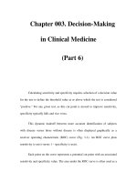
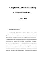
1958; Mollison 1959; Högman et al. 1960). In the
example shown in Fig. 7.2, the patient was ccddee,
with an autoantibody of apparent specificity anti-e.
The mean lifespan of transfused e-positive (DCCee)
red cells was about 8 days, which was similar to that of
the patient’s own red cells (see Dacie 1962, p. 450),
whereas the survival of e-negative (DccEE) red cells
was only slightly subnormal. For references to further
similar cases in which red cell survival has been stud-
ied, see Petz and Swisher (1989, pp. 565–567).
Specificity mimicking that of alloantibodies with
Rh specificity
A minority of warm autoantibodies at first sight
appear to have the specificity of an Rh alloantibody,
such as anti-E. For example, an eluate prepared from
the red cells of a patient of phenotype DCCee may react
more strongly with E-positive than with E-negative
cells and thus appear to contain anti-E. However,
in about 70% of such cases all antibody activity can
be absorbed completely by red cells lacking the
corresponding antigen, e.g. DCCee in the present
example. The specificity of these autoantibodies seems
in fact to be anti-Hr or anti-Hr
0
(Issitt and Pavone
1978).
The case reported by van’t Veer and co-workers
(1981) in which a negative DAT was found on the red
cells of a patient with severe haemolytic anaemia,
whereas strong autoantibodies of apparent anti-C and
anti-e specificity were present in the serum demon-
strates that such Rh specificities may be entirely
illusory: not only (1) could the autoantibodies be
absorbed with C-negative and e-negative cells, respect-
ively, but also (2) during the episode in which the
DAT was negative and the patient’s red cells (DCcee)
did not react in vitro with the patient’s own auto-
antibodies, they reacted normally with auto-anti-C
and allo-anti-e. The nature of the epitope with which
such antibodies react is not known. Neither is it clear
why the epitope should be so strongly associated with
Rh alloantigens. The case reported by Rand and co-
workers (1978) in which autoantibodies with anti-E
RED CELL ANTIBODIES AGAINST SELF-ANTIGENS, BOUND ANTIGENS AND INDUCED ANTIGENS
269
100
75
50
25
0
0
Days
10 20 30
Percentage of survival
Fig 7.2 Survival, in a ddccee patient with autoimmune
haemolytic anaemia, of e+ (DCCee) red cells (l), estimated
by differential agglutination, and of e– (DccEE) cells (×),
estimated by
51
Cr labelling and corrected for Cr elution.
The patient’s serum contained an autoantibody reacting
preferentially with e+ cells. (The legend of this figure as
published originally (Mollison 1959) stated incorrectly
that the e+ cells were autologous and were labelled
with
51
Cr.)
specificity were eluted from an E-negative patient’s
cells clearly demonstrates the mimicking nature of the
specificity of the autoantibodies. Not only could the
anti-E be absorbed to exhaustion by E-negative cells,
but also the eluate from the E-negative cells used for
absorption contained antibodies that again showed
positive reactions only with E-positive cells. A possible
explanation for this phenomenon is provided by
observations on the specificity of anti-Is reported
by Potter and co-workers (2002) who conclude that
anti-I specificity is mediated through binding to a
hydrophobic patch adjacent to the conventional anti-
gen binding site (see p. 259). It is well established that
many monoclonal anti-Ds are encoded by the same
Ig gene (V4–34) as cold agglutinins and can exhibit
cold agglutinin activity (Thorpe et al. 1998). The cold
agglutinin activity itself could account for absorption
of anti-D by D-negative red cells. Alternatively, the
unusually high positive charge of anti-Ds and/or the
considerable structural homology between D and CE
polypeptides (discussed in Chapters 3 and 5; see also
Thorpe et al. 1998) might predispose to absorption
of these antibodies on all red cells irrespective of Rh
phenotype. Some monoclonal anti-D recognized a ce
polypeptide in which Arg145 was substituted by Thr,
Thr 154 is not found in the D polypeptide. In this case,
cold reactivity was ruled out as a possible explanation
(Wagner et al. 2003).
Specificities outside the Rh system
The possible involvement of Wr
b
in the specificity of
autoantibodies was investigated by Issitt and co-workers
(1976). Of 64 sera from patients with AIHA, two
failed to react with Wr(a+ b–) cells and contained only
anti-Wr
b
; the remaining sera reacted with Wr(a+ b–)
red cells but, after absorption with these cells to
remove anti-dl, 32 could be shown to contain anti-
Wr
b
. The Wr
b
antigen is formed by the association of
band 3 with glycophorin A (see Chapter 6). Some, but
not all, warm autoantibodies capable of co-precipitating
band 3 and glycophorin A were shown to have anti-
Wr
b
specificity by Leddy et al. (1994). In patients
with warm AIHA, autoantibodies with many other
specificities are occasionally encountered, e.g. A
(Szymanski et al. 1976); K, k and Kp
b
in association
with weakening of Kell antigens (see below); Kx
(Sullivan et al. 1987); Jk
a
(van Loghem and van der
Hart 1954); Jk3 (O’Day 1987); N (Bowman et al.
1974); S (Johnson et al. 1978); U (Marsh et al. 1972);
Vel (Szaloky and van der Hart 1971); I
T
(Garratty et al.
1974); Ge (Reynolds et al. 1981); Sd
x
(Denegri et al.
1983) and Sc1 (Owen et al. 1992). For others, see
Garratty (1994).
Kell antibodies associated with autoimmune
haemolytic anaemia
Several cases have been described in which a patient
has developed a positive DAT, usually with overt
haemolytic anaemia, and has been found to have
autoantibodies of Kell specificity in the serum associ-
ated with weakening of Kell antigens. Seyfried and
co-workers (1972) described a patient with potent
anti-Kp
b
in his serum; during the period of his acute
illness his own red cells reacted with anti-Kp
b
only
after they had been treated with ficin. Sixteen weeks
later, when the patient was better, Kell antigens were
of normal strength. Beck and co-workers (1979)
described a patient with similar serological findings
but without AIHA. A patient has been described in
whom, during consecutive relapses of autoimmune
thrombocytopenia the Kell and Lutheran antigens
became virtually undetectable. It was shown that this
was due to transient absence of the Kell and Lutheran
proteins during a relapse (Williamson et al. 1994).
Other examples of weakening of red cell antigens in
association with the appearance of alloantibodies or
autoantibodies of the corresponding specificity are
given in Chapter 3.
The frequency of autoantibodies with Kell speci-
ficity in patients with warm AIHA was estimated to
be about 1 in 250 by Marsh and co-workers (1979).
Autoantibodies mimicking alloantibodies with
specificity other than Rh
Autoantibodies may mimic the specificity of anti-K
(Garratty et al. 1979; Viggiano et al. 1982); anti-Jk
b
plus anti-Jk3 (Ellisor et al. 1983); anti-Kp
b
(Manny
et al. 1983; Puig et al. 1986), anti-Fy
b
(Issitt et al. 1982;
van’t Veer et al. 1984), anti-Fy
a
plus anti-Fy
b
(Harris
1990) and anti-hr
B
-like (Vengelen-Tyler and Mogck
1991). In all of these cases, the patient was negative for
the corresponding antigen, the antibodies could be
absorbed by red cells negative for the corresponding
antigen, and eluates from such cells again showed the
mimicking specificity.
CHAPTER 7
270
Autoantibodies directed against non-
polymorphic determinants
Some warm autoantibodies are directed against
determinants that are clearly non-polymorphic. For
example, anti-phospholipid antibodies, which occur
in some patients with SLE and which may cause
haemolytic anaemia (Arvieux et al. 1991) and anti-
bodies against triosephosphate, found in some pat-
ients with falciparum malaria (see section on positive
DAT in malaria, above).
Negative direct antiglobulin test despite warm
autoantibodies in the serum
In a case reported by Seyfried and co-workers (1972),
during an episode of severe haemolysis, the DAT on
the patient’s red cells was negative despite the presence
of potent autoantibodies in the serum. The antibodies
had anti-Kp
b
specificity, and weak anti-Kp
b
could be
eluted from the patient’s red cells. The antigens of the
Kell system were severely depressed at the time when
the DAT was negative, but were of normal strength
after recovery. Cases of transient depression of LW,
associated with appearance of anti-LW in the serum,
and without haemolytic anaemia, are described in
Chapter 5. Several further cases, similar to the case of
Seyfried and co-workers, have been observed in which
the autoantibodies have had the following specificities:
anti-E (Rand et al. 1978); anti-Rh of undefined
specificity (Issitt et al. 1982; Vengelen-Tyler et al.
1983); ‘mimicking’ anti-C + anti-e (see above) (van’t
Veer et al. 1981); anti-En
a
(Garratty et al. 1983); anti-
Kp
b
(Brendel et al. 1985; Puig et al. 1986); specificity
for a high-frequency antigen in the Kell system
(Vengelen-Tyler et al. 1987); anti-Jk
a
(Ganly et al.
1988); anti-Jk3 (Issitt et al. 1990) and anti-Fy
a
+ Fy
b
(Harris 1990). In all the foregoing cases, there was
total or severe depression of the antigens, against
which the autoantibodies were directed (compare with
Chapter 3). In some cases, although the DAT was
negative, an eluate from the patient’s red cells con-
tained weak autoantibodies of the same specificity as
those in the serum. In some cases the DAT had been
positive before the episode of severe haemolysis. In other
cases the patient presented with a negative DAT and
the antibodies were first thought to be alloantibodies.
In a case reported by Herron and co-workers (1987)
the autoantibodies were found to react much more
strongly with old, i.e. relatively dense, red cells
than with young cells and it was suggested that the
DAT during an episode of severe haemolysis became
negative because only young red cells remained in the
circulation.
Role of CD47 in modulating the severity of
autoimmune haemolytic anaemia in mice
CD47 is a glycoprotein present on all cells. In human
red cells it is associated with the proteins of the band
3–Rh complex (see also Chapters 3 and 5). CD47
appears to inhibit phagocytosis of normal circulating
red cells by ligating the macrophage inhibitory receptor
signal regulator protein alpha (SIRPalpha; Oldenborg
et al. 2000). Non-obese diabetic (NOD) mice spontan-
eously develop mild AIHA aged between 300 and
550 days, whereas CD47-deficient NOD mice develop
a severe AIHA at age 180–280 days. In addition,
CD47-deficient C57BL/6 mice are much more sus-
ceptible to experimental passive AIHA induced by anti-
red cell monoclonal antibodies than their wild-type
counterparts (Oldenborg et al. 2002). These results are
consistent with a role for CD47 in antibody-mediated
phagocytosis.
Transfusion as a stimulus for allo- and
auto-antibody production
Young and co-workers (2004) carried out a retrospect-
ive analysis of blood bank records in order to deter-
mine the frequency of red cell autoimmunization
associated with alloimmunization. They found 121
out of 2618 patients with a positive direct or indirect
antiglobulin test (IAT) to have red cell autoantibodies.
Forty-one of these patients also had alloantibodies and
12 of these developed their autoantibodies in temporal
association with alloimmunization after recent blood
transfusion. These authors conclude that auto-
immunization and the development of AIHA should
be recognized as a complication of allogeneic blood
transfusion and recommend that once red cell auto-
immunization is recognized, a strategy that minimizes
exposure to allogeneic blood should be employed In
total, 6 out of 16 D-negative patients who developed
anti-D after transfusion with D-positive red cells also
made IgG autoantibody and three of these patients
suffered prolonged haemolysis (Frohn et al. 2003).
Shirey and co-workers (2002) advocate prophylactic
RED CELL ANTIBODIES AGAINST SELF-ANTIGENS, BOUND ANTIGENS AND INDUCED ANTIGENS
271
antigen-matched donor blood for patients with warm
autoantibodies in order to minimize the risk of allo-
antibody production.
Red cell transfusion and other therapy for
patients with autoimmune haemolytic anaemia
associated with warm autoantibodies
In severe AIHA, transfusion produces only a very
transient increase in Hb concentration and carries an
increased risk of: (1) inducing the formation of allo-
antibodies; (2) increasing the potency of the auto-
antibodies; and (3) inducing haemoglobinuria due to
autoantibody-mediated red cell destruction (Chaplin
1979). Accordingly, even in severely anaemic patients,
it is usually best to begin treatment with corticos-
teroids, following which the Hb concentration usually
starts to rise within 7 days (Petz and Garratty 1980,
p. 392). If the effect of corticosteroids is not satis-
factory, or if a quicker effect is needed, intravenous
immunoglobulin (IVIG) can be given which, in very
high doses (e.g. 0.4 g/kg per day) may have a very rapid
effect (MacIntyre et al. 1985; Newland et al. 1986;
Argiolu et al. 1990). However, in a study including 73
patients, IVIG had a rapid effect in only about 35% of
cases, and particularly in patients with hepatomegaly
and patients with a low pre-treatment haemoglobin.
It is recommended that this treatment should be
restricted to selected cases, for example to those in
which the pre-treatment haemoglobin level is < 60–
70 g/l or those with hepatomegaly (Flores et al. 1993).
Treatment with ciclosporin (4 mg/kg per day) can be
tried and may result in a fairly rapid increase in Hb
concentration (Hershko et al. 1990). Splenectomy is
indicated only in patients who have failed to respond
to steroids, IVIG and ciclosporin. In the patients with
complete warm haemolysins IVIG may be valuable, as
Ig has been found to inhibit complement-dependent
lysis (Frank et al. 1992). Rituximab (monoclonal anti-
CD20) has been used successfully in the treatment of
AIHA in several studies. Shanafelt and co-workers
(2003) consider that rituximab should be considered
as salvage therapy for immune cytopenias that are
refractory to both corticosteroid treatment and
splenectomy. These authors report complete remission
in 5 out of 12 patients with idiopathic thrombocytope-
nia, and two out of five patients with AIHA. However,
serious adverse effects have been reported (reviewed in
Petz 2001). Jourdan and co-workers (2003) report
a case of severe AIHA that developed following
rituximab therapy in a patient with a lymphopro-
liferative disorder.
There have been several reports of a high incidence
of alloantibodies in patients with the warm antibody
type of autoimmune haemolytic anaemia (WAIHA)
who have been transfused. In three series the frequency
was 32–38% and was as high as 75% in patients who
had received more than five transfusions (Branch and
Petz 1982; Laine and Beattie 1985; James et al. 1988;
reviewed by Garratty and Petz 1994). In these three
series, the patient’s serum was absorbed with auto-
logous red cells before being tested for alloantibodies.
In another series it was found that 44% of alloanti-
bodies could not be detected before autoabsorption
(Walhermfechtel et al. 1984). There has been one
report indicating that red cell alloimmunization is
rare in WAIHA (Salama et al. 1992) but the patients’
sera were not absorbed with autologous red cells
before being tested and alloantibodies may have been
overlooked.
The risk of haemolysis after red cell transfusions in
patients with AIHA with warm autoantibodies has
been questioned. No instance of increased haemolysis
was seen in 53 patients even in cases in which the trans-
fused red cells were incompatible with autoantibodies
detectable in the recipient’s serum (Salama et al. 1992).
Transfusion is indicated only in special circum-
stances, for example if the patient is severely anaemic
and is going into cardiac failure, or has neurological
signs, or has rapidly progressive anaemia, or is to
undergo splenectomy. In most other circumstances it is
better to use palliative measures, such as absolute bed
rest, to counteract the decreased tolerance to exercise,
while monitoring the Hb level.
If transfusions are given, it is important to group the
patient’s red cells for all clinically significant alloanti-
gens, to facilitate the identification of any alloanti-
bodies that may be produced. In patients who have
previously been transfused or have been pregnant, it is
also important to try to exclude the presence of allo-
antibodies, which may be masked by the presence of
autoantibodies. Either autoabsorption can be used or,
if sufficient autologous red cells cannot be obtained,
differential absorptions (see Chapter 8). It is helpful to
obtain red cells from the patient before the first trans-
fusion is given, and to store these at 4°C or frozen, so
as to have cells for autoabsorptions if needed (Petz and
Swisher 1989, p. 564).
CHAPTER 7
272
When the presence of an alloantibody has been
established, antigen-negative red cells must be selected
for transfusion: the practice of transfusing ‘least
incompatible red cells’ is not acceptable under these
circumstances (see Laine and Beattie 1985).
In selecting red cells for transfusion, any blood
group specificity of incomplete warm autoantibodies
should when possible also be taken into account. In Rh
D-negative females with auto-anti-e who have not yet
reached the menopause, the red cells should, if possible,
be e-negative as well as D-negative (i.e. ddccEE). In
patients with auto-anti-e, e-negative (EE) red cells may
survive better than e-positive cells (see Fig. 7.1) but may
stimulate the production of anti-E (Habibi et al. 1974).
When transfusing patients with AIHA, packed red
cells should be given in just sufficient quantities to raise
the Hb concentration to a level that will make it pos-
sible for other therapy to be applied. In acute anaemia,
oxygen may have to be given. A few patients need
regular transfusions despite all other forms of therapy.
As mentioned above, the presence of warm auto-
antibodies in the serum may make it difficult to detect
alloantibodies (see also Chapter 8).
T-cell reactivity in AIHA
Peptides corresponding to sequences in the D and CE
polypeptides stimulated proliferation of T cells from
the peripheral blood and spleen of seven out of nine
patients with AIHA. In total, four of the seven reactive
patients had autoantibody to the Rh proteins.
Multiple peptides were also stimulatory in two posit-
ive control donors who had been alloimmunized with
D-positive red cells (Barker et al. 1997). Stimulation
of peripheral blood mononuclear cells from patients
with AIHA with D polypeptide resulted in either pro-
liferation and secretion of γ-interferon or secretion of
interleukin 10 (IL-10). Peptides derived from the D
polypeptide that preferentially induced IL-10 secretion
suppressed T-cell proliferation against D polypeptide,
suggesting that it may be possible to ameliorate red
cell autoantibody responses in man with inhibitory
peptides (Hall et al. 2002). An important role for IL-10
in the function of peptide-induced regulatory T cells
in vivo is apparent from successful peptide therapy,
based on nasal administration of peptides corres-
ponding to dominant T-cell epitopes, in mouse
models of autoimmunity such as experimental allergic
encephalomyelitis, which are associated with a devia-
tion from a Th1 to a regulatory IL-10 CD4
+
T-cell
response (Sundstedt et al. 2003).
Haemolytic anaemia in recipients of allografts
Alloantibodies produced by donor lymphocytes in
grafted tissue may simulate autoantibodies in the
recipient and cause haemolytic anaemia (see Chapter 11).
Positive direct antiglobulin tests due to anti-red
cell antibodies in antilymphocyte globulin
Antilymphocyte globulin (ALG) is commonly pre-
pared in horses and the serum contains antibodies
against human red cells. Following the injection of
ALG, the recipient’s red cells acquire a positive DAT
within 1–3 days (Lapinid et al. 1984; Swanson et al.
1984). The reaction between AHG reagent and the
horse serum on the patient’s red cells can be inhibited
by adding diluted horse serum to the AHG reagent
without interfering with the reaction between the
AHG reagent and any human alloantibodies which
may be bound to the patient’s red cells (Swanson et al.
1984). In the serum of patients injected with ALG,
autoantibodies can be detected, which usually show
no obvious specificity but which occasionally have a
Lu-related pattern (Anderson et al. 1985).
Occasionally, a positive DAT in a patient who has
been injected with ALG is due to human red cell
alloantibody; the alloantibody is derived from the
plasma which has been added to the ALG to inhibit
horse antibodies against human plasma proteins
(Shirey et al. 1983).
Administration of ALG may occasionally produce
immune red cell destruction; in the case described by
Prchal and co-workers (1985) the DAT was negative
with AHG reagent but positive with anti-horse
immunoglobulin.
Antibodies against bound or induced
antigens
Drug-induced immune haemolytic anaemia
Among cases of acquired immune haemolytic anaemia
18% were due to drugs in the series of Dacie and
Worlledge (1969) and 12.4% in the series of Petz
and Garratty (1980). The great majority of cases of
drug-induced haemolytic anaemia were at one time
RED CELL ANTIBODIES AGAINST SELF-ANTIGENS, BOUND ANTIGENS AND INDUCED ANTIGENS
273
due to α-methyldopa (Worlledge 1969) but this drug is
now used much less frequently. Cases resulting from
other drugs are very rare, penicillin-induced anaemia
being the least uncommon (Petz and Garratty 1980).
Recently, four cases of haemolytic anaemia (one fatal)
have been described following piperacillin therapy
(Arndt et al. 2002), one case attributed to tazobactum
(Broadberry et al. 2004) and another to teicoplanin
(Coluccio et al. 2004).
Most drug-induced immune haemolytic anaemias
since the late 1980s have been caused by second-
and third-generation cephalosporins, cefotetan and
ceftriaxone respectively (Arndt and Garratty 2002;
Petz and Garratty 2004). In total, 10 out of 35 cases of
cefotetan-induced severe haemolytic anaemia studied
by Garratty and co-workers (1999) were in patients
who had received cefotetan prophylactically for obstetric
and gynaecological procedures. Citak and co-workers
(2002) report the development of haemolytic anaemia
in a child with no underlying immune deficiency or
haematological disease following treatment with
ceftriaxone for a urinary tract infection. The patient
had antibody against ceftriaxone and was successfully
treated with high-dose corticosteroids.
Non-steroidal anti-inflammatory drugs (NSAIDs)
can also induce very severe AIHA. Jurgensen and
co-workers (2001) describe a case of fatal AIHA
with multisystem organ failure and shock caused by
diclofenac-dependent red cell autoantibodies.
The fluoroquinolones, ciprofloxacin and levofloxacin,
have been associated with causing AIHA in single case
reports (Lim and Alam 2003; Oh et al. 2003).
Most drug molecules are not large enough to induce
an immune response but may become immunogenic
when bound to a macromolecule, for example a protein
at the surface of a cell, to form a hapten–carrier com-
plex. Antibodies formed against such a complex may
be specific for the hapten, the hapten–carrier combining
site or the carrier alone (see Shulman and Reid 1993).
There are several ways in which drugs may be
responsible for a positive DAT, often associated with
immune haemolytic anaemia (reviewed in Issitt and
Anstee 1998; Petz and Garratty 2004).
Drug adsorption mechanism
The drug may bind firmly to red cells; when an anti-
body is formed against the drug, the drug-coated cells
may be destroyed. The drug antibodies can be detected
in vitro with washed drug-coated cells. In these cases,
the antibodies are directed against the drug alone (i.e.
the hapten) and can be absorbed by the drug. This
mechanism has been called ‘the drug-adsorption
mechanism’ (Garratty and Petz 1975). Penicillin acts in
this way and so, occasionally, do other drugs, particu-
larly some of the cephalosporins (see Garratty 1994).
In about 3% of patients with bacterial endocarditis
receiving massive doses of i.v. penicillin, a positive
DAT develops but AIHA occurs only occasionally; the
first case, associated with the prolonged administra-
tion of penicillin in high dosage (20 million units or
more daily for weeks), was described by Petz and
Fudenberg (1966): the patient’s serum contained an
IgG penicillin antibody of unusual potency. If it is
necessary to continue giving penicillin to patients with
AIHA due to penicillin antibodies, transfusions may
be required. Normal red cells, uncoated with peni-
cillin, will appear to be compatible on crossmatching
but after transfusion will become coated in vivo and
destroyed in the same way as the patient’s cells.
Although penicillin antibodies are usually IgG they
may be partly IgM (Fudenberg and German 1960) or
solely IgM (Bird et al. 1975), in which case complement
is bound and the red cells are agglutinated by anti-C3.
In patients with immune haemolytic anaemia due to
penicillin antibody, the antibody can invariably be
demonstrated in high titre in the serum, using red cells
coated in vitro with penicillin (Petz and Garratty 1980;
Petz and Branch 1985).
IgM or IgG antibodies reactive with penicillin-
coated red cells have been found in the serum of about
4% of haematologically normal subjects (Fudenberg
and German 1960).
The benzyl-penicilloyl groups are the most immuno-
genic of the haptenic groups of penicillin (Garratty and
Petz 1975).
Several cases of severe or even fatal haemolytic
anaemia due to second- or third-generation
cephalosporins have been described in which the drug
adsorption mechanism was involved (see Garratty
et al. 1992). In some of the cases the immune complex
mechanism described below also seems to have been
involved (Marani et al. 1994; Ogburn et al. 1994).
Trimolecular complex mechanism
The drug does not bind firmly to red cells so that drug-
coated cells cannot be prepared. It has been suggested
CHAPTER 7
274
that in these cases, when antibodies are formed against
the drug, immune complexes attach to the red cell.
This immune complex theory has been criticized
for the following reasons: (1) certain drugs cause
haemolytic anaemia in some patients but immune
thrombocytopenia in others implying that a specific
membrane component is involved; (2) drug antibodies
attach to the cell membrane by their Fab part suggest-
ing specific binding rather than passive adsorption of
immune complexes; (3) the drug antibodies cannot be
absorbed by the drug alone and can only be detected by
bringing red cells, free drug and antibodies together;
and (4) the binding of the drug antibodies may depend
on the presence of a particular red cell antigen, which
implies that the drug binds to the cell surface, albeit
loosely (Salama and Mueller-Eckhardt 1987a). It
seems therefore more likely that a trimolecular com-
plex of the drug, the drug antibody and a component
on the red cell membrane is formed. (For a survey of
the subject, see Shulman and Reid 1993 and Garratty
1994, who also gives a list of drugs acting in this way.)
Drugs that produce red cell destruction by this mecha-
nism can do so even when given in low doses. The
haemolysis is arrested within 1–2 days of stopping the
drug. The antibodies are often IgM and complement-
activating and then only complement can be detected
on the patient’s red cells (Garratty and Petz 1975).
In some cases, the antibodies are directed against
a metabolite rather then the drug itself (Salama and
Mueller-Eckhardt 1985, 1987a,b; Kim et al. 2002).
The antibodies can then be detected by using urine
from subjects who have taken the drug. Bougie and co-
workers (1997) describe a case of haemolytic anaemia
and subsequent renal failure resulting from diclofenac
in which the patient had an antibody specific for a glu-
curonide conjugate of a known metabolite of diclofenac
(4′-OH hydroxydiclofenac). The antibody could be
demonstrated in the patient’s serum with red cells
in the presence of urine taken from individuals who
had ingested diclofenac. These authors point out that
as glucuronidation is a common pathway of drug
metabolism, studies on glucuronidation of other com-
mon medications associated with immune haemolytic
anaemia should be considered.
Drug-induced autoantibody formation
The drug does not bind firmly to red cells, antibody
against the drug is not formed, but IgG autoantibodies
are induced. α-Methyldopa and levodopa are prime
examples of drugs acting in this way. In 15–20% of
patients receiving α-methyldopa, the DAT becomes
positive after 3–6 months’ treatment; the development
of a positive DAT is dose dependent (Carstairs et al.
1966). Only about 1% of patients receiving the drug
develop haemolytic anaemia.
It has been suggested that α-methyldopa induces red
cell autoantibodies by inhibiting the activity of sup-
pressor T lymphocytes (Kirtland et al. 1980). Although
no effect on suppressor cells could be demonstrated
in one investigation (Garratty et al. 1986), a drug,
lobenzarit, which inhibits suppressor cell function, has
been found to induce α-methyldopa-type autoimmune
haemolytic anaemia (Andou et al. 1994).
α-Interferon seems to be responsible for the develop-
ment of autoantibodies to various structural proteins
or receptors and for the exacerbation of autoimmune
disease (Conlon et al. 1990). The development of warm
red cell autoantibodies has been observed in a patient
receiving α-interferon and IL-2 (Perez et al. 1991). These
various effects are believed to be due to the inhibition
of normal cellular immune suppressor mechanisms.
As stated above, some drugs that do not bind firmly
to red cells induce both anti-drug antibodies and red
cell autoantibodies, for example nomifensine (Martlew
1986; Salama and Mueller-Eckhardt 1987a), tolmetin
and suprofen (van Dijk et al. 1989).
The inference has been drawn that even when drugs
bind loosely to red cells, they can induce the formation
of antibodies against a red cell antigen alone (Salama
and Mueller-Eckhardt 1987a). Alternatively the drug
could directly influence the immune response against
autoantigens (Kirtland et al. 1980).
Non-specific adsorption of proteins
The drug may alter the red cell membrane in some
way so that proteins are adsorbed non-specifically.
Cephalosporin and cisplatin are believed to act in this
way as a rule. This mechanism has not been shown to
result in haemolytic anaemia unless antibodies against
the drug are formed.
Effect of red cell antigens on the binding of
drug–antibody complexes
In a case in which streptomycin was involved, the drug
was apparently bound to the red cell membrane through
RED CELL ANTIBODIES AGAINST SELF-ANTIGENS, BOUND ANTIGENS AND INDUCED ANTIGENS
275
chemical groups related to M and possibly D (Martinez-
Letona et al. 1977). Several similar cases in which vari-
ous drugs and different red cell antigens were involved
have been reported (for a survey, see Garratty 1994).
Treatment of drug-induced haemolytic anaemia
In cases in which antibodies are involved against a
drug that binds firmly to the red cell and in cases
in which immune complexes are responsible for the
destruction of the red cells, stopping the drug is
sufficient to arrest the haemolytic process and treat-
ment of the haemolytic anaemia is rarely necessary.
In cases in which it is impossible to stop the drug and
the patient is anaemic, red cell transfusions should
be given. In AIHA induced by α-methyldopa, the drug
must be stopped, but red cell destruction may continue
for weeks or months. If treatment is required it is the
same as for patients with drug-independent warm AIHA.
In occasional patients, autoantibodies disappear des-
pite continued administration of the drug (Habibi
1983).
Antibodies against other bound antigens
Fatty acid-dependent agglutinin (‘albumin
agglutinin’)
The serum of a small proportion of people agglutinates
red cells suspended in albumin but not those sus-
pended in saline (Weiner et al. 1956). Agglutination
is found only with caprylate-treated albumin (Golde
et al. 1969, 1973) and the antibody is in fact directed
against sodium caprylate or other fatty acid salts and
not against albumin at all (Beck et al. 1976). The term
‘fatty acid-dependent agglutinin’ is therefore prefer-
able to the previously used ‘albumin agglutinin’. Fatty
acid-dependent agglutinins may cause false-positive
reactions in slide tests in which blood grouping
reagents containing albumin are used (Reid et al.
1975; Case 1976) and in the IAT test if albumin is used
in the sensitizing phase of the reaction.
Antibiotics
Antibiotics are added to samples of red cells that
are distributed commercially for the identification of
alloantibodies. Such cells may give false-positive
results if antibodies against the relevant antibiotic are
present in a sample of serum. In a systematic search for
such antibodies, Watson and Joubert (1960) found
that 6 out of 1700 routine blood bank serum samples
agglutinated chloramphenicol-treated cells. Three
examples of an antibody of this kind were found to be
IgM and two bound complement (Beattie et al. 1976).
An IgA antibody agglutinating red cells suspended in
0.1 mg of neomycin/ml was described by Hysell and
co-workers (1975). Antibodies vs. penicillin-treated
red cells are described above.
Acriflavine
Some commercial anti-B-sera have acriflavine added
to them as a colouring agent and this may be a cause
of false-positive results if anti-acriflavine antibodies
are present in a patient’s serum, possibly as a result of
previous exposure to acriflavine. The antibodies may
cause agglutination of normal red cells in the presence
of a 1 in 150 000 dilution of acriflavine (Beattie and
Zuelzer 1968; Beattie et al. 1971).
Immune complexes adsorbed to red cells in vitro
in ulcerative colitis
In occasional patients with ulcerative colitis, the DAT
on clotted samples is positive but on anticoagulated
samples is negative. Allogeneic red cells give a positive
IAT with the patient’s serum but a negative test with
plasma. It is postulated that the patient’s plasma con-
tains an antibody against an activated coagulation
factor and that, during clotting, immune complexes
form and attach to the red cells (Garratty et al. 1980).
Lactose- or glucose-treated red cells
Antibodies have been described which agglutinated
any red cells that had been incubated with lactose
(Gray 1964) or glucose (Lewis et al. 1980). In the latter
case, red cells from patients with diabetes reacted,
although after incubation in saline, the cells were no
longer agglutinated. Examples of anti-M and anti-N
reacting only with lactose- or glucose-treated red cells
are described in the preceding chapter.
Antibody against chemically altered red cells
(the LOX antigen)
Red cells exposed to citrate–phosphate–dextrose
CHAPTER 7
276
solution in particular batches of plastic blood packs
may acquire a new red cell antigenic determinant
‘LOX’, reacting with an antibody present in normal
serum. The development of this antigen is probably
associated with sterilization of the packs with propy-
lene oxide gas (Bruce and Mitchell 1981).
Polyagglutinability
Red cells are said to be polyagglutinable when they are
agglutinated by almost all samples of normal human
serum although not by the patient’s own serum. The
commonest forms of polyagglutinability are due to
exposure, by the action of bacterial enzymes, of anti-
genic determinants (T, Tk, Th, Tx), which form part
of the structure of the normal red cell membrane, but
which are usually hidden. Another form of polyagglu-
tinability is believed to be due to somatic mutation
leading to the emergence of a line of red cells lacking an
enzyme essential for the formation of normal red cell
antigens; as a result, a normally hidden antigen, Tn, is
exposed. In all the foregoing cases, the red cells are
polyagglutinable because antibodies (anti-T, etc.) cor-
responding to the determinants are present in serum
from all normal adults (although not in serum from
newborn infants). Why these antibodies are in all nor-
mal sera is not known but, like anti-A and anti-B, this
may be related to the widespread occurrence of the
antigens in the environment. Chicks kept in germ-free
conditions developed anti-T and anti-Tn when fed
Escherichia coli O86 in their drinking water (Springer
and Tegtmeyer 1981). Tn has been found in several
helminth parasites, including Echinococcus granulosus,
Taenia hydatigena and Fasciola hepatica (Casaravilla
et al. 2003) and in human skin mites (Kanitakis et al.
1997). Further forms of polyagglutinability may be
due to the inheritance of an antigen (C3d, NOR or
HEMPAS) for which a corresponding antibody is
present in almost all normal human sera.
T activation
Exposure of T antigen in vitro
As Fig. 7.3 shows, the T determinant is normally cov-
ered by N-acetylneuraminic acid and can therefore be
described as a cryptantigen. The antigen can be exposed
by the action of bacterial or viral neuraminidases.
Anti-T and anti-Tn (see below), present in the serum
of all subjects except infants, are presumably formed as
a reaction to T and Tn present in many Gram-negative
bacteria and vaccines (Springer et al. 1979; Springer
and Tegtmeyer 1981).
Knowledge of T activation stems from the original
observation that suspensions of red cells might become
agglutinable by ABO-compatible serum after standing
for many hours at room temperature, and that this
agglutination was associated with infection of the
suspension with certain enzyme-producing bacteria
(Hübener 1925; Thomsen 1927; Friedenreich 1930).
Very many organisms, including pneumococci,
streptococci, staphylococci, clostridia, E. coli, Vibrio
RED CELL ANTIBODIES AGAINST SELF-ANTIGENS, BOUND ANTIGENS AND INDUCED ANTIGENS
277
Normal
T
Tn
NeuAc Gal
α(2–3) β(1–3)
α(2–6)
GalNAcα1
NeuAc
O
serine/threonine
Gal
β(1–3)
GalNAcα1O
serine/threonine
GalNAcα1
and
O
serine/threonine
α(2–6)
GalNAcα1
NeuAc*
O
serine/threonine
Fig 7.3 Proposed structure of
the major O-glycosidically
linked oligosaccharides of the
sialoglycoproteins in normal, T- and
Tn-exposed erythrocyte membranes
(modified from Anstee 1981). NeuAc,
N-acetylneuraminic acid; Gal, d-
galactose; GalNAcα1, N-acetyl-d-
galactosamine. *It is possible that
this NeuAc residue is present in some,
but not all, Tn structures (PD Issitt,
personal communication).
cholerae and influenza viruses are capable of produc-
ing this effect in vitro.
Preparation of T-activated red cells. Add 11 mg of CaCl
2
to
100 ml of 0.85% NaCl to provide a solution containing
approximately 10 mmol of CaCl
2
/l. Add 0.2 ml of a solution
containing 500 units of neuraminidase/ml to give an enzyme
concentration of 1 unit/ml. This solution can be stored at 4°C
for 1 year. Wash group O red cells four times in saline and
make a 25% suspension of cells in the enzyme solution.
Incubate at 37°C for 2–3 h. Remove the supernatant, wash
the red cells in saline four times, make a 5% suspension in
saline and check for T activation by testing with anti-T lectin
(Howard 1979).
T-activated red cells can be kept for several weeks in
Alsever’s solution but must be washed thoroughly in saline
before being used.
T sites on red cells
As there are 15 O-glycosidically linked oligosaccha-
rides on each molecule of glycophorin A (α-SGP), and
probably similar numbers on glycophorins C and B (β-
and δ-SGPs), there are many potential T-antigen sites
(around 20 million) on the red cell. There are also T-
active structures on red cell membrane components
other than SGPs, for example on gangliosides (Anstee
1980).
Activation of T receptor in vivo
T activation may occur in vivo. Usually, this poly-
agglutinability occurs as a transient phenomenon, dis-
appearing within a few weeks or months of the time
when it is first observed. The phenomenon is not very
common; at a large Blood Transfusion Centre, only 10
cases were observed in 12 years (Stratton and Renton
1958).
In the past, T activation was almost always detected
by finding discrepancies between the results of testing
red cells and sera in the course of ABO grouping.
Nowadays, monoclonal anti-A and -B are widely used
and so T activation seldom causes trouble in blood
grouping.
In many cases, the patient has an obvious bacterial
infection, but the phenomenon has also been observed
in apparently healthy subjects; for a review of some
of the earlier reported cases, see Henningsen (1949).
In a case reported by Reepmaker (1952), an organism
that was shown to be capable of inducing T trans-
formation was isolated from the patient’s urine.
Chorpenning and Hayes (1959) made the point that T
transformation is not the only kind of polyagglutin-
ability induced by bacterial infection, and that in many
reported cases of polyagglutinability it was simply
assumed that the change was T transformation.
Similarity of T activation to acquisition of
‘B-like’ antigen
Certain bacterial enzymes confer B-specificity on red
cells as well as rendering them polyagglutinable
(Marsh 1960; see also Chapter 4).
Reactions of T-activated cells
T-activated cells are agglutinated by all sera contain-
ing more than a trace of anti-T, that is to say, by sera
from most adults. Anti-T is absent from cord serum but
appears at or before the age of 2 months (F Stratton,
personal communication). The agglutinin reacts best
at room temperature and may be inactive at 37°C.
Agglutinates due to anti-T may be large; there are
many free cells present.
T-activated cells react most strongly with fresh
serum and sometimes fail to react with serum that has
been stored frozen (Stratton 1954). T-activated cells
react better with sera containing anti-A than with
those that do not (Race and Sanger 1975, p. 487).
T-activated cells fail to agglutinate in their own
serum (the titre of anti-T being low presumably due to
absorption by exposed T) and the cells give a negative
DAT. At 37°C they are not sensitized to an antiglobu-
lin serum by human sera that agglutinate them at room
temperature.
An anti-T lectin can be extracted from the peanut,
Arachis hypogaea (Bird 1964). Testing with peanut
anti-T lectin is far more sensitive than testing with
adult serum (Seger et al. 1980). Reactions of various
types of polyagglutinable red cells with different
lectins and with polybrene are shown in Table 7.3.
Use of polybrene in testing for T. Normal red cells
(negatively charged) are agglutinated by polybrene
(positively charged), but red cells, such as T-activated
cells, which are deficient in sialic acid and thus have
a reduced negative charge, are not. For a method of
testing red cells with polybrene, see Issitt and Issitt
(1975).
CHAPTER 7
278
The deficiency of red cell sialic acid must exceed
about 12% before cells fail to be agglutinated by
0.1 g/dl of polybrene in a test tube (EA Steane, personal
communication, 1978), although with very dilute poly-
brene solutions (< 0.003 g/dl), red cells that have only a
10% reduction in sialic acid are agglutinated (Cartron
et al. 1978). When polybrene in as high a concentra-
tion as 0.6 g/dl is used, prozones are observed, for
example cells with 30% or more loss of sialic acid are
not agglutinated (Cartron et al. 1978).
A typical case. In the recent past, when normal sera
containing anti-T, in addition to polyclonal anti-A and
-B, were used for ABO grouping, T-activated cells
were recognized by finding discrepancies between the
results of testing red cells and serum, as in the follow-
ing case:
Mrs S was admitted to hospital with a septic abortion: mixed
coliforms and non-haemolytic streptococci were grown from
a vaginal swab. Her red cells were strongly agglutinated by
anti-A serum and partially agglutinated by anti-B; her serum
agglutinated and lysed B cells but failed to agglutinate A cells,
suggesting that she really belonged to group A. On further
testing, her red cells were found to be agglutinated by four
adult AB sera, the reactions being strongest at 4°C and
weakest at 37°C; the cells were not agglutinated by several
samples of cord blood serum from group A infants.
The patient made a good recovery and 1 week after admis-
sion her cells were only very weakly polyagglutinable.
Polyagglutinability is frequent in newborn infants
with necrotizing enterocolitis (Bird 1982) but will not
be detected when monoclonal anti-A and -B are used
for grouping. The diagnosis can be made with anti-T
lectin.
Leucocytes and platelets also become T activated;
platelet function is not impaired (Hysell et al. 1976).
Haemolytic syndromes due to T activation?
Most patients with T-activated cells do not have an
associated haemolytic process. Although several cases
have been reported in which such an association has
been observed, it is difficult in some of the cases to
be sure that T activation has been responsible. The
difficulty of incriminating anti-T seems particularly
great in infants, in whom anti-T, if present at all, is
not strong. Moreover, in many of the cases described
the patients have had an infection with Clostridium
perfringens, an organism notorious for producing vio-
lent haemolytic syndromes.
In four cases described by van Loghem (1965), the Hb con-
centration was between 4.5 and 6.9 g/dl and serum hap-
toglobin was reduced. Two of the patients showed red cell
autoagglutination. In the three cases in which organisms were
isolated they were C. perfringens, Staphylococcus aureus and
pneumococcus. In a patient reported by Moores and co-
workers (1975), with a presumed lung infection following a
stab wound, there was rapid improvement on treatment with
antibiotics, but after 7 days there was a sudden deterioration
and fall in Hb concentration to 3.4 g/dl and a reticulocytosis
of 25%. T activation was demonstrated and anti-T eluted
from the patient’s red cells. F Stratton (personal communica-
tion) has seen four infants of 2 months old or less with T activa-
tion, associated with the development of severe anaemia.
E. coli was implicated in one of the cases. In a child aged
14 months described by Rickard and co-workers (1969) the
Hb concentration fell to 3.7 g/dl: the blood film showed
spherocytes and Schumm’s test was positive.
RED CELL ANTIBODIES AGAINST SELF-ANTIGENS, BOUND ANTIGENS AND INDUCED ANTIGENS
279
Arachis Dolichos Glycine Vicia Salvia Leonurus
hypogaea biflorus* soja
†
GS II
‡
cretica sclarea cardiaca Polybrene
T + – + – + – +
§
–
Tk + ––+ –– – +
Th + –––+ –– +
Tx + –––––– +
Tn – ++–– + ––
Cad – ++–– – ++
* Tests with this lectin can be used only when the cells are group O or B.
†
May not react with weaker examples of Cad.
‡
Griffonia simplicifolia II.
§
Weak reaction.
Table 7.3 Reactions of different kinds
of polyagglutinable red cells (based on
the publications of GWG Bird and
co-workers).
Further cases were described by Bird and Stephenson
(1973) and by Tanaka and Okubo (1977).
In one case, the transfusion of normal plasma (con-
taining anti-T) appeared to be the cause of a severe
haemolytic transfusion reaction (HTR) (van Loghem
et al. 1955). Among six further cases, there was severe
haemolysis in one and mild haemolysis in three. It was
recommended that patients with T activation should
be transfused with washed red cells or platelets
(William et al. 1989).
Scepticism about the haemolytic potential of anti-T
was expressed by Heddle and co-workers (1977), who
found no evidence of significant red cell destruction
in three premature infants with T-activated red cells
following the transfusion of blood components con-
taining anti-T.
Haemolytic syndromes associated with polyagglutinability
have been produced experimentally in guinea pigs follow-
ing the injection of pneumococcal cultures (Ejby-Poulsen
1954a,b), and have been shown to occur spontaneously in
rabbits in association with enteritis (Evans et al. 1963).
Other kinds of polyagglutinability due to bacterial
enzymes or bacteria
Tk activation
This form of polyagglutinability of the red cells is
similar to T activation in that it is a transient
phenomenon associated with infection. The red
cells are agglutinated by the Tk-specific lectin GS II,
isolated from Griffonia simplificifolia seeds (Bird
and Wingham 1972); they also react with peanut
lectin, the reaction being greatly enhanced if the red
cells are first treated with papain (Bird and Wingham
1972).
Tk cells have normal amounts of sialic acid, as
indicated by the fact that they are agglutinated by
polybrene (see Table 7.3). Further work indicates that
Tk is exposed by the action of an endo-β-galactosidase
produced by Bacteroides fragilis (Inglis et al. 1975a,b)
or derived from Escherichia freundii (Doinel et al.
1980) or Flavobacterium keratolyticus (Liew et al.
1982). Endo-β-galactosidase exposes a terminal N-
acetylglucosamine residue on carbohydrate chains of
long-chain glycolipids and glycoproteins carrying
highly branched N-glycans, notably band 3 (Doinel
et al. 1980).
T and Tk activation associated with acquired B
In patients with acquired B the red cells often exhibit
Tk polyagglutination with or without T activation.
In reporting three patients it was pointed out that
the changes in each were due to different bacterial
enzymes and that, depending on the relative amounts
of each of these enzymes, different phenotypes were
produced, for example in one case T activation might
predominate and in another, Tk (Judd et al. 1979 – see
also Mullard et al. 1978; Janot et al. 1979).
In Tk polyagglutination H and A red cell antigens
are weakened (Inglis et al. 1978), as are I and i (Andreu
et al. 1979). These observations are consistent with the
location of the majority of ABH and Ii antigens on
complex N-glycans on the normal red cell (see Chap-
ter 3).
VA polyagglutination
This condition is considered here for convenience although
there is no evidence that it is caused by bacterial enzymes. The
condition is characterized by persistent polyagglutination
associated with haemolytic anaemia; the red cells are weakly
agglutinated by almost all adult sera but only up to a temper-
ature of 18°C. The abnormalities in the red cells include a
slight reduction in sialic acid (3.7%) and a depression of H
receptors (Graninger et al. 1977a,b). VA accompanied by Tk
was reported by Beck et al. (1978). VA may represent one end
of a Tk spectrum (Bird 1980).
Th activation
The Th antigen is exposed in infection with
Corynebacterium aquaticum (Levene 1984). A neura-
minidase that can be isolated from culture super-
natant of C. aquaticum was shown to be responsible.
The release of less than 20 µg of sialic acid per 10
10
red cells appeared to lead to Th reactivity, whereas
hydrolysis of greater amounts of sialic acid activates T
(Sondag-Thull et al. 1989).
When Th is exposed (Bird et al. 1978), the red cells
are agglutinated by peanut lectin, extracts from Vieia
eretica (Bird and Wingham 1981), Medicago disciformis
(Bird and Wingham 1983) and by polybrene, but not
by lectins from Glycine sofa, GS II, Salvia sclarea or
Salvia horminin (Bird et al. 1978); see Table 7.3.
In studying 200 paired samples of maternal and
cord blood, the incidence of Th activation was found
CHAPTER 7
280
to be much higher in newborn infants (11% and their
mothers 13%) than in blood donors (Wahl et al.
1989), in whom the incidence was 1.5% (Herman
et al. 1987). In none of the cases were the red cells
polyagglutinable, which shows that Th activation
leads to polyagglutinability only in some cases.
Tx and Tr activation
The Tx antigen is exposed on red cells by pneumococ-
cal enzymes (Bird et al. 1982). Tx cells are agglutinated
by peanut lectin but not by other lectins. Transient Tx
polyagglutination lasting 4–5.5 months was described
in three siblings of one family by Wolach and co-
workers (1987).
Reid and co-workers (1998) reported on an individual
with an unusual form of polyagglutination, denoted
Tr, the red cells giving a unique reaction pattern when
tested with lectins.
Polyagglutinability due to adsorbed bacteria
Many bacteria or their thermostable products will
adhere to red cells (Keogh et al. 1948; Jochem 1958a,b).
The red cells will be polyagglutinable when the cor-
responding antibody is present in most samples of
human serum. Antibodies to some bacteria, for ex-
ample Bacillus cereus, do not agglutinate red cells
coated with the bacteria but sensitize the cells to aggluti-
nation by antiglobulin serum (Weeden et al. 1960).
Tn red cells
Tn red cells, like T-activated cells, are deficient in
sialic acid (Bird et al. 1971) and are polyagglutinable
(Dausset et al. 1959), as anti-Tn, like anti-T, is present
in all normal adult sera.
Apart from the fact that T and Tn are quite separate
antigenic structures (see Fig. 7.2) Tn polyagglutination
differs in three important respects from T polyaggluti-
nation or any other polyagglutination associated with
infection: first, Tn agglutination is persistent; second,
it is associated with haematological abnormalities; and
third, affected subjects have two populations of red
cells, one normal and one showing the Tn change. The
condition (sometimes referred to as persistent mixed
field polyagglutination) appears to be due to somatic
mutation occurring in stem cells, leading to the emer-
gence of a population of abnormal (Tn) red cells (as
originally suggested by Bird et al. 1971, 1976b). Data
supporting this concept are as follows: in subjects with
Tn red cells there are also two populations of platelets,
Tn positive and Tn negative (Cartron and Nurden
1979). Only Tn-positive platelets contain glycoprotein
1b with a modified oligosaccharide chain structure
responsible for the expression of Tn antigen (Nurden
et al. 1982). A similar abnormality is present on Tn-
positive granulocytes (Cartron et al. 1981). It has also
been found that a sizeable fraction of erythrocyte,
granulocyte and megakaryocyte colonies grown from
the bone marrow of a patient with the Tn syndrome
appear to consist exclusively of either Tn-positive or
Tn-negative cells, demonstrating the clonal origin of
Tn cells (Vainchenker et al. 1982). Tn-positive B and T
cells can also be demonstrated in patients with the Tn
syndrome (Brouet et al. 1983) and, finally, expression
of the Tn antigen has been demonstrated at a very early
stage of differentiation, i.e. in colony-forming units
(Vainchenker et al. 1985).
Initially, N-acetylgalactosamine bound glycosidic-
ally to serine and threonine of red cell glycoproteins
(GalNAcα1-O-serine/threonine) was considered to
be the only major antigen on Tn cells (Dahr et al.
1975). A deficiency in Tn cells of the GalNAc (β1,3)-d-
galactosyl transferase, which normally generates the
O-glycosidically linked oligosaccharides attached to
the red cell sialoglycoproteins was described (Cartron
et al. 1978). An explanation for the deficiency of this
galactosyl transferase in Tn syndrome is provided by
Ju and Cummings (2002). These authors show that
expression of an active GalNAc(beta1,3)-d-galactosyl
transferase requires the presence of a chaperone pro-
tein (Cosmc) encoded by a gene on the X chromosome
at Xq23. In the Jurkat cell line that expresses Tn
antigen, the galactosyl transferase gene is normal, but
Cosmc has an inactivating mutation. Formal proof
is required from analysis of Cosmc in DNA derived
from patients with Tn syndrome but the analogy with
paroxysmal nocturnal haemoglobinuria (PNH) is very
compelling. Like Tn syndrome, PNH is an acquired
clonal disorder affecting a subset of all blood cells.
PNH results from inactivating mutations in a gene
on the X chromosome (PIG-A on Xp22.1, reviewed
in Young et al. 2000). Sialosyl-Tn (NeuAcα2–
6GalNAcα1-o-serine/threonine) is also present in Tn
cells (Kjeldsen et al. 1989). This disaccharide is absent
in normal glycophorins and therefore the responsible
transferase must be induced in Tn cells (Blumenfeld
RED CELL ANTIBODIES AGAINST SELF-ANTIGENS, BOUND ANTIGENS AND INDUCED ANTIGENS
281
et al. 1992). The induction of this transferase is thus
the second abnormality in Tn cells.
Although Tn polyagglutination usually persists for
long periods, it has been known to disappear in four
subjects; two of these disappearances were spontan-
eous, as neither patient was receiving cytotoxic ther-
apy (Bird et al. 1976b). Although Tn polyagglutination
is usually associated with neutropenia and thrombocy-
topenia (Gunson et al. 1970; Haynes et al. 1970) and
may be associated with haemolytic anaemia (Bird et al.
1971), it is also found in normal subjects (Myllylä et al.
1971; Bird et al. 1976c). In a subject investigated by
Myllylä and co-workers (1971) it was shown that the
survival of the subject’s red cells in his own circulation
was normal and that no anti-Tn was demonstrable
in the serum; when the red cells were injected into the
circulation of a normal subject (i.e. with anti-Tn in the
plasma) the cells were rapidly destroyed.
When normal red cells are transfused to a subject
with Tn polyagglutination the transfused cells do not
become Tn positive (Haynes et al. 1970).
Tn red cells are agglutinated by an extract of
Dolichos biflorus (Gunson et al. 1970) and by snail
anti-A but not by purified human anti-A (Bird 1978).
They are also agglutinated by the lectins from Salvia
sclarea, Helix pomatia and Glycine sofa (Bird 1978).
Tn red cells are best diagnosed by testing with an
extract of Salvia sclarea (see Table 7.3). The lectin
must be diluted to avoid non-specific activity but
then reacts strongly with Tn cells and not at all with
T-activated cells.
Exposure of T, Tn, sialylated Tn and Tk in
malignant cells
Immunoreactive T antigen is present in the cytoplasm
and on the outer cell membrane of about 90% of the
major forms of carcinoma and T lymphoma, as deter-
mined by absorption of human anti-T antibodies and
immunohistochemistry (Springer et al. 1974, 1983). In
addition to T, carcinoma cells express Tn antigen. Tn
antigen was detected by absorption of human anti-Tn
antibody in 46 out of 50 primary breast carcinomas
and in all six metastases originating from Tn-positive
primary carcinomas. In total, 13 out of 25 (52%)
anaplastic carcinomas, but only 2 out of 15 (13%)
well-differentiated carcinomas had more Tn than T;
one anaplastic carcinoma had neither antigen. More-
over, 18 out of 20 benign breast lesions had no Tn; the
two with Tn were premalignant. Tissue from 18 breast
carcinomas reacted strongly with anti-Tn (Springer
et al. 1985). Carcinoma-associated T antigen stimulates
profound cellular and humoral immune responses in
the patient, early in the disease and throughout its
course (Springer et al. 1983). Monoclonal anti-T and
anti-Tn, which reacted with T- and Tn-positive carci-
noma cells, were prepared by Springer and co-workers
(1983). Several monoclonal antibodies to T, Tn and
sialylated Tn have been produced and used to explore
the prognostic value of the respective antigens in cancer
(O’Boyle et al. 1996; Rittenhouse-Diakun et al. 1998).
Meichenin and co-workers (2000) used a monoclonal
anti-Tk (LM389) to show that Tk is a colorectal
carcinoma-associated antigen.
NOR, an inherited form of polyagglutinability
The red cells of a healthy young male were found to be
agglutinable by 75 of 100 ABO-compatible sera; the
red cell characteristic, NOR, associated with polyag-
glutinability was shown to be inherited in an appar-
ently dominant manner by four other family members
in two generations (Harris et al. 1982). The only other
inherited characteristics associated with polyagglutin-
ability are C3d and HEMPAS, described in the previ-
ous chapter. NOR can be distinguished from C3d by
the failure of NOR red cells to react with an extract of
D. biflorus and can be distinguished from the acquired
forms of polyagglutinability, T, Tk, Th and Tn, by the
failure of NOR red cells to react with an extract either
of Arachis hypogaea or of S. sclarea (Harris et al. 1982).
A second case was reported in a Polish family (Kusnierz-
Alejska et al. 1999). Subsequent studies on the red cells
of NOR+ individuals from this second family identified
two neutral glycolipids unique to NOR+ cells reactive
with anti-NOR and the lectin Griffonia simplicifolia
IB4 (GSL-IB4). The structure of one of these glycolipids
(NOR1) was determined to be Galα1–4GalNAcβ1–
3Galα1–4Galβ1–4Glcβ1-CER(α-galactosyl-globoside;
Duk et al. 2001). Duk and co-workers (2002) point
out that GSL-IB4 can be used in a simple serological
test with papain-treated red cells to detect NOR+ cells
in individuals of group A and O but not of group B
because the lectin also recognizes the Gal alpha1–3
structure. However, these authors found it necessary
to absorb their GSL-IB4 preparation with normal group
A1 cells to remove traces of other lectins found in GSL
in order to render it useful for detection of NOR antigen.
CHAPTER 7
282
Agglutinins for other normally
hidden antigens
Antigens on enzyme-treated red cells
Agglutinins for red cells treated with various enzymes
are found in all normal sera; for example, if trypsinized
red cells are mixed with normal human serum and
incubated for not more than 20 min and then centri-
fuged they will usually be found to be agglutinated,
although if incubation is continued for 1 h only about
1% of samples will still be agglutinated (Rosenthal
and Schwartz 1951; Rosenfield and Vogel 1951).
If normal serum is heated to 60°C for 2 h the agglu-
tinin, which is IgM (Mellbye 1966), is inactivated; the
heated serum can now be shown to contain a factor
‘reversor’, which renders cells non-agglutinable by
normal serum (Spaet and Ostrom 1952). ‘Reversor’ is
histidine (Mellbye 1967).
Although trypsin is adsorbed to red cells during
enzyme treatment, the receptor with which the ‘trypsin
agglutinin’ reacts is not trypsin itself but is probably a
glycoprotein (Mellbye 1969a). Problems caused by
such antibodies can be avoided by use of crystalline
enzymes.
According to Mellbye (1969b), agglutinins specific for
trypsin-, papain-, bromelin-, neuraminidase- and periodate-
treated red cells can all be found in normal serum; each agglu-
tinin can be removed only by absorption with the appropriate
red cells. The agglutinin for trypsin-treated red cells is the
only one found in cord serum and the only one whose reac-
tions are reversed by the addition of histidine. In testing a
very large series of normal samples, a warm haemolysin for
papain-treated red cells was found in 0.1%. There was some
crossreaction with trypsinized cells but none with bromelin-
treated cells. The antibody did not affect the survival of red
cells in vivo (Bell et al. 1973b). In one series in which the
serum of normal donors was tested in the autoanalyzer,
agglutinins reacting with bromelin-treated red cells were
found in 2% of donors (Ranadazzo et al. 1973).
Autohaemolysin reacting with trypsinized red cells
Heistö and co-workers (1965) found that the serum of
94 out of 961 normal donors would haemolyse the
subject’s own trypsinized red cells; the haemolysin
was twice as common in women as in men and was
shown to be inherited; it was not inhibited by trypsin
itself.
Antigens demonstrable on stored red
cells and on freshly washed cells
A cold agglutinin reacting only with stored
red cells
In the first example described, red cells became agglu-
tinable after 4–7 days’ storage at room temperature
(Brendemoen 1952). Three further examples were
found in women with severe haemolytic anaemia; the
antibodies were active at 37°C as well as at lower tem-
peratures. Fresh untreated red cells never reacted but
they became agglutinable after brief enzyme treatment
(Jenkins and Marsh 1961). A very thorough study of
another case was published by Ozer and Chaplin (1963).
Evidence that the antigen on stored red cells might
resemble low-density lipoprotein was found in one
case (Beaumont et al. 1976) but a more recent study of
two further cases indicates that during storage there
is a gradual expression of galactose- or mannose-
containing epitopes and that these are the determin-
ants involved (Krugluger et al. 1994). Incidentally,
these three patients did not have haemolytic anaemia.
An antibody reacting only with freshly
washed red cells
This was described by Freiesleben and Jensen (1959).
The donor’s plasma would no longer agglutinate red
cells after they had stood at room temperature for per-
iods between 5 min and 4 h after washing (Allan et al.
1972). A later example not only agglutinated freshly
washed red cells, but also bound complement to them;
washed red cells were found to have a shortened
51
Cr
survival time (Davey et al. 1979).
The changes in the red cell membrane induced by
washing in sodium chloride, which renders the cells
agglutinable, remain to be determined. It should be
added that there is no reason to suppose that the phe-
nomenon is related in any way to an enzyme, and that
it is included in this section simply for lack of any more
convenient place for it.
Red cell agglutination not due to
antibodies
Rouleaux formation
If red cells are allowed to sediment in their own plasma,
RED CELL ANTIBODIES AGAINST SELF-ANTIGENS, BOUND ANTIGENS AND INDUCED ANTIGENS
283
they tend to adhere together in a characteristic way, ‘like
a pile of coins’. The rate of sedimentation of the red cells
depends upon the degree of this tendency to aggregate
so that rapid, intense rouleaux formation and a high
erythrocyte sedimentation rate (ESR) go hand in hand.
The relation between the ESR and the plasma con-
centrations of 20 different proteins was determined
by Scherer and co-workers (1975). The correlation
coefficient was highest with fibrinogen, α-1-acid gly-
coprotein, α-2-macroglobulin, α-1-antitrypsin, caeru-
loplasmin and IgM. The best correlation between the
concentration of various plasma proteins and ESR was
obtained when the molar concentrations of fibrinogen,
α-2-macroglobulin and IgM were summed.
Occasional samples of serum with high levels of
immunoglobulin, as in myelomatosis, even when
diluted with an equal volume of saline, may cause
strong rouleaux formation. On the other hand, most
samples of human serum, when diluted with an equal
volume of saline, will not cause rouleaux formation – a
fact that is of great value in distinguishing rouleaux
formation from agglutination due to antibodies.
Dextran molecules produce rouleaux formation only
when they exceed a certain size (Bull et al. 1949). It
has been postulated that a monolayer of large dextran
molecules serves to increase the distance between cells,
so that there is weaker electrical repulsion, and at the
same time provides a large absorption area on the cell
surface, which provides a bridging force (Chien and
Kung-Ming 1973).
It is often thought that rouleaux formation can be
distinguished from true agglutination by simple micro-
scopic examination, but in fact the distinction may be
difficult to make. In large rouleaux, the cells do not
all adhere together in neat piles but tend instead to
form large clumps, which may easily be mistaken for
agglutinates. Conversely, weak agglutination in colloid
media can closely resemble rouleaux formation; this
may be observed, for example, in the titration of par-
tially neutralized immune anti-A sera against A cells in
a medium of serum.
Other causes of non-specific agglutination of
red cells
Colloidal silica
When solutions are autoclaved or stored in glass
bottles, the solution may become contaminated by
colloidal silica, particularly if the solution is alkaline.
Colloidal silica is adsorbed by red cells and may be
the cause of false-positive serological tests. Red cells
suspended in a 1 in 200 dilution of plasma are com-
pletely protected against this effect (see previous
editions of this book for references). The potential
adverse effects of colloidal silica in serological tests
have become of much less importance now that solu-
tions are often stored in plastic rather than glass.
Chromic chloride, etc.
Many other substances cause red cells to agglutinate
non-specifically, for example multivalent metallic ions
such as Cr
3+
or tannic acid. The antibiotic vancomycin,
a polycation, also induces red cell aggregation (Williams
and Domen 1989).
Wharton’s jelly
Samples of blood contaminated with Wharton’s jelly
may agglutinate spontaneously (Wiener 1943, p. 49).
Contamination of cord blood with a 1 in 1000 dilu-
tion of the jelly is enough to cause red cell clumping
(Flanagan and Mitoma 1958). The clumping can be
dispersed by adding hyaluronidase (Killpack 1950).
The phenomenon is likely to cause trouble only when
cord blood is collected by cutting the cord and allow-
ing the blood to drain into the tube. For many reasons
this is a very unsatisfactory way of obtaining a sample.
A far better way is to take a blood sample with a
syringe and needle from the umbilical vein.
References
Abdalla S, Weatherall DJ (1982) The direct antiglobulin test
in P. falciparum malaria. Br J Haematol 51: 415–425
Adinolfi M (1965a) Anti-I in normal newborn infants.
Immunology 9: 43
Adinolfi M (1965b) Some serological characteristics of the
normal incomplete cold antibody. Immunology 9: 31
Adinolfi M, Polley MJ, Hunter DA et al. (1962) Classification
of blood-group antibodies as β
2
M or gamma globulin.
Immunology 5: 566
Adinolfi M, Daniels C, Mollison PL et al. (1963) Evidence
that ‘normal incomplete cold antibody’ is not a gamma
globulin. Nature (Lond) 199: 389
Allan J, Garratty G (1980) Positive direct antiglobulin tests in
normal blood donors. In: Abstracts, 16th Congress of the
International Society of Blood Transfusion, Montreal
CHAPTER 7
284