ABC OF RESUSCITATION - PART 5 docx
Bạn đang xem bản rút gọn của tài liệu. Xem và tải ngay bản đầy đủ của tài liệu tại đây (256.38 KB, 13 trang )
Resuscitation in pregnancy
37
diaphragm by the abdominal contents. Observing the rise and
fall of the chest in such patients is also more difficult.
Circulation
Circulatory arrest is diagnosed by the absence of a palpable
pulse in a large artery (carotid or femoral). Chest compressions
at the standard rate (see Chapter 1) and ratio of 15 : 2 are
given. Chest compression on a pregnant woman is made
difficult by flared ribs, raised diaphragm, obesity, and breast
hypertrophy. Because the diaphragm is pushed cephalad by the
abdominal contents the hand position for chest compressions
should similarly be moved up the sternum, although currently
no guidelines suggest exactly how far. In the supine position an
additional factor is compression of the inferior vena cava by the
gravid uterus, which impairs venous return and so reduces
cardiac output; all attempts at resuscitation will be futile unless
the compression is relieved. This is achieved either by placing
the patient in an inclined lateral position by using a wedge or
by displacing the uterus manually. Raising the patient’s legs will
improve venous return.
Lateral displacement of the uterus
Effective forces for chest compression can be generated with
patients inclined at angles of up to 30Њ, but pregnant women
tend to roll into a full lateral position when inclined at angles
greater than this, making chest compression difficult. The
Cardiff resuscitation wedge is not commercially available, so
other techniques need to be used. One technique is the “human
wedge,” in which the patient is tilted onto a rescuer’s knees to
provide a stable position for basic life support. Alternatively, the
patient can be tilted onto the back of an upturned chair.
Purpose-made wedges are available in maternity units, but any
available cushion or pillow can be used to wedge the patient
into the left inclined position. An assistant should, however,
move the uterus further off the inferior vena cava by bimanually
lifting it to the left and towards the patient’s head.
Advanced life support
Intubation
Tracheal intubation should be carried out as soon as facilities
and skill are available. Difficulty in tracheal intubation is more
common in pregnant women, and specialised equipment for
advanced airway management may be required. A short obese
neck and full breasts due to pregnancy may make it difficult to
insert the laryngoscope into the mouth. The use of a short
handled laryngoscope or one with its blade mounted at more
than 90Њ (polio or adjustable blade) or demounting the blade
from the handle during its insertion into the mouth may help.
Mouth-to-mouth or bag and mask ventilation is best
undertaken without pillows under the head and with the head
and neck fully extended. The position for intubation, however,
requires at least one pillow to flex the neck and extend the
head. The pillow removed to facilitate initial ventilation must,
therefore, be kept at hand for intubation.
In the event of failure to intubate the trachea or ventilate
the patient’s lungs with a bag and mask, insertion of a laryngeal
mask airway (LMA) should be attempted. Cricoid pressure
must be temporarily removed in order to place the LMA
successfully. Once the LMA is in place, cricoid pressure should
be reapplied.
Defibrillation and drugs
Defibrillation and drug administration is in accordance with
advanced life support recommendations. On a practical note,
Manual displacement of uterus
Cardiff wedge
Alternative method for lateral position
ABC of Resuscitation
38
it is difficult to apply an apical defibrillator paddle with the
patient inclined laterally, and great care must be taken to
ensure that the dependant breast does not come into contact
with the hand holding the paddle. This problem is avoided if
adhesive electrodes are used.
Increasingly, magnesium sulphate is used for the treatment
and prevention of eclampsia. If a high serum magnesium
concentration has contributed to the cardiac arrest, consider
giving calcium chloride. Tachyarrhythmias due to toxicity by
the anaesthetic agent bupivacaine are probably best treated
by electrical cardioversion or with bretylium rather than
lidocaine (lignocaine).
Caesarean section
This is not merely a last ditch attempt to save the life of the
fetus, but it plays an important part in the resuscitation of the
mother. Many successful resuscitations have occurred after
prompt surgical intervention. The probable mechanism for the
favourable outcome is that occlusion of the inferior vena cava is
relieved completely by emptying the uterus, whereas it is only
partially relieved by manual uterine displacement or an
inclined position. Delivery also improves thoracic compliance,
which will improve the efficacy of chest compressions and the
ability to ventilate the lungs.
After cardiac arrest, non-pregnant adults suffer irreversible
brain damage from anoxia within three to four minutes, but
pregnant women become hypoxic more quickly. Although
evidence shows that the fetus can tolerate prolonged periods of
hypoxia, the outlook for the neonate is optimised by immediate
caesarean section.
If maternal cardiac arrest occurs in the labour ward,
operating theatre, or accident and emergency department, and
basic and advanced life support are not successful within
five minutes, the uterus should be emptied by surgical
intervention. Given the time taken to prepare theatre packs,
this procedure is probably best carried out with just a scalpel.
Time will pass very quickly in such a high-pressure situation,
and it is advisable to practise this scenario, particularly in the
accident and emergency department. Cardiopulmonary
resuscitation must be continued throughout the operation and
afterwards because this improves the prognosis for mother and
child. If necessary, transabdominal open cardiac massage can
be performed. After successful delivery both mother and infant
should be transferred to their appropriate intensive care units
as soon as clinical conditions permit. The key factor for
successful resuscitation in late pregnancy is that all midwifery,
nursing, and medical staff concerned with obstetric care should
be trained in cardiopulmonary resuscitation.
Retention of cardiopulmonary resuscitation skills is poor,
particularly in midwives and obstetricians who have little
opportunity to practise them. Regular short periods of practice
on a manikin are therefore essential.
Members of the public and the ambulance service should
be aware of the additional problems associated with
resuscitation in late pregnancy. The training of ambulance staff
is of particular importance as paramedics are likely to be the
primary responders to community obstetric emergency calls.
Further reading
● Department of Health. Report on Confidential enquiry into
maternal deaths in the United Kingdom 1997–1999. London:
HMSO, 2001.
● European Resuscitation Council. Part 8: Advanced challenges in
resuscitation. Section 3: Special challenges in ECC. 3F: Cardiac
arrest associated with pregnancy. Resuscitation 2000;46:293-5.
● Goodwin AP, Pearce AJ. The human wedge: a manouevre to
relieve aortocaval compression in resuscitation during late
pregnancy. Anaesthesia 1992;47:433-4.
● Page-Rodriguez A, Gonzalez-Sanchez JA. Perimortem cesarean
section of twin pregnancy: case report and review of the
literature. Acad Emerg Med 1999;6:1072-4.
● Whitten M, Irvine LM. Postmortem and perimortem cesarean
section: what are the indications? J R Soc Med 2000;93:6-9.
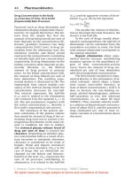
The timing of caesarean section and the
speed with which surgical delivery is
carried out is critical in determining the
outcome for mother and fetus. Most of the
children and mothers who survive
emergency caesarean deliveries are
delivered within five minutes of maternal
cardiac arrest
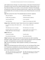
Paramedics are often the primary responders to obstetric emergency calls,
and so awareness of problems associated with resuscitation in late pregnancy
is important
39
The first priority for all those responsible for the care of babies
at birth must be to ensure that adequate resuscitation facilities
are available. Sadly, some babies have irreversible brain damage
by the time of delivery, but it is unacceptable that any damage
should occur after delivery due to inadequate equipment or
insufficiently trained staff. For this reason, there should always
be at least two healthcare professionals at all deliveries—one
who is primarily responsible for the care of the mother, and the
other, who must be trained in basic neonatal resuscitation, to
look after the baby.
All babies known to be at increased risk should be delivered
in a unit with full respiratory support facilities and must always be
attended by a doctor who is skilled in resuscitation and solely
responsible for the care of that baby. Whenever possible, there
should also be a trained assistant who can provide additional help
if necessary. Babies at increased risk make up about a quarter of
all deliveries and about two thirds of those requiring resuscitation;
the remaining one third are babies born after a normal
uneventful labour who have no apparent risk factors. Staff on
labour wards must, therefore, always be prepared to provide
adequate resuscitation until further help can be obtained.
Equipment
The padded platform on which the baby is resuscitated can
either be flat or have a head-down tilt. It can be wall mounted
or kept on a trolley, provided that one is available for each
delivery area. It is essential that there should be an overhead
heater with an output of 300-500 Watts mounted about 1 m
above the platform. This must have a manual control because
servo systems are slow to set up and likely to malfunction when
the baby’s skin is wet. These heaters are essential, as even in
environments of 20-24 ЊC the core temperature of an
asphyxiated wet baby can drop by 5 ЊC in as many minutes.
Facilities must be available for facemask and tracheal
tube resuscitation. The laryngeal mask airway is also
potentially useful. The use of oxygen versus air during
resuscitation at birth is controversial because high
concentrations of oxygen may be toxic in some circumstances.
The current international recommendation is that 100%
oxygen should be used initially if it is available. As the latest
generation of resuscitation systems have air and oxygen mixing
facilities it will usually be possible to reduce the inspired
oxygen fraction to a lower level once the initial phase of
resuscitation is over. Additional equipment needed includes an
overhead light, a clock with a second hand, suction equipment,
stethoscope, an electrocardiogram (ECG) monitor, and an
oxygen saturation monitor.
Procedure at delivery
It is common practice during labour to aspirate the pharynx
with a catheter as soon as the face appears. But this is almost
always unnecessary unless the amniotic fluid is stained with
meconium or blood. Aggressive pharyngeal suction can delay
the onset of spontaneous respiration for a considerable time.
Once the baby is delivered the attendant should wipe any
9 Resuscitation at birth
Anthony D Milner
High-risk deliveries
Delivery
● Fetal distress
● Reduced fetal movement
● Abnormal presentation
● Prolapsed cord
● Antepartum haemorrhage
● Meconium staining of liquor
● High forceps
● Ventouse
● Caesarean section under general
anaesthetic
Maternal
● Severe pregnancy-induced hypertension
● Heavy sedation
● Drug addiction
● Diabetes mellitus
● Chronic illness
Fetal
● Multiple pregnancy
● Pre-term (Ͻ 34/52)
● Post-term (Ͼ 42/52)
● Small for dates
● Rhesus isoimmunisation
● Hydramnios and oligohydramnios
● Abnormal baby
Resuscitation equipment
● Padded shelf or resuscitation trolley
● Overhead heater
● Overhead light
● Oxygen and air supply
● Clock
● Stethoscope
● Airway pressure manometer and pressure
relief valve
● Facemask
● Oropharyngeal airways 00, ϩ0
● Resuscitation system (facemask, T-piece,
bag and mask)
● Suction catheters (sized 5, 8, 10 gauge)
● Mechanical and/or manual suction with
double trap
● Two laryngoscopes with spare blades
● Tracheal tubes 2, 2.5, 3, 3.5, and 4 mm,
introducer
● Laryngeal masks
● Umbilical vein catheterisation set
● 2, 10, and 20 ml syringes with needles
● Intraosseous needle
● ECG and transcutaneous oxygen saturation
monitor
● Note: capnometers are a strongly
recommended optional extra
excess fluid off the baby with a warm towel to reduce
evaporative heat loss, while examining the child for major
external congenital abnormalities such as spina bifida and
severe microcephaly. Most babies will start breathing during
this period as the median time until the onset of spontaneous
respiration is only 10 seconds. They can then be handed to
their parents. If necessary, the baby can be encouraged to
breathe by skin stimulation—for example, flicking the baby’s
feet; those not responding must be transferred immediately to
the resuscitation area.
Resuscitation procedure
Once it is recognised that the newborn baby is failing to
breathe spontaneously and adequately, the procedures
standardised in the International Resuscitation Guidelines
published in 2000 should be followed. These guidelines
acknowledge that few resuscitation interventions have been
subjected to randomised controlled trials. However, there have
been a number of small physiological studies on the effects of
these interventions.
Check first for respiratory efforts and listen and feel for air
movement. If respiratory movements are present, even if they
are vigorous, but there is no tidal exchange, then the airway is
obstructed. This can usually be overcome by placing the head
in a neutral position (which may require a small roll of cloth
under the shoulders) and gently lifting the chin. An
oropharyngeal airway may occasionally be required, particularly
if the baby has congenital upper airway obstruction, such as
choanal atresia.
If respiratory efforts are feeble or totally absent, count the
heart rate for 10-15 seconds with a stethoscope over the
praecordium. If the heart rate is higher than 80 beats/min it is
sufficient to repeat skin stimulation, but if this fails to improve
respiration then proceed to facemask resuscitation.
Facemask resuscitation
Only facemasks with a soft continuous ring provide an
adequate seal. Most standard devices for manual resuscitation
of the neonate fail to produce adequate tidal exchange when
the pressure-limiting device is unimpeded. Thus, a satisfactory
outcome almost always depends on the inflation pressure
stimulating the baby to make spontaneous inspiratory efforts
(Head’s paradoxical reflex). Tidal exchange can be increased
by using a 500 ml rather than a 250 ml reservoir, which allows
inflation pressure to be maintained for up to one second.
More satisfactory tidal exchange can be achieved with a
T-piece system. In this system, a continuous flow of air and
oxygen is led directly into the facemask at 4-6 l/min; the lungs
are inflated by intermittently occluding the outlet from the
mask. It is essential to incorporate a pressure valve into the
fresh gas tubing so that the pressure cannot exceed 30 cmH
2
O.
The baby’s lungs are inflated at a rate of about 30/min, allowing
one second for each part of the cycle. Listen to the baby’s chest
after 5-10 inflations to check for bilateral air entry and a
satisfactory heart rate. If the heart rate falls below 80 beats/min
proceed immediately to tracheal intubation.
Tracheal intubation
Most operators find a straight-bladed laryngoscope preferable
for performing neonatal intubation. This is held in the left
hand with the baby’s neck gently extended, if necessary by the
assistant. The laryngoscope is passed to the right of the tongue,
ensuring that it is swept to the left of the blade, which is
advanced until the epiglottis comes into view. The tip of the
ABC of Resuscitation
40
Neonatal resuscitation trolley
Dry the baby. Remove any wet towels and cover. Start the clock or
note the time Assess colour, tone, breathing, and heart rate
If still not breathing. Give five inflation breaths. Look for a response.
If no increase in heart rate look for chest movement
If no response. Recheck head position. Apply jaw thrust.
Repeat inflation breaths Look for a response.
If no increase in heart rate look for chest movement
If still no response. Try alternative airway opening manoeuvres.
Repeat inflation breaths Look for a response.
If no increase in heart rate look for chest movement
If not breathing. Open the airway
When chest is moving. Give ventilation breaths. Check the heart rate
If heart rate is not detectable or slow (<60) and not increasing.
Start chest compressions. Three compressions to each breath
Reassess heart rate every 30 seconds. Consider venous access and drugs
Algorithm for newborn life support. Adapted from Newborn Life Support
Manual, London: Resuscitation Council (UK)
blade can then be positioned either proximal to or just under
the epiglottis so that the cords are brought into view. Gentle
backward pressure over the larynx may be needed at this stage.
As the upper airway tends to be filled with fluid it may have to
be cleared with the suction catheter held in the right hand.
Once the cords are visible, pass the tracheal tube with the
right hand and remove the laryngoscope blade, taking care that
this does not displace the tube out of the larynx. Most people
find it necessary to use an introducer to stiffen straight tracheal
tubes. It is then essential to ensure that the tip of the
introducer does not protrude, to avoid tracheal and
mediastinal perforation. If intubation proves difficult, because
the anatomy of the upper airway is abnormal or because of a
lack of adequately trained personnel, then a laryngeal mask
may be inserted.
Attach the tracheal tube either to a T-piece system
incorporating a 30-40 cmH
2
O blow-off valve (see above) or to a
neonatal manual resuscitation device. If a T-piece is used,
maintain the initial inflation pressure for two to three seconds.
This will help lung expansion. The baby can subsequently be
ventilated at a rate of 30/min, allowing about one second for
each inflation.
Inspect the chest during the first few inflations, looking for
evidence of chest wall movement, and confirm by auscultation
that gas is entering both lungs. If no air is entering the lungs
then the most likely cause is that the tip of the tracheal tube is
lying in the oesophagus. If this is suspected, remove the tube
immediately and oxygenate with a mask system. If auscultation
shows that gas is entering one lung only, usually the right,
withdraw the tube by 1 cm while listening over the lungs. If this
leads to improvement, the tip of the tracheal tube was lying in
the main bronchus. If no improvement is seen then the
possible causes include pneumothorax, diaphragmatic hernia,
or pleural effusion.
Severe bradycardia
If the heart rate falls below 60 beats/min, chest compression
must be started by pressing with the tips of two fingers over
sternum at a point that is one finger’s breadth below an
imaginary line joining the nipples. If there are two rescuers it is
preferable for one to encircle the chest with the hands and
compress the same point with the thumbs, while the other
carries out ventilation. The chest should be compressed by about
one third of its diameter. Give one inflation for every three chest
compressions at a rate of about 120 “events” per minute. This
will achieve about 90 compressions each minute. Those babies
who fail to respond require 10 mcg/kg (0.1 ml/kg of 1/10 000
solution) of adrenaline (epinephrine) given down the tracheal
tube. If no improvement is seen within 10-15 seconds the
umbilical vein should be catheterised with a 5 French gauge
catheter. This is best achieved by transecting the cord 2-3cm
away from the abdominal skin and inserting a catheter until
blood flows freely up the catheter. The same dose of adrenaline
(epinephrine) can then be given directly into the circulation.
Although evidence shows that sodium bicarbonate can
make intracellular acidosis worse, its use can often lead to
improvement, and the current recommendation is that the
baby should then be given 1-2 mmol/kg of body weight over
two to three minutes. This should be given as 2-4 ml/kg of 4.2%
solution. Those who fail to respond, or who are in
asystole, require further doses of adrenaline (epinephrine)
(10-30 mcg/kg). This can be given either intravenously or
injected down the tracheal tube.
It is reasonable to continue with alternate doses of
adrenaline (epinephrine) and sodium bicarbonate for
20 minutes, even in those who are born in apparent asystole,
Resuscitation at birth
41
Neonatal tracheal intubation equipment
Bag mask for neonatal resuscitation
Paediatric face masks.
ABC of Resuscitation
42
provided that a fetal heart beat was noted at some time within
15 minutes of delivery. Resuscitation efforts should not be
continued beyond 20 minutes unless the baby is making at least
intermittent respiratory efforts.
Naloxone therapy
Intravenous or intramuscular naloxone (100 mcg/kg) should
be given to all babies who become pink and have an obviously
satisfactory circulation after positive pressure ventilation but fail
to start spontaneous respiratory efforts. Often the mothers have
a history of recent opiate sedation. Alternatively, naloxone can
be given down the tracheal tube. If naloxone is effective then
an additional 200 micrograms/kg may be given intramuscularly
to prevent relapse. Naloxone must not be given to infants of
mothers addicted to opiates because this will provoke severe
withdrawal symptoms.
Meconium aspiration
A recent large, multicentre, randomised trial has shown that
vigorous babies born through meconium should be treated
conservatively. The advice for babies with central nervous
system depression and thick meconium staining of the liquor
remains—that direct laryngoscopy should be carried out
immediately after birth. If this shows meconium in the pharynx
and trachea, the baby should be intubated immediately and
suction applied directly to the tracheal tube, which should then
be withdrawn. Provided the baby’s heart rate remains above
60 beats/min this procedure can be repeated until meconium
is no longer recovered.
Hypovolaemia
Acute blood loss from the baby during delivery may complicate
resuscitation. It is not always clear that the baby has bled, so it is
important to consider this possibility in any baby who remains
pale with rapid small-volume pulses after adequate gas
exchange has been achieved. Most babies respond well to a
bolus (20-25 ml/kg) of an isotonic saline solution. It is rarely
necessary to provide the baby with blood in the labour suite.
Pre-term babies
Babies with a gestation of more than 32 weeks do not differ
from full-term babies in their requirement for resuscitation.
At less than this gestation they may have a lower morbidity and
mortality if a more active intervention policy is adopted.
However, no evidence has been found to show that a rigid
policy of routine intubation for all babies with a gestation of
less than 28 or 30 weeks leads to an improved outcome.
Indeed, unless the operator is extremely skilful, this
intervention may produce hypoxia in a previously lively pink
baby and predispose to intraventricular haemorrhage. A
reasonable compromise is to start facemask resuscitation after
15-30 seconds, unless the baby has entirely adequate respiratory
efforts, and proceed to intubation if the baby has not achieved
satisfactory respiratory efforts by 30-60 seconds. This policy may
need to be modified for the delivery of prophylactic surfactant
therapy, or if the neonatal unit is a considerable distance from
the labour suite.
Evidence is increasing to show that the pre-term baby is at
greatest risk from overinflation of the lungs immediately after
birth, and inflation volumes as little as 8 ml/kg may be capable
of producing lung damage. The lowest inflation pressure
compatible with adequate chest wall expansion should
therefore be used. Sometimes, however, pressures in excess of
30 cmH
2
0 will be necessary to inflate the surfactant-deficient
lungs.
Pharyngeal suction
● Rarely necessary unless amniotic fluid
stained with meconium or blood and the
baby asphyxiated
● Can delay onset of spontaneous respiration
for a long time if suction is aggressive
● Not recommended by direct mouth suction
or oral mucus extractors because of
congenital infection
Further reading
● International guidelines 2000 for cardiopulmonary resuscitation
and emergency cardiac care—a consensus on science. Part 11
neonatal resuscitation. Resuscitation 2000;46:401-6.
● Niermeyer S, Kattwinkel J, Van Reempts P, Nadkarni V, Philips B,
Zideman D, et al. International guidelines for neonatal
resuscitation: an excerpt from the guidelines 2000 for
cardiopulmonary resuscitation and emergency cardiac care:
Contributors and reviewers for the neonatal resuscitation
guidelines. Pediatrics 2000;106:E29.
● Ellemunter H, Simma B, Trawoger R, Maurer H. Intraosseous
lines in preterm and full term neonates. Arch Dis Child
1999;80:F74-F75.
● Field DJ, Milner AD, Hopkin IE. Efficacy of manual resuscitation
at birth. Arch Dis Child 1986;61:300-2.
● Saugstad OD, Roorwelt T, Aalen O. Resuscitation of asphyxiated
newborn infants with room air or oxygen: an international
controlled trial: the Resair 2 Study. Pediatrics 1998:102:e1.
● Saugstad OD. Mechanisms of tissue injury by oxygen radicals:
implications for neonatal disease. Acta Pediatr 1996;85:1-4.
● Vyas H, Field DJ, Milner AD, Hopkin IE. Physiological responses
to prolonged and slow rise inflation. J Pediatr 1981;99:635-9.
The goal of all deliveries—a healthy new born baby. With permission from
Steve Percival/Science Photo Library
43
The aetiology of cardiac arrest in infants and children is
different from that in adults. Infants and children rarely have
primary cardiac events. In infants the commonest cause of
death is sudden infant death syndrome, and in children aged
between 1 and 14 years trauma is the major cause of death. In
these age groups a primary problem is found with the airway.
The resulting difficulties in breathing and the associated
hypoxia rapidly cause severe bradycardia or asystole. The poor
long-term outcome from many cardiac arrests in childhood is
related to the severity of cellular anoxia that has to occur
before the child’s previously healthy heart succumbs. Organs
sensitive to anoxia, such as the brain and kidney, may be
severely damaged before the heart stops. In such cases
cardiopulmonary resuscitation (CPR) may restore cardiac
output but the child will still die from multisystem failure in the
ensuing days, or the child may survive with serious neurological
or systemic organ damage. Therefore, the early recognition of
the potential for cardiac arrest, the prevention and limitation
of serious injury, and earlier recognition of severe illness is
clearly a more effective approach in children.
Paediatric basic life support
Early diagnosis and aggressive treatment of respiratory or
cardiac insufficiency, aimed at avoiding cardiac arrest, are the
keys to improving survival without neurological deficit in
seriously ill children. Establishment of a clear airway and
oxygenation are the most important actions in paediatric
resuscitation. These actions are prerequisites for other forms of
treatment.
Resuscitation should begin immediately without waiting for
the arrival of equipment. This is essential in infants and
children because clearing the airway may be all that is required.
Assessment and treatment should proceed simultaneously to
avoid losing vital time. As in any resuscitation event, the
Airway-Breathing-Circulation sequence is the most appropriate.
If aspiration of a foreign body is strongly suspected, because
of sudden onset of severe obstruction of the upper airway, the
steps outlined in the section on choking should be taken
immediately.
Assess responsiveness
Determine responsiveness by carefully stimulating the child.
If the child is unresponsive, shout for help. Move the child only
if he or she is in a dangerous location.
Airway
Open the airway by tilting the head and lifting the lower jaw.
Care must be taken not to overextend the neck (as this may
cause the soft trachea to kink and obstruct) and not to press on
the soft tissues in the floor of the mouth. Pressure in this area
will force the tongue into the airway and cause obstruction.
The small infant is an obligatory nose breather so the patency
of the nasal passages must be checked and maintained.
Alternatively, the jaw thrust manoeuvre can be used when a
10 Resuscitation of infants and children
David A Zideman, Kenneth Spearpoint
Definitions
● An infant is a child under one year of age
● A child is aged between one and eight years
● Children over the age of eight years should
be treated as adults
Stimulate and check responsiveness
Open airway. Head tilt, chin lift (jaw thrust)
Check breathing. Look, listen, feel
If breathing, place
in recovery position
If no chest rise
- reposition airway
- re-attempt up to five times
If no success
- treat as for
airway obstruction
Breathe. Two effective breathes
No
No
Yes
Yes
Assess for signs of a circulation
Check pulse (10 seconds maximum)
Compress chest. Five compressions:
One ventilation, 100 compressions/minute
Continue resuscitation
Algorithm for paediatric basic life support
Opening infant airway
history of trauma or damage to the cervical spine is suspected.
Maintaining the paediatric airway is a matter of trying various
positions until the most satisfactory one is found. Rescuers
must be flexible and willing to adapt their techniques.
Breathing
Assess breathing for 10 seconds while keeping the airway open by:
● Looking for chest and abdominal movement
● Listening at the mouth and nose for breath sounds
● Feeling for expired air movement with your cheek.
If the child’s chest and abdomen are moving but no air can
be heard or felt, the airway is obstructed. Readjust the airway
and consider obstruction by a foreign body. If the child is not
breathing, expired air resuscitation must be started
immediately. With the airway held open, the rescuer covers the
child’s mouth (or mouth and nose for an infant) with their
mouth and breathes out gently into the child until the chest is
seen to rise. Minimise gastric distension by optimising the
alignment of the airway and giving slow and steady inflations.
Give two effective breaths, each lasting about 1-1.5 seconds, and
note any signs of a response (the child may cough or “gag”).
Up to five attempts may be made to achieve two effective
breaths when the chest is seen to rise and fall.
Circulation
Recent evidence has questioned the reliability of using a pulse
check to determine whether effective circulation is present.
Therefore, the rescuer should observe the child for 10 seconds
for “signs of a circulation.” This includes any movement,
coughing, or breathing (more than an odd occasional gasp).
In addition, healthcare providers are expected to check for the
presence, rate, and volume of the pulse. The brachial pulse is
easiest to feel in infants, whereas for children use the carotid
pulse. The femoral pulse is an alternative for either. If none of
the signs of a circulation have been detected, then start chest
compressions without further delay and combine with
ventilation. Immediate chest compressions, combined with
ventilation, will also be indicated when a healthcare provider
detects a pulse rate lower than 60 beats/min.
In infants and children the heart lies under the lower third
of the sternum. In infants, compress the lower third of the
sternum with two fingers of one hand; the upper finger should
be one finger’s breadth below an imaginary line joining the
nipples. When more than one healthcare provider is present,
the two-thumbed (chest encirclement) method of chest
compression can be used for infants. The thumbs are aligned
one finger’s breadth below an imaginary line joining the
nipples, the fingers encircle the chest, and the hands and
fingers support the infant’s rib cage and back. In children,
the heel of one hand is positioned over a compression point
two fingers’ breadth above the xiphoid process. In both infants
and children the sternum is compressed to about one third of
the resting chest diameter; the rate is 100 compressions/min.
The ratio of compressions to ventilations should be 5 : 1,
irrespective of the number of rescuers. The compression phase
should occupy half of the cycle and should be smooth, not jerky.
In larger, older children (over the age of eight years) the
adult two-handed method of chest compression is normally
used (see Chapter 1). The compression rate is 100/min and
the compression to ventilation ratio is 15 : 2, but the
compression depth changes to 4-5 cm.
Activation of the emergency medical services
When basic life support is being provided by a lone rescuer the
emergency medical services must be activated after one minute
ABC of Resuscitation
44
Mouth-to-mouth and nose ventilation
Chest compression in infants and children
because the provision of advanced life support procedures is
vital to the child’s survival. The single rescuer may be able to
carry an infant or small child to the telephone, but older
children will have to be left. Basic life support must be restarted
as soon as possible after telephoning and continued without
further interruption until advanced life support arrives. In
circumstances in which additional help is available or the child
has known heart disease, then the emergency medical services
should be activated without delay.
Activate emergency services after one minute.
Choking
If airway obstruction caused by aspiration of a foreign body is
witnessed or strongly suspected, special measures to clear the
airway must be undertaken. Encourage the child, who is
conscious and is breathing spontaneously, to cough and clear
the obstruction themselves. Intervention is only necessary if
these attempts are clearly ineffective and respiration is
inadequate. Never perform blind finger sweeps of the pharynx
because these can impact a foreign body in the larynx. Use
measures intended to create a sharp increase in pressure within
the chest cavity, such as an artificial cough.
Back blows
Hold the infant or child in a prone position and deliver up to
five blows to the middle of the back between the shoulder
blades. The head must be lower than the chest during this
manoeuvre. This can be achieved by holding a small infant
along the forearm or, for older children, across the thighs.
Chest thrusts
Place the child in a supine position. Give up to five thrusts to
the sternum. The technique of chest thrusts is similar to that
for chest compressions. The chest thrusts should be sharper
and more vigorous than compressions and carried out at a
slower rate of 20/min.
Check mouth
Remove any visible foreign bodies.
Open airway
Reposition the head by the head tilt and chin lift or jaw thrust
manoeuvre and reassess air entry.
Breathe
Attempt rescue breathing if there are no signs of effective
spontaneous respiration or if the airway remains obstructed.
It may be possible to ventilate the child by positive pressure
expired air ventilation when the airway is partially obstructed,
but care must be taken to ensure that the child exhales most of
this artificial ventilation after each breath.
Repeat
If the above procedure is unsuccessful in infants it should be
repeated until the airway is cleared and effective respiration
established. In children, abdominal thrusts are substituted for
chest thrusts after the second round of back blows.
Subsequently, back blows are combined with chest thrusts or
abdominal thrusts in alternate cycles until the airway is cleared.
Paediatric advanced life support
The use of equipment in paediatric resuscitation is fraught with
difficulties. Not only must a wide range be available to
correspond with different sized infants and children but the
rescuer must also choose and use each piece accurately.
Resuscitation of infants and children
45
Back blows for choking infants and children are delivered between the
shoulder blades with the subject prone
Abdominal thrusts
● In children over one year deliver up to five
abdominal thrusts after the second five
back blows. Use the upright position
(Heimlich manoeuvre) if the child is
conscious
● Unconscious children must be laid supine
and the heel of one hand placed in the
middle of the upper abdomen. Up to five
sharp thrusts should be directed upwards
toward the diaphragm
● Abdominal thrusts are not recommended
in infants because they may cause damage
to the abdominal viscera
Effective basic life support is a prerequisite for successful
advanced life support.
Airway and ventilation management
Airway and ventilation management is particularly important in
infants and children during resuscitation because airway and
respiratory problems are often the cause of the collapse. The
airway must be established and the infant or child should be
ventilated with high concentrations of inspired oxygen.
Airway adjuncts
Use an oropharyngeal (Guedel) airway if the child’s airway
cannot be maintained adequately by positioning alone during
bag-valve-mask ventilation. A correctly sized airway should
extend from the centre of the mouth to the angle of the jaw
when laid against the child’s face. A laryngeal mask can be used
for those experienced in the technique.
Tracheal intubation is the definitive method of securing the
airway. The technique facilitates ventilation and oxygenation
and prevents pulmonary aspiration of gastric contents, but it
does require training and practice. A child’s larynx is narrower
and shorter than that of any adult and the epiglottis is relatively
longer and more U-shaped. The larynx is also in a higher, more
anterior, and more acutely angled position than in the adult.
A straight-bladed laryngoscope and plain plastic uncuffed
tracheal tubes are therefore used in infants and young
children. In children aged over one year the appropriate size of
tracheal tube can be assessed by the following formula:
Internal diameter (mm) ϭ (age in years/4) ϩ 4
Infants in the first few weeks of life usually require a tube of
size 3-3.5 mm, increasing to a size 4 when aged six to
nine months.
Basic life support must not be interrupted for more than
30 seconds during intubation attempts. After this interval the
child must be reoxygenated before a further attempt is made.
If intubation cannot be achieved rapidly and effectively at this
stage it should be delayed until later in the advanced life
support protocol. Basic life support must continue.
Oxygenation and ventilation adjuncts
A flowmeter capable of delivering 15 l/min should be attached
to the oxygen supply from either a central wall pipeline or an
independent oxygen cylinder. Facemasks for mouth-to-mask or
bag-valve-mask ventilation should be made of soft clear plastic,
have a low dead space, and conform to the child’s face to form
a good seal. The circular design of facemask is recommended,
especially when used by the inexperienced resuscitator. The
facemask should be attached to a self-inflating bag-valve-mask of
either 500 ml or 1600 ml capacity. The smaller bag size has a
pressure-limiting valve attached to limit the maximum airway
pressure to 30-35 cm H
2
O and thus prevent pulmonary damage.
Occasionally, this pressure-limiting valve may need to be
overridden if the child has poorly compliant lungs. An oxygen
reservoir system must be attached to the bag-valve-mask system,
thereby enabling high inspired oxygen concentrations of over
80% to be delivered. The Ayre’s T-piece with the open-ended
bag (Jackson Reece modification) is not recommended because
it requires specialist training to be able to operate it safely and
effectively.
Management protocols for advanced life support
Having established an airway and effective ventilation with high
inspired oxygen, the next stage of the management depends on
the cardiac rhythm. The infant or child must therefore be
attached to a cardiac monitor or its electrocardiogram (ECG)
monitored through the paddles of a defibrillator.
ABC of Resuscitation
46
Assess rhythm
Basic life support algorithm
Ventilate/oxygenate
Attach defibrillator/monitor
± Check pulse
Non VF/VT
Asystole;
Pulseless
electrical
activity
VF/VT
CPR 3 minutes
CPR
1 minute
Defibrillate
as necessary
Adrenaline
(epinephrine)
During CPR
• Attempt/verify:
Tracheal intubation
Intraosseous/vascular access
• Check
Electrode/paddle positions and contact
• Give
Adrenaline (epinephrine) every 3 minutes
• Consider anti-arrhythmics
• Consider acidosis
Consider giving bicarbonate
• Correct reversible causes
Hypoxia
Hypovolaemia
Hyper- or hypokalaemia
Hypothermia
Tension pneumothorax
Tamponade
Toxic/therapeutic disturbances
Thromboemboli
Algorithm for paediatric advanced life support
Guedel oropharyngeal airways
Laerdal face masks
Non-ventricular fibrillation/non-ventricular tachycardia
Asystole is the commonest cardiac arrest rhythm in infancy and
childhood. It is the final common pathway of respiratory or
circulatory failure and is usually preceded by an agonal
bradycardia.
The diagnosis of asystole is made on electrocardiographic
evidence in a pulseless patient. Care must be taken to ensure
that the electrocardiograph leads are correctly positioned and
attached and that the monitor gain is turned up. Effective basic
life support and ventilation with high-flow oxygen through a
patent airway are essential. Having established a secure airway
and intravenous or intraosseous access, 10 mcg/kg (0.1 ml/kg of
1 : 10 000) of adrenaline (epinephrine) is administered followed
by three minutes of basic life support. If asystole persists then a
further dose of 0.1 ml/kg of 1:10 000 adrenaline (epinephrine)
should be administered during the subsequent three minute
period of CPR. If asystole persists, further three-minute
sequences of CPR with adrenaline (epinephrine) at doses of
10-100 mcg/kg (0.1 ml/kg of 1:1000) may be given while
considering other drugs and interventions.
Alkalising agents are of unproven benefit and should be
used only after clinical diagnosis of profound acidosis in
patients with respiratory or circulatory arrest if the first dose of
adrenaline (epinephrine) has been ineffective. The dose of
bicarbonate is 1 mmol/kg and is given as a single bolus by slow
intravenous injection, ideally before the second dose of
adrenaline (epinephrine). If an alkalising agent is used then
the cannula must be thoroughly flushed with normal saline
before any subsequent dosing with adrenaline (epinephrine)
because this drug will be chemically inactivated by the
alkalising agent. Subsequent treatment with alkalising agents
should be guided by the blood pH.
A bolus of normal saline should follow the intravenous or
intraosseous injection of any drug used in resuscitation,
especially if the injection site is peripheral. The amount should
be 5-20 ml, depending on the size of the child. When cardiac
arrest has resulted from circulatory failure a larger bolus of
fluid should be given if no response or only a poor response to
the initial dose of adrenaline (epinephrine) is seen. Examples
of such cases are children with hypovolaemia from blood loss,
gastroenteritis, or sepsis when a profound distributive
hypovolaemic shock may occur. These children require
20 ml/kg of a crystalloid (normal saline or Ringer’s lactate)
or a colloid (5% human albumin or an artificial colloid).
Pulseless electrical activity
Formerly known as electromechanical dissociation, pulseless
electrical activity (PEA) is described as a normal (or near
normal) ECG in the absence of a detectable pulse. If not
treated, this rhythm will soon degenerate through agonal
bradycardia to asystole. It is managed in the same way as
asystole, with oxygenation and ventilation accompanying basic
life support and adrenaline (epinephrine) to support coronary
and cerebral perfusion.
Ventricular fibrillation and pulseless ventricular tachycardia
Ventricular fibrillation is relatively rare in children, but it is
occasionally seen in cardiothoracic intensive care units or in
patients being investigated for congenital heart disease. In
contrast to the treatment of asystole, defibrillation takes
precedence. Defibrillation is administered in a series of
three energy shocks followed by one minute of basic life
support. The defibrillation energy is 2 J/kg for the first shock,
2 J/kg for the second rising to 4 J/kg for the third and all
subsequent defibrillation attempts. For defibrillators with
Resuscitation of infants and children
47
Two arrest rhythms
● Non-VF/VT: asystole or pulseless electrical
activity
● Ventricular fibrillation or pulseless
ventricular tachycardia
Asystole
● Common arrest rhythm in children
● ECG evidence in a pulseless patient
PEA
● Absence of cardiac output with normal or
near normal ECG
● ECG evidence in pulseless patient
Ventricular fibrillation and pulseless
ventricular tachycardia
● Characteristic ECG in pulseless patient
● Relatively rare in children
● Treatment is immediate defibrillation
Asystole in an infant or child
Broad and slow rhythm is associated with pulseless electrical activity
stepped current levels the nearest higher step to the calculated
energy level required should be selected.
Ventilation and chest compressions should be continued at all
times except when shocks are being delivered or the ECG is
being studied for evidence of change. Paediatric paddles
should be used in children below 10 kg, but in bigger children
the larger adult electrode will minimise transthoracic
impedance and should be used when the child’s thorax is
broad enough to permit electrode-to-chest contact over the
entire paddle surface. One paddle should be placed over the
apex of the heart and one beneath the right clavicle.
Alternatively, a front-to-back position can be used.
Consider giving adrenaline (epinephrine) every
three minutes during resuscitation. In ventricular fibrillation
adrenaline (epinephrine) should be administered as
10 mcg/kg initially followed by 10-100 mcg/kg for all
subsequent administrations.
Other considerations
As mentioned previously, it is rare for infants and children to
have a primary cardiac arrest. Therefore, it is important to seek
out and treat the initial cause of the cardiorespiratory collapse.
This cause should be sought while basic and advanced life
support continues. The most common causes can be
summarised as the 4Hs and 4Ts.
When detected, the underlying cause must be treated
rapidly and appropriately.
ABC of Resuscitation
48
4Hs and 4Ts
● Hypoxia
● Hypovolaemia
● Hyper- or hypokalaemia
● Hypothermia
● Tension pneumothorax
● Tamponade
● Toxic or therapeutic disturbances
● Thromboembolism
Drug doses and equipment sizes
An important consideration when managing cardiac arrest in
children is the correct estimation of drug doses, fluid volumes,
and equipment sizes. There are two systems in current use. The
first entails a calculation based on the length of the child and a
specifically designed tape measure (the Broselow tape. The
other uses a length-weight-age nomogram chart (the Oakley
chart). It is important to become familiar with and to use one
of these systems.
Audit of results
The future development of paediatric guidelines will be
determined by an examination of published scientific evidence.
The Utstein Template has aided the uniform collection of data
from paediatric resuscitation attempts.
Drugs and fluid administration
If venous access has not been established before the
cardiorespiratory collapse, peripheral venous access should be
attempted. This is notoriously difficult in small ill children.
Central venous access is also difficult except in the hands of
experts, is hazardous in children, and is unlikely to provide a
more rapid route for drugs. If venous access is not gained
within 90 seconds, the intraosseous route should be attempted.
50
Age (years)
51020304050
14
18-21
18
17
16
15
14
13
12
10
7.5-8.0 (cuffed)
Oral
length
(cm)
Internal
diameter
(mm)
Endotracheal tube
Length
Weight
0.5 1 2 3 4 5Adrenaline/epinephrine (ml of 1 in 10 000)
intravenous or intraosseous
1246810*Atropine (ml of 100µg/ml)
intravenous or intraosseous
0.8 1.5 3.5 5 6.5
50
mmol
*Bicarbonate (mmol)
intravenous or intraosseous
-2.55 5 55mg*Salbutamol (mg nebuliser solution) by
nebuliser (dilute to 2.5-5 ml in physiological saline)
100 200 400 600 800 1000**Initial fluid bolus in shock (ml)
intravenous or intraosseous (crystalloid or colloid)
* Caution! Non-standard drug concentrations may be available:
Use atropine 100 µg/ml or prepare by diluting 1 mg to 10 ml or 600 µg to 6 ml in 0.9% saline
Bicarbonate is available in various concentrations (8.4% has 1 mmol/ml; 4.2% has
0.5 mmol/ml; 1.26% has 0.15 mmol/ml). In infants, avoid 8.4% or dilute to at least 4.2%.
Note that 1 ml of calcium chloride 10% is equivalent to 3 ml of calcium gluconate 10%
Use lidocaine/lignocaine (without adrenaline/epinephrine) 1% or give half the volume of 2%
(or dilute appropriately)
In the initial nebulised dose of salbutamol, ipratropium may be added to the nebuliser in
doses of 250 µg for a 10 kg child and 500 µg for an older child. Salbutamol may also
be given by slow intravenous injection (5 µg/kg over 5 minutes), but beware of the different
concentrations available (eg 50 and 500 µg/ml)
** In uncontrolled haemorrhage, give fluid in careful, repeated increments (eg 5 ml/kg
rather than 20 ml/kg at once) to maintain a palpable pulse and minimum acceptable
blood pressure until bleeding is controlled
0.5*Calcium chloride (ml of 10%)
intravenous or intraosseous
- 0.3 0.7 1 1.3 1.7Atropine (ml of 600µg/ml)
*Amiodarone (ml of 50µg/ml concentrated
solution)
0.5 1 2 3 4 5Lorazepam (ml of 5mg diluted to 5ml in
0.9% saline)
intravenous or intraosseous
- - 0.4 0.6 0.8 1Lorazepam (ml of 5mg/ml neat)
2.5 5 - - - -Naloxone neonatal (ml of 20µg/ml)
intravenous or intraosseous
- 0.25 0.5 0.75 1 1.25Naloxone adult (ml of 400µg/ml)
0.5 1 2 3 4 5
12 3 4 5
0.5*Lidocaine/lignocaine (ml of 1%)
intravenous or intraosseous
12 3 4 5
25Glucose (ml of 10%)
intravenous or intraosseous
50 100 150 200 250
Adrenaline/epinephrine (ml of 1 in 1000)
endotracheal
2.5 5 10 10 10 10mgDiazepam (mg rectal tube solution)
(if lorazepam or intravenous access not available)
rectal
10 20 40 60 80 100JInitial DC defibrillation (J) for ventricular
fibrillation or pulseless ventricular tachycardia
5 5 10 15 20 25JInitial DC cardioversion (J) for supraventricular
tachycardia with shock (synchronous) or ventricular
tachycardia with shock (non-synchronous)
kg
cm
7.0 (uncuffed)
6.5
6.0
5.5
5.0
4.5
4.0
3.5
3.0-3.5
12
10
8
6
4
2
1
9 months
6 months
3 months
60 80 100120 140 150
510203040
8.5ml
0.5 1 2 3
dilute appropriately in 5% glucose
dilute appropriately in 5% glucose
45ml
*Amiodarone (ml of 30µg/ml prefilled)
(bolus in cardiac arrest, slowly over 3 minutes if not)
intravenous or intraosseous
The Oakley chart
ABCR-10.qxd 10/21/03 3:39 PM Page 48
Intraosseous access is a safe, simple, and rapid means of
circulatory access for infants and children. Resuscitation drugs,
fluid, and blood can be safely given via this route and rapidly
reach the heart. Complications are uncommon and usually
result from prolonged use of the site or poor technique.
Marrow aspirate can be drawn and used to estimate
concentrations of haemoglobin, sodium, potassium, chloride,
glucose, venous pH, and blood groups.
If circulatory access proves impossible to achieve within
two to three minutes, some drugs, including adrenaline
(epinephrine) and atropine, can be given down the tracheal
tube. Data from studies on animals and humans suggest that
the endotracheal dose of adrenaline (epinephrine) should be
10 times the standard dose, but doubts have been cast on the
reliability of this route and intravenous or intraosseous drug
administration is preferable.
Resuscitation of infants and children
49
Intraosseous infusion needle placed in the upper tibia
Further reading
● APLS Working Group. Advanced paediatric life support. The practical
approach. 3rd ed. London: BMJ Publishing Group, 2001.
● European Resuscitation Council. Guidelines 2000 for
cardiopulmonary resuscitation and cardiovascular care—an
international consensus on science. Resuscitation 2000;46:301-400.
● Nadkarni V, Hazinski MF, Zideman DA, Kattwinkel K, Quan L,
Bingham R, et al. Paediatric life support: an advisory statement
by the Paediatric Life Support Working Group of the
International Liaison Committee on Resuscitation. Resuscitation
1997;34:115-27.
● Luten R, Wears R, Broselow J, Zaritsky A, Barnett T, Lee T.
Length based endotracheal tube and emergency equipment
selection in paediatrics. Ann Emerg Med 1992;2:900-4.
● Oakley P. Inaccuracy and delay in decision making in paediatric
resuscitation and a proposed reference chart to reduce error.
BMJ 1988;297:817-9.
● Oakley P, Phillips B, Molyneux E, Mackway-Jones K. Paediatric
resuscitation. BMJ 1994;306:1613.
● Zaritsky A, Nadkarni V, Hanzinski MF, Foltin G, Quan L, Wright
J, et al. Recommended guidelines for uniform reporting of
paediatric advanced life support: the paediatric utstein style.
Resuscitation 1995;30:95-116.
The algorithms for paediatric basic life support and paediatric
advanced life support are adapted from Resuscitation Guidelines
2000, London: Resuscitation Council (UK), 2000.
The diagrams
of Guedel oropharyngeal airways and Laerdal masks are adapted
from Newborn Life Support Manual, London: Resuscitation Council
(UK). The diagram of and intraosseous infusion needle is courtesy
of Cook Critical Care (UK).