LAST MINUTE EMERGENCY MEDICINE - PART 3 pdf
Bạn đang xem bản rút gọn của tài liệu. Xem và tải ngay bản đầy đủ của tài liệu tại đây (600.4 KB, 62 trang )
VALVULAR DISORDERS 111
TABLE 3-10 NATIVE VALVE DISORDE RS (CONTINUED)
VALVULAR
ABNOR MALITY ETIOLOGY PR ESENTATION HEART SOUNDS NOTES
Mitral valve
prolapse
(MVP)
r
The most common
valvular heart
abnormality in U.S.
r
Exact cause is uncertain
but presumed to be
either congenital or due
to myxomatous
degeneration of the
mitral valve
r
Most cases are
asymptomatic
r
Presenting
symptoms often
include atypical
chest pain, dyspnea,
palpitations
r
Increased incidence
of panic disorder,
TIAs,
tachydysrhythmias,
sudden death
r
Early, or mid-systolic
click
r
Click is often
followed by
high-pitched
late-systolic murmur
r
Increases in left
ventricular volume
(e.g., squatting,
Trendelenburg)
move the click/
murmur closer to S
2
and decrease the
intensity/duration of
the murmur
r
Decreases in left
ventricular volume
(e.g., standing,
valsalva) move the
click/murmur closer
to S
1
and increase
the intensity/
duration of the
murmur
r
Advanced disease
may be associated
with MR
r
Treatment:
beta-blocking
medications relieve
atypical chest pain
and atrial
dysrhythmias
r
Antibiotic
prophylaxis against
infective
endocarditis
indicated only if
murmur (or
echocardiographic
evidence of MR)
present
Right-sided
valvular
heart
disease
r
Isolated tricuspid valve
disease is the most
common right-sided
valvular disorder,
usually caused by
intravenous drug use
r
Right-sided valvular
disease may also be
caused by rheumatic
heart disease
r
The most common
symptoms are
related to infective
endocarditis: fevers,
myalgias, dyspnea
r
Tricuspid
regurgitation
presents with a
holosystolic murmur
heard best at the left
lower sternal border
r
Treatment of
tricuspid
regurgitation is
focused on treating
underlying
endocarditis, surgery
if significant
dysfunction occurs
(Continued )
112 CHAPTER 3 / CARDIOVASCULAR DISORDERS
TABLE 3-10 NATIVE VALVE DISORDE RS (CONTINUED)
VALVULAR
ABNOR MALITY ETIOLOGY PR ESENTATION HEART SOUN DS NOTES
Aortic
stenosis
(AS)
r
Most common causes
are congenital bicuspid
valve and rheumatic
heart disease
r
Most common cause in
the elderly is idiopathic
calcification/
degeneration
r
Obstruction to outflow
produces low cardiac
output and leads to
symptoms once the
valve lumen is reduced
to 25% of normal
r
Classic triad is
angina, syncope,
CHF
r
5–10% incidence
of sudden death
r
Systolic crescendo–
decrescendo
murmur
r
Paradoxical splitting
of S
2
r
Best heard at right
upper sternal border
r
Usually murmur
radiates to carotids
r
ECG usually
demonstrates
evidence of left
ventricular
hypertrophy
r
CXR demonstrates
cardiomegaly,
evidence of CHF
r
Narrow pulse
pressure in severe
disease
r
Best treatment is
valve replacement
r
The use of preload
reducers, afterload
reducers, and
inotropes must be
done with extreme
caution, can cause
hemodynamic
decompensation
Aortic
regurgitation
(AR)
r
Most common causes
of chronic AR are
rheumatic heart
disease and congenital
causes
r
Chronic cases are
associated with
development of left
ventricular
hypertrophy and
dilation, which over
many year results in
CHF
r
Chronic AR
produces a
high-pitched
decrescendo
diastolic murmur
r
ECG usually
demonstrates
evidence of left
ventricular
hypertrophy in
chronic cases
VALVULAR DISORDERS 113
TABLE 3-10 NATIVE VALVE DISORDE RS (CONTINUED)
VALVULAR
ABNOR MALITY ETIOLOGY PR ESENTATION HEART SOUN DS NOTES
r
Most common causes
of acute AR are
endocarditis
r
Proximal thoracic aortic
dissection can also
cause acute AR
r
Acute AR is the most
common valvular
disorder caused by
blunt chest trauma
r
Peripheral signs of
chronic AR include
Water-hammer
pulse, “pistol shot”
femoral pulses,
pulsating nail beds,
and head-bobbing
with systole
r
Acute cases are
associated with
chest pain and
pulmonary edema;
patients are
sometimes
hypotensive
r
Best heard along the
left sternal border
r
Acute AR produces
a faint short diastolic
murmur, which is
often inaudible
r
CXR in chronic cases
demonstrates
cardiomegaly, CHF
r
CXR in acute cases
demonstrates
normal sized heart
with pulmonary
edema
r
Patients with chronic
AR develop a wide
pulse pressure
r
Treatment of chronic
AR is based on
treating the CHF
r
Acute AR requires
treatment with
inotropes, afterload
reduction, and
surgery
114 CHAPTER 3 / CARDIOVASCULAR DISORDERS
TABLE 3-11 PROSTHETIC VALVE COMPLICATIONS
COMPLICATION NOTES
Thromboemboli
r
Thrombus formation can cause acute valvular dysfunction
r
Patients may present with acute heart failure, hypotension, loss of the
expected abnormal valve sounds
r
More common with mechanical valves than with tissue valves
Endocarditis
r
See section on Infective Endocarditis
Hemolysis
r
If mild, iron supplementation is adequate treatment
r
If severe, consider paravalvular leak
r
Patients may present with anemia symptoms and jaundice
r
More common with mechanical valves than with tissue valves
Paravalvular leak
r
Occurs when part of the valve becomes displaced
r
Patients may present with acute heart failure and/or hemolytic anemia
r
Regurgitant murmur is usually present
r
More common with mechanical valves than with tissue valves
Primary valve failure
r
Patients may present with evidence of embolization of a prosthetic fragment,
hemolysis, acute valvular occlusion
FURTHER READING
Antman EM, Anbe DT, Armstrong PW, et al. ACC/AHA Guidelines for the Management of Patients with ST-Elevation
Myocardial Infarction—Executive Summary. Circulation 2004;110:588–636.
Chen K, Varon J, Wenker OC, et al. Acute Thoracic Aortic Dissection: The Basics. J Emerg Med 1997;15:859–867.
Fox KA. Management of Acute Coronary Syndromes: An Update. Heart 2004;90:698–706.
Gropper MA, Wiener-Kronish JP, Hashimoto S. Acute Cardiogenic Pulmonary Edema. Clin Chest Med 1994;15:501–15.
Lange RA, Hillis LD: Clinical practice: acute pericarditis. N Engl J Med 2004;351:2195–2202.
Ma OJ, Cline DM, Tintinalli JE, et al. (eds.). Emergency Medicine: Just the Facts, 2nd ed. New York: McGraw-Hill,
2004.
Marx JA, Hockberger RS, Walls RM, et al. (eds.). Rosen’s Emergency Medicine: Concepts and Clinical Practice, 5th ed.
St. Louis: Mosby, 2002.
Mattu A. Cardiogenic pulmonary edema. Current Opinion in Cardiovascular, Pulmonary, and Renal Investigational
Drugs 2000;2:9–16.
Mattu A, Brady WJ: ECGs for the Emergency Physician. London: BMJ Publishing Company, 2003.
CHAPTER 4
CUTANEOUS DISORDERS
GENERAL TERMINOLOGY AND DESCRIPTORS
TABLE 4-1 DEFINITION OF SKIN LESIONS
TERM LATIN MEANING DESCRIPTION SIZE EXAM PLE
Macule Spot Flat, nonpalpable well
defined
<1 cm Vitiligo
Papule Pimple Palpable <1 cm Mollususcum contagiousum
Plaque Confluence of papules
Plateau-like elevation that is
well defined
Psoriasis, Lichenification of
atopic dermatitis
Nodule Small knot Solid, palpable lesions (hard
or soft)
>1 cm Wart
Wheal Flat-topped papule or plaque
that last for 24–48 h
Urticaria
Vesicles Blister Elevated, superficial,
fluid-filled cavities
<0.5 cm Herpes zoster
Bullae Bubble Elevated, superficial,
fluid-filled cavities
>0.5 cm Bullous impetigo
Pustule Circumscribed skin cavity
filled with purulent exudates,
may arise from a hair follicle
Variable Folliculitis
Purpura Red-purple lesions that do
not blanch with pressure
>0.3 mm ITP, TTP, meningiococcemia
Petechaie Skin spot Red-purple lesions that do
not blanch with pressure
<0.3 mm Seen on the arm distal to
the BP cuff in patients on
anticoagulants
115
Copyright © 2007 by The McGraw-Hill Companies, Inc. Click here for terms of use.
116 CHAPTER 4 / CUTANEOUS DISORDERS
Specific Lesions and Management
ERYTHEMA MULTIFORME
Erythema multiforme (EM) is a continuum of pathology including EM minor and EM major. EM major
encompasses Stevens-Johnson syndrome (SJ) and toxic epidermal necrolysis (TEN).
ERYTHEMA MULTIFORME MINOR
Etiology: Over 50% of cases are idiopathic. Of known etiologies, drugs are the most common cause.
Common offenders include penicillin, sulfonamides, and anticonvulsants. Infections are also a common
cause and are more commonly the etiology in children. HSV I and II, Influenza A, and mycoplasma have
been implicated in EM. Recurrent EM has been associated with HSV I and II. It may present with mucosal
lesions, possibly causing confusion with EM major.
Clinical Presentation: EM Minor is an acute, self-limited process. An inflammatory process yields the
pathognomonic target, or “iris” lesion. The lesions evolve over 1–2 days, are symmetrically distributed and
are found on the hands, feet, and extensor surfaces of the extremities. Lesions persist for about 7 days and
tend to resolve after 1 to 2 weeks with minimal or no sequelae. EM Minor has little or no mucus membrane
involvement.
Diagnosis: Clinical.
Treatment: Supportive care and symptomatic treatment. Discontinue offending agent if possible. Topical
steroids may be used on all lesions except eroded areas. Control of HSV using oral antiviral therapy when
indicated may prevent recurrent EM.
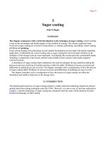
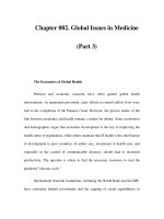
FIGURE 4-1. ERYTHEMA MULTIFORME MINOR.
ERYTHEMA MULTIFORME MAJOR
Two main syndromes include SJS and TEN (see Figure 4-2).
Etiology: The same drugs have been implicated as the cause of EM major as in EM minor, though some
cases are idiopathic. SJS is considered a maximal variant of EM Major, while TEN is considered a maximal
variant of SJS.
Clinical Presentation: With EM major, the patient is clinically ill appearing. A prodrome of fever
and flu-like symptoms occur for 1–3 days prior to mucocutaneous manifestations. Target lesions become
confluent, progress to diffuse erythema, and later become bullous. Necrosis of epidermis occurs followed by
sheet-like loss of the epidermis. The epidermis of bullous lesions will dislodge with minimal lateral pressure
(positive Nikolsky sign). EM Major is associated with mucus membrane lesions in all cases. It may present
with odynophagia or dysuria secondary to oropharyngeal and genital involvement. Epithelial erosions of
trachea, bronchi, and GI tract may also occur. Fever is usually higher with TEN.
Diagnosis: The diagnosis is made clinically. A differential diagnosis includes graft-versus-host-disease, ther-
mal burns, toxic shock syndrome (TSS), scarlet fever, and staphylococcal scalded-skin syndrome (SSSS)
(children).
GENERAL TERMINOLOGY AND DESCRIPTORS 117
–FIGURE4-1— Erythema multiforme minor—Iris and target-like lesions with concentric macules and papules on
the palm.
Reprinted from Fitzpatrick’s Color Atlas and Synopsis of Clinical Dermatology. 5th ed. New York: McGraw Hill, 2005, Fig. 7-20,
p. 141.
Treatment: Despite aggressive treatment, SJS and TEN have a mortality rate anywhere from 5–30%, de-
pending on severity. Early withdrawal of the suspected drug is essential. IV fluids and electrolyte management
is done similar to a burn victim, thus patients with SJS or TEN with diffuse skin involvement are probably
best cared for in the ICU or burn care center setting. Administration of systemic glucocorticoids has not
been proven to be beneficial, although it is recommended. Intravenous immunoglobulin (IVIG) has been
shown to be beneficial in TEN if given early.
TOXIC SHOCK SYNDROME
Etiology: The source of infection is often a foreign body such as a tampon (85%), nasal packing, or
indwelling catheter. Staph aureus produces an exotoxin that is responsible for the clinical syndrome. Nearly
one-third of reported patients with TSS are men.
Clinical Presentation: Patients generally have a history of indwelling foreign body. Fever, hypotension,
and diffuse erythroderma should prompt clinical suspicion for TSS. Multiorgan dysfunction is common
with severe laboratory abnormalities noted. The rash has been described as “painless sunburn” that fades
and is followed by full-thickness desquamation, especially on the palms and soles. There is a spectrum of
this illness that ranges from mild illness with no organ involvement to the severe cases that meet the strict
diagnostic criteria for TSS.
118 CHAPTER 4 / CUTANEOUS DISORDERS
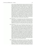
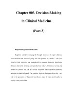
– FIGURE 4-2 — Erythema multiforme major (toxic epidermal necrolysis)—Generalized, macular eruption with
some target-like lesions which rapidly developed epidermal necrosis, positive Nikolsky sign, bulla formation, and
denuded erosive areas. On the back this eruption looks like scalding. It was due to a sulfonamide.
Reprinted from Fitzpatrick’s Color Atlas and Synopsis of Clinical Dermatology. 5th ed. New York: McGraw Hill, 2005, Fig. 7-24,
p. 147.
Diagnosis: The CDC maintains strict criteria for the diagnosis of TSS: including fever over 102
◦
F, multi-
organ involvement, hypotension, and a rash that desquamates 1–2 weeks following onset of disease.
Treatment: Treatment should include removal of the foreign body. Parenteral antibiotic administration
is indicated although they have not been shown to affect outcomes. A reasonable treatment could include
dicloxacillin and vancomycin for MRSA coverage. In penicillin allergic patients, clindamycin is an alternative
to dicloxacillin. IV steroids and IVIG have been used with some success in severe cases. Care depends largely
on the severity of disease.
STREPTOCOCCAL TOXIC SHOCK SYNDROME
Etiology: Less common than TSS caused by Staph. aureus, Group A streptoccocus (GAS) also known
as Strep. pyogenes, produces an exotoxin that is responsible for the clinical syndrome. Invasive soft tissue
streptococcal infections such as cellulitis are common precipitating factors.
Clinical Presentation: Patients present with fever, hypotension, skin edema, erythema, or bullae. Multi-
system organ involvement is the rule. Desquamation occurs less commonly than with Staph TSS. The criteria
used to diagnose Staph TSS are applicable to the GAS variant. A thorough search for inciting infections
such as myositis, fasciitis, or cellulitis is essential.
Diagnosis: Clinical. See criteria for TSS.
GENERAL TERMINOLOGY AND DESCRIPTORS 119
Treatment: Supportive care and antibiotic therapy similar to staph TSS. If deep soft tissue infection is the
inciting cause, incision and drainage may be indicated.
STAPHYLOCOCCAL SCALDED SKIN SYNDROME
Etiology: Stap. aureus produces exfoliative toxins responsible for the clinical syndrome. This most com-
monly occurs in infants <3 months of age but can occur in children up to 5 years old. It is considered a
severe variant of bullous impetigo.
Clinical Presentation: The site of the Staphylococcus infection is often not obvious. Conjunctivitis,
occult nasopharyngeal infection, or umbilical stump infection are common sites. Localized exfoliative toxin
is responsible for the lesions of bullous impetigo, while SSSS is secondary to hematogenous spread of toxin
with diffuse skin effects. The skin manifestations of SSSS classically start in the perioral area. Diffuse, tender
erythroderma with a sandpaper appearance progress to large, fluid-filled bullae with a positive Nikolsky sign.
Desquamation then follows though mucus membranes are spared.
Diagnosis: Clinical findings followed by bacterial cultures and skin biopsy.
Treatment: Fluid resuscitation is the top priority. An attempt should be made to localize the source of
infection and antistaphylococcal antibiotics should be given.
NECROTIZING FASCIITIS
Etiology: Aerobic and anaerobic bacteria in the polymicrobial form or Group A streptococcus can cause
this infection. There is an increased risk in the immunocompromised patient population. Fournier’s gangrene
is a severe variant involving the perineum. Concomitant Varicella infection increases the risk of GAS type
infection.
Clinical Presentation: With the polymicrobial form pain out of proportion to exam is the classic pre-
sentation. Erythema and edema are seen initially, followed by discoloration, vesicles, and crepitus later on
with development of reddish-purple patches and bullae. Low-grade fever and tachycardia are common. This
infection can progress within hours. The GAS form has a similar presentation to the polymicrobial form,
but is usually more rapidly progressive and associated with higher mortality due to more virulent bacteria.
Diagnosis: Wound and blood cultures. Lack of bleeding and presence of cloudy, foul fluid after incision
into wound suggests this disease process. Finger or surgical instrument passes easily through planes of fascia
and soft tissue is easily dissected away from the fascia. X-ray or CT may demonstrate subcutaneous gas, but
may be normal. MRI is more sensitive for detecting deep soft-tissue infection.
Treatment: Early surgical consultation is essential. Antibiotics should be initiated early in suspected cases.
Penicillin alone is often not adequate in severe cases. Broad-spectrum antibiotic administration is indicated
to cover anaerobes as well. Vancomycin and pipericillin/tazobactam would be a good choice for empiric
coverage. A floroquinolone with clindamycin is an alternative for penicillin allergic patients. Hyperbaric
oxygen therapy (HBO) following surgical therapy may be beneficial.
GAS GANGRENE (CLOSTRIDIAL AND NONCLOSTRIDIAL MYONECROSIS)
Etiology: The majority of cases are caused by the spore-forming clostridial species. Clostridium perfringens
is responsible for most. The nonclostridial form is caused by a mixed infection of anaerobic and aerobic
organisms. This infection can be avoided by proper wound care with crushed or dead-tissue debridement at
initial evaluation of a wound.
120 CHAPTER 4 / CUTANEOUS DISORDERS
Clinical Presentation: Gangrene is a rapidly progressive infection of the deep subcutaneous tissues with
severe myonecrosis and sepsis. Physical examination is significant for pain out of proportion to examination.
Patients may report a sensation of heaviness in affected part. Edema and crepitance appear with progression
of infection. Brown discoloration, bullae, and serosanguinous discharge may be present. Low-grade fever and
tachycardia are common. The patient with gas gangrene may develop irritability, confusion, anddeterioration
of mental status.
Diagnosis: Gram stain of bullae may reveal pleomorphic gram-positive bacilli with or without spores. X-ray
or CT may reveal gas in the muscle and surrounding soft tissue. Surgical exploration may reveal nonbleeding
muscle in later stages of disease as well as loss of contractility and presence of gas bubbles.
Treatment: Early surgical intervention is a necessity. Early antibiotic therapy should be started with peni-
cillin G plus clindamycin. Ceftriaxone or erythromycin are alternative choices. HBO following surgical
therapy may be beneficial.
RASHES ASSOCIATED WITH BACTERIAL INFECTIONS
TABLE 4-2. RASHES WITH ASSOCIATED BACTERIAL INFECTIONS (OUTPATIENT TREATMENT)
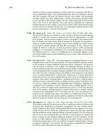
FIGURE 4-3. IMPETIGO (HONEY CRUSTED LESIONS).
TABLE 4-2 RASHES WITH ASSOCIATED BACTERIAL INFECTIONS (OUT-PATIENT TREATMENT)
DISEASE ETIOLOGY CHARACTERISTIC RASH PR ESENTATION TREATMENT COMMENTS
Scarlet fever Group A Streptococcus
Group C Streptococcus
Scarlet macules over
erythema evolving to
punctate lesions cause
the classic “sandpaper”
rash
Fever, sore throat,
headache
“Pastia’s lines” are
lines along skin folds
due to petechiae
Petechiae and red
macules on hard
palate
Circumoral pallor
Penicillin or
Erythromycin
Treatment will not
decrease incidence of
nephritis
Erythrogenic toxin
causes rash
Desquamation occurs
after rash fades
Associated
post-streptococcus
GN
Impetigo
(Figure 4-3)
Staph. aureus
Group A Streptococcus
(nephrogenic strain)
Small pustules or
vesicles with
erythematous margins
After rupture, “honey
crusted” lesions persist
No systemic
symptoms
Topical mupirocin
For systemic coverage—
cephalosporins,
beta-lactamase penicillin
and erythromycin
Associated
post-streptococcus
GN
Bullous impetigo Staph. aureus (80%) Localized bullae without
surrounding erythema
“Coin” lesions after
rupture—shiny, rounded
erosions with peeling
skin
Topical mupirocin
For systemic coverage—
cephalosporins,
beta-lactamase penicillin,
and erythromycin
If toxin is
hematogenously
spread, it causes
SSSS in children
Erysipelas Group A Streptococcus
Also Staph. aureus and
Haemophilius
influenza
Sharply demarcated
edematous plaque with
raised borders
(St. Anthony’s fire)
Fever, associated
lymph obstruction
Rash most commonly
seen on face
May recur
Penicillin or ery thromycin Newborns are
susceptible to
erysipelas from
Group B
Streptococcus
121
122 CHAPTER 4 / CUTANEOUS DISORDERS
– FIGURE 4-3 — Impetigo (honey crusted lesions)—Crusted erythematous erosions becoming confluent on the
nose, cheek, lips, and chin in a child with nasal carriage of Staph. aureus and mild facial eczema.
Reprinted from Fitzpatrick’s Color Atlas and Synopsis of Clinical Dermatology. 5th ed. New York: McGraw Hill, 2005, Fig. 22-10,
p. 589.
VIRAL EXANTH EMS
TABLE 4-3 VIRAL DISEASE AND ASSOCIATED RASH
DISEASE ETIOLOGY CHARACTERISTIC RASH P RESENTATION COMMENTS
Varicella Varicella zoster
virus
Pruritic erythematous
papules evolving into
“dew drop” vesicles and
then pustules
Lesions of various ages
Starts on face and scalp
with centripetal spread
Fever and malaise is
present
Mucosal involvement in
many patients
Some will develop
varicella pneumonia
Resolution of the
lesions seen after
2 weeks
Measles
(Rubeola)
Paramyxovirus Maculopapular
beginning on the face
and neck, spreading to
the trunk and
extremities in a
centifugal fashion
Koplik’s spots (white
1-mm lesions) on the
buccal mucosa are
pathognomonic
Fever “3 C’s”–cough,
coryza, conjunctivitis
Symptoms begin 2–4
days prior to rash
Complications include
pneumonia and
encephalitis
German
Measles
(Rubella)
Togavirus Pink macules and
papules which become
confluent to form a
scarletiniform rash.
Spread is from the face
caudally.
Forscheimer spots are
petechiae on the soft
palate
Malaise, fever,
headache, mild
conjunctivitis,
arthralgias, and
prominent adenopathy
Symptoms precede
rash by 1–5 days
In rare cases,
thrombocytopenia
Strong risk of congenital
deformities in the fetus
of women who contract
rubella in their first
trimester
Erythema
Infectiosum
(Fifth disease)
Parvovirus B19 Classic appearance of
the “slapped cheek”
erythema on the face
initially
Then, maculopapular
rash beginning on the
upper extremities and
spreading proximally
and distally
Fading macular rash will
appear “lace-like”
Arthralgias and arthritis
Circumoral pallor
Fever and malaise
Pregnant women
exposed are at risk for
fetal hydrops
Patients with sickle cell
disease at risk for
aplastic anemia
(Continued )
124 CHAPTER 4 / CUTANEOUS DISORDERS
TABLE 4-3 VIRAL DISEASE AND ASSOCIATED RASH (CONTINUED)
DISEASE ETIOLOGY CHARACTERISTIC RASH PRESENTATION COMMENTS
Roseola
(Exanthem
subitum)
Human
herpesvirus 6
Pink maculopapular
beginning on the trunk
and spreading outward
Onset of rash with drop
in fever
High fever (up to
105
◦
C) in a classically
well-appearing infant
before the appearance
of the rash
Lymphadenopathy
Leukopenia on CBC
Associated with
intussusception due to
lymphoid hyperplasia in
GI tract
Hand, foot,
and mouth
disease
Enteroviruses:
Coxsackie virus
Echovirus
Oral vesicles which
ulcerate
Papules which prog ress
to vesicles on palms
and soles
Fever, anorexia, malaise
Oral lesions appear 1–2
days after fever; then
extremity lesions appear
Decreased oral intake
due to pain
Lesions typically heal in
7–10 days
Herpangina Enteroviruses Vesicular eruptions on
posterior pharynx that
ulcerate leaving small
craters
Fever, headache, sore
throat
Lesions heal in 5–10
days
Molluscum
Contagiosum
Poxvirus White waxy papules
with central
umbilication
Few patients have
puritis
Lesions can be more
extensive in patients
with eczema or
immunocompromised
patients
Individual lesions
resolve in 2 months.
Disease can persist for
more than a year.
Mycoplasma Mycoplasma Erythematous,
maculopapular rash
inconsistently seen
Frequent cause of
exanthems associated
with URIs in children
Also associated with EM
minor and major
Mononucleosis Epstein-Barr
virus
Generalized
erythematous rash
Petechaie on the soft
palate
Fever, malaise
Adenopathy
Rash only seen in 5%
cases primarily
Seen in 100% patients
treated with ampicillin
or similar agents
RASHES ASSOCIATED WITH AUTOIMMUNE DISEASES 125
RASHES ASSOCIATED WITH AUTOIMMUNE DISEASES
HENOCH-SCHONLEIN PURPURA
Etiology: Henoch-schonlein purpura (HSP) is a hypersensitivity vasculitis believed to be mediated by IgA
immune complexes, most often affecting children, but may present at any age.
Clinical Presentation: The cause is unknown, but HSP classically follows a URI. The rash is character-
ized by pink macules that blanch and progress to nonblanching purpura distributed on the lower extremities,
perineum, buttocks, elbows, and lower trunk. Hematuria may occur as a result of glomeruli involvement.
Arthralgias are common. GI tract vasculature involvement causes colicky abdominal pain and may result in
GI bleeding. HSP is also associated with intussusception.
Diagnosis: Other causes of purpura must be eliminated since the diagnosis of HSP made based on clinical
findings.
Treatment: The treatment for HSP is generally supportive.
PEMPHIGUS VULGARIS
Etiology: This autoimmune disorder causes loss of cell-to-cell adhesions in the epidermis as a result of IgG
autoantibodies, resulting in a serious and often fatal disease of the skin and mucus membranes. It usually
occurs in patients 40–60 years of age.
Clinical Presentation: Pemphigus vulgaris (PV) presents with painful round or oval vesicles and bullae
with serous fluid that are easily ruptured, positive Nikolsky sign, with predilection for the scalp, face, chest,
axillae, and groin. Lesions can become confluent and bleed with minimal trauma. The lesions first arise in
the oral mucosa and months later appear in a random pattern on normal skin. Due to the ease with which
vesicles rupture, often only the erosions are seen, and bullae are rarely seen on the mucosa.
Diagnosis: Dermatology referral is recommended for work up since it can be a difficult diagnosis if only
mouth lesions are present.
Treatment: Aggressive treatment with immunosupressants.
BULLOUS PEMPHIGOID
Etiology: This autoimmune disorder presents as chronic bullous disease in the elderly. It is the most
common bullous autoimmune disease.
Clinical Presentation: This disorder begins with erythematous, papular lesions that may precede bullae
over months. Eruption may be localized or generalized. Bullae rupture less easily than in PV. There is oral
involvement in up to one-third of cases, but is typically less severe and less painful than PV.
Diagnosis: Clinical appearance along with histopathology determines the diagnosis.
Treatment: Bullous pemphigoid is treated with systemic steroids in conjunction with a dermatologist
consultation.
KAPOSI’S SARCOMA
Etiology: Caused by human herpesvirus-8. Age of onset varies depending on type, with HIV associated
form affecting young adults, and most commonly in homosexual males, while classic Kaposi’s sarcoma (KS)
126 CHAPTER 4 / CUTANEOUS DISORDERS
form has peak incidence in patients greater than 50 years of age. Classic KS is rare in the United States. Risk
is 20,000 times greater in HIV-infected individuals.
Clinical Presentation: These skin lesions are painless raised, brown-black, or purple nodules and papules
that do not blanch. Lesions are commonly found on the face, genitals, chest, and oral cavity, but may be
found anywhere in widespread disease. Lesions may even be found on the internal organs.
Diagnosis: Diagnosis should be made by skin biopsy.
Treatment: Treatment is focused on painful or cosmetically disfiguring lesions. Treatment modalities
include cryotherapy and radiation for localized disease and chemotherapy for widespread disease.
OTHER COMMON CHILDHOOD RASHES
TABLE 4-4. OTHER COMMON CHILDHOOD RASHES
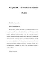
FIGURE 4-4.
PITYRIASIS ROSEA (HERALD PATCH)
Lesions and Pearls
HERPES SIMPLEX
HSV-1. Involves the lips (herpes labialis), tip of the finger (herpetic whitlow), and the eyes (herpetic keratitis)
and is also responsible for the minority of urogenital lesions.
HSV-2. Usually involves the genital area.
Skin lesions begin as groups of vesicles on an erythematous base that rupture, ulcerate, and become
crusted and are very painful and spread by direct contact. Tzanck smear will show multinucleated giant
cells.
HERPES ZOSTER
Herpes zoster is the reactivation of the latent varicella-zoster virus (VZV). Painor parasthesias in a dermatomal
pattern may herald the future skin lesions. Clusters of vesicles erupt in a dermatomal pattern. The thoracic
dermatomes are most commonly involved. Lesions at the tip of the nose (Hutchinson sign) should guide the
clinician to rule out corneal involvement because of involvement of the nasociliary branch of the trigeminal
nerve. Immunocompromised patients should be treated with IV acyclovir in the inpatient setting.
TINEA VERSICOLOR
This opportunistic, benign, cutaneous infection with Malassezia furfur most often affects otherwise healthy
individuals. Patients present with scaly macules or papules on the skin. Versi meaning several and the lesions
are often of differing pigments. Treat with topical selenium sulfide or topical antifungals.
SEBORRHEIC DERMATITIS
This dermatitis is known as “cradle cap” in infants and appears as erythema and yellow-orange scales and
crust on the scalp. There may be an association with fungus such as Malassezia furfur. Selenium sulfide
shampoos, topical steroids, and antifungal shampoos are effective treatments.
OTHER COMMON CHILDHOOD RASHES 127
TABLE 4-4 OTHER COMMON CHILDHOOD RASHES
DISEASE ETIOLOGY RASH TREATMENT COMMENTS
Pityriasis Rosea
(Figure 4-4)
Unknown
Herpes virus
7 suspected
Fine scaly
papulosquamous
eruption in “Christmas
tree” distribution on
the trunk
“Marginal collarette”
describes the scale as
attached on the
periphery and loose in
the middle
“Herald patch” first
lesion seen is an oval
salmon-colored plaque
Oral antihistamines
and topical steroids
for itching
Type B UV light
decreases disease
severity if used in first
week of eruption
Seen in the spring
and fall
Bacterial and fungal
cultures are negative
Dermatophytosis
(Ringworm)
Dermatophytes-
Tinea
Circular erythematous
scaling lesion with
central clearing and
raised borders
Treat with antifungal
cream
Scabies Sarcoptes
scabiei mite
Small erythematous
burrows in/on web
spaces and
intertrigenous regions
as well as the flexor of
the wrists surfaces. The
head is spared
Permethrin 5% is the
treatment of choice
All bedding and
clothing should be
washed in hot water
A delayed
hypersensitivity
reaction is
responsible for the
intense puritis
PSORIASIS
Salmon-colored plaques covered with silvery-white scales are seen predominately on the extensor surfaces
of the arms and legs.
ATOPIC DERMATITIS
Atopic dermatitis, also known as eczema, usually begins in infancy with dry skin and pruritis. Chronic
rubbing and scratching leads to lichenification and further pruritis and scratching (itch–scratch cycle). IgE
is elevated in 85% of patients. Of infants with atopic dermatitis, 35% will develop asthma later in life.
EXFOLIATIVE ERYTHRODERMA
Exfoliative erythroderma is the result of an underlying systemic disease or reaction to drugs or chemicals.
Most or all of the patient’s skin is covered with a red, scaling rash that is warm, but not tender. Diagnosis is
made with skin biopsy. Patients typically need admission to work up and treat underlying cause. Symptomatic
treatment is indicated with systemic steroids for severe disease which can be life threatening in these cases.
128 CHAPTER 4 / CUTANEOUS DISORDERS
– FIGURE 4-4
— Pityriasis rosea (herald patch).
Reprinted from Fitzpatrick’s Color Atlas and Synopsis of Clinical Dermatology. 5th ed. New York: McGraw Hill, 2005, Fig. 7-1,
p. 119.
CUTANEOUS MANIFESTATIONS OF SYSTEMIC ILLNESSES
TABLE 4-5 RASHES WITH LESIONS ON SOLES AN D PALMS
Rocky mountain spotted fever
Neisseria meningitis/meningococcemia
Secondary syphilis
Disseminated gonococcal infection
Hand, Foot, and Mouth disease
Urticaria
Erythema multiforme
Dermatophytosis (Ringworm)
Toxic Shock syndrome (desquamation phase)
Smallpox
The following illnesses have cutaneous manifestations of systemic illness which may be discussed in more
depth under another appropriate chapter. The skin lesions are highlighted here.
CUTANEOUS MANIFESTATIONS OF SYSTEMIC ILLNESSES 129
Primary Syphilis
The chancre of primary syphilis is a painless, indurated ulcer found at the site of inoculation: the genitals or
mucus membranes of the mouth. These lesions can be associated with painless regional lymphadenopathy.
Secondary Syphilis
Symmetric maculopapular rash with confluence on the trunk and extremities develops in secondary syphilis.
The rash also involves the palms and soles. Generalized lymphadenopathy and malaise can accompany this
stage of syphilis (see Table 10-2).
Disseminated Gonorrhea
Most cases are seen in young, sexually active women (men are more likely to seek treatment early). Erythe-
matous macules of 1–5 mm progress to hemorrhagic pustules with a “red halo” and grey necrotic centers.
Arms are involved more often than legs, with lesions typically near small joints of hands or feet and may
–FIGURE4-5— Disseminated gonococcal infection—Hemorrhagic, painful pustules on erythematous bases on
the palm and the finger of the other hand.
Reprinted from Fitzpatrick’s Color Atlas and Synopsis of Clinical Dermatology. 5th ed. New York: McGraw Hill, 2005, Fig. 27-17,
p. 910.
130 CHAPTER 4 / CUTANEOUS DISORDERS
be in web spaces. The illness is characterized by fever, arthralgias, and possibly septic arthritis (see also
Chapter 10).
Rocky Mountain Spotted Fever
Rocky mountain spotted fever (RMSF) is caused by Rickettsia rickettsii, an intracellular organism introduced
via the Dermacentor tick. The characteristic rash begins on the wrists, forearms, and ankles and affects
the palms and soles later in the illness. Lesions are initially macular, progress to papules, and become
nonblanching and hemorrhagic. Treatment is with doxycycline.
Children should also be treated with doxycycline. It has been reported that children with recurrent
RMSF may be treated with up to five courses of doxycycline with minimal risk of dental staining (Cale and
McCarthy, 1997) (see Table 10-2).
Meningococcemia
FIGURE 4-6. MENINGOCOCCEMIA.
Neisseria meningitides is transmitted through respiratory secretions and usually affects patients younger than
20 years, with highest rates of infection in the winter and spring. Patients present with headache, fever,
– FIGURE 4-6 — Meningococcemia—Acute meningococcemia: purpura fulminans. Maplike, gray-to-black areas of
cutaneous infarction of the leg in a child with NM meningitis and disseminated intravascular coagulation with
purpura fulminans.
Reprinted from Fitzpatrick’s Color Atlas and Synopsis of Clinical Dermatology. 5th ed. New York: McGraw Hill, 2005, Fig. 22-43,
p. 643.
FURTHER READING 131
vomiting, and possibly with meningeal signs. Petechial rash classically found on the extremities and trunk
and progress to palpable purpura with grey necrotic centers (see also Chapter 10).
Kawasaki’s Disease
Kawasaki’s disease is an idiopathic disease characterized by cutaneous and mucosal erythema and edema with
subsequent desquamation. The rash can be erythematous, morbilliform, urticarial, or similar in appearance to
erythema multiforme. Criteria for diagnosis include fever for 5 days and four of the following: conjunctivitis,
rash, lymphadenopathy, oropharynx changes (strawberry tongue), and extremity erythema/edema. Kawasaki’s
disease is associated with coronary aneurysms if undiagnosed and untreated (see also Chapter 14). Initial
treatment should include aspirin and intravenous immunoglobulin (IVIG) to decrease the incidence of
coronary aneurysms.
REFERENCES
Cale DF, McCarthy MW. Treatment of Rocky Mountain Spotted Fever in Children. Ann Phamacother 1997Apr;31
(4):492–494.
FURTHER READING
Fitzpatrick TB, Johnson RA, Suurmond D, et al. Color Atlas and Synopsis of Clinical Dermatology. 4th ed. New York:
McGraw Hill, 2001.
Wolff K, Johnson RA, Suurmond D. Fitzpatrick’s Color Atlas and Synopsis of Clinical Dermatology. 5th ed. New York:
McGraw Hill, 2005.
Tintinalli JE, Kelen GD, Stapczinski JS. Emergency Medicine A Comprehensive Study Guide. 5th ed. New York: McGraw
Hill, 2000.
Tintinalli JE, Kelen GD, Stapczinski JS. Emergency Medicine A Comprehensive Study Guide. 6th ed. New York: McGraw
Hill, 2004.
Cline DM, Ma OJ, Tintinalli JE, et al. Just the Facts in Emergency Medicine. New York: McGraw Hill, 2001.
Cydulka RK, Stewart MH. Dermatologic Presentations. In Marx JA, Hockberger RS, Walls RM, et al. (eds). Rosen’s
Emergency Medicine Concepts and Clinical Practice. 5th ed. St. Louis, MO: Mosby, 2001.
Shoff WH. Dermatologic Emergencies. In Rivers CS, Dorfman T (eds). Preparing for the Written Board Exam in
Emergency Medicine. Ohio: Emergency Medicine Educational Enterprises, Inc., 2001.
Fleisher GR, Ludwig S, Henretig FM, et al. Textbook of Pediatric Emergency Medicine. Philadelphia: Lippincott Williams
and Wilkins, 2000.
Schofield JK, Tatnall FM, Leigh IM. Recurrent Erythema Multiforme: Clinical Features and Treatment in a Large
Series of Patients. Br J Dermatol 1993;128(5):542–545.
CHAPTER 5
ENDOCRINE, METABOLIC,
AND NUTRITIONAL
DISORDERS
ENDOCRINE
The Thyroid Gland
The thyroid gland secretes thyroid hormone (thyroxine) in the form of tetroiodothyronine (T4) and tri-
iodothyronine (T3). T3 is largely produced in the peripheral tissues via monodeiodination and is more
metabolically active than T4. Thyroid hormone is largely carried in the bloodstream by thyroid-binding
globulin (TBG). It is, however, the free hormone concentration that is kept constant by the feedback regu-
latory system and that determines thyroid status. The principal role of thyroid hormone is to regulate tissue
metabolism. Adequate levels are necessary for the normal development of the CNS during the first 2 years
of life. Inadequate hormone levels during this period lead to cretinism, a syndrome of dwarfism and mental
retardation. Normal thyroid function is also required for normal growth and bone maturation in children.
The feedback loop for the regulation of thyroid function begins when thyroid-releasing hormone (TRH) is
secreted by the hypothalamus. It stimulates the synthesis and release of thyroid-stimulating hormone (TSH)
from the anterior pituitary gland. TSH stimulates the thyroid gland to synthesize and release thyroxine. In
turn, T3 and T4 suppress the release of TSH competing with TRH to complete the feedback loop.
The most severe forms of thyroid dysfunction are dramatic and unmistakable. However mild degrees of
thyroid dysfunction, which are more common, present with symptoms that are more subtle and nonspecific
and may be difficult to recognize.
HYPERTHYROIDISM
Definition: Hyperthyroidism is characterized by excessive quantities of circulating thyroid hormone. Al-
though serum catecholamine levels are not elevated, many of the signs and symptoms of hyperthyroidism are
those of adrenergic hyperactivity. Thyroid storm is a life-threatening hypermetabolic state. It likely represents
the addition of adrenergic hyperactivity, induced by stress, onto the setting of unrecognized or undertreated
hyperthyroidism.
Etiology:
TABLE 5-1. CAUSES OF HYPERTHYROIDISM
Clinical Presentation:
TABLE 5-2. SIGNS AND SYMPTOMS OF HYPERTHYROIDISM
132
Copyright © 2007 by The McGraw-Hill Companies, Inc. Click here for terms of use.
ENDOCRINE 133
TABLE 5-1 CAUSES OF HYPERTHYROIDISM
r
Toxic diffuse goiter or Graves disease
᭺
Accounts for 85% of cases
᭺
Stimulation of the thyroid gland by TSH thyroid receptor antibodies
᭺
Thyroid hormone levels are highest with this form of hyperthyroidism
᭺
Women > men
᭺
Third and four th decades
r
Apathetic hyperthyroidism
᭺
Most common in elderly population
᭺
Develops slowly over time
᭺
Cardiovascular symptoms prominent
᭺
Weight loss often >40 lbs
᭺
Depressed mental function
r
Toxic multinodular goiter (Plummer disease)
᭺
Most common in elderly population (>50 years)
᭺
More common in areas of iodine deficiency
᭺
Develops slowly over time
᭺
Symptoms are generally mild
r
Toxic uninodular goiter
r
Thyrotoxicosis
r
Hashimoto thyroiditis
᭺
Most common thyroid disease of childhood
᭺
Autoimmune mediated infiltration of the thyroid gland with lymphocytes
᭺
Parenchymal destruction leads to hypothyroidism
r
De Quervain thyroiditis
᭺
Possibly secondary to viral infection
᭺
Usually self-limiting
r
Factitious thyrotoxicosis
᭺
Exogenous thyroid hormone
r
Metastatic follicular thyroid carcinoma
r
TSH producing pituitary tumors
r
Iodide-induced hyperthyroidism (Jod-Basedow syndrome)
᭺
Excess iodine intake
᭺
Found in a number of medications
(Continued )
134 CHAPTER 5 / ENDOCRINE, METABOLIC, AND NUTRITIONAL DISORDERS
TABLE 5-1 CAUSES OF HYPERTHYROIDISM (CONTINUED)
r
Expectorants
r
Amiodarone
r
Lithium
r
Seaweed
r
Iodinated radiocontrast
r
Choriocarcinoma (uterine or testicular origin) or hydatiform mole
᭺
High levels of circulating beta human chorionic gonadotropin (βHCG) which can
weakly activate the TSH receptor
r
Struma ovarii
᭺
Ectopic thyroid tissue associated with dermoid tumors or ovarian teratomas
Diagnosis: Free T4 and T3 levels will generally be elevated and TSH will be low in patients with hyper-
thyroidism. Patients with pituitary adenomas as the cause of their hyperthyroidism will have an elevated
TSH.
Treatment: Mild hyperthyroidism does not require emergent treatment or hospital admission, and further
evaluation and treatment can be done on an outpatient basis. Beta blockers may be used to control symptoms.
Thyroid storm, on the other hand, requires immediate treatment.
There are 5 goals of therapy:
1. Inhibit hormone synthesis
a. Propylthiouracil (PTU) preferred or methimazole
2. Block hormone release
a. Iodine therapy
b. Lithium carbonate (Note: Thionamides should be given 1 hour prior to blocking hormone release.)
3. Prevent peripheral conversion of T3 to T4
a. Dexamethasone
b. PTU—minor effect
4. Block peripheral adrenergic effects of thyroid hormone
a. Propanolol or esmolol
b. Guanethidine
c. Reserpine
5. Supportive care
a. Fever
i. Acetaminophen
ii. Cooling methods
b. Congestive heart failure (CHF)
i. Digitalis
ii. Diuretics
ENDOCRINE 135
TABLE 5-2 SIGNS AND SYMPTOMS OF HYPERTHYROIDISM
SYMPTOMS:
Palpitations
Heat intolerance
Increased perspiration
Weight loss (often >40 lbs) with increased appetite
Fatigue
Anxiety
Emotional liability
Diminished or absent menstruation
Frequent bowel movements
SIGNS:
Goiter or thyroid mass
Sinus tachycardia (most common rhythm disturbance)
Supraventricular tachycardia or atrial fibrillation
Systolic hypertension with widened pulse pressure
Fever
Stare
Extension tremor
Warm, moist, and smooth skin
Large muscle weakness
SIGNS ASSOCIATED SPECIFICALLY WITH GRAVES DISEASE:
Ophthalmopathy
r
Periorbital edema
r
Chemosis
r
Proptosis
r
Extraocular muscle dysfunction/diplopia
Dermopathy
r
Nonpitting pretibial edema associated with erythema and thickening of the skin
Diffusely enlarged thyroid gland
r
± Thyroid bruit