Wound Healing and Ulcers of the Skin - part 1 pps
Bạn đang xem bản rút gọn của tài liệu. Xem và tải ngay bản đầy đủ của tài liệu tại đây (484.79 KB, 28 trang )
A. Shai
H. I. Maibach
Wound Healing and Ulcers of the Skin
Diagnosis and Therapy –
The Practical Approach
00_001_022_Titelei*** 01.09.2004 13:46 Uhr Seite I
A. Shai
H. I. Maibach
Wound Healing
and Ulcers
of the Skin
Diagnosis and Therapy –
The Practical Approach
With 115 Figures and 25 Tables
00_001_022_Titelei*** 01.09.2004 13:46 Uhr Seite III
Dr. Avi Shai
Department of Dermatology
Soroka University Medical Center
Faculty of Health Sciences
Ben-Gurion University of the Negev
Beer-Sheva 84105
Israel
Professor Howard I. Maibach
Department of Dermatology
University of California
San Francisco School of Medicine
Box 0989
San Francisco, CA 94143-0989
USA
Library of Congress Control Number: 2004104389
ISBN 3-540-21275-2 Springer Berlin Heidelberg New York
This work is subject to copyright. All rights are reserved, whether the whole or part of the material is
concerned, specifically the rights of translation, reprinting, reuse of illustrations, recitation, broadcast-
ing, reproduction on microfilm or in any other way, and storage in data banks. Duplication of this pub-
lication or parts thereof is permitted only under the provisions of the German Copyright Law of
September 9, 1965, in its current version, and permission for use must always be obtained from
Springer. Violations are liable to prosecution under the German Copyright Law.
Springer is a part of Springer Science+Business Media
springeronline.com
© Springer-Verlag Berlin Heidelberg 2005
Printed in Germany
The use of general descriptive names, registered names, trademarks, etc. in this publication does not
imply, even in the absence of a specific statement, that such names are exempt from the relevant pro-
tective laws and regulations and therefore free for general use.
Product liability: the publishers cannot guarantee the accuracy of any information about dosage and
application contained in this book. In every individual case the user must check such information by
consulting the relevant literature.
Editor: Marion Philipp
Desk Editor: Irmela Bohn
Production: ProEdit GmbH, 69126 Heidelberg, Germany
Cover: Frido Steinen-Broo, EStudio Calamar, Spain
Typesetting: K. Detzner, 67346 Speyer, Germany
Printed on acid-free paper 21/3150 ML 5 4 3 2 1 0
00_001_022_Titelei*** 01.09.2004 13:46 Uhr Seite IV
In recent years, the amount of knowledge surrounding the processes of wound heal-
ing has significantly increased, resulting in a vast array of therapeutic options. The
assortment of preparations currently available may become somewhat perplexing
to physicians and medical personnel.
We have become aware of the difficulty involved in selecting the most appropri-
ate therapy for a specific type of wound. Our main purpose in writing this book,
therefore, is to present a step-by-step algorithmic approach to the treatment of
chronic wounds.
The caring of wounds has always been the realm of the various branches of sur-
gery. Dermatology, on the other hand, being the medical science that specializes in
skin and cutaneous physiology, deals with the essential and fundamental aspects of
wound healing. Due to its very nature, wound healing overlaps into the many disci-
plines of medicine in general. Internists, diabetologists, and geriatricians are be-
coming increasingly involved in the field of wound care. General practitioners and
family physicians are frequently required to treat acute and chronic wounds.
In this book, we introduce the dermatologic perspective of wound healing which
applies to the diagnosis of cutaneous ulcers,based on history, physical examination,
biopsy, and laboratory tests. We also present a therapeutic approach to ulcers ac-
cording to their appearance.
We believe that this guidebook will assist physicians in the treatment of chronic
wounds,and that it will ultimately serve to reduce the immense suffering of those af-
flicted.
Note to the Reader. Neither the authors nor the publishers are liable for any con-
sequences arising from the use of information presented in this book. The readers
are advised to check for up-dated information provided by the manufacturers,
including dosage and safety regulations, for each of the products described in this
book. Ultimate responsibility rests with the treating physician.
Some of the chapters include lists of commercial names of preparations used in
the healing of chronic ulcers. This is by no means intended as a commercial recom-
mendation. It is simply intended to provide the readers with a guide to the range of
brand names in use for a certain biologic substance. We have done our best to pro-
vide up-dated and accurate lists. However, this area is subject to frequent changes,
and the readers are advised to gather information from other currently available
sources.
Preface
00_001_022_Titelei*** 01.09.2004 13:46 Uhr Seite V
Acknowledgements
The authors wish to thank the following for providing this book with illustrations
and pictures: Dr. Emanuela Cagnano for Figs. 12.1 (modified by Inanit Ashtamker as
Fig. 1.1), 2.2, 6.1, 6.3, and 6.6; Dr. Oren Lapid for Figs. 12.3 and 12.4; Dr. David Vigoda
for Fig. 12.5; Dr. Tidhar Steiner and the Semmelweis Museum of the History of Med-
icine for Figs. 3.5 and 3.6; Dr. Alex Zvulunov for Fig. 4.2; Dr. Kosta Mumcuoglu for
Figs. 9.6–9.8; Professor Sima Halevy for Figs. 14.1–14.7; Audra J. Gera and Novartis
for Figs. 2.6 and 3.9, from Dermatology: A Medical Artist’s Interpretation, copyright
1990 by Sandoz Pharma LTD; The Wellcome Library, London, for Figs. 3.1–3.4, 3.7,
and 8.8; Taylor & Francis Publishing House for Fig. 8.6, reprinted from Jacobsson et
al: A new principle for the cleansing of infected wounds. Scandinavian Journal of
Plastic & Reconsructive Surgery, 10 : 65–72, 1976; Taylor & Francis Publishing House
for Fig. 20.2, reproduced from Handbook of Cosmetic Skin Care, published by Mar-
tin Dunitz, 2001. Figure 18.1 is reprinted from T.J. Ryan: Wound healing and current
dermatologic dressings. Clinics in Dermatology 8 : 21–29, copyright 1990, with per-
mission from Elsevier Science; Fig. 2.5 is reprinted from Germain et al: Human
wound healing fibroblasts have greater contractile properties than dermal fibro-
blasts. Journal of Surgical Research 57 : 268–273, copyright 1994, with permission
from Elsevier Science; Fig. 18.2 is reprinted from Dermatologic Therapy in General
Practice, by M. Sulzberger and J. Wolf, (p 116), published by The Year-Book Publish-
ers, copyright 1943, with permission from Elsevier Science; Fig. 6.2 is reprinted from
Falanga et al: The cutaneous manifestations of cholesterol crystal embolization,
Archives of Dermatology 122 : 1194–1198, copyright 1986, with permission from the
American Medical Association; Fig. 4.4 is reprinted from S.W. Graeca et al: A painful
precursor for necrosis. Postgraduate Medicine 106 : 249–250, copyright 1999, with
permission from Postgraduate Medicine (photographed by Scott Dornbaser).
Fig. 6.4 is reprinted from J. Lima-Maribona et al: Self-assessment examination. The
American Academy of Dermatology Journal 29:803, 1993, with permission from
Mosby-Year Book, Inc.; part of Fig. 2.1 is reprinted from CIBA Clinical Symposia on
common bleeding disorders, vol 35, no 3, p 8, copyright 1983, with permission from
Novartis; part of Fig. 2.1 is taken from Dermatology: A Medical Artist’s Interpreta-
tion, copyright 1990 by Sandoz Pharma LTD.
Many thanks are due to the following for their assistance in the preparation of the
text and for their valuable comments: Dr. Gary Zentner; Professor Ilana Harman-
Bohem; Professor Pablo Yagupsky; Dr. Batya Davidovici; Dr. Marcelo H. Grunwald;
Dr. Dafna Hallel-Halevy; and Dr. Emmilia Hodak. Our particular thanks go to all the
reviewers of the chapters in this book for their efforts and assistance (see below); to
Professor Sima Halevy, for advancing the field of wound healing in Soroka Uni-
versity Medical Center and for actively supporting the production of this book; to
Mrs. Rina Ben-Zeev for her assistance in the preparation of the Appendix section of
the book and for constructive collaboration at the Chronic Wound Clinic; to Dr.Alex
00_001_022_Titelei*** 01.09.2004 13:46 Uhr Seite VII
Zvulunov and Mr. Naftali Oron for their most valuable ongoing advice stemming
from sheer wisdom and clear reason. We would like to especially thank Miss
Kristina Hawthorne for contributing her vast experience in the production of
books, for her creative ideas, and for her indispensable support and assistance
throughout the whole course of this project.
Acknowledgements
VIII
00_001_022_Titelei*** 01.09.2004 13:46 Uhr Seite VIII
Contents
1 Basic Definitions and Introduction . . . . . . . . . . . . . . . . . . . . . . . . . . . . 1
1.1 Definitions . . . . . . . . . . . . . . . . . . . . . . . . . . . . . . . . . . . . . . . . . . . . . . . . 1
1.2 Three Aspects of Treatment in Wounds and Ulcers . . . . . . . . . . . . . . . 2
1.2.1 Etiology . . . . . . . . . . . . . . . . . . . . . . . . . . . . . . . . . . . . . . . . . . . . . . . . . . . 2
1.2.2 Clinical Appearance of the Ulcer . . . . . . . . . . . . . . . . . . . . . . . . . . . . . . . 3
1.2.3 Adjuvant Therapy . . . . . . . . . . . . . . . . . . . . . . . . . . . . . . . . . . . . . . . . . . . 3
1.3 Ulcer Depth . . . . . . . . . . . . . . . . . . . . . . . . . . . . . . . . . . . . . . . . . . . . . . . . 3
1.4 Comments on Current Treatments . . . . . . . . . . . . . . . . . . . . . . . . . . . . . 4
References . . . . . . . . . . . . . . . . . . . . . . . . . . . . . . . . . . . . . . . . . . . . . . . . . 4
2 Natural Course of Wound Repair Versus Impaired Healing
in Chronic Skin Ulcers . . . . . . . . . . . . . . . . . . . . . . . . . . . . . . . . . . . . . . . 7
2.1 Overview . . . . . . . . . . . . . . . . . . . . . . . . . . . . . . . . . . . . . . . . . . . . . . . . . . 7
2.2 Inflammation Phase . . . . . . . . . . . . . . . . . . . . . . . . . . . . . . . . . . . . . . . . . 8
2.2.1 Vasoconstriction and Hemostasis . . . . . . . . . . . . . . . . . . . . . . . . . . . . . . 8
2.2.2 Vasodilatation and Increased Permeability . . . . . . . . . . . . . . . . . . . . . . 9
2.2.3 Chemotactic Growth Factors and Phagocytosis . . . . . . . . . . . . . . . . . . 9
2.3 Tissue Formation Phase . . . . . . . . . . . . . . . . . . . . . . . . . . . . . . . . . . . . . . 9
2.3.1 Angiogenesis and Granulation Tissue Formation . . . . . . . . . . . . . . . . . 9
2.3.2 Extracellular Matrix Formation . . . . . . . . . . . . . . . . . . . . . . . . . . . . . . . . 10
2.3.3 Re-epithelialization . . . . . . . . . . . . . . . . . . . . . . . . . . . . . . . . . . . . . . . . . . 11
2.3.4 Wound Contraction . . . . . . . . . . . . . . . . . . . . . . . . . . . . . . . . . . . . . . . . . 11
2.3.5 Role of Nitric Oxide in Wound Healing . . . . . . . . . . . . . . . . . . . . . . . . . 12
2.4 Tissue Remodeling Phase . . . . . . . . . . . . . . . . . . . . . . . . . . . . . . . . . . . . 12
2.5 Types of Repair . . . . . . . . . . . . . . . . . . . . . . . . . . . . . . . . . . . . . . . . . . . . . 13
2.6 Chronic Ulcers and Protracted Inflammation . . . . . . . . . . . . . . . . . . . . 13
2.6.1 Increased Enzymatic Activity of Matrix Proteases . . . . . . . . . . . . . . . . 13
2.6.2 Reduced Responsiveness to Growth Factors . . . . . . . . . . . . . . . . . . . . . 13
2.6.3 Cell Senescence . . . . . . . . . . . . . . . . . . . . . . . . . . . . . . . . . . . . . . . . . . . . . 14
2.7 Concluding Remarks . . . . . . . . . . . . . . . . . . . . . . . . . . . . . . . . . . . . . . . . 15
References . . . . . . . . . . . . . . . . . . . . . . . . . . . . . . . . . . . . . . . . . . . . . . . . . 15
00_001_022_Titelei*** 01.09.2004 13:46 Uhr Seite IX
Contents
X
3 Milestones in the History of Wound Healing . . . . . . . . . . . . . . . . . . . . 19
3.1 Overview . . . . . . . . . . . . . . . . . . . . . . . . . . . . . . . . . . . . . . . . . . . . . . . . . . 19
3.2 The Ancient World . . . . . . . . . . . . . . . . . . . . . . . . . . . . . . . . . . . . . . . . . . 19
3.2.1 Medicine in Mesopotamia . . . . . . . . . . . . . . . . . . . . . . . . . . . . . . . . . . . . 20
3.2.2 Ancient Egypt . . . . . . . . . . . . . . . . . . . . . . . . . . . . . . . . . . . . . . . . . . . . . . 20
3.3 Inflammation, Infection and the Attitude to Appearance
of Purulent Discharge in the Past . . . . . . . . . . . . . . . . . . . . . . . . . . . . . . 21
3.4 Renaissance Era . . . . . . . . . . . . . . . . . . . . . . . . . . . . . . . . . . . . . . . . . . . . 22
3.5 Antiseptics, Identification of Bacteria and the Use of Antibiotics . . . 23
3.5.1 Ignatz Phillip Semmelweis . . . . . . . . . . . . . . . . . . . . . . . . . . . . . . . . . . . . 23
3.5.2 Joseph Lister . . . . . . . . . . . . . . . . . . . . . . . . . . . . . . . . . . . . . . . . . . . . . . . 23
3.5.3 Other Researchers . . . . . . . . . . . . . . . . . . . . . . . . . . . . . . . . . . . . . . . . . . . 25
3.5.4 Antibiotics . . . . . . . . . . . . . . . . . . . . . . . . . . . . . . . . . . . . . . . . . . . . . . . . . 26
3.6 Investigation of Wound Healing Processes . . . . . . . . . . . . . . . . . . . . . . 26
3.7 The Significance of a Moist Wound Environment . . . . . . . . . . . . . . . . . 26
3.8 Keratinocyte Cultures and Advanced Skin Substitutes . . . . . . . . . . . . 27
3.9 Recent Developments . . . . . . . . . . . . . . . . . . . . . . . . . . . . . . . . . . . . . . . . 27
3.10 Future Directions in Wound Healing . . . . . . . . . . . . . . . . . . . . . . . . . . . 27
References . . . . . . . . . . . . . . . . . . . . . . . . . . . . . . . . . . . . . . . . . . . . . . . . . 28
4 Etiology and Mechanisms of Cutaneous Ulcer Formation . . . . . . . . 31
4.1 Overview: Etiologies of Cutaneous Ulcers . . . . . . . . . . . . . . . . . . . . . . . 31
4.2 Mechanisms of Ulcer Formation . . . . . . . . . . . . . . . . . . . . . . . . . . . . . . 31
4.3 Mechanisms of Formation of Specific Types of Cutaneous Ulcers . . . 36
4.3.1 Ulceration Following Injury/External Damage to the Skin . . . . . . . . . . 36
4.3.2 Infections . . . . . . . . . . . . . . . . . . . . . . . . . . . . . . . . . . . . . . . . . . . . . . . . . . 37
4.3.3 Vascular Disease . . . . . . . . . . . . . . . . . . . . . . . . . . . . . . . . . . . . . . . . . . . . 41
4.3.4 Leukocytoclastic Vasculitis . . . . . . . . . . . . . . . . . . . . . . . . . . . . . . . . . . . 44
4.3.5 Connective Tissue and Multisystem Diseases . . . . . . . . . . . . . . . . . . . . 44
4.3.6 Hypercoagulable States . . . . . . . . . . . . . . . . . . . . . . . . . . . . . . . . . . . . . . 44
4.3.7 Metabolic Disorders: Diabetes Mellitus . . . . . . . . . . . . . . . . . . . . . . . . . 45
4.3.8 Hematologic Abnormalities . . . . . . . . . . . . . . . . . . . . . . . . . . . . . . . . . . . 47
4.3.9 Nutritional Disorders . . . . . . . . . . . . . . . . . . . . . . . . . . . . . . . . . . . . . . . . 48
4.3.10 Other Causes . . . . . . . . . . . . . . . . . . . . . . . . . . . . . . . . . . . . . . . . . . . . . . . 48
References . . . . . . . . . . . . . . . . . . . . . . . . . . . . . . . . . . . . . . . . . . . . . . . . . 48
5 Determining Etiology: History and Physical Examination . . . . . . . . . 53
5.1 Diagnostic Approach: Overview . . . . . . . . . . . . . . . . . . . . . . . . . . . . . . . 53
5.2 Incidence by Age: Common Causes of Ulcers in Adults and Children 54
5.2.1 Adults . . . . . . . . . . . . . . . . . . . . . . . . . . . . . . . . . . . . . . . . . . . . . . . . . . . . . 54
5.2.2 Children . . . . . . . . . . . . . . . . . . . . . . . . . . . . . . . . . . . . . . . . . . . . . . . . . . . 54
5.3 Typical Location of Various Cutaneous Ulcers . . . . . . . . . . . . . . . . . . . 56
5.3.1 Lower Legs . . . . . . . . . . . . . . . . . . . . . . . . . . . . . . . . . . . . . . . . . . . . . . . . . 56
00_001_022_Titelei*** 01.09.2004 13:46 Uhr Seite X
Contents
XI
5.3.2 Fingers and Toes . . . . . . . . . . . . . . . . . . . . . . . . . . . . . . . . . . . . . . . . . . . . 59
5.3.3 Soles . . . . . . . . . . . . . . . . . . . . . . . . . . . . . . . . . . . . . . . . . . . . . . . . . . . . . . 59
5.3.4 Facial Ulcers . . . . . . . . . . . . . . . . . . . . . . . . . . . . . . . . . . . . . . . . . . . . . . . 59
5.3.5 Genital Ulcers . . . . . . . . . . . . . . . . . . . . . . . . . . . . . . . . . . . . . . . . . . . . . . 60
5.4 The Ulcer’s Appearance and Its Surroundings . . . . . . . . . . . . . . . . . . . 61
5.4.1 The Ulcer’s Margin . . . . . . . . . . . . . . . . . . . . . . . . . . . . . . . . . . . . . . . . . . 61
5.4.2 The Skin that Surrounds the Ulcer . . . . . . . . . . . . . . . . . . . . . . . . . . . . . 62
5.5 The Primary Lesion from Which the Ulcer Originates . . . . . . . . . . . . . 63
5.5.1 Ulcers Originating from a Plaque or a Nodule . . . . . . . . . . . . . . . . . . . . 63
5.5.2 Ulcers that May Originate from a Vesicle or a Pustule . . . . . . . . . . . . . 63
5.5.3 Erythematous Area that Gradually Darkens . . . . . . . . . . . . . . . . . . . . . . 63
5.6 Infectious Ulcers in Various Geographical Areas . . . . . . . . . . . . . . . . . 64
5.7 Additional Points . . . . . . . . . . . . . . . . . . . . . . . . . . . . . . . . . . . . . . . . . . . 65
5.8 Addendum: Details Regarding Venous and Arterial Ulcers . . . . . . . . . 66
5.8.1 Venous Ulcers . . . . . . . . . . . . . . . . . . . . . . . . . . . . . . . . . . . . . . . . . . . . . . 66
5.8.2 Arterial Ulcers . . . . . . . . . . . . . . . . . . . . . . . . . . . . . . . . . . . . . . . . . . . . . . 67
References . . . . . . . . . . . . . . . . . . . . . . . . . . . . . . . . . . . . . . . . . . . . . . . . . 67
6 Determining Etiology: Biopsy and Laboratory Investigation . . . . . . 71
6.1 Overview . . . . . . . . . . . . . . . . . . . . . . . . . . . . . . . . . . . . . . . . . . . . . . . . . . 71
6.2 A Cutaneous Ulcer in Which the Clinical Diagnosis
Is Not Established . . . . . . . . . . . . . . . . . . . . . . . . . . . . . . . . . . . . . . . . . . . 72
6.2.1 Possibilities of Histologic Picture . . . . . . . . . . . . . . . . . . . . . . . . . . . . . . 72
6.2.2 Intravascular Occlusion . . . . . . . . . . . . . . . . . . . . . . . . . . . . . . . . . . . . . . 72
6.2.3 Vasculitis . . . . . . . . . . . . . . . . . . . . . . . . . . . . . . . . . . . . . . . . . . . . . . . . . . 76
6.2.4 Other Histologic Patterns . . . . . . . . . . . . . . . . . . . . . . . . . . . . . . . . . . . . . 79
6.2.5 Insufficient Histologic Data . . . . . . . . . . . . . . . . . . . . . . . . . . . . . . . . . . . 80
6.3 A Non-Healing Ulcer . . . . . . . . . . . . . . . . . . . . . . . . . . . . . . . . . . . . . . . . 80
6.3.1 The Various Histologic Patterns . . . . . . . . . . . . . . . . . . . . . . . . . . . . . . . 80
6.3.2 Histologic Characteristics of Venous Ulcers . . . . . . . . . . . . . . . . . . . . . . 80
6.3.3 Histologic Characteristics of Ischemic Ulcers . . . . . . . . . . . . . . . . . . . . 82
6.3.4 ‘Unexpected’ Histologic Findings in Certain Types
of Cutaneous Ulcers . . . . . . . . . . . . . . . . . . . . . . . . . . . . . . . . . . . . . . . . . 82
6.4 Suspected Malignancy . . . . . . . . . . . . . . . . . . . . . . . . . . . . . . . . . . . . . . . 82
6.4.1 When Should Malignancy Be Suspected? . . . . . . . . . . . . . . . . . . . . . . . . 82
6.4.2 Epithelioma as a Primary Lesion . . . . . . . . . . . . . . . . . . . . . . . . . . . . . . . 83
6.4.3 Epithelioma Developing in a Long-Standing Cutaneous Ulcer . . . . . . . 83
6.5 An Ulcerated Nodule or Plaque . . . . . . . . . . . . . . . . . . . . . . . . . . . . . . . . 84
6.5.1 Ulcers Developing Within a Nodule or a Plaque . . . . . . . . . . . . . . . . . . 84
6.5.2 Granulomatous Histologic Pattern . . . . . . . . . . . . . . . . . . . . . . . . . . . . . 84
6.5.3 Seeking an Infectious Cause . . . . . . . . . . . . . . . . . . . . . . . . . . . . . . . . . . 84
6.6 Pyoderma Gangrenosum . . . . . . . . . . . . . . . . . . . . . . . . . . . . . . . . . . . . . 85
References . . . . . . . . . . . . . . . . . . . . . . . . . . . . . . . . . . . . . . . . . . . . . . . . . 86
00_001_022_Titelei*** 01.09.2004 13:46 Uhr Seite XI
Contents
XII
7 Ulcer Measurement and Patient Assessment . . . . . . . . . . . . . . . . . . . 89
7.1 Introduction . . . . . . . . . . . . . . . . . . . . . . . . . . . . . . . . . . . . . . . . . . . . . . . 89
7.2 Ulcer/Wound Measurements . . . . . . . . . . . . . . . . . . . . . . . . . . . . . . . . . . 90
7.2.1 Precise Anatomic Site . . . . . . . . . . . . . . . . . . . . . . . . . . . . . . . . . . . . . . . . 90
7.2.2 Measurement of the Ulcer Area . . . . . . . . . . . . . . . . . . . . . . . . . . . . . . . . 91
7.2.3 Assessment of Depth . . . . . . . . . . . . . . . . . . . . . . . . . . . . . . . . . . . . . . . . 93
7.2.4 Undermining . . . . . . . . . . . . . . . . . . . . . . . . . . . . . . . . . . . . . . . . . . . . . . . 94
7.2.5 Measurement in Cases of Infection or Suspected Infection . . . . . . . . . 94
7.2.6 Appearance of the Ulcer Surface and Spectrophotometry . . . . . . . . . . 95
7.3 Patient Assessment . . . . . . . . . . . . . . . . . . . . . . . . . . . . . . . . . . . . . . . . . . 95
7.3.1 General . . . . . . . . . . . . . . . . . . . . . . . . . . . . . . . . . . . . . . . . . . . . . . . . . . . 95
7.3.2 Nutritional Deficits . . . . . . . . . . . . . . . . . . . . . . . . . . . . . . . . . . . . . . . . . . 96
7.3.3 Drugs . . . . . . . . . . . . . . . . . . . . . . . . . . . . . . . . . . . . . . . . . . . . . . . . . . . . . 96
7.3.4 Edema . . . . . . . . . . . . . . . . . . . . . . . . . . . . . . . . . . . . . . . . . . . . . . . . . . . . 96
7.3.5 Other Factors to Be Considered . . . . . . . . . . . . . . . . . . . . . . . . . . . . . . . 98
7.4 Summary Tables . . . . . . . . . . . . . . . . . . . . . . . . . . . . . . . . . . . . . . . . . . . . 100
References . . . . . . . . . . . . . . . . . . . . . . . . . . . . . . . . . . . . . . . . . . . . . . . . . 100
8 Dressing Materials . . . . . . . . . . . . . . . . . . . . . . . . . . . . . . . . . . . . . . . . . . 103
8.1 Overview . . . . . . . . . . . . . . . . . . . . . . . . . . . . . . . . . . . . . . . . . . . . . . . . . . 103
8.2 Traditional Dressings: Non-Resorbable Gauze/Sponge Dressings . . . 103
8.3 Development of Advanced Dressing Modalities . . . . . . . . . . . . . . . . . . 104
8.4 Features of Dressings . . . . . . . . . . . . . . . . . . . . . . . . . . . . . . . . . . . . . . . . 104
8.4.1 Transparency . . . . . . . . . . . . . . . . . . . . . . . . . . . . . . . . . . . . . . . . . . . . . . . 104
8.4.2 Adhesiveness . . . . . . . . . . . . . . . . . . . . . . . . . . . . . . . . . . . . . . . . . . . . . . . 105
8.4.3 Form of Dressing . . . . . . . . . . . . . . . . . . . . . . . . . . . . . . . . . . . . . . . . . . . 105
8.4.4 Absorptive Capacity . . . . . . . . . . . . . . . . . . . . . . . . . . . . . . . . . . . . . . . . . 105
8.4.5 Permeability/Occlusiveness . . . . . . . . . . . . . . . . . . . . . . . . . . . . . . . . . . . 105
8.4.6 Antimicrobial Effect . . . . . . . . . . . . . . . . . . . . . . . . . . . . . . . . . . . . . . . . . 106
8.5 Advanced Dressing Modalities . . . . . . . . . . . . . . . . . . . . . . . . . . . . . . . . 106
8.5.1 Occlusive Dressings: Films, Hydrocolloids, Foams . . . . . . . . . . . . . . . . 106
8.5.2 Hydrogels . . . . . . . . . . . . . . . . . . . . . . . . . . . . . . . . . . . . . . . . . . . . . . . . . . 110
8.5.3 Hydrophilic/Absorptive Dressings . . . . . . . . . . . . . . . . . . . . . . . . . . . . . 111
8.6 Other Types of Dressings . . . . . . . . . . . . . . . . . . . . . . . . . . . . . . . . . . . . . 114
8.6.1 Dressings Combining Two of the Above Groups . . . . . . . . . . . . . . . . . . 114
8.6.2 Interactive Dressings . . . . . . . . . . . . . . . . . . . . . . . . . . . . . . . . . . . . . . . . 114
8.6.3 Dressings with Unique Features . . . . . . . . . . . . . . . . . . . . . . . . . . . . . . . 115
8.6.4 Biological Dressings . . . . . . . . . . . . . . . . . . . . . . . . . . . . . . . . . . . . . . . . . 115
8.7 Summary . . . . . . . . . . . . . . . . . . . . . . . . . . . . . . . . . . . . . . . . . . . . . . . . . . 115
References . . . . . . . . . . . . . . . . . . . . . . . . . . . . . . . . . . . . . . . . . . . . . . . . . 116
9 Debridement . . . . . . . . . . . . . . . . . . . . . . . . . . . . . . . . . . . . . . . . . . . . . . 119
9.1 Definition of Debridement . . . . . . . . . . . . . . . . . . . . . . . . . . . . . . . . . . . 119
9.2 Appearance of Necrotic Material on an Ulcer’s Surface . . . . . . . . . . . . 119
00_001_022_Titelei*** 01.09.2004 13:46 Uhr Seite XII
Contents
XIII
9.3 Why Should Ulcers Be Debrided? . . . . . . . . . . . . . . . . . . . . . . . . . . . . . . 120
9.4 Methods of Debridement . . . . . . . . . . . . . . . . . . . . . . . . . . . . . . . . . . . . . 121
9.4.1 Surgical Debridement . . . . . . . . . . . . . . . . . . . . . . . . . . . . . . . . . . . . . . . . 122
9.4.2 Mechanical Debridement . . . . . . . . . . . . . . . . . . . . . . . . . . . . . . . . . . . . . 125
9.4.3 A Variant of Mechanical Debridement: Absorptive Debridement . . . . 126
9.4.4 Chemical Debridement . . . . . . . . . . . . . . . . . . . . . . . . . . . . . . . . . . . . . . 127
9.4.5 Autolytic Debridement . . . . . . . . . . . . . . . . . . . . . . . . . . . . . . . . . . . . . . . 129
9.4.6 Maggot Therapy . . . . . . . . . . . . . . . . . . . . . . . . . . . . . . . . . . . . . . . . . . . . 129
9.5 Disadvantages of and Contraindications to Debridement:
Final Comments . . . . . . . . . . . . . . . . . . . . . . . . . . . . . . . . . . . . . . . . . . . . 131
9.6 Summary . . . . . . . . . . . . . . . . . . . . . . . . . . . . . . . . . . . . . . . . . . . . . . . . . . 131
References . . . . . . . . . . . . . . . . . . . . . . . . . . . . . . . . . . . . . . . . . . . . . . . . . 132
10 Antibiotics, Antiseptics, and Cutaneous Ulcers . . . . . . . . . . . . . . . . . . 136
10.1 Overview: Detrimental Effects of Bacteria on Wound Healing . . . . . . 136
10.2 Antibiotics and Antiseptics: Definitions and Properties . . . . . . . . . . . 136
10.3 Infected Ulcers, Clean Ulcers, and Non-Healing ‘Unclean’ Ulcers . . . . 137
10.3.1 Infected Ulcers . . . . . . . . . . . . . . . . . . . . . . . . . . . . . . . . . . . . . . . . . . . . . 137
10.3.2 Clean Ulcers . . . . . . . . . . . . . . . . . . . . . . . . . . . . . . . . . . . . . . . . . . . . . . . 138
10.3.3 The Broad Spectrum Between Clean Ulcers and Infected Ulcers . . . . . 138
10.3.4 Non-Healing ‘Unclean’ Ulcers . . . . . . . . . . . . . . . . . . . . . . . . . . . . . . . . . 139
10.4 Systemic Antibiotics for Cutaneous Ulcers . . . . . . . . . . . . . . . . . . . . . . 139
10.4.1 General . . . . . . . . . . . . . . . . . . . . . . . . . . . . . . . . . . . . . . . . . . . . . . . . . . . 139
10.4.2 Clinical Studies . . . . . . . . . . . . . . . . . . . . . . . . . . . . . . . . . . . . . . . . . . . . . 140
10.4.3 Arguments Against the Use of Systemic Antibiotics
for Non-Healing ‘Unclean’ Cutaneous Ulcers . . . . . . . . . . . . . . . . . . . . . 140
10.4.4 Arguments Supporting the Use of Systemic Antibiotics
for Non-Healing ‘Unclean’ Cutaneous Ulcers . . . . . . . . . . . . . . . . . . . . . 141
10.5 Topical Preparations for Infected Cutaneous Ulcers
and ‘Unclean’ Ulcers . . . . . . . . . . . . . . . . . . . . . . . . . . . . . . . . . . . . . . . . . 141
10.5.1 Topical Antibiotics . . . . . . . . . . . . . . . . . . . . . . . . . . . . . . . . . . . . . . . . . . 142
10.5.2 Topical Antiseptics . . . . . . . . . . . . . . . . . . . . . . . . . . . . . . . . . . . . . . . . . . 142
10.5.3 Allergic Reactions to Topical Antibiotics and Antiseptics . . . . . . . . . . . 143
10.5.4 When to Consider the Use of Antiseptics or Topical Antibiotic
Preparations . . . . . . . . . . . . . . . . . . . . . . . . . . . . . . . . . . . . . . . . . . . . . . . 143
10.6 Guidelines for the Use of Topical Antibiotics
and Antiseptic Preparations in the Management of Cutaneous Ulcers 144
10.6.1 Avoid Toxic Antiseptics . . . . . . . . . . . . . . . . . . . . . . . . . . . . . . . . . . . . . . 144
10.6.2 Base Selection of Antibiotics on Clinical Grounds . . . . . . . . . . . . . . . . 144
10.6.3 Consider Carefully the Type of Antibiotic Preparation . . . . . . . . . . . . . 144
10.6.4 Take a Careful History Regarding Allergic Reactions . . . . . . . . . . . . . . 145
10.6.5 Avoid Spreading Infection . . . . . . . . . . . . . . . . . . . . . . . . . . . . . . . . . . . . 145
10.6.6 Cleanse and Debride the Ulcer . . . . . . . . . . . . . . . . . . . . . . . . . . . . . . . . 145
10.6.7 Final Comment . . . . . . . . . . . . . . . . . . . . . . . . . . . . . . . . . . . . . . . . . . . . . 145
10.7 Addendum A: Collection and Identification of Pathogenic Bacteria .145
10.7.1 Swabbing . . . . . . . . . . . . . . . . . . . . . . . . . . . . . . . . . . . . . . . . . . . . . . . . . . 145
00_001_022_Titelei*** 01.09.2004 13:46 Uhr Seite XIII
Contents
XIV
10.7.2 Deep-Tissue Biopsy . . . . . . . . . . . . . . . . . . . . . . . . . . . . . . . . . . . . . . . . . 146
10.7.3 Needle Aspiration . . . . . . . . . . . . . . . . . . . . . . . . . . . . . . . . . . . . . . . . . . . 146
10.7.4 Curettage . . . . . . . . . . . . . . . . . . . . . . . . . . . . . . . . . . . . . . . . . . . . . . . . . . 146
10.7.5 Conclusion . . . . . . . . . . . . . . . . . . . . . . . . . . . . . . . . . . . . . . . . . . . . . . . . . 146
10.8 Addendum B: Biofilms . . . . . . . . . . . . . . . . . . . . . . . . . . . . . . . . . . . . . . . 147
References . . . . . . . . . . . . . . . . . . . . . . . . . . . . . . . . . . . . . . . . . . . . . . . . . 147
11 Topical Antibacterial Agents . . . . . . . . . . . . . . . . . . . . . . . . . . . . . . . . . 151
11.1 Overview . . . . . . . . . . . . . . . . . . . . . . . . . . . . . . . . . . . . . . . . . . . . . . . . . . 151
11.2 Oxidizing Agents . . . . . . . . . . . . . . . . . . . . . . . . . . . . . . . . . . . . . . . . . . . 151
11.2.1 Hydrogen Peroxide . . . . . . . . . . . . . . . . . . . . . . . . . . . . . . . . . . . . . . . . . . 151
11.2.2 Potassium Permanganate . . . . . . . . . . . . . . . . . . . . . . . . . . . . . . . . . . . . . 152
11.3 Iodines . . . . . . . . . . . . . . . . . . . . . . . . . . . . . . . . . . . . . . . . . . . . . . . . . . . . 152
11.3.1 Povidone-Iodine . . . . . . . . . . . . . . . . . . . . . . . . . . . . . . . . . . . . . . . . . . . . 152
11.3.2 Other Iodine Compounds . . . . . . . . . . . . . . . . . . . . . . . . . . . . . . . . . . . . 153
11.4 Chlorines . . . . . . . . . . . . . . . . . . . . . . . . . . . . . . . . . . . . . . . . . . . . . . . . . . 153
11.5 Silver . . . . . . . . . . . . . . . . . . . . . . . . . . . . . . . . . . . . . . . . . . . . . . . . . . . . . 154
11.5.1 General Comments . . . . . . . . . . . . . . . . . . . . . . . . . . . . . . . . . . . . . . . . . . 154
11.5.2 Silver Sulfadiazine . . . . . . . . . . . . . . . . . . . . . . . . . . . . . . . . . . . . . . . . . . . 154
11.6 Other Antiseptics . . . . . . . . . . . . . . . . . . . . . . . . . . . . . . . . . . . . . . . . . . . 155
11.6.1 Antiseptic Dyes . . . . . . . . . . . . . . . . . . . . . . . . . . . . . . . . . . . . . . . . . . . . . 155
11.6.2 Burow’s Solution . . . . . . . . . . . . . . . . . . . . . . . . . . . . . . . . . . . . . . . . . . . . 156
11.7 Conclusion . . . . . . . . . . . . . . . . . . . . . . . . . . . . . . . . . . . . . . . . . . . . . . . . 156
References . . . . . . . . . . . . . . . . . . . . . . . . . . . . . . . . . . . . . . . . . . . . . . . . . 156
12 Skin Grafting . . . . . . . . . . . . . . . . . . . . . . . . . . . . . . . . . . . . . . . . . . . . . . 159
12.1 Introduction . . . . . . . . . . . . . . . . . . . . . . . . . . . . . . . . . . . . . . . . . . . . . . . 159
12.2 Split-Thickness Skin Graft and Full-Thickness Skin Graft . . . . . . . . . 160
12.3 Preparing a Cutaneous Ulcer for Grafting . . . . . . . . . . . . . . . . . . . . . . . 160
12.4 Forms of Autologous Grafting . . . . . . . . . . . . . . . . . . . . . . . . . . . . . . . . 161
12.5 Conclusion . . . . . . . . . . . . . . . . . . . . . . . . . . . . . . . . . . . . . . . . . . . . . . . . 162
References . . . . . . . . . . . . . . . . . . . . . . . . . . . . . . . . . . . . . . . . . . . . . . . . . 163
13 Skin Substitutes and Tissue-Engineered Skin Equivalents . . . . . . . . 165
13.1 Overview . . . . . . . . . . . . . . . . . . . . . . . . . . . . . . . . . . . . . . . . . . . . . . . . . . 165
13.2 ‘Non-Living’ Skin Substitutes . . . . . . . . . . . . . . . . . . . . . . . . . . . . . . . . . 165
13.2.1 General Functions . . . . . . . . . . . . . . . . . . . . . . . . . . . . . . . . . . . . . . . . . . . 165
13.2.2 Allogeneic Cadaver Skin . . . . . . . . . . . . . . . . . . . . . . . . . . . . . . . . . . . . . 165
13.2.3 Xenografts . . . . . . . . . . . . . . . . . . . . . . . . . . . . . . . . . . . . . . . . . . . . . . . . . 166
13.2.4 Naturally Occurring Collagen Matrix
and Collagen-Containing Dressings . . . . . . . . . . . . . . . . . . . . . . . . . . . . 166
13.2.5 Conclusion . . . . . . . . . . . . . . . . . . . . . . . . . . . . . . . . . . . . . . . . . . . . . . . . . 168
00_001_022_Titelei*** 01.09.2004 13:46 Uhr Seite XIV
Contents
XV
13.3 ‘Living’ Skin Substitutes . . . . . . . . . . . . . . . . . . . . . . . . . . . . . . . . . . . . . 168
13.3.1 General . . . . . . . . . . . . . . . . . . . . . . . . . . . . . . . . . . . . . . . . . . . . . . . . . . . 168
13.3.2 Epidermal: Keratinocyte Grafts . . . . . . . . . . . . . . . . . . . . . . . . . . . . . . . . 169
13.3.3 Dermal Grafting . . . . . . . . . . . . . . . . . . . . . . . . . . . . . . . . . . . . . . . . . . . . 172
13.3.4 Composite Grafts . . . . . . . . . . . . . . . . . . . . . . . . . . . . . . . . . . . . . . . . . . . 172
13.4 Summary . . . . . . . . . . . . . . . . . . . . . . . . . . . . . . . . . . . . . . . . . . . . . . . . . . 173
References . . . . . . . . . . . . . . . . . . . . . . . . . . . . . . . . . . . . . . . . . . . . . . . . . 174
14 Human Skin Equivalents: When and How to Use . . . . . . . . . . . . . . . . 177
14.1 General Structure and Mechanism of Action . . . . . . . . . . . . . . . . . . . . 177
14.2 Product Description . . . . . . . . . . . . . . . . . . . . . . . . . . . . . . . . . . . . . . . . . 178
14.2.1 Apligraf . . . . . . . . . . . . . . . . . . . . . . . . . . . . . . . . . . . . . . . . . . . . . . . . . . . 178
14.2.2 OrCel . . . . . . . . . . . . . . . . . . . . . . . . . . . . . . . . . . . . . . . . . . . . . . . . . . . . . 178
14.3 Indications . . . . . . . . . . . . . . . . . . . . . . . . . . . . . . . . . . . . . . . . . . . . . . . . 178
14.4 Instructions for Use . . . . . . . . . . . . . . . . . . . . . . . . . . . . . . . . . . . . . . . . . 179
14.4.1 Preparing the Ulcer Bed . . . . . . . . . . . . . . . . . . . . . . . . . . . . . . . . . . . . . . 179
14.4.2 Steps to Take Prior to Applying the Product to the Ulcer Bed . . . . . . . 179
14.4.3 Grafting Procedure . . . . . . . . . . . . . . . . . . . . . . . . . . . . . . . . . . . . . . . . . . 180
14.4.4 Dressing the HSE Layer . . . . . . . . . . . . . . . . . . . . . . . . . . . . . . . . . . . . . . 180
14.4.5 Following Grafting . . . . . . . . . . . . . . . . . . . . . . . . . . . . . . . . . . . . . . . . . . 180
14.5 Contraindications . . . . . . . . . . . . . . . . . . . . . . . . . . . . . . . . . . . . . . . . . . . 181
14.6 Efficacy . . . . . . . . . . . . . . . . . . . . . . . . . . . . . . . . . . . . . . . . . . . . . . . . . . . 181
14.7 Concluding Remark . . . . . . . . . . . . . . . . . . . . . . . . . . . . . . . . . . . . . . . . . 181
References . . . . . . . . . . . . . . . . . . . . . . . . . . . . . . . . . . . . . . . . . . . . . . . . . 183
15 Growth Factors . . . . . . . . . . . . . . . . . . . . . . . . . . . . . . . . . . . . . . . . . . . . . 185
15.1 Overview . . . . . . . . . . . . . . . . . . . . . . . . . . . . . . . . . . . . . . . . . . . . . . . . . . 185
15.2 What Are Growth Factors? . . . . . . . . . . . . . . . . . . . . . . . . . . . . . . . . . . . . 185
15.3 Beneficial Effects of Growth Factors on Acute Wounds
and Chronic Cutaneous Ulcers . . . . . . . . . . . . . . . . . . . . . . . . . . . . . . . . 186
15.4 Recombinant Human Platelet-Derived Growth Factor:
rhPDGF (Becaplermin) . . . . . . . . . . . . . . . . . . . . . . . . . . . . . . . . . . . . . . 186
15.5 Research Studies Using Recombinant Human PDGF . . . . . . . . . . . . . . 187
15.6 PDGF: Indications and Contraindications . . . . . . . . . . . . . . . . . . . . . . 187
15.7 Mode of Using PDGF Gel Preparation . . . . . . . . . . . . . . . . . . . . . . . . . . 188
15.8 Topical Use of Other Growth Factors . . . . . . . . . . . . . . . . . . . . . . . . . . . 188
15.8.1 Granulocyte-Macrophage Colony-Stimulating Factor . . . . . . . . . . . . . . 189
15.8.2 Epidermal Growth Factor . . . . . . . . . . . . . . . . . . . . . . . . . . . . . . . . . . . . 189
15.9 Anti-Infective Effects of Growth Factors . . . . . . . . . . . . . . . . . . . . . . . . 190
15.10 Summary and Future Research . . . . . . . . . . . . . . . . . . . . . . . . . . . . . . . . 190
References . . . . . . . . . . . . . . . . . . . . . . . . . . . . . . . . . . . . . . . . . . . . . . . . . 190
00_001_022_Titelei*** 01.09.2004 13:46 Uhr Seite XV
Contents
XVI
16 Drugs, Wound Healing and Cutaneous Ulcers . . . . . . . . . . . . . . . . . . . 193
16.1 Overview . . . . . . . . . . . . . . . . . . . . . . . . . . . . . . . . . . . . . . . . . . . . . . . . . . 193
16.2 Ulceration at the Injection Site . . . . . . . . . . . . . . . . . . . . . . . . . . . . . . . . 194
16.2.1 Injections for Therapeutic Purposes –
Subcutaneous or Intramuscular . . . . . . . . . . . . . . . . . . . . . . . . . . . . . . . 194
16.2.2 Injection for Therapeutic Purposes –
Extravasation . . . . . . . . . . . . . . . . . . . . . . . . . . . . . . . . . . . . . . . . . . . . . . 196
16.2.3 Accidental Injections . . . . . . . . . . . . . . . . . . . . . . . . . . . . . . . . . . . . . . . . 196
16.2.4 Drug Abuse . . . . . . . . . . . . . . . . . . . . . . . . . . . . . . . . . . . . . . . . . . . . . . . . 196
16.2.5 Self-Inflicted Ulcers . . . . . . . . . . . . . . . . . . . . . . . . . . . . . . . . . . . . . . . . . 197
16.3 Direct Cutaneous Exposure . . . . . . . . . . . . . . . . . . . . . . . . . . . . . . . . . . . 198
16.4 Systemic Drugs that Directly Induce Ulceration . . . . . . . . . . . . . . . . . . 198
16.4.1 Causing or Aggravating Certain Diseases . . . . . . . . . . . . . . . . . . . . . . . . 198
16.4.2 Induction of Vasculitis . . . . . . . . . . . . . . . . . . . . . . . . . . . . . . . . . . . . . . . 199
16.4.3 Vasospasm . . . . . . . . . . . . . . . . . . . . . . . . . . . . . . . . . . . . . . . . . . . . . . . . . 199
16.4.4 Drugs Affecting Coagulability . . . . . . . . . . . . . . . . . . . . . . . . . . . . . . . . . 199
16.4.5 Drugs Causing Bullae . . . . . . . . . . . . . . . . . . . . . . . . . . . . . . . . . . . . . . . . 200
16.4.6 Unidentified Mechanisms . . . . . . . . . . . . . . . . . . . . . . . . . . . . . . . . . . . . 200
16.5 Interference with Normal Mechanisms of Wound Healing . . . . . . . . . 200
16.5.1 Glucocorticoids . . . . . . . . . . . . . . . . . . . . . . . . . . . . . . . . . . . . . . . . . . . . . 201
16.5.2 Non-Steroidal Anti-Inflammatory Drugs . . . . . . . . . . . . . . . . . . . . . . . . 202
16.5.3 Anti-Neoplastic and Immunosuppressive Drugs . . . . . . . . . . . . . . . . . . 202
16.5.4 Other Drugs that Interfere with Healing . . . . . . . . . . . . . . . . . . . . . . . . 202
16.6 Drugs that Adversely Affect Skin Quality . . . . . . . . . . . . . . . . . . . . . . . 202
16.6.1 Leg Edema . . . . . . . . . . . . . . . . . . . . . . . . . . . . . . . . . . . . . . . . . . . . . . . . . 202
16.6.2 Skin Atrophy or Scleroderma-Like Reactions . . . . . . . . . . . . . . . . . . . . 203
References . . . . . . . . . . . . . . . . . . . . . . . . . . . . . . . . . . . . . . . . . . . . . . . . . 203
17 Alternative Topical Preparations . . . . . . . . . . . . . . . . . . . . . . . . . . . . . . 209
17.1 Overview . . . . . . . . . . . . . . . . . . . . . . . . . . . . . . . . . . . . . . . . . . . . . . . . . . 209
17.2 Herbal and Traditional Home Remedies . . . . . . . . . . . . . . . . . . . . . . . . 210
17.2.1 Aloe Vera . . . . . . . . . . . . . . . . . . . . . . . . . . . . . . . . . . . . . . . . . . . . . . . . . . 211
17.2.2 Calendula . . . . . . . . . . . . . . . . . . . . . . . . . . . . . . . . . . . . . . . . . . . . . . . . . . 211
17.2.3 Other Herbal Extracts . . . . . . . . . . . . . . . . . . . . . . . . . . . . . . . . . . . . . . . 211
17.2.4 Balsam of Peru . . . . . . . . . . . . . . . . . . . . . . . . . . . . . . . . . . . . . . . . . . . . . 212
17.2.5 Clay . . . . . . . . . . . . . . . . . . . . . . . . . . . . . . . . . . . . . . . . . . . . . . . . . . . . . . 212
17.3 Honey . . . . . . . . . . . . . . . . . . . . . . . . . . . . . . . . . . . . . . . . . . . . . . . . . . . . . 212
17.3.1 General . . . . . . . . . . . . . . . . . . . . . . . . . . . . . . . . . . . . . . . . . . . . . . . . . . . 212
17.3.2 Mode of Action: Why Does Honey Have a Beneficial Effect? . . . . . . . . 212
17.3.3 Research . . . . . . . . . . . . . . . . . . . . . . . . . . . . . . . . . . . . . . . . . . . . . . . . . . . 213
17.3.4 Mode of Use . . . . . . . . . . . . . . . . . . . . . . . . . . . . . . . . . . . . . . . . . . . . . . . 214
17.3.5 Summary . . . . . . . . . . . . . . . . . . . . . . . . . . . . . . . . . . . . . . . . . . . . . . . . . . 214
17.4 Conclusion . . . . . . . . . . . . . . . . . . . . . . . . . . . . . . . . . . . . . . . . . . . . . . . . 214
References . . . . . . . . . . . . . . . . . . . . . . . . . . . . . . . . . . . . . . . . . . . . . . . . . 214
00_001_022_Titelei*** 01.09.2004 13:46 Uhr Seite XVI
Contents
XVII
18 Additional Topical Preparations . . . . . . . . . . . . . . . . . . . . . . . . . . . . . . 217
18.1 Overview . . . . . . . . . . . . . . . . . . . . . . . . . . . . . . . . . . . . . . . . . . . . . . . . . . 217
18.2 Vitamins and Trace Elements . . . . . . . . . . . . . . . . . . . . . . . . . . . . . . . . . 217
18.2.1 Topical Vitamin A and Derivatives . . . . . . . . . . . . . . . . . . . . . . . . . . . . . 217
18.2.2 Topical Zinc . . . . . . . . . . . . . . . . . . . . . . . . . . . . . . . . . . . . . . . . . . . . . . . . 218
18.3 Scarlet Red . . . . . . . . . . . . . . . . . . . . . . . . . . . . . . . . . . . . . . . . . . . . . . . . 219
18.4 Hyaluronic Acid Derivatives . . . . . . . . . . . . . . . . . . . . . . . . . . . . . . . . . . 220
18.5 Biafine® . . . . . . . . . . . . . . . . . . . . . . . . . . . . . . . . . . . . . . . . . . . . . . . . . . . 220
References . . . . . . . . . . . . . . . . . . . . . . . . . . . . . . . . . . . . . . . . . . . . . . . . . 221
19 Nutrition and Cutaneous Ulcers . . . . . . . . . . . . . . . . . . . . . . . . . . . . . . 223
19.1 Overview . . . . . . . . . . . . . . . . . . . . . . . . . . . . . . . . . . . . . . . . . . . . . . . . . . 223
19.2 Malnutrition . . . . . . . . . . . . . . . . . . . . . . . . . . . . . . . . . . . . . . . . . . . . . . . 223
19.2.1 Assessment of Nutritional Status . . . . . . . . . . . . . . . . . . . . . . . . . . . . . . 224
19.2.2 Protein Depletion . . . . . . . . . . . . . . . . . . . . . . . . . . . . . . . . . . . . . . . . . . . 224
19.2.3 Supplementation of Amino Acids . . . . . . . . . . . . . . . . . . . . . . . . . . . . . . 225
19.2.4 Caloric- and Lipid-Deficient States . . . . . . . . . . . . . . . . . . . . . . . . . . . . . 225
19.2.5 Practical Conclusions . . . . . . . . . . . . . . . . . . . . . . . . . . . . . . . . . . . . . . . . 226
19.2.6 Maintaining Appropriate Hydration . . . . . . . . . . . . . . . . . . . . . . . . . . . . 226
19.2.7 Specific Types of Ulcers Directly Associated with Malnutrition . . . . . 226
19.3 Vitamins . . . . . . . . . . . . . . . . . . . . . . . . . . . . . . . . . . . . . . . . . . . . . . . . . . 226
19.3.1 Vitamin A . . . . . . . . . . . . . . . . . . . . . . . . . . . . . . . . . . . . . . . . . . . . . . . . . 228
19.3.2 Vitamin C . . . . . . . . . . . . . . . . . . . . . . . . . . . . . . . . . . . . . . . . . . . . . . . . . 230
19.3.3 Vitamin E . . . . . . . . . . . . . . . . . . . . . . . . . . . . . . . . . . . . . . . . . . . . . . . . . 231
19.4 Trace Elements . . . . . . . . . . . . . . . . . . . . . . . . . . . . . . . . . . . . . . . . . . . . . 231
19.4.1 Zinc . . . . . . . . . . . . . . . . . . . . . . . . . . . . . . . . . . . . . . . . . . . . . . . . . . . . . . 231
19.4.2 Iron . . . . . . . . . . . . . . . . . . . . . . . . . . . . . . . . . . . . . . . . . . . . . . . . . . . . . . 233
19.4.3 Other Vitamins and Trace Elements . . . . . . . . . . . . . . . . . . . . . . . . . . . . 233
19.4.4 Vitamin and Trace Element Supplementation in Patients
with Cutaneous Ulcers . . . . . . . . . . . . . . . . . . . . . . . . . . . . . . . . . . . . . . . 234
19.5 Summary . . . . . . . . . . . . . . . . . . . . . . . . . . . . . . . . . . . . . . . . . . . . . . . . . . 234
References . . . . . . . . . . . . . . . . . . . . . . . . . . . . . . . . . . . . . . . . . . . . . . . . . 235
20 Therapeutic Approach to Cutaneous Ulcers According
to Appearance . . . . . . . . . . . . . . . . . . . . . . . . . . . . . . . . . . . . . . . . . . . . . 241
20.1 Overview . . . . . . . . . . . . . . . . . . . . . . . . . . . . . . . . . . . . . . . . . . . . . . . . . . 241
20.2 Secreting ‘Yellow’ Ulcers . . . . . . . . . . . . . . . . . . . . . . . . . . . . . . . . . . . . . 242
20.2.1 Ulcers with Profuse and/or Purulent Secretion . . . . . . . . . . . . . . . . . . . 242
20.2.2 Ulcers with Mild to Moderate Secretion . . . . . . . . . . . . . . . . . . . . . . . . . 242
20.2.3 Additional Comments . . . . . . . . . . . . . . . . . . . . . . . . . . . . . . . . . . . . . . . 244
20.3 Dry ‘Black’ Ulcers . . . . . . . . . . . . . . . . . . . . . . . . . . . . . . . . . . . . . . . . . . . 244
20.4 ‘Sloughy’ Ulcers . . . . . . . . . . . . . . . . . . . . . . . . . . . . . . . . . . . . . . . . . . . . 245
00_001_022_Titelei*** 01.09.2004 13:46 Uhr Seite XVII
Contents
XVIII
20.5 Clean ‘Red’ Ulcers . . . . . . . . . . . . . . . . . . . . . . . . . . . . . . . . . . . . . . . . . . . 247
20.5.1 Ulcers Advancing Towards Healing . . . . . . . . . . . . . . . . . . . . . . . . . . . . . 248
20.5.2 ‘Stagnant’ Ulcer . . . . . . . . . . . . . . . . . . . . . . . . . . . . . . . . . . . . . . . . . . . . . 248
20.6 ‘Unresponsive’ Ulcers . . . . . . . . . . . . . . . . . . . . . . . . . . . . . . . . . . . . . . . . 249
20.7 ‘Mixed’ Ulcers . . . . . . . . . . . . . . . . . . . . . . . . . . . . . . . . . . . . . . . . . . . . . . 250
20.8 Additional Comments . . . . . . . . . . . . . . . . . . . . . . . . . . . . . . . . . . . . . . . 250
20.9 Treating Hypergranulation Tissue . . . . . . . . . . . . . . . . . . . . . . . . . . . . . 250
20.10 Addendum: Dressings that Apply Topical Negative Pressure . . . . . . . . 251
References . . . . . . . . . . . . . . . . . . . . . . . . . . . . . . . . . . . . . . . . . . . . . . . . . 252
21 Appendix: Guidelines for Patients and Medical Staff . . . . . . . . . . . . . 255
21.1 General Patient Guidelines for Treatment of Ulcers
or Wounds at Home . . . . . . . . . . . . . . . . . . . . . . . . . . . . . . . . . . . . . . . . . 255
21.2 Patient Guidelines for the Management of Skin Ulcers Caused
by Venous Insufficiency . . . . . . . . . . . . . . . . . . . . . . . . . . . . . . . . . . . . . . 256
21.3 Patient Guidelines for the Management of Skin Ulcers Caused
by Diabetes or Peripheral Arterial Disease . . . . . . . . . . . . . . . . . . . . . . 256
21.4 Treatment of Edema . . . . . . . . . . . . . . . . . . . . . . . . . . . . . . . . . . . . . . . . . 257
References . . . . . . . . . . . . . . . . . . . . . . . . . . . . . . . . . . . . . . . . . . . . . . . . . 257
21.5 Guidelines for Nurses: Outpatient Management of Cutaneous Ulcers 258
References . . . . . . . . . . . . . . . . . . . . . . . . . . . . . . . . . . . . . . . . . . . . . . . . . 259
Subject Index . . . . . . . . . . . . . . . . . . . . . . . . . . . . . . . . . . . . . . . . . . . . . . 261
List of Products . . . . . . . . . . . . . . . . . . . . . . . . . . . . . . . . . . . . . . . . . . . . 269
00_001_022_Titelei*** 01.09.2004 13:46 Uhr Seite XVIII
List of Reviewers
Natural Course of Wound Repair versus Impaired Healing
in Chronic Cutaneous Ulcers
Theodora Mauro, MD
Associate Professor in Residence and Vice-Chairman
Department of Dermatology
University of California, San Francisco
Service Chief, Dermatology
San Francisco Veteran’s Hospital
San Francisco, California
USA
Milestones in the History of Wound Healing
David T. Rovee, PhD
Cambridge, MA
USA
Etiology and Mechanisms of Cutaneous Ulcer Formation
Jürg Hafner, MD, PD
Dermatology, Vascular Medicine, Phlebology, Dermatologic Surgery
Senior Staff Member
Department of Dermatology
University Hospital of Zürich
Zürich
Switzerland
Determining Etiology: History and Physical Examination
Irwin M. Braverman, MD
Professor of Dermatology
Department of Dermatology
Yale Medical School
New Haven, CT
USA
Determining Etiology: Biopsy and Laboratory Investigation
Robert S. Kirsner, MD
Department of Dermatology and Cutaneous Surgery
Department of Epidemiology and Public Health
University of Miami School of Medicine
Veterans Administration Medical Center
Miami, Florida
USA
00_001_022_Titelei*** 01.09.2004 13:46 Uhr Seite XIX
List of Reviewers
XX
Ulcer Measurement and Patient Assessment
Marco Romanelli, MD, PhD
Consultant Dermatologist
Department of Dermatology
University of Pisa
Pisa
Italy
Dressing Materials
Marketa Limova, MD
Associate Clinical Professor
Wound and Ulcer Clinic
University of California, San Francisco
San Francisco, CA
USA
Debridement
William H. Eaglstein, MD
Harvey Blank Professor
Department of Dermatology and Cutaneous Surgery
University of Miami School of Medicine
Miami, FL
USA
Antibiotics, Antiseptics and Cutaneous Ulcers
Keith G. Harding, MD MB ChB MRCGP FRCS
Professor of Rehabilitation Medicine (Wound Healing)
Head of Department of Surgery
Wound Healing Research Unit
University of Wales College of Medicine
Cardiff
UK
Topical Antibacterial Agents
Raza Aly, PhD
Professor of Dermatology and Microbiology
Department of Dermatology
University of California
San Francisco, CA
USA
Skin Grafting
I. Kelman Cohen, MD
Emeritus Professor
Plastic and Reconstructive Surgery
Virginia Commonwealth University
Health Science Center
Richmond,Virginia
USA
00_001_022_Titelei*** 01.09.2004 13:46 Uhr Seite XX
List of Reviewers
XXI
Skin Substitutes and Tissue-Engineered Skin Equivalents
Tania J. Phillips, MD FRCPC
Professor of Dermatology
Boston University School of Medicine
Department of Dermatology
Boston, MA
USA
Growth Factors
Martin C. Robson, MD
Emeritus Professor of Surgery
Department of Surgery
University of South Florida, Tampa
Florida
USA
Drugs,Wound Healing and Cutaneous Ulcers
Sima Halevy, MD
Professor of Dermatology
Ben-Gurion University
Chairman, Department of Dermatology
Soroka University Medical Center
Beer Sheva
Israel
Nutrition and Cutaneous Ulcers
Nancy Collins, PhD, RD, LD/N
Nutrition Guidance Center
Pembroke Pines
Florida
USA
00_001_022_Titelei*** 01.09.2004 13:46 Uhr Seite XXI
1.1 Definitions
Dorland’s Medical Dictionary defines ‘wound’
as “a disruption in the normal continuity of a
body structure”. The term ‘wound’, as found in
dictionaries and in the commonly accepted ter-
minology, usually relates to an acute injury or
an acute mechanical trauma, such as a gunshot
wound, a stab wound, etc.
The accepted definition of ‘chronic wound’
relates to any wound that fails to heal within a
reasonable period. There is no clear-cut defini-
Contents
1.1 Definitions 1
1.2 Three Aspects of Treatment in Wounds
and Ulcers 2
1.2.1 Etiology 2
1.2.2 Clinical Appearance of the Ulcer 3
1.2.3 Adjuvant Therapy 3
1.3 Ulcer Depth 3
1.4 Comments on Current Treatments 4
References 4
T
he treatment of leg ulcers is gener-
ally looked upon as an inferior
branch of practice, an unpleasant
and unglorious task where much
labor must be bestowed, and little
honor gained.
(Edinb Med Surg, 1805)
’’
tion that points to the chronicity of a wound.
However, most physicians would agree that a
wound that fails to heal within 3–4 months may
be regarded as chronic. The estimated time for
healing is not arbitrary but depends on factors
such as the size of the wound, its cause, and the
patient’s general clinical status.
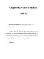
In dermatology,the preferred term for ‘chron-
ic wound’ is ‘chronic cutaneous ulcer’. An ulcer,
in turn, is defined as a depressed lesion in
which the epidermis and at least the upper der-
mis have been destroyed [1] (Figs. 1.1, 1.2). An
‘erosion’, on the other hand, is a focal loss of the
epidermis without involvement of the dermis
(Fig. 1.3).
Note that a cutaneous ulcer is not a primary
lesion.An ulcer does not develop de novo,from
Fig. 1.1. Schematic illustration of an ulcer. There is in-
volvement of the epidermis and at least part of the der-
mis
Basic Definitions and Introduction
1
01_001_006* 01.09.2004 13:50 Uhr Seite 1
intact normal skin. It is preceded by another in-
itial pathologic lesion, such as a papule or a
pustule, from which the ulcer evolves.
1.2 Three Aspects of Treatment
in Wounds and Ulcers
In recent years, accumulating knowledge re-
garding wound healing processes has led to the
development of numerous therapies. A bewil-
dering plethora of novel topical preparations,
dressing materials, and advanced methods of
debridement are now at the hands of physicians
and medical personnel. In many cases, even
those who specialize in the field of wound heal-
ing, such as dermatologists or plastic surgeons,
may find it difficult to choose the most appro-
priate treatment.
This book places this flood of information
and the many modes of therapy currently sug-
gested into some order and offers a reasonable
approach to the treatment of cutaneous ulcers.
The following chapters put forward a practical
and algorithmic therapeutic approach, accord-
ing to specific features of the ulcer.
As a rule, the treatment of a cutaneous ulcer
is determined by three aspects:
5 Etiology
5 Clinical appearance of the ulcer
5 Adjuvant therapy
1.2.1 Etiology
The treatment modality used is directed
specifically to the pathologic process which
caused the ulceration. For example:
5 Glucocorticoid therapy should be
considered when the ulcer is attrib-
uted to a vasculitic process or to a
certain connective tissue disease. It
is often advisable for pyoderma
gangrenosum. However, since gluco-
corticoids have an inhibitory effect
on wound-healing processes, their
use is not desirable for other kinds
of cutaneous ulcers.
5 In an ulceration diagnosed as
caused by leishmaniasis, one may
consider applying a topical prepara-
tion containing parmomycin. In cas-
es of unresponsive or destructive ul-
ceration, intravenous pentostam
may be considered.
5 Splenectomy may be considered for
recalcitrant ulcerations due to he-
reditary spherocytosis.
Due to the vast array of diseases and patholog-
ic processes characterized by the appearance of
cutaneous ulcers, a comprehensive dermatolo-
Chapter 1 Basic Definitions and Introduction
2
1
Fig. 1.2. A cutaneous ulcer. Note that the destruction ex-
tends deeper into the epidermis
Fig. 1.3. An erosion that developed following blister rup-
ture
t
t
01_001_006* 01.09.2004 13:50 Uhr Seite 2
gy textbook would be needed to fully cover
each issue. In this book we limit the discussion
to the appropriate identification of an ulcer’s
cause based on clinical features, histology, and
laboratory tests (see Chaps. 5 and 6). A struc-
tured diagnostic process is suggested as an al-
gorithmic approach.
1.2.2 Clinical Appearance of the Ulcer
The currently accepted classification of cutane-
ous ulcers is based on their clinical appearance
[2–9]. A practical distinction is made between
‘yellow’, ‘black’, and ‘red’ ulcers. In most cases,
the topical therapeutic method to be used de-
pends on the ulcer’s clinical appearance.
In Chap. 20, a flow chart is presented for the
treatment of cutaneous ulcers, when the ulcer’s
clinical appearance is the major determinant
regarding choice of topical treatment.
For the time being, there is no evidence that
a certain dressing type or a certain method of
debridement is more beneficial for a cutaneous
ulcer of specific etiology (e.g., venous ulcers or
diabetic ulcers).
1.2.3 Adjuvant Therapy
Modalities of adjuvant therapy are those in-
tended to improve a patient’s general condition,
thereby providing wounds with better healing
conditions.
Improvement of a patient’s nutritional status
and supplementation of certain nutritional in-
gredients, as described in Chap. 19, is applicable
here. The treatment of a patient’s other medical
problems that can increase the severity of the
ulcer can be included in this category. For ex-
ample,treatment of congestive heart failure can
reduce edema in the lower limbs, allowing en-
hanced healing of leg ulcers.
Similarly, hyperbaric-oxygen therapy can be
regarded as another mode of adjuvant therapy
that may have a beneficial effect on the healing
of a large spectrum of cutaneous ulcers. It
should always be considered in cases of diffi-
cult-to-heal cutaneous ulcers, in which ische-
mia is involved in the pathogenesis.
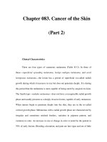
1.3 Ulcer Depth
A further classification of ulcers refers to their
depth. Definitions of ulcer staging, based on
depth and severity, were originally used for
pressure ulcers (Figs. 1.4–1.7). The staging
1.3Ulcer Depth
3
Fig. 1.4. A stage-I pressure ulcer
Fig. 1.5. A stage-II pressure ulcer
Fig. 1.6. A stage-III pressure ulcer
01_001_006* 01.09.2004 13:50 Uhr Seite 3
system was developed with the objective of
creating better communication between medi-
cal personnel. Obviously, these definitions can
be implemented for any other sorts of cutane-
ous ulcers as well.
A commonly accepted system was developed
in 1987 in the USA by The National Pressure
Ulcer Advisory Panel (NPUAP) [10], as fol-
lows:
5 Stage I: Blanchable erythema of in-
tact skin
5 Stage II: Partial-thickness skin loss
involving the epidermis and dermis,
presenting clinically as an abrasion
5 Stage III: Full-thickness skin loss,
including the subcutaneous layer
with extension down to (but not
through) the underlying fascia
5 Stage IV: Full-thickness skin loss
with involvement of muscle, bone,
or other deep structures such as
tendons or joint capsules
1.4 Comments on Current Treatments
Accepted modes of treatment are discussed in
various chapters of this book, including dress-
ing materials, methods of debridement, biolog-
ical dressings and skin substitutes, and growth
factors. Other therapeutic measures have not
been sufficiently established, thus they cannot
be recommended as evidence-based methods
in the treatment of cutaneous ulcers for the
time being. These are mainly physical thera-
peutic modalities such as infrared light,low-en-
ergy laser irradiation, ultrasonography, and
electrical stimulation. These modalities are not
discussed in this book.
As a rule, it is difficult to accurately deter-
mine the efficacy of various treatments for cu-
taneous ulcers on the basis of current data. Re-
searchers have indicated that in some studies,
basic information such as history of previous
ulceration, ulcer duration, or its appearance,
has not been provided [11, 12]. Similarly, other
studies have included too small a sample of pa-
tients or have not been controlled. Neverthe-
less, some idea of the efficacy of current treat-
ments may be obtained, allowing a treatment
approach to be suggested, as presented in sub-
sequent chapters.
References
1. Stewart MI, Bernhard JD, Cropley, Fitzpatrick TB:
The structure of skin lesions and fundamentals of
diagnosis. In: Freedberg IM, Eisen AZ,Wolff K, Aus-
ten KF, Goldsmith LA, Katz SI (eds) Fitzpatrick’s
Dermatology in General Medicine, 6th edn. New
York: McGraw-Hill. 2003; pp 11–30
2. Hellgren L,Vincent J: Debridement: an essential step
in wound healing. In: Westerhof W (ed) Leg Ulcers:
Diagnosis and Treatment. Amsterdam: Elsevier.
1993; pp 305–312
3. Hellgren L, Vincent J: A classification of dressings
and preparations for the treatment of wounds by
second intention based on stages in the healing pro-
cess. Care Sci Pract 1986; 4: 13–17
4. Stotts NA: Seeing red and yellow and black. The three-
color concept of wound care. Nursing 1990; 20 :59–61
5. Eriksson G: Local treatment of venous leg ulcers.Ac-
ta Chir Scand [Suppl] 1988; 544 : 47–52
6. Lorentzen HF, Holstein P, Gottrup F: Interobserver
variation in the red-yellow-black wound classifica-
tion system. Ugeskr Laeger 1999; 161 :6045–6048.
7. Goldman RJ, Salcido R: More than one way to meas-
ure a wound: An overview of tools and techniques.
Adv Skin Wound Care 2002; 15: 236–243
8. Findlay D: Modern dressings: what to use. Aust Fam
Phys 1994; 23 :824–839
9. Romanelli M, Gaggio G, Piaggesi A, et al: Technolog-
ical advances in wound bed measurements. Wounds
2002; 14 :58–66
Chapter 1 Basic Definitions and Introduction
4
1
Fig. 1.7. A stage-IV pressure ulcer. Note that the bone is
exposed
t
01_001_006* 01.09.2004 13:50 Uhr Seite 4
10. Pressure ulcer prevalence, cost and risk assessment:
consensus development conference statement. The
National Pressure Ulcer Advisory Panel. Decubitus
1989; 2: 24–28
11. Nelson EA, Bradley MD: Dressing and topical agents
for arterial leg ulcers (Cochrane Review). In: The
Cochrane Library, issue 1, 2003.Oxford: Update Soft-
ware
12. Stephens P, Wall IB, Wilson MJ, et al: Anaerobic coc-
ci populating the deep tissues of chronic wounds
impair cellular wound healing responses in vitro.Br
J Dermatol 2003; 148 : 456–466
References
5
01_001_006* 01.09.2004 13:50 Uhr Seite 5
2.1 Overview
Wound healing is a complex alignment of vari-
ous dynamic processes which are not yet fully
understood. The natural processes that occur
during normal wound healing, including the
various aspects of molecular and cellular
events, will be briefly discussed in this chapter,
the aim of which is to describe the practical ba-
sis for treating acute and chronic cutaneous
ulcers.
It is recommended that whoever is interest-
ed in a wider view of the basic scientific aspects
of this matter consult Cutaneous Wound Heal-
ing, edited by Vincent Falanga (Martin Dunitz);
The Molecular and Cellular Biology of Wound
Repair, edited by R.A.F. Clark (Plenum Press);
and The Epidermis in Wound Healing by David
T. Rovee and Howard I. Maibach (CRC Press).
In its usual schematic presentation,the course
of normal wound healing is divided into
three phases (Fig. 2.1):
5 Inflammation phase
(also called ‘lag phase’)
5 Tissue formation phase
(‘proliferative phase’)
5 Tissue remodeling phase
Note that this traditional division is somewhat
arbitrary and these phases partially overlap.
For example, processes of tissue formation be-
gin while active events of the inflammation
phase are still occurring.
Wound healing is regulated and synchron-
ized by a unique group of cytokines, known as
growth factors, secreted from thrombocytes,
Contents
2.1 Overview 7
2.2 Inflammation Phase 8
2.2.1 Vasoconstriction and Hemostasis 8
2.2.2 Vasodilatation and Increased Permeability 9
2.2.3 Chemotactic Growth Factors
and Phagocytosis 9
2.3 Tissue Formation Phase 9
2.3.1 Angiogenesis and Granulation Tissue
Formation 9
2.3.2 Extracellular Matrix Formation 10
2.3.3 Re-epithelialization 11
2.3.4 Wound Contraction 11
2.3.5 Role of Nitric Oxide in Wound Healing 12
2.4 Tissue Remodeling Phase 12
2.5 Types of Repair 13
2.6 Chronic Ulcers
and Protracted Inflammation 13
2.6.1 Increased Enzymatic Activity of Matrix
Proteases 13
2.6.2 Reduced Responsiveness
to Growth Factors 13
2.6.3 Cell Senescence 14
2.7 Concluding Remarks 15
References 15
W
hat wound did ever heal but
by degrees?
(William Shakespeare,
Othello II: iii, 379)
’’
Natural Course of Wound Repair
Versus Impaired Healing
in Chronic Skin Ulcers
2
t
02_007_018* 01.09.2004 13:50 Uhr Seite 7