Wound Healing and Ulcers of the Skin - part 2 pptx
Bạn đang xem bản rút gọn của tài liệu. Xem và tải ngay bản đầy đủ của tài liệu tại đây (750.35 KB, 28 trang )
ciency of one of these cofactors may result in
impaired healing [8].
As previously mentioned, TGF-β induces ex-
tracellular matrix deposition. In addition, re-
cent studies have indicated the main role of ac-
tivins, i.e., members of the TGF-β superfamily,
in various processes of wound healing. Animal
studies suggest that activins may affect dermal
components with the induction of matrix for-
mation and dermal fibrosis [15, 25].
2.3.3 Re-epithelialization
Re-epithelialization is achieved by migration,
proliferation, and differentiation of epidermal
keratinocytes. The overall purpose is complete
ulcer healing, when the whole ulcer surface ar-
ea is covered by a layer of epithelium.
Note that in most cases, epithelial cells tend
to behave as stationary cells. Yet, they may be-
come migratory cells under certain unique
conditions: embryonic development, the nor-
mal course of wound healing, and malignancy
[26, 27].
Migration. Initial re-epithelialization of a
cutaneous wound is discerned several hours af-
ter wounding, when a gradual flattening and
pseudopodium-like projections are seen in epi-
dermal cells adjacent to the wound margin.
Within 24 h, epidermal cells detach themselves
from the basal lamina to which they are at-
tached. The movement, or migration, of epider-
mal cells is seen from the margins of the wound
towards the wound matrix [13]. This type of
movement is obtained by contraction and re-
insertion of intracellular filaments of actinom-
yosin [28]. The ameboid motion of each cell is
in the form of a unique pattern called lamello-
podial crawling. The advancing epithelializa-
tion also combines movement of cells in groups
or sheets, with sliding over other epidermal
cells [29,30]. Under optimal conditions, a single
cell does not advance more than two or three
cell diameters from its original, initial location
[31]. Therefore, appropriate epidermal coverage
has to be accomplished by proliferation.
Proliferation. A few hours following initial
migration,epithelial cells in this area undergo a
phenomenon called proliferative burst [1, 32,
33]. In the following days, due mainly to the
stimulus of growth factors, epidermal cells pro-
liferate, forming and producing new epidermal
cells and enabling the process of epithelializa-
tion to be completed [12, 13].
In a simple incisional/surgical wound, re-epi-
thelialization is expected to be completed with-
in 24 h, when cells from both sides of the wound
margin touch one another and seal the area.
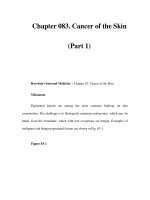
2.3.4 Wound Contraction
Wound contraction is a major process that fur-
ther contributes to wound closure (Fig. 2.5).
This process does not involve the formation of
2.3Tissue Formation Phase
11
Fig. 2.5a, b. a. A cutaneous ulcer. b. A scar following
complete healing of the same ulcer. From the size of the
scar, it is clear that a significant part of the healing pro-
cess is achieved by contraction
02_007_018* 01.09.2004 13:51 Uhr Seite 11
new tissues, as discussed above. It is based on
the centripetal movement of healthy tissues pe-
ripheral to the site of injury, so that when the
wound is eventually closed, the scar in its center
will be of the minimal possible size. Wound
contraction begins a few days after injury, si-
multaneous to the tissue remodeling phase.
This process is conducted via modified fibro-
blasts, called myofibroblasts. Certain growth
factors, such as TGF-β1, regulate the conversion
of fibroblasts to contractile myofibroblasts
[1, 34]. Myofibroblasts resemble smooth muscle
cells; having actin-containing contractile fila-
ments,they can induce contractile forces on the
edges of a wound towards its center [35–38].
The rate of contraction is dependent on all
factors that dictate the ability to heal in general,
such as the patient’s general and nutritional
condition, the etiology of the wound, and the
presence of local infection. It is also determined
by the geometric shape of the healing wound.
In round wounds, for example, the process of
contraction tends to be slower.
2.3.5 Role of Nitric Oxide
in Wound Healing
Nitric oxide (NO) is a free radical synthesized
from
L-arginine. In recent years, data have been
accumulating on the significant role of NO in
the processes of wound healing. NO is a vasodi-
lator and apparently regulates proliferation and
differentiation of several cell types such as
macrophages, keratinocytes, fibroblasts, and
endothelial cells during the inflammatory and
proliferative phases of wound healing. Hence, it
affects angiogenesis, collagen deposition, and
wound contraction [39–41]. Most evidence sug-
gests that a certain increase in NO production
may be beneficial to normal healing [42].
Further research is required to identify the
exact mechanisms by which NO affects healing.
The clinical implications of the above have not
yet been determined.
2.4 Tissue Remodeling Phase
The tissue remodeling phase represents the late
processes of healing, taking place up to two
years following injury in normal healing condi-
tions.A continuous process of dynamic equilib-
rium between the synthesis of new stable colla-
gen and the lysis of old collagen is the hallmark
of this phase. Collagen type III, synthesized in
the first few weeks, is replaced by the more
stable collagen type I. The fibers of collagen are
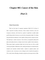
arranged in a desired alignment. These pro-
cesses lead, eventually, to the formation of scar
tissue (Fig. 2.6).
The increasing amount of stable collagen
and the alignment of its fibers gradually in-
crease the strength of the healing wound [13,
43]. Two weeks after injury, an average wound
has about 5% of its original strength; after one
month, it reaches about 40% of its original
strength. A healed wound will never regain
more than 80% of its original strength. It al-
ways has a higher risk of breakdown compared
with intact skin.
Chapter 2 Natural Course of Wound Repair
12
2
Fig. 2.6. Formation of scar tissue. (From [76])
02_007_018* 01.09.2004 13:51 Uhr Seite 12
2.5 Types of Repair
From the surgical point of view,one may distin-
guish between three different modes of wound
management,relating mainly to approximation
of the wound’s edges:
Repair by Primary Intention. Repair by pri-
mary intention is intended for acute, clean sur-
gical wounds. The skin edges are approximated
to each other, either by suturing, by staples, or
by adhesive plasters. This procedure facilitates
a relatively rapid process of wound healing
[44].
Repair by Secondary Intention. In the case
of chronic ulcers, or in wounds that have a
higher probability of developing infection, re-
pair should be achieved by secondary inten-
tion. The edges of such wounds should not be
approximated. Closure and complete healing is
achieved gradually by granulation tissue for-
mation and re-epithelialization [44].
Repair by Tertiary Intention. Tertiary inten-
tion, also called delayed primary closure, is in-
tended for wounds where the surgeon approxi-
mates the wound edges only after a few days.
The delay allows natural physiological process-
es to take place, such as drainage of exudates or
reduction in the extent of edema [44, 45].
2.6 Chronic Ulcers and Protracted
Inflammation
In contrast to the normal, natural course of
wound repair described above, chronic cutane-
ous ulcers are considered to be arrested and
‘trapped’ in an ongoing inflammatory phase
[46–48]. A protracted inflammatory process
develops in ulcers where normal mechanisms
of wound healing are not sufficient to enable
the wound to heal completely. This may occur
due to bacterial infection or to the presence of
foreign material that cannot be removed, solu-
bilized or phagocytized.
Clinically, the bed of a chronic cutaneous ul-
cer tends to appear fibrotic and to contain a
variable amount of necrotic debris. It cannot be
regarded as an appropriate matrix for the pro-
cesses of normal wound healing, such as migra-
tion of keratinocytes or epithelialization of the
wound surface.
The main features that characterize chronic
ulcers are as follows:
5 Increased enzymatic activity of ma-
trix proteases
5 Reduced response to growth factors
5 Cell senescence
2.6.1 Increased Enzymatic Activity
of Matrix Proteases
Chronic ulcers have been shown to have high
enzymatic activity of matrix metalloproteases
(MMP), which act to degrade growth factors
and extracellular matrix components such as
collagen, fibronectin, and vitronectin [47,
49–53].
At the same time, the activity of MMP inhib-
itors, which could neutralize those unwanted
effects, is reduced [54, 55]. The ongoing degra-
dation of a newly formed matrix by MMP im-
pairs and prevents normal wound healing, per-
petuating the continuous inflammatory pro-
cesses that characterize chronic ulcers.
2.6.2 Reduced Responsiveness
to Growth Factors
The level of growth factors is not necessarily
lower in chronic ulcers than in acute lesions.
Numerous studies of growth factor levels in
chronic ulcers have reported a wide range of re-
sults [47, 54–58]. Nevertheless, the general im-
pression is that the growth factors of chronic
ulcers are subjected to ongoing degradation
due to increased protease activity, as described
above. Accumulating evidence suggests that in
chronic ulcers there may be reduced expression
of growth factor receptors [59,60].It seems that
2.6Chronic Ulcers and Protracted Inflammation
13
t
02_007_018* 01.09.2004 13:51 Uhr Seite 13
these pathophysiologic changes are, at least in
part, an expression of cell senescence that oc-
curs in the chronic ulcer bed.
2.6.3 Cell Senescence
Recently, research studies have focused on the
issue of cellular senescence. The term ‘senes-
cence’ is derived from the Latin word senescere,
meaning to grow old. According to Dorland’s
Medical Dictionary, ‘senescence’ indicates the
process of growing old, especially the condition
resulting from the transitions and accumula-
tions of the deleterious aging process.
Old cells, in general, are characterized by re-
duced proliferative capacity [61–64]. The cur-
rent concept suggests that each human cell is
programmed to have a limited number of cellu-
lar divisions, determined by its specific origin
and nature. Following a finite number of divi-
sions, the cells reach a state of senescence, with
subsequent reduced proliferative capacity. An
in-vivo model of neonatal fibroblasts demon-
strated that these cells reached growth arrest
after 40–60 population doublings [65].
Senescent cells have characteristic morpho-
logical features; i.e., they tend to be larger than
cells that have not undergone such changes [66,
67]. In addition, they have specific biochemical
changes, such as an over-expression of matrix
proteins (e.g., cellular fibronectin). Senescent
cells have a decreased response to growth fac-
tors [66].
Mendez et al. [66] and Vande-Berg et al. [67]
demonstrated that fibroblasts derived from the
margins and beds of chronic cutaneous ulcers
become prematurely senescent. It is logical to
assume that the presence of senescent cells on
the surface and edges of a cutaneous ulcer re-
sults in impaired healing.
Agren et al. [68] demonstrated that fibro-
blasts obtained from chronic cutaneous ulcers
showed characteristics of senescence; their in-
vitro growth was significantly slower compared
with that of fibroblasts isolated from acute
wounds or normal skin.
Possible explanations for the presence of
senescent cells in cutaneous ulcers are as fol-
lows:
1. Cells within the surface or margin of a cuta-
neous ulcer are continuously stimulated to
proliferate (since the ulcer is not closed). On
the other hand,the basic pathologic process-
es leading to ulceration (e.g., infection, poor
vascularization, external pressure) still exist
and prevent healing. Mendez [66] suggests
that in these cases, cells undergo many un-
necessary futile divisions and gradually lose
their proliferative capacity.
2. It is suggested that chronic wound fluid and
the ulcer microenvironment contain certain
components that lead to cellular senescence.
Certain cytokines [69] or bacterial toxins
[70] may be involved in this process. Re-
search studies have shown that chronic
wound fluid suppresses in-vitro prolifera-
tion of fibroblasts, keratinocytes and endo-
thelial cells [70].
There are several clinical implications aris-
ing from the fact that cell senescence could
be an important factor in the failure of
ulcers to heal.
5 Meticulous debridement has an im-
portant part in the optimal treat-
ment of a chronic ulcer.
Debridement helps to remove se-
nescent cells from the ulcer’s sur-
face and margin. The value of
debridement procedures prior to
applications of growth factors, kera-
tinocyte transplantation, and the
use of composite grafts has been
documented [71–75].
5 Autologous skin grafting should be
considered for chronic ulcers that
are relatively large. As described in
Chap. 13, the main mechanism by
which allogeneic grafting is consid-
ered to exert its beneficial effect is
via the production of growth fac-
tors, which, in turn, enhance prolife-
ration of epithelial cells, fibroblasts,
and endothelial cells of the ulcer
bed. However, it is reasonable to as-
sume that in large, long-standing ul-
Chapter 2 Natural Course of Wound Repair
14
2
t
02_007_018* 01.09.2004 13:51 Uhr Seite 14
cers cell senescence has occurred.
Consequently, the patient’s own cells
would not be able to heal and close
a relatively large ulcer. Moreover, in
such cases, growth factors do not
actually have an appropriate and
functional target tissue to affect.
Therefore, under appropriate condi-
tions, it may be preferable to consid-
er using autologous skin grafting,
which may ‘take’ and cover the ulcer
bed, rather than allogeneic grafting.
5 Future research studies may identify
specific components that lead to se-
nescence, which would then enable
the development of new treatment
modalities specifically aimed at pre-
venting senescence and thereby im-
proving the healing of cutaneous
ulcers.
2.7 Concluding Remarks
In contrast to the normal healing of an acute
wound, chronic ulcers tend to be ‘stuck’ in an
ongoing inflammatory process. Today, chronic
ulcers are considered to represent a unique
pathophysiologic entity, in which the precise
process remains an enigma.
The optimal treatment of a chronic ulcer re-
quires appropriate ulcer bed preparation, fol-
lowed by advanced therapeutic measures such
as cultured keratinocyte grafts, composite
grafts, or preparations containing growth fac-
tors. These steps are aimed at breaking the cycle
of futile events that occur in a chronic ulcer and
to divert its course to a pathway of normal
wound healing.
References
1. Mehendale F, Martin P: The cellular and molecular
events of wound healing. In: Falanga V (ed) Cutane-
ous Wound Healing, 1st edn. London: Martin Du-
nitz. 2001; pp 15–37
2. Clark RAF: Wound repair: overview and general
considerations. In: Clark RAF (ed) The Molecular
and Cellular Biology of Wound Repair, 2nd edn.
New York: Plenum Press. 1996; pp 3–50
3. Harding K: Introduction to growth factors.In: Meet-
ing the challenge of managing the diabetic foot: use
of growth factor therapy. Proceedings from a sym-
posium preceding the 35th Annual Meeting of the
European Association for the Study of Diabetes.
Antwerp, Belgium. 1999; pp 31–40
4. Cohen K: An overview of wound healing biology. In:
Ziegler TR, Pierce GF, Herndon DN (eds) Growth
Factors and Wound Healing: Basic Science and Po-
tential Clinical Applications. Berlin Heidelberg New
York: Springer. 1997; pp 3–7
5. Kiritsy CP, Lynch AB, Lynch SE: Role of growth fac-
tors in cutaneous wound healing: a review. Crit Rev
Oral Biol Med 1993; 4: 729–760
6. Schaffer CJ, Nanney LB: Cell biology of wound heal-
ing. Int Rev Cytol 1996; 169: 151–181
7. Bennett NT, Schultz GS: Growth factors and wound
healing. Role in normal and chronic wound healing,
part II. Am J Surg 1993; 166 :74–81
8. Iocono JA, Ehrlich HP, Gottrup F, et al: The biology
of healing. In: Leaper DJ,Harding KG (eds).Wounds:
Biology and Management. Oxford, New York: Ox-
ford University Press. 1998; pp 10–22
9. Bryant WM: Wound healing. Clin Symp 1977; 29 :
1–36
10. Ross R, Benditt EP: Wound healing and collagen for-
mation. 1. Sequential changes in components of
guinea pig skin wounds observed in the electron mi-
croscope. J Biophys Biochem Cytol 1961; 11: 677–700
11. Ross R: The fibroblast and wound repair. Biol Rev
Camb Philos Soc 1968; 43 : 51–96
12. Clark RAF: Cutaneous tissue repair. Basic biologic
considerations. I. J Am Acad Dermatol 1985; 13 :
701–725
13. Kanzler MH, Gorsulowsky DC, Swanson NA: Basic
mechanisms in the healing cutaneous wound. J Der-
matol Surg Oncol 1986; 12 : 1156–1164
14. Diegelmann RF, Cohen IK, Kaplan AM: The role of
macrophages in wound repair: a review. Plast Re-
constr Surg 1981; 68: 107–113
15. Werner S, Grose R: Regulation of wound healing by
growth factors and cytokines. Physiol Rev 2002; 83 :
835–870
16. Falanga V, Shen J: Growth factors, signal transduc-
tion and cellular responses. In: Falanga V (ed) Cuta-
neous Wound Healing. 1st edn. London: Martin Du-
nitz. 2001; pp 81–93
17. Marikovsky M, Rosenblum CI, Faltin Z, et al: Ap-
pearance of leptin in wound fluid in response to in-
jury.Wound Rep Reg 2002; 10 :302–307
18. Murad A, Nath AK, Cha ST, et al: Leptin is an auto-
crine/paracrine regulator of wound healing. FASEB J
2003; 17 :1895–1897
19. Varghese MC, Balin AK, Carter DM, et al: Local
wound environment under synthetic dressings. J In-
vest Dermatol 1984; 82 : 395–396
References
15
t
02_007_018* 01.09.2004 13:51 Uhr Seite 15
20. Varghese MC, Balin AK, Carter DM, et al: Local envi-
ronment of chronic wounds under synthetic dress-
ings. Arch Dermatol 1986; 122 : 52–57
21. Eckes B,Aumailley M, Kreig T: Collagens and the re-
establishment of dermal integrity. In: Clark RAF
(ed) The molecular and cellular biology of wound
repair, 2nd edn. New York: Plenum Press, 1996;
pp 493–512
22. Micera A,Vigneti E, Pickholtz D, et al: Nerve growth
factor displays stimulatory effects on human skin
and lung fibroblasts, demonstrating a direct role for
this factor in tissue repair. Proc Natl Acad Sci USA
2001; 98: 6162–6167,
23. Liu M, Warn JD, Fan Q, et al: Relationships between
nerves and myofibroblasts during cutaneous wound
healing in the developing rat. Cell Tissue Res 1999;
297: 423–433
24. Smith PG, Liu M: Impaired cutaneous wound heal-
ing after sensory denervation in developing rats: ef-
fects on cell proliferation and apoptosis. Cell Tissue
Res 2002; 307 : 281–291
25. Beer HD, Gassmann MG, Munz B, et al: Expression
and function of keratinocyte growth factor and acti-
vin in skin morphogenesis and cutaneous wound
repair.J Investig Dermatol Symp Proc 2000; 5: 34–39
26. Gumbiner BM: Cell adhesion: the molecular basis of
tissue architecture and morphogenesis. Cell 1996;
84 : 345–357
27. Schmitz AA, Govek EE, Bottner B, et al: Rho GTPas-
es: signaling, migration, and invasion. Exp Cell Res
2000;261 : 1–12
28. Mitchison TJ, Cramer LP: Actin-based cell motility
and cell locomotion. Cell 1996; 84 : 371–379
29. Zhao M, Song B, Pu J, et al: Direct visualization of a
stratified epithelium reveals that wounds heal by
unified sliding of cell sheets. FASEB J 2003; 17: 397–
406
30. Jacinto A, Martinez-Arias A, Martin P: Mechanisms
of epithelial fusion and repair. Nat Cell Biol 2001; 3 :
E117–E123
31. Winter GD: Epidermal regeneration studied in the
domestic pig. In: Maibach HI, Rovee DT (eds) Epi-
dermal wound healing. Chicago: Year Book Medical
Publishers, Inc. 1972; pp 71–112
32. Potten CS, Allen TD: The fine structure and cell ki-
netics of mouse epidermis after wounding. J Cell Sci
1975; 17: 413–447
33. Garlick JA, Taichman LB: Fate of human keratinocy-
tes during reepithelialization in an organotypic cul-
ture model. Lab Invest 1994; 70 : 916–924
34. Desmouliere A, Geinoz A, Gabbiani F, et al: Trans-
forming growth factor-β 1 induces α-smooth muscle
actin expression in granulation tissue myofibro-
blasts and in quiescent and growing cultured fibro-
blasts. J Cell Biol 1993; 122: 103–111
35. Wrobel LK, Fray TR, Molloy JE, et al: Contractility of
single human dermal myofibroblasts and fibro-
blasts. Cell Motil Cytoskeleton 2002; 52 : 82–90
36. Serini G, Gabbiani G: Mechanisms of myofibroblast
activity and phenotypic modulation. Exp Cell Res
1999; 250: 273–283
37. Tomasek JJ, Gabbiani G, Hinz B, et al: Myofibroblasts
and mechano-regulation of connective tissue re-
modelling. Nat Rev Mol Cell Biol 2002; 3 :349–363
38. Majno G, Gabbiani G, Hirschel BJ, et al: Contraction
of granulation tissue in vitro: similarity to smooth
muscle. Science 1971; 173 :548–550
39. Weller R: Nitric oxide: a key mediator in cutaneous
physiology. Clin Exp Dermatol 2003; 28 : 511–514
40. Schentker A, Billiar TR: Nitric oxide and wound re-
pair. Surg Clin North Am 2003; 83 : 521–530
41. Witte MB, Barbul A: Role of nitric oxide in wound
repair.Am J Surg 2002; 183: 406–412
42. Efron DT, Most D, Barbul A: Role of nitric oxide in
wound healing. Curr Opin Clin Nutr Metab Care
2000; 3: 197–204
43. Levenson SM, Geever EF, Crowley LV, et al: The heal-
ing of rat skin wounds.Ann Surg 1965; 161 :293–308
44. Cohen IK, Diegelmann RF, Yager DR, et al: Wound
care and wound healing. In: Schwartz SI, Shires GT,
Spencer FC, et al (eds) Principles of Surgery. 7th
edn. New York: McGraw-Hill. 1999; pp 263–295
45. Verrier ED, Bossart KJ,Heer FW: Reduction of infec-
tion rates in abdominal incisions by delayed wound
closure techniques. Am J Surg 1979; 138: 22–28
46. Bello YM, Phillips TJ: Recent advances in wound
healing. JAMA 2000; 283: 716–718
47. Konig M, Peschen M,Vanscheidt W: Molecular biol-
ogy of chronic wounds. In: Hafner J, Ramelet AA,
Schmeller W, Brunner UV (eds) Current problems in
dermatology.Management of leg ulcers.Basel: Karg-
er. 1999; pp 8–12
48. Kloth LC, McCulloch JM: The inflammatory re-
sponse to wounding. In: McCulloch JM, Kloth LC,
Feedar JA (eds) Wound Healing: Alternatives in
Management, 2nd edn. Philadelphia: F.A. Davis.
1995; pp 3–15
49. Rao CN, Ladin DA, Liu YY, et al: α-1-antitrypsin is
degraded and non-functional in chronic wounds
but intact and functional in acute wounds: the in-
hibitor protects fibronectin from degradation by
chronic wound fluid enzymes. J Invest Dermatol
1995; 105: 572–578
50. Herrick S, Ashcroft G, Ireland G, et al: Up-regulation
of elastase in acute wounds of healthy aged humans
and chronic venous leg ulcers are associated with
matrix degradation. Lab Invest 1997; 77 :281–288
51. Lauer G, Sollberg S, Cole M, et al: Expression and
proteolysis of vascular endothelial growth factor is
increased in chronic wounds. J Invest Dermatol
2000; 115: 12–18
52. Grinnell F, Zhu M: Fibronectin degradation in
chronic wounds depends on the relative levels of
elastase, α1-proteinase inhibitor and α2-macroglob-
ulin. J Invest Dermatol 1996; 106: 335–341
53. Palolahti M, Lauharanta J, Stephens RW, et al: Prote-
olytic activity in leg ulcer exudate. Exp Dermatol
1993; 2: 29–37
54. Trengove NJ, Stacey MC, MacAuley S, et al: Analysis
of the acute and chronic wound environments: The
role of proteases and their inhibitors. Wound Rep
Reg 1999; 7 :442–452
Chapter 2 Natural Course of Wound Repair
16
2
02_007_018* 01.09.2004 13:51 Uhr Seite 16
55. Bullen EC, Longaker MT, Updike DL, et al: TIMP-1 is
decreased and activated gelatinases are increased in
chronic wounds. J Invest Dermatol 1995; 104 :
236–240
56. Mast B, Schultz GS: Interactions of cytokines,
growth factors, and proteases in acute and chronic
wounds.Wound Rep Reg 1996; 4: 411–420
57. Peschen M, Grenz H, Grothe C, et al: Patterns of epi-
dermal growth factor receptor, basic fibroblast
growth factor and transforming growth factor-β
3
expression in the skin with chronic venous insuffi-
ciency. Eur J Dermatol 1998; 8 : 334–338
58. Harris IR, Yee KC, Walters CE, et al: Cytokine and
protease levels in healing and non-healing chronic
venous leg ulcers. Exp Dermatol 1995; 4 :342–349
59.Cowin AJ, Hatzirodos N, Holding CA, et al: Effect of
healing on the expression of transforming growth
factor βs and their receptors in chronic venous leg
ulcers. J Invest Dermatol 2001; 117: 1282–1289
60. Jude EB, Blakytny R, Bulmer J, et al: Transforming
growth factor-β 1,2,3 and receptor type I and II in di-
abetic foot ulcers. Diabet Med 2002; 19 :440–447
61. Martin GM, Sprague CA, Epstein CJ: Replicative life
span of cultivated human cells. Effects of donor’s
age, tissue and genotype. Lab Invest 1970; 23: 86–92
62. Schneider EL, Mitsui Y: The relationship between in
vitro cellular aging and in vivo human age. Proc Natl
Acad Sci USA 1976; 73 :3584–3588
63. Schneider EL, Epstein CJ: Replication rate and life
span of cultured fibroblasts in Down’s syndrome.
Proc Soc Exp Biol Med 1972; 141 : 1092–1094
64. Elmore E,Swift M: Growth of cultured cells from pa-
tients with ataxia-telangiectasia. J Cell Physiol 1976;
89 : 429–431
65. Raffetto JD, Mendez MV, Phillips TJ, et al: The effect
of passage number on fibroblast cellular senescence
in patients with chronic venous insufficiency with
and without ulcer.Am J Surg 1999; 178 :107–112
66. Mendez MV, Stanley A, Park HY, et al: Fibroblasts
cultured from venous ulcers display cellular charac-
teristics of senescence. J Vasc Surg 1998; 28 : 876–883
67. Vande-Berg JS, Rudolph R, Hollan C, et al: Fibroblast
senescence in pressure ulcers. Wound Repair Reg
1998; 6: 38–49
68. Agren MS, Steenfos HH, Dabelsteen S, et al: Prolife-
ration and mitogenic response to PDGF-BB of fibro-
blasts isolated from chronic venous leg ulcers is ul-
cer-age dependent. J Invest Dermatol 1999; 112:
463–469
69. Mendez MV, Raffetto JD,Phillips T, et al: The prolife-
rative capacity of neonatal skin fibroblasts is re-
duced after exposure to venous ulcer wound fluid: A
potential mechanism for senescence in venous ul-
cers. J Vasc Surg 1999; 30: 734–743
70. Bucalo B,Eaglstein WH,Falanga V: Inhibition of cel-
lular proliferation by chronic wound fluid. Wound
Rep Reg 1993; 1 : 181–186
71. Steed DL, Donohoe D, Webster MW, et al: Effect of
extensive debridement and treatment on the healing
of diabetic foot ulcers. J Am Coll Surg 1996; 183:
61–64
72. Fisher JC: Skin grafting. In: Georgiades GS, Riefkohl
R, Levin LS (eds) Plastic, Maxillofacial and Recon-
structive Surgery, 3rd edn. Baltimore: Williams &
Wilkins. 1996; pp 13–18
73. Marcusson JA,Lindgren C, Berghard A,et al: Alloge-
neic cultured keratinocytes in the treatment of leg
ulcers: A pilot study. Acta Derm Venereol (Stockh)
1992; 72: 61–64
74. Teepe RG, Roseeuw DI, Hermans J, et al: Random-
ized trial comparing cryopreserved cultured epider-
mal allografts with hydrocolloid dressings in heal-
ing chronic venous ulcers.J Am Acad Dermatol 1993;
29 : 982–988
75. Pham HT, Rosenblum BI, Lyons TE, et al: Evaluation
of a human skin equivalent for the treatment of dia-
betic foot ulcers in a prospective, ramdomized, clin-
ical trial. Wounds 1999; 11: 79–86
76. Geras AJ: Dermatology: A Medical Artist’s Interpre-
tation. Sandoz Pharma Ltd. 1990
References
17
02_007_018* 01.09.2004 13:51 Uhr Seite 17
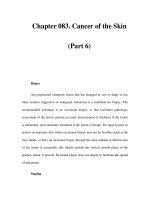
3.1 Overview
The history of wound healing is as old as the
history of medicine and probably as mankind
itself. In light of its magnitude, we shall not cov-
er the whole subject in this chapter. We shall fo-
cus rather on the principal milestones in the
history of wound healing.
In the past centuries and in recent decades,
there have been breakthroughs which have
made significant changes in our scientific under-
standing of wound repair processes. These
events have influenced the currently accepted
approach to treating wounds and ulcers.
This historical survey is an overview of the
treatment of wounds and skin lesions in gener-
al. In the medical literature, one can find histor-
ical surveys of specific types of cutaneous ul-
cers, especially venous leg ulcers, since they are
common [1, 2].
3.2 The Ancient World
Naturally, the topic has no clear starting point. It
may be attributed to that ancient father of hu-
manity who once used leaves as a dressing and
then even washed his wound in water – blissfully
unaware of the fact that he was opening up new
horizons in the history of medicine and of hu-
manity.
Later, though still well prior to documenta-
tion by clear historical records, various sub-
stances were rubbed on wounds or skin lesions;
natural materials were used, such as mud, vari-
ous plant extracts, or honey. Throughout histo-
ry, the putting together of these remedies be-
came more complex, requiring exact notation
of the mixtures that were used, as well as of just
how they were to be prepared.
Milestones in the History of Wound Healing
3
Contents
3.1 Overview 19
3.2 The Ancient World 19
3.2.1 Medicine in Mesopotamia 20
3.2.2 Ancient Egypt 20
3.3 Inflammation, Infection and the Attitude
to Appearance of Purulent Discharge
in the Past 21
3.4 Renaissance Era 22
3.5 Antiseptics, Identification of Bacteria
and the Use of Antibiotics 23
3.5.1 Ignatz Phillip Semmelweis 23
3.5.2 Joseph Lister 24
3.5.3 Other Researchers 25
3.5.4 Antibiotics 26
3.6 Investigation of Wound Healing Processes 26
3.7 The Significance
of a Moist Wound Environment 26
3.8 Keratinocyte Cultures
and Advanced Skin Substitutes 27
3.9 Recent Developments 27
3.10 Future Directions in Wound Healing 27
References 28
A
s no man can say who it was that
first invented the use of clothes and
houses against the inclemency of
the weather, so also can no investi-
gator point out the origin of Medi-
cine – mysterious as the source of
the Nile. There has never been a
time when it was not.
Thomas Sydenham
(Medical Observations)
’’
03_019_030* 01.09.2004 13:54 Uhr Seite 19
Magical and religious connotations were al-
ways dominant features of ancient medicine.
These elements have accompanied medicine
since the dawn of history, and only with the ad-
vent of modern medicine have they begun to
fade.
A unique aspect in the history of medicine is
the attempt to explain ancient healing rituals by
relying on modern medical knowledge and
technological capabilities. Thus, for example,
the Greeks used to scrape the point of a lance
over a wound, so that some metal powder was
sprinkled on it. It has been suggested that me-
tallic copper, when combined with vinegar,pro-
duces copper acetate, which has antibacterial
properties that could help in the treatment of
wounds and cutaneous ulcers [3, 4].
Similarly, inscriptions and marble carvings
found in shrines to the Greek god Asklepios (or
to Aesculapius, in the Roman world) associate
healing with having been in contact with the oral
cavity of non-poisonous serpents.Angeletti et al.
[5] have suggested that salivary growth factors
may have contributed to the healing process.
It is impossible to evaluate these and other
suppositions today, since the ancients neither
conducted nor documented strict clinical trials.
It is nonetheless reasonable to assume that such
magical or ritualistic treatments had signifi-
cant psychological consequences.
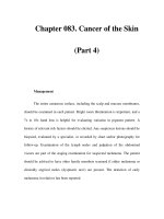
3.2.1 Medicine in Mesopotamia
The first written historical record was found on
a Sumerian clay tablet from ca. 2100
BC
(Fig. 3.1). This is actually the world’s oldest
medical manuscript. The “three healing ges-
tures” described in this tablet are: washing the
wound, applying dressings/plasters, and band-
aging the wound. These constitute the basic
principles of wound treatment today.
In his book The Healing Hand: Man and
Wound in the Ancient World [6], Guido Majno
states that there were 15 prescriptions recorded
on the tablet, without indication of the diseases
for which they were intended. Twelve of the 15
were for external use, eight being plasters, indi-
cating that they may have been used for local
diseases. Majno presents several examples of
these prescriptions, such as [6]:“Pound togeth-
er: dried wine dregs, juniper and prunes, pour
beer on the mixture. Then rub (the diseased
part) with oil, and bind on (as a plaster).”
Beer was widely used in Sumerian treat-
ments and it is likely that, owing to the antisep-
tic ingredients it contains, it did have some
beneficial effect in the treatment of wounds
and skin lesions [6].
However, it is impossible to assess today the
beneficial effect, if any, these remedies had on
the treated lesions. In fact, the Sumerians had a
variety of topical agents that could have been
useful. Oils may have been beneficial in sooth-
ing dry wounds.As mud and inorganic salts ab-
sorb water, they could have dried out wounds
and thus prevented proliferation of bacteria.
Certain plant extracts could also have had some
antibacterial effect. At present, nobody knows
whether the Sumerians actually made reason-
able use of the materials at hand.
3.2.2 Ancient Egypt
The information we have on medicine in an-
cient Egypt is based on the Smith and the Ebers
Chapter 3 Milestones in the History of Wound Healing
20
3
Fig. 3.1. Cuneiform medical clay tablet. (From The Well-
come Library, London)
03_019_030* 01.09.2004 13:54 Uhr Seite 20
papyri, dating from around 1650 BC and 1550 BC,
respectively (Fig. 3.2). The information seems
to be based on older papyri that were probably
written a thousand years earlier.
The ancient Egyptians made use of mixtures
with substances such as honey, grease, and lint
for topical application to wounds. Lint was made
from vegetable fibers and apparently helped in
the absorption of secretions from the wound’s
surface.Whether honey has a beneficial effect on
the processes of wound healing is still controver-
sial (see Chap. 17).
The Egyptian science of bandaging wounds
was similar to that used in bandaging the dead
during the process of mummification. Prior to
bandaging, the materials were dipped in vari-
ous preparations, including herbal extracts,
gums, and resins. Gum applied to bandage
strips was also used to draw and to approxi-
mate wound margins.This procedure can be re-
garded as the first adhesive bandage [7, 8].
3.3 Inflammation, Infection
and the Attitude to Appearance
of Purulent Discharge in the Past
The Sumerian and ancient Egyptian docu-
ments include the terms ummu and shememet,
respectively, which are understood today as in-
dicating the presence of inflammation. The
Egyptians distinguished between two types of
wounds: ‘Good wounds’ were treated according
to the principles described above, including
dressing with topical preparation and bandag-
ing. On the other hand, ‘bad wounds’ were
affected by a ‘whirl of inflammation,’ identified
by touching the wound edge and by their ten-
dency to secrete pus. These wounds were left
open [7].
However, the earliest description of the ‘four
cardinal signs of inflammation’ was set down
by Aulus Cornelius Celsus (42?
BC–37 AD), who
wrote a comprehensive eight-volume compen-
dium of medicine (De re Medicina). This book
was based on the Hippocratic Canon and other
classical sources. De re Medicina was forgotten
some years after its writing, only to be rediscov-
ered after a long period, in 1426. It was one of
the first medical books to be printed, appearing
in 1478. Thereafter, it enjoyed great success; new
editions were published even in the nineteenth
century [9]. It was here that Celsus first de-
scribed the four cardinal signs of inflamma-
tion, namely, rubor (redness), tumor (swelling),
calor (heat) and dolor (pain).
The Egyptians recognized that a suppurat-
ing wound should be drained [10]. Later, Galen
indicated that when infection was localized in a
wound, the discharge of pus might be followed
by healing. This observation was misinterpret-
ed in a dogmatic and rigid manner during the
following 1500 years [3, 11].
During this period, pus secreted by a surgi-
cal wound was considered to be beneficial in
cases where the amount of secretions gradually
decreased and the patient recuperated. The
presence of purulent discharge was considered
to be auspicious; the ancient expression pus bo-
num et laudabile reflects this concept.
In contrast, in cases of brown, thin, and foul-
smelling discharge, patients usually died. This
3.3Inflammation, Infection and the Attitude
21
Fig. 3.2.A piece of the Edwin Smith papyrus. (From The
Wellcome Library, London)
03_019_030* 01.09.2004 13:54 Uhr Seite 21
type of discharge was, most probably, a mani-
festation of invasive infection.
Many topical preparations were introduced
into wounds with the objective of encouraging
suppuration, a mistaken treatment that could
actually increase the risk of spreading infection
with subsequent mortality [3, 11].
It would take until the nineteenth century for
it to be understood that the presence of pus in a
wound was undesirable. Not until the break-
through discoveries of Semmelweis, Lister, and
others (see below) was it possible to prevent the
development of pus in surgical wounds with
any degree of efficiency. These principles
played a significant role in the treatment of
wounds and cutaneous ulcers.
3.4 Renaissance Era
Ambroise Paré (1509–1590) was one of the
greatest physicians of the Renaissance and in
the entire history of medicine. His broad
knowledge of medicine and surgery, stemming
from his unique skills and many years of ser-
vice in the French army as a military surgeon,
resulted in significant changes in the medical
conceptions of those times. His scientific initia-
tives helped to direct traditional medieval med-
icine towards modern medicine.
Paré was chief surgeon to four kings of
France [12]: Henry II (1547–1559), Francis II
(1559–1560), Charles IX (1560–1574), and Henry
III (1574–1589). As a military surgeon he saved
the lives of thousands of soldiers, and in so
doing, changed the previous approach, which
was to simply leave the wounded soldiers be-
hind to die on the battlefield. He wrote two
books: Treatment of Gunshot Wounds and The
Method of Treating Wounds Made by Arquebus-
es, in which he summarized the surgical tech-
niques of his era and introduced those he had
developed. His books were translated into sev-
eral languages from the French (Fig. 3.3).
His unique contribution to the field of
wound healing prevented the suffering of many
a wounded soldier.At that time, gunshot wounds
were considered to be ‘poisoned wounds’ due to
their direct contact with gunpowder. The ac-
cepted approach was to treat these wounds by
cauterizing them with a red-hot iron or with
boiling oil.
During a military expedition to Turin led by
King Francis I (1536–1537), Paré gained impor-
tant experience (Fig. 3.4). In one of the battles,
the oil he used to treat gunshot wounds ran out.
He had no option but to improvise a mixture
that included egg yolks, oil of roses, and tur-
pentine. When he changed their dressings on
Chapter 3 Milestones in the History of Wound Healing
22
3
Fig. 3.3. The cover picture of Paré’s book. (From The
Wellcome Library, London)
Fig. 3.4.Paré in the battlefield: A wood engraving of that
period. (By C. Maurand; from The Wellcome Library,
London)
03_019_030* 01.09.2004 13:54 Uhr Seite 22
the following day, he was surprised to see that
the wounds treated with the improvised mix-
ture were greatly improved, compared with
those treated with the usual boiling oil. The re-
covery of those not cauterized with the oil was
faster and with fewer complications.Of this dis-
covery, Paré wrote [12]:
“I slept badly that night, as I greatly feared that,
when I would come to examine the wounded
the following morning, I should find that those
whose wounds I had failed to treat with boiling
oil will have died from poisoning. I arose at a
very early hour,and was much surprised to dis-
cover that the wounds to which I had applied
the egg and turpentine mixture were doing
well: they were quite free from swelling and
from all evidence of inflammatory action: and
the patients themselves, who showed no signs
of feverishness, said that they had experienced
little or no pain and had slept quite well. On the
other hand, the men to whom I had applied the
boiling oil, said that they had experienced dur-
ing the night, and were still suffering from,
much pain at the seat of the injury; and I found
that they were feverish and that their wounds
were inflamed and swollen. After thinking the
matter over carefully, I made up my mind that
thenceforward I would abstain wholly from the
painful practice of treating gunshot wounds
with boiling oil.”
This observation, which Paré published, yield-
ed significant improvement in the treatment of
gunshot wounds.
Paré was responsible for two further signifi-
cant contributions: The first was the use of a
ligature to stop bleeding, rather than cauteriza-
tion. The second was the development of an ar-
tificial hand, the prosthesis.Although it was not
particularly efficient, it allowed the disabled
person a degree of ability to function. Paré may
therefore be viewed as the father of medical re-
habilitation. His most memorable statement re-
flects his modesty:“Je le pansay, Dieu le quarit”:
“I dressed him, God healed him.”
3.5 Antiseptic, Identification
of Bacteria and the Use
of Antibiotics
A further aspect of the history of wound heal-
ing took place in parallel to that of the use of
antiseptics, identification of bacteria and the
development of antibiotic preparations. Break-
throughs in this field led to the prevention of
serious complications in acute and chronic
wounds.
3.5.1 Ignatz Phillip Semmelweis
The pioneer in this area was Ignatz Phillip Sem-
melweis (1818–1865). He was born in Hungary
of German parentage (Fig. 3.5) and studied at
the medical school in Vienna.Among his teach-
ers were Rokitansky and Skoda [13]. At that
time, ‘puerperal fever’ was the cause of many
deaths of women after childbirth throughout
Europe, and there was no reasonable explana-
tion for this phenomenon. Some doctors be-
lieved that insufficient ventilation was respon-
sible, and therefore many large skylights were
constructed with ventilation apertures in the
ceilings, still to be seen in European hospitals.
The mortality was higher in units where de-
liveries were carried out by obstetricians and
medical students than in units where deliveries
3.5Antiseptics, Identification of Bacteria
23
Fig. 3.5. Semmelweis house in Budapest. The building
now serves as the Semmelweis Museum, Library and
Archives of Medical History
03_019_030* 01.09.2004 13:54 Uhr Seite 23
were carried out by midwives. Semmelweis be-
gan to think that some substance found in
corpses was being transmitted by the doctors
and medical students handling autopsies to the
women giving birth [13].
Because Semmelweis noticed that chlorine
eliminated the smell typical of corpses, he de-
manded that the hands of anyone about to ex-
amine a woman after carrying out an autopsy
or examining a sick woman be washed in a
chlorine solution (Fig. 3.6). This policy reduced
the mortality among the women giving birth in
his department.However, in the early years,this
approach was ignored by all the medical jour-
nals. Only in December 1847 did Von Hebra
publish Semmelweis’s discovery in a brief edi-
torial in a local Viennese medical journal [14].
Throughout his life, his concept met with se-
rious opposition.In 1858,Semmelweis published
an article entitled “Etiology of Puerperal Fever”
in the weekly Hungarian medical journal Orvosi
Hetilap [13]. This was his first written article
presenting his approach. His book The Etiology,
the Concept and the Prevention of Puerperal Fe-
ver was published in 1860 [15]. However, after
the book appeared, the medical establishment
still failed to support his ideas.
Towards the end of his life, Semmelweis lost
his ability to reason. Researchers have found
that certain characteristics of his behavior
point to the Alzheimer syndrome. He died in a
psychiatric hospital, and there are findings that
may indicate he was beaten to death by hospital
attendants.
It is noted that the possibility of transmis-
sion of some pathogenic agent causing puer-
peral fever was identified, at almost the same
time, by Semmelweis and Oliver Wendell Hol-
mes, Professor of Anatomy at Harvard. Holmes
spoke of such matters at the Boston Society for
Medical Improvement in 1843 [16]. He suspect-
ed a possible association between the mortality
of mothers giving birth and the presence of
physicians in autopsies, and he recommended
that doctors avoid carrying out autopsies prior
to treating the mothers. However, Holmes did
not offer a practical solution to the problem
(washing the hands in a chlorine solution) as
did Semmelweis, and his statements failed to
result in any response.
The scientific basis for understanding Sem-
melweis’s observations was to be established in
the years that followed by Pasteur and Joseph
Lister, as described below.
3.5.2 Joseph Lister
In the middle of the nineteenth century, when
Joseph Lister (Fig. 3.7) began his medical career,
amputations were the most common form of
surgery. However, a high percentage of the
wounds became gangrenous. The mortality of
patients undergoing amputation was generally
higher than 40%, as a result of surgical contam-
ination [17–19].
In 1865, Lister happened upon the work of
Louis Pasteur. Pasteur had rejected the theory
that had supported the spontaneous appear-
ance of bacteria, and related the phenomena of
decay and fermentation to microbial action.
Lister came to the conclusion that the suppurat-
ing inflammation of wounds had a similar eti-
ology. In contrast to the then-accepted notion
that such bacteria originated in the patient’s
body, Lister was impressed by Pasteur’s claim
that they existed everywhere, including the at-
Chapter 3 Milestones in the History of Wound Healing
24
3
Fig. 3.6. A porcelain device for washing the hands in the
Semmelweis era. (In the Semmelweis Museum of the
History of Medicine, Budapest)
03_019_030* 01.09.2004 13:54 Uhr Seite 24
mosphere and the bodies and clothes of the
doctors, and that they could contaminate
wounds. Lister wrote [20]:
“But when it had been shown by Pasteur’s re-
searchers that the septic property of the atmos-
phere depended, not upon the oxygen or any
gaseous constituent, but on minute organisms
suspended in it, which owed their energy to
their vitality,it occurred to me that decomposi-
tion of the injured part might be avoided with-
out excluding the air, by applying as a dressing
some material capable of destroying the life of
the floating particles.”
In order to prevent the contamination of
wounds, he began to wrap them in many layers
of gauze which he had first immersed in a car-
bolic acid solution [21]. Between the gauze
layers and the wound, he would place a layer of
relatively impermeable silk, which he called
‘protective silk’, in order to prevent damage to
the tissues by the carbolic acid.
Later, he also applied these principles in the
operating theater. He would cover the area of
the operation in a piece of cloth dipped in car-
bolic acid, which he removed only when the
surgical incision was made. He steeped the sur-
gical instruments, as well as his hands, in a car-
bolic acid solution.Thereafter,he devised a car-
bolic acid spray, and the surgical area was
sprayed with the solution in order to destroy
the air-borne bacteria.
After a few months of carrying out these
practices, the level of contamination in his unit
in a Glasgow hospital dropped considerably.
However, the excessive exposure to the carbolic
acid was detrimental to the doctors’ health.
Damage to their lungs and those of the medical
staff was described as being so severe that they
had to stop working.
For the rest of his life, Lister tried to discover
the ideal bandage that would contain antiseptic
but non-irritant material – a worthy mission
indeed, since papers discussing damage to the
processes of wound healing caused by anti-bac-
terial agents are still being published today (see
Chaps. 10 and 11).
Only in the 1890s, more than 20 years after
the discovery that the source of contamination
is external, did the use of antiseptics become
universal.
3.5.3 Other Researchers
Similar to the observations of Semmelweis and
Holmes, described above, a British surgeon,
Spencer Wells, published an article in 1864,
entitled: ‘Some Causes of Excessive Mortality
After Surgical Operation’ [22]. Wells also re-
ferred to Pasteur’s work, and in light of it pro-
posed that bacteria settle on wounds and cause
the appearance of pus and sepsis. Wells insisted
on thorough washing with cold water and the
use of fresh towels when operating. Only spec-
tators who testified in writing that they had not
been in an autopsy room during the preceding
seven days were allowed to enter his operating
theater [17]. Nevertheless, Well’s conjectures
caused little reverberation,and he did not apply
his ideas beyond the practice noted above.
Following Pasteur’s discoveries, the science
of bacteriology developed. Koch noted that
there was a major transfer of bacteria during
surgery or treatment from the surgeon’s hands,
the instruments and bandages, and from the
patient himself. In order to destroy such germs,
he proposed the use of substances such as io-
dine and alcohol.
3.5Antiseptics, Identification of Bacteria
25
Fig. 3.7. Joseph Lister (From The Wellcome Library,
London)
03_019_030* 01.09.2004 13:54 Uhr Seite 25
Other antiseptic preparations included sodi-
um hypochloride, used by Alexis Carrell, and
mercuric chloride, used by William Halsted [3].
A major breakthrough, however, took place lat-
er with the discovery of antibiotics.
3.5.4 Antibiotics
In 1928, Alexander Fleming discovered a blue
mold growing in a Petri dish which had been
accidentally exposed to its spores. He noted
that all the bacteria surrounding the mold had
been killed. However, this discovery found no
application until it attracted the attention of
Chain and Florey in 1938, an event that led di-
rectly to the isolation of penicillin in 1940 [23,
24]. The first article describing the treatment of
streptococcal meningitis by penicillin was pub-
lished in 1943 [25].
The development of antibiotic medicines is
of primary importance in the treatment of
acute wounds and chronic lesions and the pre-
vention of possible complications such as cellu-
litis, osteomyelitis, and sepsis.
3.6 Investigation of Wound Healing
Processes
The establishment of the unique scientific
branch of histopathology by Virchow [26] in
the middle of the nineteenth century is the ba-
sis for our understanding of the processes of
wound healing as they are known today. The
development of antiseptic preparations con-
tributed significantly to the understanding of
these processes, since this meant that it was
possible for the first time to examine wounds
without accompanying contamination, and
thus to identify specific inflammatory charac-
teristics.
From initial breakthroughs achieved in the
works of Metchnikoff [27], our knowledge of
the complex processes of wound healing has
gradually increased.
3.7 The Significance
of a Moist Wound Environment
In the 1950s, physicians noticed that blistered
skin achieved re-epithelialization and complete
healing if the blister roof was left intact, func-
tioning as a natural biological dressing, provid-
ed that the blister content was not infected [28].
In 1962, Winter et al. [29] presented a model
that changed the traditional concept of wound
healing. Instead of letting a wound dry out and
be covered by a dry scab, it was demonstrated
that keeping the wound environment moist
would yield much better clinical results. This
approach was confirmed in 1963 by Hinman
and Maibach [30]. They conducted a study on
human subjects with superficial wounds and
confirmed the beneficial effects of a moist envi-
ronment on wounds, compared with wounds
exposed to the air (Fig. 3.8).
Chapter 3 Milestones in the History of Wound Healing
26
3
Fig. 3.8. The significance of a moist wound environ-
ment. Left: In a moist environment,epithelialization oc-
curs along the wound surface.Right: Abnormal and pro-
longed course of healing under a dry scab. Epidermal
cells are forced to advance under the crust; the metabol-
ic expense is higher. (From the book ‘Epidermal Wound
Healing’ by Maibach & Rovee, 1972)
03_019_030* 01.09.2004 13:54 Uhr Seite 26
3.8 Keratinocyte Cultures
and Advanced Skin Substitutes
In 1975, Rheinwald and Green [31] presented a
method of culturing keratinocytes from single
cell suspensions of human epidermal cells.
Their breakthrough opened up new and chal-
lenging possibilities in the field of skin re-
search. The technique enabled the transplanta-
tion of keratinocytes and the development of
skin substitutes containing living cells. This
topic is described in detail in Chap. 13.
3.9 Recent Developments
Growth Factors. There is an ever-increasing
knowledge of various cytokines that function
as growth factors, and the way they exert their
effect on wound healing. Thus, while other cur-
rently accepted modes of therapy may provide
optimal conditions for healing, preparations
containing growth factors actually accelerate
the wound repair process.
Epidermal growth factor was first isolated in
1962 from the submaxillary glands of adult
male mice [32]. Initially, it was defined as a
polypeptide hormone and was given the title of
‘growth factor’, since it stimulated mitosis and
epidermal hypertrophy when injected subcuta-
neously into neonatal mice [33–35]. It was later
isolated from human urine, saliva, breast milk,
and amniotic fluid and, in 1975, it was the first
true growth factor to be biochemically identi-
fied [36]. Pursuant to this discovery, the Nobel
Prize for Physiology and Medicine was awarded
to Rita Levi-Montalcini and Stanley Cohen in
1986 for the identification of nerve growth fac-
tor and epidermal growth factor.
Currently, platelet-derived growth factor
(PDGF) is in practical use. One may expect that
in the near future other cytokines will also be-
come available for routine clinical purposes.
Cell Senescence. Recent research has fo-
cused on the field of cellular senescence, trying
to identify the complex processes by which old
cells gradually lose their proliferative capacity
[37–40]. Cells from the margins and beds of
chronic cutaneous ulcers become prematurely
senescent [41, 42]. This topic is discussed in
Chap. 2.
Research studies are currently focusing on
this issue, trying to identify specific processes
that lead to senescence. Identification of these
processes may be followed by the development
of new modes of treatment aimed at preventing
senescence. These may be implemented in the
field of wound repair.
3.10 Future Directions
in Wound Healing
Since the 1980s, there has been a growing
awareness and understanding of the subject of
wound repair. A number of medical associa-
tions that specifically address the area wound
healing have been established, such as the
Wound Healing Society and The European
Tissue Repair Society. Specialized journals are
now published, such as Wou nd s, The Journal of
Wound Care, Advances in Wound Care,and
Wound Repair and Regeneration.
Future directions in this fascinating field
may include the incorporation of further
growth factors into clinical use and a better
understanding of the conditions under which
they should be utilized, e.g.,the possible combi-
nation of certain growth factors and various
skin substitutes, better matching and adapta-
tion of various treatments to the etiology, heal-
ing phase, and clinical appearance of the
wound.
We can also expect to have better ways to
cope with infection. Moreover, the enormous
progress now being made in gene therapy is
opening new possibilities in the field of wound
healing.
In certain respects, the significant progress
in the field of wound healing is rapidly ap-
proaching the realm of science fiction. Fig-
ure 3.9 clearly illustrates this.
3.10Future Directions in Wound Healing
27
03_019_030* 01.09.2004 13:54 Uhr Seite 27
References
1. Scholz A: Historical aspects. In: Westerhof W (ed)
Leg Ulcers – Diagnosis and Treatment, 1st edn. Am-
sterdam: Elsevier Science Publishers. 1993; pp 5–18
2. Quintal D, Jackson R: Leg ulcers: a historical per-
spective. Clin Dermatol 1990; 8 :4–12
3. Caldwell MD: Topical wound therapy – an historical
perspective. J Trauma 1990; 30: S116–S122
4. The Iatros (Greece).In: Majno G: The Healing Hand.
Man and Wound in the Ancient World, 2nd edn.
Cambridge, Massachusetts: Harvard University
Press. 1975; pp 141–205
5. Angeletti LR,Agrimi U, Curia C, et al: Healing rituals
and sacred serpents. Lancet 1992; 340 :223–225
6. The Asu (Mesopotamia). In: Majno G: The Healing
Hand. Man and Wound in the Ancient World, 2nd
edn. Cambridge, Massachusetts: Harvard University
Press. 1975; pp 29–67
7. The Swnw (Egypt). In: Majno G: The Healing Hand.
Man and Wound in the Ancient World, 2nd edn.
Cambridge Massachusetts: Harvard University
Press. 1975; pp 69–139
8. Witkowski JA, Parish LC: Cutaneous ulcer therapy.
Int J Dermatol 1986; 25: 420–426
9. Clendening L: Celsus. In: Clendening L. Source book
of medical history. New York: Over Publications.
1960; pp 58–61
10. Ebbell B: The Ebers papyrus. The greatest Egyptian
medical document. Oxford: Oxford University
Press. 1937
11. Leaper DJ: History of wound healing. In: Leaper DJ,
Harding KG (eds) Wounds: Biology and Manage-
ment. New York: Oxford University Press. 1998;
pp 5–9
12. The development of Surgery in France (continued) –
Ambroise Paré. In: Buck AH: The Growth of Medi-
cine. From the Earliest Times to About 1800. New
Haven: Yale University Press. 1917; pp 499–515
Chapter 3 Milestones in the History of Wound Healing
28
3
Fig. 3.9.
Processes of wound healing
(From the book
‘Dermatology: A Medical
Artist’s Interpretation’
by Geras AJ, 1990)
03_019_030* 01.09.2004 13:54 Uhr Seite 28
13. Nuland SB: The enigma of Semmelweis – an inter-
pretation. J Hist Med Allied Sci 1979; 34 :255–272
14. Von Hebra F: Hochst wichtige Ehrfahrungen über
die Aetiologie der Gebäranstalten epidemischen
Puerperalfieber, KK. Ges Aerzte Wien 1847; 4 :
242–244
15. Semmelweis IP: Die Aetiologie, der Begriff und die
Prophylaxis des Kindbettfiebers. Vienna and Leip-
zig, 1861
16. Holmes OW: The contagiousness of puerperal fever
– 1843. Medical Essays. Boston. 1895; pp 103–172
17. Lawrence G: Surgery (traditional). In: Bynum WF,
Porter R (eds) Companion Encyclopedia of the His-
tory of Medicine. London New York: Routledge. 1993;
vol 2, pp 961–983
18. Lyell A: Alexander Ogston, micrococci, and Joseph
Lister. J Am Acad Dermatol 1989; 20 :302–310
19. Godlee RJ: Lord Lister. 3rd edn. Oxford: Clarendon
Press. 1924
20. Lister J: An address on the antiseptic management of
wounds. Br Med J 1893; 1: 161–162, 277–278, 337–339
21. Savin JA: Joseph Lister: a neglected master of in-
vestigative dermatology. Br J Dermatol 1995; 132 :
1003–1007
22. Wells TS: Some causes of excessive mortality after
surgical operation. Medical Times and Gazette. Oc-
tober 1, 1864; pp 349–352
23. Fleming A: On the antibacterial action of cultures of
a penicillium with special reference to their use in
the isolation of B. influenzae. Br J Exp Pathol 1929;
10 : 226–232
24. Chain E, Florey HW, Gardner AD, et al: Penicillin as
a chemotherapeutic agent. Lancet 1940; 2: 226–236
25. Fleming A: Streptococcal meningitis treated with
penicillin. Lancet 1943; 2: 434–438
26. Virchow R: Cellular Pathology. London: John
Churchill. 1860; pp 283–315
27. Metchnikoff E: Immunity in Infective Diseases
(translated by Binnie FG). London: Cambridge Uni-
versity Press. 1905
28. Gimbel NS, Kapetansky DI, Weissmen F, et al: A sto-
ry of epithelization in blistered burns. Arch Surg
1957; 74: 800–803
29. Winter GD: Formation of the scab and the rate of
epithelization of superficial wounds in the skin of
the young domestic pig. Nature 1962; 193 : 293–294
30. Hinman CD, Maibach H: Effect of air exposure and
occlusion on experimental human skin wounds. Na-
ture 1963; 200: 377–378
31. Rheinwald JG, Green H: Serial cultivation of strains
of human epidermal keratinocytes: The formation
of keratinizing colonies from single cells. Cell 1975;
6 : 331–343
32. Cohen S: Isolation of a mouse submaxillary gland
protein accelerating incisor eruption and eyelid
opening in the new-born animal. J Biol Chem 1962;
237 : 1555–1562
33. Carpenter G, Cohen S: Epidermal growth factor. J
Biol Chem 1990; 265 : 7709–7712
34. Cohen S, Taylor JM: Epidermal growth factor:
Chemical and biological characterization. In: Mai-
bach HI, Rovee DT (eds): Epidermal Wound Heal-
ing. Chicago: Year Book Medical Publishers, Inc.
1972; pp 203–218
35. Tranuzzer RW, Macauley SP, Mast BA, et al: Epider-
mal growth factor in wound healing: A model for the
molecular pathogenesis of chronic wounds. In: Zie-
gler TR, Pierce GF, Herndon DN (eds) Growth Fac-
tors and Wound Healing. Berlin Heidelberg New
York: Springer. 1997. pp 206–228
36. Starkey RH, Cohen S, Orth DN: Epidermal growth
factor: Identification of a new hormone in human
urine. Science 1975; 189: 800–802
37. Martin GM, Sprague CA, Epstein CJ: Replicative life
span of cultivated human cells. Effects of donor age,
tissue and genotype. Lab Invest 1970; 23: 86–92
38. Schneider EL, Mitsui Y: The relationship between in
vitro cellular aging and in vivo human age. Proc Natl
Acad Sci USA 1976; 73 :3584–3588
39. Schneider EL, Epstein CJ: Replication rate and life
span of cultured fibroblasts in Down’s syndrome.
Proc Soc Exp Biol Med 1972; 141 : 1092–1094
40. Elmore E, Swift M: Growth of cultured cells from pa-
tients with ataxia-telangiectasia. J Cell Physiol 1976;
89 : 429–431
41. Mendez MV, Stanley A, Park HY, et al: Fibroblasts
cultured from venous ulcers display cellular charac-
teristics of senescence. J Vasc Surg 1998; 28 : 876–883
42. Vande-Berg JS, Rudolph R, Hollan C, et al: Fibroblast
senescence in pressure ulcers. Wound Repair Regen
1998; 6: 38–49
References
29
03_019_030* 01.09.2004 13:54 Uhr Seite 29
4.1 Overview: Etiologies
of Cutaneous Ulcers
Identifying the cause of a cutaneous ulcer is of-
ten a multi-staged, challenging process. Not in-
frequently, it requires considerable expertise in
general medicine as well as in dermatology and
a thorough laboratory investigation. This chap-
ter, together with Chaps. 5 and 6, reviews etiolo-
Etiology and Mechanisms
of Cutaneous Ulcer Formation
4
Contents
4.1 Overview: Etiologies of Cutaneous Ulcers 31
4.2 Mechanisms of Ulcer Formation 31
4.3 Mechanisms of Formation
of Specific Types of Cutaneous Ulcers 36
4.3.1 Ulceration Following Injury/
External Damage to the Skin 36
4.3.2 Infections 37
4.3.3 Vascular Disease 41
4.3.4 Leukocytoclastic Vasculitis 44
4.3.5 Connective Tissue
and Multisystem Diseases 44
4.3.6 Hypercoagulable States 44
4.3.7 Metabolic Disorders: Diabetes Mellitus 45
4.3.8 Hematologic Abnormalities 47
4.3.9 Nutritional Disorders 48
4.3.10 Other Causes 48
References 48
I
t is better not to stand in the case of
an ulcer on the leg.
(Hippocrates)
’’
gies of skin ulcers and discusses the basic pro-
cesses underlying these mechanisms.
It is a well-known axiom in medicine that
things do not always turn out to be as they first
appear. A cutaneous ulcer that seems to result
from venous insufficiency may, following
thorough investigation, turn out to be a mani-
festation of carcinoma or the outcome of an
occult infectious process. In some cases, the
underlying disease is rare, requiring consider-
able clinical experience for its identification.
Etiologies of cutaneous ulcers are presented
in Table 4.1. Note that in many patients, there
are several co-existing etiologies. Moreover, in
certain conditions, such as venous insufficien-
cy, lymphedema, peripheral arterial disease, or
diabetes, the skin is much more vulnerable. In
these cases, development of a cutaneous ulcer is
not necessarily ‘spontaneous’– the skin tends to
ulcerate following various triggers such as pen-
etrating injury, blunt trauma,or contact derma-
titis. One should distinguish between underly-
ing conditions that gradually affect the quality
of the skin and actual triggers that result direct-
ly in ulceration [1].
4.2 Mechanisms of Ulcer Formation
As Table 4.1 indicates, there is a wide array of
possible etiologies of cutaneous ulcers. Howev-
er, in many cases, it is not sufficient to merely
‘label’ or classify an ulcer as being caused by a
specific disease. In a given disease, or for any
given ‘etiology’, the ulcer may be formed by a
series of complex mechanisms.Hence,the exact
mechanism by which the ulcer was formed
should also be considered.
One may assume that the main mechanism
of ulceration in an infectious process is a direct
04_031_052 01.09.2004 13:55 Uhr Seite 31
Chapter 4 Cutaneous Ulcer Formation
32
4
Following injury
Trauma (including self-inflicted ulcers)
Burns
Injection sites (e.g., heroin, cocaine, steroids)
Severe contact dermatitis (e.g., chrome ulcers)
Cold exposure: frostbite; chillblain (perniosis); Raynaud’s phenomenon
Radiation dermatitis
Bites (spider, scorpion, snake)
Pressure ulcers (presented here as a model of ongoing injury)
Infections
Bacterial
In the course of bullous erysipelas (or cellulitis)
Necrotising fasciitis
Skin over a bone affected by osteomyelitis
Ecthyma
Ecthyma gangrenosum
Meleney’s ulcer (progressive bacterial synergistic gangrene)
Noma (chancrum oris)
Tropical ulcer (tropical sloughing phagedena)
Mycobacterial
Tuberculosis (M. tuberculosis)
Swimming pool granuloma (M. marinum)
Buruli ulcer (M. ulcerans)
Leprosy (M. leprae)
Spirochetal
Syphilis
Ya w s
Tularemia (Francisella tularensis)
Anthrax (Bacillus anthracis)
Viral
Herpes genitalis
CMV
Ecthymatous varicella zoster
Fungal
Deep fungal infections
Protozoal
Leishmaniasis
Venereal ulcers
Syphilis
Chancroid
Lymphogranuloma venereum
Granuloma inguinale
Herpes genitalis
Table 4.1. Etiologies of cutaneous ulcers
04_031_052 01.09.2004 13:55 Uhr Seite 32
4.2Mechanisms of Ulcer Formation
33
Table 4.1. Etiologies of cutaneous ulcers (Continued)
Vascular
Ve n o u s
Arterial
Peripheral arterial disease
Thromboangiitis obliterans (Buerger’s disease)
Embolus: atheromatous,
cholesterol,
cardiac origin
Lymphedema
Va s c u l iti s
Leukocytoclastic vasculitis
Nodular vasculitis (erythema induratum)
Serum sickness
Cryoglobulinemia (mixed, type II & III)
Erythema elevatum diutinum
Connective tissue/multisystem diseases
Rheumatoid arthritis
Systemic lupus erythematosus
Dermatomyositis
Polyarteritis nodosa
Wegener’s granulomatosis
Churg-Strauss syndrome
Systemic sclerosis
Behçet’s disease
Sjögren’s syndrome
Temporal arteritis
Takayasu disease
Anti-phospholipid syndrome
Dysproteinemias
Cryoglubulinemia (monoclonal, type 1)
Cryofibrinogemia
Waldenstrom’s macroglubulinemia
Hypercoagulable states
Coumarin-induced necrosis
Heparin necrosis
Disseminated intravascular coagulation
Purpura fulminans
Protein C deficiency
Activated protein C resistance
Protein S deficiency
Anti-thrombin III deficiency
Hyperhomocystinemia
04_031_052 01.09.2004 13:55 Uhr Seite 33
Chapter 4 Cutaneous Ulcer Formation
34
4
Table 4.1. Etiologies of cutaneous ulcers (Continued)
Hematologic abnormalities
Hemolytic anemia
Hemoglobinopathies
Sickle cell anemia
Thalassemia
Hereditary spherocytosis
Pyruvate kinase deficiency
Paroxysmal nocturnal hemoglobinuria
Myeloproliferative diseases
Polycythemia vera
Essential thrombocytosis
Tumoral/malignant diseases
Lymphoproliferative diseases
B-cell lymphoma
Mycosis fungoides
Leukemia
Acute and chronic leukemia
Epithelial tumors
Basal cell carcinoma
Squamous cell carcinoma
Keratoacanthoma
Other epidermal tumors
Malignant melanoma
Merkel cell carcinoma
Tumors of skin appendages
(i.e., sebaceous carcinoma)
Lymph vessel/Vascular tumors
Hemangioma
Lymphangioma
Sarcomas
Kaposi’s sarcoma and other sarcomatous tumors
Other soft tissue tumors
Histiocytosis syndromes
Malignant peripheral nerve tumors
Other tumors (non-cutaneous) that may affect skin
By direct invasion into the skin
Skin metastases originating from an internal malignant tumor
04_031_052 01.09.2004 13:55 Uhr Seite 34
4.2Mechanisms of Ulcer Formation
35
Table 4.1. Etiologies of cutaneous ulcers (Continued)
Neuropathic ulcers
Diabetes mellitus
Leprosy
Tabes dorsalis
Syringomyelia
Poliomyelitis
Hereditary peripheral sensory neuropathy type 4 (insensitivity to pain)
Metabolic disorders
Diabetes mellitus
Prolidase deficiency
Calciphylaxis
Ulcerating panniculitis
Pancreatic fat necrosis
Weber-Christian disease
Histiocytic cytophagic panniculitis
α 1 anti-trypsin deficiency panniculitis
Nodular vasculitis
Nutritional disorders
Noma (chancrum oris)
Tropical ulcer (Tropical sloughing phagedena)
Vitamin C deficiency
Others
Pyoderma gangrenosum
Metastatic Crohn’s disease
Necrobiosis lipoidica diabeticorum
Atrophie blanche
Kawasaki syndrome
Klinefelter’s syndrome
Insect infestation
Drugs
Drugs that induce ulceration directly
Drugs that interfere with normal mechanisms of wound healing
Drugs that adversely affect the quality of the skin
(detailed in chapter 16)
04_031_052 01.09.2004 13:55 Uhr Seite 35
toxic effect and its subsequent abnormal local
inflammatory processes [2, 3]. However, some
infectious diseases may result in ulcers by other
mechanisms. For example, hepatitis B may in-
duce leukocytoclastic vasculitis, cryoglobuline-
mia, or periarteritis nodosa, all of which may
lead to the development of cutaneous ulcers.
Moreover, in certain infectious diseases such as
syphilis, tuberculosis, and leprosy, there are
unique pathways by which cutaneous ulcers
may appear, as detailed below.
Similarly, several pathways may lead to ulcer-
ation in connective-tissue diseases. Ulcers may
be caused by vasculitis, Raynaud’s phenome-
non, secondary anti-phospholipid syndrome, or
other mechanisms, as discussed below.
4.3 Mechanisms of Formation
of Specific Types
of Cutaneous Ulcers
Since the subject of cutaneous ulcer formation
is extensive, this section limits the discussion to
the mechanisms of formation in certain types of
cutaneous ulcers, with reference to the data pre-
sented in the table above. We shall concentrate
here on those topics particularly important for
establishing an etiologic diagnosis clinically.
4.3.1 Ulceration Following Injury/
External Damage to the Skin
4.3.1.1 Trauma and Ulcers
Mechanical trauma or some other injury may
result in a cutaneous ulcer. In some cases, the
external trauma may be minor. Although the
patient may consider it to be the cause of the ul-
cer, the skin may have been previously affected
by some underlying process (e.g., deep fungal
infection or malignancy) which may well be the
actual cause of the ulcer and should be identi-
fied.
Note that a skin injury may become a portal
of entry for bacterial or fungal infection, with
subsequent ulceration. Even a superficial ero-
sion due to trauma may become an ulcer fol-
lowing bacterial infection. The chances of this
occurring are much higher when there is an
underlying problem such as diabetes or periph-
eral arterial disease.
Self-Inflicted Ulcers. In most cases, self-
inflicted ulcers are induced by continuous
scratching, rubbing, or cutting of the skin. Once
an ulcer exists, the continuing ‘fiddling’ with it
by the patient interferes with its normal heal-
ing process.
The clinical appearance of such ulcers de-
pends on the way in which they were created.
For example, they may present sharp angular
borders as a result of the device or instrument
that was used. In some cases, self-inflicted ul-
cers are induced by injecting foreign material
into the skin. This issue is discussed in Chap. 16.
4.3.1.2 Contact Dermatitis
Patients with leg ulcers often suffer from con-
tact dermatitis. Leg ulceration has been report-
ed following exposure to numerous topical
preparations applied to cutaneous ulcers such
as topical antibiotics, certain vehicles (e.g., lan-
olin), or preservatives [4–6]. In these patients,
the application of such preparations may sig-
nificantly aggravate the ulcers. Be aware of this
possibility; topical treatment should be re-eval-
uated and patch tests performed when needed.
Moreover, skin areas affected by contact der-
matitis may be secondarily infected, with the
subsequent formation of cutaneous ulcers.
In addition, the following should be noted
with regard to cutaneous ulcers and contact
dermatitis:
5 A few substances such as chromates
[7, 8] or sodium silicate [9] are well-
known inducers of contact derma-
titis. They have a highly irritant and
corrosive effect, and patients may
present with severe dermatitis char-
acterized by the appearance of cuta-
neous ulcers. Skin ulceration has al-
Chapter 4 Cutaneous Ulcer Formation
36
4
t
04_031_052 01.09.2004 13:55 Uhr Seite 36
so been documented following the
use of povidone iodine [10].
5 The scrotal skin is highly vulner-
able. Certain topical preparations
that would not normally cause con-
tact dermatitis elsewhere may in-
duce severe irritant contact derma-
titis with ulceration when applied to
the scrotal skin [11].
5 Jacquet’s erosive diaper dermatitis
has been attributed to the presence
of various chemicals used in home
diapering. This problem is much
less common these days due to the
widespread use of disposable dia-
pers [12].
4.3.1.3 Pressure Ulcers
Pressure ulcers are presented here as a model of
continuous injury. The prevalence of pressure
ulcers varies from population to population.
Elderly patients, or patients who are bed-rid-
den or bound to a wheelchair, are particularly
prone to pressure ulcers. They may occur in an
immobilized or bed-ridden patient of any age
[13–15]. For example, among patients admitted
to hospital following spinal cord injury, the
prevalence, 1–5 years after the initial injury, is
between 20% and 30% [16].
Two other commonly accepted terms for
pressure ulcers are ‘pressure sores’and ‘decubit-
us ulcers’.
Mechanism of Formation. Local pressure on
the affected site is the most important etiolog-
ic factor, with the degree of pressure and its du-
ration being the most significant variables [14,
17]. Pressure ulcers tend to develop in areas
where soft tissues are compressed for pro-
longed periods between a bony prominence
and any external object, but they may also
evolve over any cutaneous region that is ex-
posed to continuous pressure. Pressures ex-
ceeding normal capillary pressure result in re-
duced oxygenation and impair the microcircu-
lation of the affected tissue. Garfin et al. [18] de-
termined that 30 mmHg was the critical value
for ischemia. However, high pressures of
70 mmHg or more, which may occur when a
patient lies on a standard hospital mattress,
may induce tissue death within a few hours [14,
19–21]. Most of the potential damage may be
prevented by relieving the pressure intermit-
tently [22, 23]. Therefore, prolonged immobil-
ization is a significant risk factor [24, 25]. This
emphasizes the importance of frequently mov-
ing and repositioning immobilized patients in
order to prevent the formation of pressure
ulcers.
Apart from pressure, there are other me-
chanical factors such as shearing forces and
friction which may enhance ulceration [14,
26–29]. Significant topical factors include ex-
cessive moisture and exposure to irritant sub-
stances. Both the general condition and nutri-
tional status of the patient play a major role in
the formation of pressure ulcers.
Location. More than 90% of pressure ulcers
appear on the lower parts of the body – 65% in
the pelvic region and 30% on the lower legs.
Heels and malleoli are also frequently affected
[14, 30].
Clinical Appearance. The clinical appear-
ance of a pressure ulcer depends on its severity.
Several gradation scales have been proposed. A
commonly accepted system was developed in
the USA in 1987 by The National Pressure Ulcer
Advisory Panel (NPUAP) (see Chap. 1) [31].
Pressure ulcers are characterized by periph-
eral undermining. At any stage, the initial im-
pression of the depth of a pressure ulcer may be
misleading and the ulceration of the tissue may
be much deeper and more extensive than origi-
nally thought.
4.3.2 Infections
Mild injuries may be aggravated when they be-
come secondarily infected by bacteria such as
Staphylococci or Streptococci. In some cases, an
ulcer caused by a relatively minor trauma may
become deeper as a result of infection.
4.3Mechanisms of Formation of Specific Types
37
t
04_031_052 01.09.2004 13:55 Uhr Seite 37