Wound Healing and Ulcers of the Skin - part 10 pdf
Bạn đang xem bản rút gọn của tài liệu. Xem và tải ngay bản đầy đủ của tài liệu tại đây (679.28 KB, 26 trang )
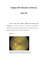
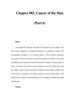
20.2 Secreting ‘Yellow’ Ulcers
It is the presence of a purulent or seropurulent
discharge that imparts a characteristic yellow-
ish appearance to these ulcers (Fig. 20.1 a,b).
The secretions may vary from thin and relative-
ly clear to heavy and thick.
When secretions are seen on the ulcer bed,
they can be removed by irrigation with saline,
which should be done as gently as possible.
However, the purpose is not only to treat obvi-
ous secretions, but also to prevent their ongo-
ing formation within the ulcer bed. We shall
distinguish between an ulcer with profuse
and/or purulent secretion and an ulcer with
mild secretion (Fig. 20.2).
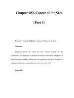
20.2.1 Ulcers with Profuse
and/or Purulent Secretion
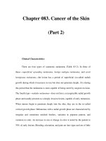
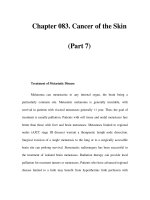
The primary objective here is to dry the ulcer.
Traditionally, the simplest way of doing this is
by repeated wetting. This can be done by gentle
saline irrigation, several times a day. Alterna-
tively, a wet dressings may be equally effective.
This is done by applying a damp sterile cloth
soaked in saline or Ringer’s lactate solution, a
few times a day, each time for 10–20 min (see
Fig. 20.3).
In these cases, the added water cannot bind
to the skin and it evaporates. In so doing, it
‘pulls’ water from the outer layers of the ulcer
bed. We are not aware at present of any scientif-
ically based research to explain this phenome-
non, but as much as it seems paradoxical, re-
peated wetting or frequent washing does have a
drying effect. Apart from its drying effect, fre-
quent saline washing mechanically removes
bacteria from the ulcer’s surface.
The effectiveness of this technique depends
on its being carried out correctly. For example,
if the damp cloth/gauze is covered by a plastic
wrap, evaporation will not be possible and the
ulcer will not dry. Similarly, if, instead of using
one layer of gauze or a thin cloth, one applies
several layers of gauze which are repeatedly wet
with a large amount of saline, a drying effect
will not be achieved. On the contrary, excessive
wetting may result in maceration and signifi-
cant damage to the tissue.
Antiseptic solutions, such as potassium per-
manganate or chlorine-based solutions, may be
used instead of saline to achieve some degree of
antibacterial effect. Antibiotic solutions may al-
so be considered, taking into account the con-
troversy that surrounds this issue (see Chap. 10).
20.2.2 Ulcers with Mild
to Moderate Secretion
Possible treatment for a mildly secreting ulcer
is a hydrophilic dressing to absorb secretions.
Preparations containing dextranomer gran-
ules, charcoal dressings, or alginate dressings
may be used.
Chapter 20 Therapeutic Approach to Cutaneous Ulcers
242
20
Fig 20.1 a,b. ‘Yellow’ ulcers
20_241_254* 01.09.2004 14:08 Uhr Seite 242
Another reasonable form of treatment is to
use a dressing that exerts topical negative pres-
sure (vacuum-assisted closure®). This assists in
absorbing fluid and debris from the ulcer bed
with subsequent reduction of wound edema. In
addition, it may draw the ulcer’s edges towards
its center, thereby enhancing wound contrac-
tion. (Note: All the methods described above
are discussed in Chap. 8. The issue of negative
topical pressure in presented in the Addendum
to this chapter.)
It is reasonable to assume that, in many cas-
es, the presence of secretions may represent a
mild degree of bacterial infection that inter-
feres with the processes of wound healing, even
though there may be no clear signs of clinical
infection (i.e., cellulitis or erysipelas). There-
fore, if the amount of secretion is not very ex-
cessive, one may consider applying an antibac-
terial cream such as silver-sulfadiazine onto the
ulcer. It may have some drying effect and can be
combined with wetting at every dressing re-
moval. In any case,an ointment should never be
applied to a secreting wound.
20.2Secreting ‘Yellow’ Ulcers
243
Fig. 20.3. Wetting a secreting lesion with a damp cotton
cloth
Fig. 20.2. Therapeutic approach to a secreting ulcer
20_241_254* 01.09.2004 14:08 Uhr Seite 243
20.2.3 Additional Comments
When treating secreting ulcers, the physician
has to ascertain that there is no wound infec-
tion, i.e., cellulitis or erysipelas, that requires
systemic antibiotics. It is also generally accept-
ed that the presence of thick purulent secre-
tions on an ulcer bed should be regarded as ev-
idence of infection [9–13]. In such cases, occlu-
sive dressings should be avoided.
A large amount of relatively thin and clear
secretion from an ulcer is not necessarily the
result of infection, but may represent edema-
tous extracapillary fluid, which is released
through the ulcer.Further investigation will de-
termine the cause of the edema and its appro-
priate treatment.
Whenever a ‘yellow’ulcer becomes clean and
red, treatment should be re-evaluated.
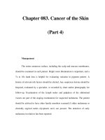
20.3 Dry ‘Black’ Ulcers
A dry ulcer is covered by black necrotic materi-
al, i.e., eschar composed mainly of devitalized
tissues or an ‘eschar-like’ crust (Fig. 20.4a, b).
The accepted approach 40–50 years ago, was to
allow wounds to dry out, enabling them to form
a crust, as part of what was considered then to
be a healthy process of wound healing. Winter
et al. [14], followed by Hinman and Maibach
[15], demonstrated the importance of moist
healing on wounds and cutaneous ulcers.
It is now understood that creating a moist en-
vironment enables the black crust to gradually
separate from the ulcer bed, thereby creating
better conditions for healing.A suitable degree of
moisture within an ulcer’s environment creates a
desirable biologic medium that provides optimal
conditions for the processes of healing.It enables
a more efficient metabolic activity, cellular inter-
action, and growth-factor activities that cannot
occur within a dry environment.
Ointments and Hydrogel Preparations. In
most cases, application of an ointment may be
beneficial. The occlusive fatty layer above the
ulcer prevents water evaporation; thus, the tis-
sues become saturated with water. When the
tissues become well hydrated, the black crust
may gradually separate from the ulcer bed.
Some use antibiotic ointments in cases requir-
ing an additional antibacterial effect. Hydrogel
preparations may also be considered in view of
their water-donating properties.
Soaking/Hydration. Soaking the affected
limb (and ulcer) in a bath of water may soften
the crust. However, for patients with leg ulcers,
especially those with diabetes, this procedure is
not desirable, since it may result in maceration
and damage to healthy skin.
In order to limit water exposure to the ulcer
area, hydration can be carried out as demon-
strated in Fig. 20.5:Apply several layers of gauze
or cloth (not one layer only, as in the case of a
secreting ulcer) saturated with water, Ringer’s
lactate or saline solution, in the form of a com-
press.Wetting should be done several times per
day, each time for 15–20 min. In this way, evapo-
ration is not possible and the crust becomes hy-
Chapter 20 Therapeutic Approach to Cutaneous Ulcers
244
20
Fig. 20.4a, b. Black ulcers
20_241_254* 01.09.2004 14:08 Uhr Seite 244
drated and soft. This can be combined with the
use of ointments on the ulcer bed.
Surgical Debridement. In more severe cases,
surgical debridement is the treatment of choice.
Hydration with saturated gauzes, as described
above, or application of fatty ointment prior to
the surgical procedure may help soften the dry
material and ease its removal. See Fig. 20.6 for a
therapeutic approach to a dry black ulcer.
20.4 ‘Sloughy’ Ulcers
In addition to the three classical types of ulcers,
as previously mentioned, we feel that an addi-
tional type should be included to complete the
classification. The term 'sloughy ulcer' has al-
ready been described by others [8]. We refer to
these as ‘sloughy’ ulcers, whose surface is cov-
ered with material, which may be yellow, green
or gray/white in appearance. It is usually soft in
consistency, ranging from a liquefied mass to
semi-solid or relatively solid material; it is com-
posed of necrotic proteins, devitalized collagen
and fibrin (Fig. 20.7a, b). It is essential to re-
move or dissolve the necrotic layer to enable
appropriate healing of the ulcer.
When there is clearly defined devitalized
material, which can be cut away and removed
20.4‘Sloughy’ Ulcers
245
Fig. 20.5. Hydration, using several layers of gauze satu-
rated with water
Fig. 20.6. Therapeutic approach to a black, dry ulcer
Fig. 20.7a,b. Sloughy ulcers (Note: Sometimes it is diffi-
cult to differentiate between a picture of a sloughy ulcer
and a picture of a yellow ulcer, as opposed to seeing
them in real life)
b
20_241_254* 01.09.2004 14:08 Uhr Seite 245
without damaging healthy and vital tissue, sur-
gical debridement may be carried out. When
nearing vital tissue, the procedure should be
discontinued and further debridement may be
accomplished by using an alternative method.
However, in many cases, there is no clear
border between the sloughy and healthy tissue.
Surgical debridement, in that situation, may re-
sult in the unnecessary loss of healthy tissue.
Note that in cases where the amount of
slough is minimal and the ulcer seems to ap-
pear relatively clean, one may consider shaving
surgical debridement (which is immediately
followed by the application of growth factors or
composite grafting). This method is detailed
below in the section on a clean red ulcer.
When surgical debridement cannot be used,
other methods should be employed to dissolve
the necrotic material. The therapeutic options
presented below (and in Fig. 20.8) should be
considered in accordance with the ulcer type,
etiology, the patient’s general health, and the
availability of each method.
Soaking/Hydration. Soaking the ulcer in
water (or hydration, as described above) may
soften the necrotic material and ease its remov-
al. A modification of this method is the use of a
product which combines multi-layered polyac-
rylate dressing with Ringer’s lactate solution
(Tenderwet®, see Chap. 8). The Ringer’s lactate
solution creates a moist environment, and may
Chapter 20 Therapeutic Approach to Cutaneous Ulcers
246
20
Fig. 20.8. Therapeutic approach to a sloughy ulcer
20_241_254* 01.09.2004 14:08 Uhr Seite 246
soften and loosen the slough, resulting in its
detachment from the ulcer bed.
Topical Negative Pressure. Another reason-
able method of treatment is to use a dressing ap-
plying topical negative pressure (vacuum-assist-
ed closure®). This method helps to absorb ne-
crotic material, secretions, and debris from the
ulcer bed.
Chemical or Autolytic Debridement. Chem-
ical or autolytic debridement may also be used
(see Chap. 9).
Antibacterial Preparations. In most cases,
this wound type is associated with bacterial
colonization. Therefore, antibacterial prepara-
tions may be used, according to the general
guidelines detailed in Chap. 10.
20.5 Clean ‘Red’ Ulcers
A clean red ulcer is the so-called ‘ideal’ ulcer a
physician would like to achieve, with the best
chances for complete healing. When dealing
with the three other types of ulcers described
above, the purpose is to convert them to this
clean red form. The desired hue lies somewhere
in the spectrum between dark-red and purple
(Fig. 20.9 a,b). Red ulcers may manifest a scale
of hydration states – from relatively dry red to
moist red.
The surface area of a ‘red’ ulcer, i.e., the ulcer
bed, is covered by granulation tissue, which is
composed mainly of numerous blood vessels,
leukocytes (mainly macrophages), and fibro-
blasts. It serves as a substrate on which the
healing proceeds, until the whole ulcer bed is
covered by epithelial cells. The various cells of
the granulation tissue secrete growth factors
that regulate and enhance the healing process-
es.
The term ‘granulation’ is derived from the
general appearance of the tissue. On close in-
spection, the tissue seems to contain numerous
tiny granules, which are actually young blood
vessels.
Normal granulation tissue is dark red to
purple. This is in contrast to ischemic ulcers,
which occur in elderly patients suffering from
peripheral vascular disease, where the granula-
tion tissue tends to be relatively bright red or
even pink.
Note that certain infected ulcers may mani-
fest an exuberant deep reddish-brown granula-
tion tissue, which tends to bleed easily [9, 16].
This is not the desired red-to-purple color of
clean red wounds.
The decision on how to treat a clean red
wound should be determined by the speed (if at
all) at which the ulcer heals. It is important to
distinguish between an ulcer that gradually
improves and advances towards healing and a
‘stagnant’ ulcer, which does not.
20.5Clean ‘Red’ Ulcers
247
Fig. 20.9a, b. Clean red ulcers
20_241_254* 01.09.2004 14:08 Uhr Seite 247
20.5.1 Ulcers Advancing Towards
Healing
When positive parameters such as re-epithe-
lialization and progressive wound contraction
are observed, it may suffice to merely supply an
ideal moist environment. The significance of
the moist environment in the ulcer area for
normal healing is detailed in Chap. 8
There are several methods for providing a
moist environment:
Saline or Ringer’s Lactate Solution. An ulcer
can be kept moist by applying a moistened
woven gauze to the surface. The following sim-
ple traditional method was presented by Pham
et al. [17]: A layer of saline-moistened gauze is
placed over the wound bed, followed by a layer
of dry gauze. A layer of petrolatum gauze (or a
plastic wrap) is then placed over that and the
area is wrapped with a layer of conforming
gauze bandage. The secondary dressing should
be changed twice a day.
Since, under these circumstances, the ulcer is
occluded, it is important to keep a close watch
on the area to identify and prevent maceration
or infection. Moreover, when using saline solu-
tion on an ulcer bed,a layer of a protective prep-
aration (such as zinc paste) should be applied to
the healthy skin around the ulcer to prevent
maceration of intact surrounding skin.
A similar therapeutic approach uses a very
slow, continuous drip of saline solution which
provides a moist environment and also re-
moves bacteria from the ulcer’s surface [18].
The rate of the drip should be adjusted to the
level of hydration of the ulcer – the dryer the
ulcer the faster the drip rate should be. Fre-
quent monitoring is mandatory. The continu-
ous drip is a relatively old method. Similar
techniques were developed at the beginning of
the twentieth century, as shown in Fig. 20.10.
Hydrocolloid or Hydrogel Dressings. The
more widely accepted approach to achieving a
relatively moist environment is to use occlu-
sives such as hydrocolloid dressings. Certain
hydrogel dressings may also be used for this
purpose, due to their water-donating proper-
ties (see Chap. 8).
20.5.2 ‘Stagnant’ Ulcers
When dealing with ulcers that do not show any
sign of improvement, a more active approach is
needed. Significant enhancement of healing
may be achieved by the application of prepara-
tions containing growth factors. Alternatively,
other advanced treatment modalities may be
used, such as keratinocyte grafts, autologous
skin grafts, or composite grafts.
Note: Advanced therapeutic modalities such
as growth factors or composite grafting are in-
tended for clean red ulcers. There is no justifi-
cation for using them on a secreting ulcer or on
an infected ulcer. Nevertheless, there is docu-
mented evidence that such treatments may
have some antibacterial effect, or that they may
enhance the patient’s immune function [19–21].
Therefore, one may consider using these treat-
ment modalities even for ulcers that are not
‘perfectly clean’, preferably combined with one
of the treatments for ‘yellow’ or ‘sloughy’ ulcers,
as discussed above.Figure 20.11 summarizes the
therapeutic approach to a clean red ulcer.
Chapter 20 Therapeutic Approach to Cutaneous Ulcers
248
20
Fig. 20.10. A device for instilling antiseptic liquid under
the dressing. The preparation used in this case is
Dakin’s solution. (From The Treatment of Infected
Wound s, by Carrel & Dehelly, published by The Macmil-
lan Company of Canada, 1917)
20_241_254* 01.09.2004 14:08 Uhr Seite 248
20.6 ‘Unresponsive’ Ulcers
We do not always have a clear and scientific ex-
planation as to why, in some cases, certain
modes of therapy do not improve healing,
whereas in other cases they do. For the time be-
ing, there are several ‘black holes’ in the under-
standing of wound healing.
Certain sloughy ulcers benefit from autolyt-
ic debridement,while others benefit from enzy-
matic debridement. Certain clean wounds im-
prove only when treated with saline-moistened
gauze and do not heal when treated with hydro-
colloid dressings. In many cases, there is an ele-
ment of trial and error in the treatment of cuta-
neous ulcers. When a certain regimen aggra-
vates the ulcer, treatment should be changed. If
an ulcer does not improve within 10–14 days
with one mode of therapy, another approach
should be considered. However, the treatment
should not be changed too often; a reasonable
amount of time is required to let a certain treat-
ment take effect.
For an unresponsive ulcer, consider one of
the following options:
5 Hospitalizing patients whose self-
treatment seems to be inadequate.
In many cases, cutaneous ulcers do
not respond to accepted treatment
because it is carried out inappropri-
ately [22]. In cases where a patient is
not capable of treating the ulcer as
required, the ulcer may deepen and
worsen.
20.6‘Unresponsive’ Ulcers
249
Fig. 20.11. Therapeutic approach to a red clean ulcer
t
20_241_254* 01.09.2004 14:08 Uhr Seite 249
5 Hyperbaric oxygen therapy, when
appropriate.
5 An alternative/additional topical
therapy (see Chaps. 17 and 18).
As stated above, for a stagnant red ulcer, one
should consider the use of growth factors or
composite grafting.
In any case the workup to determine the
ulcer’s etiology should be revised. Complica-
tions such as osteomyelitis should be ruled out
(see Table 7.3).
20.7 ‘Mixed’ Ulcers
Often, cutaneous ulcers are not uniform in col-
or. Some ulcers may present slough together
with black crusting on the surface. In others,
one may discern clean red areas as well as yel-
low secreting or sloughy areas.
In these cases, the guiding principles are as
follows:
5 Avoid any damage to healthy granu-
lation tissue; e.g., clean red areas of
the ulcer bed should not be exposed
to enzymatic preparations.
5 Secreting or sloughy areas should be
treated first, since these areas are
more prone to the development of
infection.
20.8 Additional Comments
5 Healing of a cutaneous ulcer is a dy-
namic process, subject to changes.
Treatment should be adjusted ac-
cording to the ulcer’s current clini-
cal appearance. When a yellow se-
creting wound becomes clean and
red, the therapeutic approach
should be modified accordingly.
5 Consider combining some of the
treatment modalities as presented
above. For example, repeated wetting
together with application of an anti-
bacterial cream, or with special
dressings.
5 Several researchers have suggested
that Ringer’s lactate solution may be
preferable to saline for rinsing and/or
wetting wounds and cutaneous ul-
cers. It is considered to be more ‘fri-
endly’ to the ulcer tissue in respect to
pH and electrolyte content (e.g. calci-
um). Currently, there are insufficient
data to confirm this approach.
20.9 Treating Hypergranulation Tissue
Hypergranulation tissue above a wound or
ulcer’s surface may impair normal healing (Fig.
20.12 a). This is superfluous tissue that impedes
epithelialization and wound closure. Thus, the
excess tissue should be removed. This may be
done surgically (preferably followed by advanced
therapeutic modalities such as growth factors,
or keratinocyte grafting, or skin grafting).
Alternative methods are as follows [23–25]:
5 Applying a preparation containing a
low-potency corticosteroid for a
short period, once or twice daily,
which may decrease the amount of
excessive granulation tissue
(Fig. 20.12b).
5 Some suggest using semipermeable
instead of impermeable dressings,
since low oxygen tension may en-
hance the formation of granulation
tissue. As described in Chap. 8, this
may be so when dealing with acute
wounds, but not necessarily for
chronic ulcers.
Chapter 20 Therapeutic Approach to Cutaneous Ulcers
250
20
t
t
t
t
t
20_241_254* 01.09.2004 14:08 Uhr Seite 250
20.10 Addendum: Dressings
that Apply Topical Negative
Pressure
Topical negative pressure (TNP) (vacuum-assi-
sted closure®) is currently being used on acute
traumatic wounds as well as on chronic cutane-
ous ulcers and has already been studied by
many investigators [26–31].
At present,not all mechanisms by which TNP
exerts its beneficial effects have been identi-
fied. The main mechanisms suggested are as
follows [32, 33]:
5 Absorption of fluid and debris from
the ulcer bed, with subsequent re-
duction of wound edema
5 Increasing blood flow and dermal
perfusion, with enhancement of
granulation tissue formation
5 Mechanical effect, intended to draw
the ulcer’s edges towards its center,
thereby accelerating wound contrac-
tion
5 Reducing the amount of stagnant
fluid and bacterial load
The TNP dressing is a porous foam material.
Tubes are embedded in the dressing, while their
proximal part is connected to an adjustable
vacuum pump (Fig. 20.13). The dressing should
be trimmed to conform to the shape of the ul-
cer into which it is inserted.While activated,the
vacuum device creates a continuous and con-
trolled negative pressure.
In our experience, the TNP dressing has
shown a relatively high level of efficacy in
‘cleaning’ the chronic ulcer bed, leading to the
development of healthy granulation tissue. This
is especially evident in venous ulcers and ulcers
related to lymphedema. However, TNP has also
been documented as accelerating healing of ul-
cers of various other etiologies. In any case,
more research studies are required to examine
the most appropriate guidelines for TNP use.
During TNP therapy patients are immobi-
lized and continuously attached to the TNP de-
vice. Therefore, during the treatment period,
patients (especially elderly patients for whom
immobilization carries a risk of deep venous
thrombosis or pneumonia) should be encour-
aged to detach themselves from the device a few
times each day, to walk and activate their legs.
20.10Dressings that Apply Topical Negative Pressure
251
Fig. 20.12. a. Excessive granulation tissue. b. The same
ulcer following two weeks’ daily application of a prepar-
ation containing low-potency corticosteroids
tt
5 A polyurethane foam dressing has
been shown to have a beneficial ef-
fect.
20_241_254* 01.09.2004 14:08 Uhr Seite 251
References
1. Hellgren L, Vincent J: A classification of dressings
and preparations for the treatment of wounds by
second intention based on stages in the healing pro-
cess. Care Sci Pract 1986; 4 : 13–17
2. Stotts NA: Seeing red and yellow and black. The
three-color concept of wound care. Nursing 1990;
20 : 59–61
3. Hellgren L,Vincent J: Debridement: an essential step
in wound healing. In: Westerhof W (ed) Leg Ulcers:
Diagnosis and Treatment. Amsterdam: Elsevier.
1993; pp 305–312
4. Eriksson G: Local treatment of venous leg ulcers.Ac-
ta Chir Scand 1988; [Suppl] 544 : 47–52
5. Goldman RJ, Salcido R: More than one way to meas-
ure a wound: An overview of tools and techniques.
Adv Skin Wound Care 2002; 15: 236–245
6. Findlay D: Modern dressings: What to use.Aust Fam
Physician 1994; 23: 824–839
7. Romanelli M, Gaggio G, Piaggesi A, et al: Technolog-
ical advances in wound bed measurements.Wounds
2002; 14 : 58–66
8. Thomas S: Wound dressings. In: Rovee DT, Maibach
HI (eds) The Epidermis in Wound Healing. Boca Ra-
ton: CRC Press. 2004; pp 215–241
9. Browne A,Dow G, Sibbald RG. Infected wounds; def-
initions and controversies. In: Falanga V (ed) Cuta-
neous Wound Healing, 1st edn. London: Martin Du-
nitz. 2001; pp 203–219
10. Parish LC, Witkowski JA: The infected decubitus ul-
cer. Int J Dermatol 1989; 28: 643–647
11. Niedner R, Schopf E. Wound infections and antibac-
terial therapy. In: Westerhof W (ed) Leg Ulcers: Di-
agnosis and Treatment. Amsterdam: Elsevier. 1993;
pp 293–303
12. Lipsky BA, Berendt AR: Principles and practice of
antibiotic therapy of diabetic foot infections. Dia-
betes Metab Res Rev 2000; 16 [Suppl 1] : S42–S46
13. Robson MC: Wound Infection: a failure of wound
healing caused by an imbalance of bacteria. Surg
Clin North Am 1997; 77 :637–650
14. Winter GD: Formation of the scab and the rate of
epithelization of superficial wounds in the skin of
the young domestic pig. Nature 1962; 193: 293–294
15. Hinman CD, Maibach H: Effect of air exposure and
occlusion on experimental human skin wounds. Na-
ture 1963; 200 : 377–378
16. Cutting KF, Harding KG: Criteria to identify wound
infection. J Wound Care 1994; 3: 198-201
17. Pham HT, Rosenblum BI, Lyons TE, et al: Evaluation
of a human skin equivalent for the treatment of dia-
betic foot ulcers in a prospective, randomized, clini-
cal trial. Wounds 1999; 11: 79–86
18. Marquez RR: Wound debridement and hydrothera-
py. In: Gogia PP (ed) Clinical Wound Management,
1st edn. New Jersey: Slack Incorporated. 1995;
pp 115–130
19. Schmid P: Apligraf – phenotypic characteristics and
their potential implications for the treatment of dia-
betic foot ulcers. A satellite symposium at the 36th
annual meeting of the European association for the
study of diabetes (EASD).Jerusalem, Israel. Septem-
ber 2000
20. Gough A, Clapperton M, Rolando N, et al: Random-
ized placebo-controlled trial of granulocyte- colony
stimulating factor in diabetic foot infection. Lancet
1997; 350 : 855–859
21. De Lalla F, Pellizzer G, Strazzabosco M, et al: Ran-
domized prospective controlled trial of recombi-
nant granulocyte colony- stimulating factor as ad-
junctive therapy for limb- threatening diabetic foot
infection. Antimicrob Agents Chemother 2001; 45:
1094–1098
22. Haram RB, Dagfinn N: Errors and discrepancies: a
patient perspective on leg ulcer treatment at home. J
Wound Care 2003; 12: 195–199
23. Feedar JA: Clinical management of chronic wounds.
In: McCulloch JM, Kloth LC, Feedar JA (eds) Wound
Healing – Alternatives in Management, 2nd edn.
Philadelphia: FA Davis. 1995; pp 137–185
Chapter 20 Therapeutic Approach to Cutaneous Ulcers
252
20
Fig. 20.13a,b. A dressing applying negative pressure.
Tubes connect the vacuum device to the porous dress-
ing covering the ulcer. a. Before applying negative pres-
sure. b. Flattening of the dressing following the applica-
tion of negative pressure
20_241_254* 01.09.2004 14:08 Uhr Seite 252
24. Harris A, Rolstad BS: Hypergranulation tissue: a
nontraumatic method of management. Ostomy
Wound Manage 1994; 40 : 20–22,24,26–30
25. Thomas S, Leigh IM: Wound dressing. In: Leaper DJ,
Harding KG (eds) Wounds: Biology and Manage-
ment. Oxford: Oxford University Press. 1998;
pp 166–183
26. Evans D, Land L: Topical negative pressure for treat-
ing chronic wounds: a systematic review. Br J Plast
Surg 2001; 54 : 238–242
27. Argenta LC, Morykwas MJ: Vacuum-assisted clo-
sure: A new method for wound control and treat-
ment: Clinical experience. Ann Plast Surg 1997; 38 :
563–576
28. Banwell PE, Teot L: Topical negative pressure (TNP):
the evolution of a novel wound therapy. J Wound
Care 2003; 12: 22–28
29. McCallon SK, Knight CA,Valiulus JP, et al: Vacuum-
assisted closure versus saline-moistened gauze in
the healing of postoperative diabetic foot wounds.
Ostomy Wound Manage 2000; 46: 28–32,34
30. Joseph E, Hamori CA, Bergman S, et al: A prospec-
tive, randomized trial of vacuum-assisted closure
versus standard therapy of chronic non-healing
wounds.Wounds 2000; 12 : 60–67
31. Evans D, Land L: Topical negative pressure for treat-
ing chronic wounds (Cochrane Review). In: The Co-
chrane library, Issue 4, 2001. Oxford: Update soft-
ware
32. Morykwas MJ, Argenta LC, Shelton-Brown EI, et al:
Vacuum assisted closure: a new method control and
treatment: animal studies and basic foundation.
Ann Plast Surg 1997; 38 : 553–562
33. Mendez-Eastman S: Guidelines for using negative
pressure wound therapy. Adv Skin Wound Care
2001; 14 : 314–322
References
253
20_241_254* 01.09.2004 14:08 Uhr Seite 253
21.1 General Patient Guidelines
for the Treatment of Ulcers
or Wounds at Home
5 Take care to wash your hands with
soap and water before and after the
treatment.
5 When the dressing is changed,
any materials removed from the
wound should be placed directly
into a plastic bag set aside earlier
for that purpose. Make sure that
the infected dressings do not
come into contact with the floor,
the furniture, or any other object,
so as to avoid as far as possible
any spread of infectious bacteria
to the surroundings.
Appendix: Guidelines for Patients
and Medical Staff
21
Contents
21.1 General Patient Guidelines for the Treatment
of Ulcers or Wounds at Home 255
21.2 Patient Guidelines for the Management
of Skin Ulcers Caused
by Venous Insufficiency 256
21.3 General Guidelines for Patients with Diabetes
or Peripheral Arterial Disease 256
21.4 Treatment of Edema 257
References 257
21.5 Guidelines for Nurses:
Outpatient Management
of Cutaneous Ulcers 258
References 259
5 The skin around the wound may be
cleaned using a very dilute, mild
soap. Ensure that all the residual
soap is thoroughly rinsed off, using
a gentle stream of lukewarm water.
Avoid using soap directly on the
wound.
5 After being rinsed with water, the
wound should be dried by gentle
patting/dabbing only. Never scrub
or rub the wound.
5 Any medical substance that needs to
be placed on the wound bed should
be applied using an object such as a
spatula, or tongue depressor. Never
apply the substance directly from its
container, and never use bare fin-
gers to apply it.
5 To remove dressings that have be-
come stuck to the wound because of
dried secretions on the wound sur-
face, moisten them with saline or
tap water and leave them wet for a
few minutes. They can usually then
be removed relatively easily without
causing any damage to the bed of
the wound.
5 Ensure that the dressing is not too
tight or pressing too hard on the
wound, since a tightly applied dress-
ing may interfere with the blood
flow.
5 Use an elastic net dressing (e.g., fle-
xible elastic net bandages; stockin-
ette) to hold the dressing on the
wound. Avoid the use of adhesive
plaster directly on the skin.
5 Smoking is strictly forbidden!!!
t
t
21_255_260* 01.09.2004 14:10 Uhr Seite 255
21.2 Patient Guidelines
for the Management
of Skin Ulcers Caused
by Venous Insufficiency
5 Avoid standing as much as pos-
sible.
5 While seated, make sure your legs
are elevated, if possible slightly
above the level of your buttocks.
5 When lying down, it is advisable to
elevate the legs slightly on a cushion
or folded blanket.
5 It is advisable to walk every day for
at least 30–60 min; do not stand still
during this time.
5 When seated or lying down, move
your legs (twisting, cycling motion,
etc.) several times a day for
10–15 min each time.
5 If your doctor advises you to use
support stockings, you should put
them on in the morning, after your
legs have been elevated for a few
hours, so that your legs will not be
swollen when you put the stockings
on.
5 Smoking is strictly forbidden!!!
21.3 General Guidelines
for Patients with Diabetes
or Peripheral Arterial Disease
The following guidelines should be imple-
mented by patients with diabetes or peri-
pheral arterial disease, in order to prevent
formation of cutaneous ulcers.
5 Wash your feet at least once a day
with a mild soap under running
water. Make sure you rinse off all
traces of the soap, particularly
between the toes.
5 Use only lukewarm, not hot water.
5 After washing, dry your feet
thoroughly, including between the
toes, since moisture in that area
can lead to infections. Usually a
normal towel is not adequate; use
absorbent paper in addition.
5 It is recommended you walk at
least 30–60 min every day. It is also
advisable to move your legs while
lying down or sitting (twisting or
cycling movements, etc.) several
times a day, for 10–15 min each
time. In the case of a neuropathic
ulcer consult your doctor as to the
appropriate mode of physical
activity and pressure off-loading.
5 Cutting toenails: Avoid cutting the
corners; trim the toenails straight
across. If necessary, get someone to
help you, so you don’t injure your-
self. Do not ‘dig’ with a sharp in-
strument in the angle between the
nail and the flesh.
5 Examine your legs and feet once a
day, using a mirror, or get someone
to help if necessary.
5 Treatment of wounds, corns, blis-
ters should be carried out only
under medical supervision. Should
areas of dry skin, rashes, scaling,
or changes in the skin color ap-
pear, seek medical advice.
5 Cold and heat injury: Avoid expo-
sure to cold; keep the feet warm in
winter. However, do not sit too close
to a heater or fire. Don’t use a hot
water bottle. Do not use an electric
blanket without a thermostat. Be-
fore washing your feet, test the wa-
ter temperature with your hand.
5 Wear comfortable, well-fitting
shoes. A poorly fitted shoe may
cause corns, because of uneven
pressure distribution on the foot
when walking. Before putting your
shoes on, shake them out to re-
move any small stones. Don’t wear
shoes without socks. Cotton socks
Chapter 21 Appendix: Guidelines for Patients and Medical Staff
256
21
t
t
t
21_255_260* 01.09.2004 14:10 Uhr Seite 256
are preferable. Change your socks
at least once a day. Avoid sandals
or thongs, since they do not pro-
tect the foot from injury.
5 Smoking is strictly forbidden!!!
21.4 Treatment of Edema
Once the cause of edema has been identified,
treatment should be done accordingly (see
Chap. 7).
In addition, the following steps should be
considered:
5 Elevation of the extremities: The pa-
tient should be given clear instruc-
tions to raise the affected extremity.
In the case of leg ulcers the patient
should be instructed to avoid pro-
longed standing as much as possible
and to raise the leg while sitting or
lying down.
5 Physical activity: The patient should
be advised to engage in physical ac-
tivity and to exercise the leg mus-
cles. Physical activity can reduce the
degree of leg edema [1, 2].
5 Physical therapy: The patient should
be referred for a special form of
therapy known as manual lymph
drainage. This is a massage tech-
nique designed to reduce edema. It
is effective for edema of the lower
extremities and can aid in the heal-
ing of cutaneous ulcers associated
with edema [2–9]. Kurtz et al. [8]
found that this procedure may lead
to the mobilization of up to 1 l of
urine from reabsorption and trans-
port of interstitial fluid. Gentle pres-
sure is applied to lymph nodes and
lymph vessels with the finger tips to
evacuate lymphatic content. The
rhythm of applied pressure is differ-
ent from that of traditional massage
techniques [3, 4]. The basic principle
is that central regions of lymph
drainage, such as the lower abdo-
men and proximal thigh, should be
emptied initially to provide space
for the drainage of peripheral fluid.
After the proximal region of a limb
has been cleared of its lymph, distal
areas are massaged to mobilize the
fluid proximally. The scheduling of
therapeutic sessions depends on the
size of the ulcer, the amount of ede-
ma, and the patient’s general condi-
tion. It is usual to conduct two to
three sessions per week [4], but dai-
ly sessions should be considered
under certain circumstances.
5 Compression therapy is done using
stockings or elastic bandages. Com-
pression therapy has a proven value
as a means of treating venous insuf-
ficiency and its ensuing leg ulcers
[10–13].
5 Administration of diuretics should
be considered, depending on the cli-
nical data.
References
1. Ciocon JO, Galindo Ciocon D,Galindo DJ: Raised leg
exercises for leg edema in the elderly. Angiology
1995; 46: 19–25
2. Casley-Smith JR, Casley-Smith JR: The nature of
complex physical therapy. In: Casley-Smith JR, Cas-
ley-Smith JR (eds) Modern Treatment for Lymphoe-
dema, 5th revised edn. Adelaide: The Lymphoedema
Association of Australia. 1997; p 131
3. Casley-Smith JR, Casley-Smith JR: Massage tech-
niques for lymphoedema. In: Casley-Smith JR, Cas-
ley-Smith JR (eds) Modern Treatment for Lymphoe-
dema, 5th revised edn. Adelaide: The Lymphoedema
Association of Australia. 1997; pp 134–152
4. Stahel HU: Manual Lymph Drainage. Curr Probl
Dermatol 1999; 27: 148–152
5. Hoffmann A, Petzoldt D: Manual lymph drainage.
Hautarzt 1978; 29: 463–466
6. Evrard-Bras M, Coupe M, Laroche JP, et al: Manual
lymphatic drainage. Rev Prat 2000; 50: 1199–1203
7. Derdeyn A,Aslam M, Pflug JJ: Manual lymph drain-
age – mode of action. Lymphology 1994; 27 [Suppl]:
527–529
References
257
tt
t
21_255_260* 01.09.2004 14:10 Uhr Seite 257
8. Kurz W, Kurz R, Litmanovitch YI, et al: Effect of
manual lymph drainage massage on blood compo-
nents and urinary neurohormones in chronic lym-
phedema. Angiology 1981; 32: 119–127
9. Francois A, Richaud C, Bouchet JY, et al: Does medi-
cal treatment of lymphedema act by increasing
lymph flow? Vasa 1989; 18: 281–286
10. Partsch H: Compression therapy for venous ulcers.
Curr Probl Dermatol 1999; 27 : 130–140
11. Cullum N, Nelson EA, Fletcher AW, et al: Compres-
sion for venous leg ulcers (Cochrane Review). In:
The Cochrane Library. Issue 1, 2003; Oxford: Update
Software
12. Phillips TJ, Dover JS: Leg ulcers. J Am Acad Derma-
tol 1991; 25:965–987
13. Yasuhara H, Shigematsu H, Muto T: A study of the
advantages of elastic stockings for leg lymphedema.
Int Angiol 1996; 15 :272–277
21.5 Guidelines for Nurses:
Outpatient Management
of Cutaneous Ulcers
5 Warn the patient not to touch any
object, furniture, or any other item
in the clinic unnecessarily, to reduce
the spread of bacteria to the sur-
roundings.
5 Both the patient and the nurse
should wash their hands before and
after the treatment. Show the pa-
tient how to wash his/her hands
correctly. Hands should be washed
under running water, for about
15–20 sec, with 3–5 ml of a cleaning
agent [1]. Another accepted disin-
fecting method is to use an alcohol-
based hand-rub solution (contain-
ing about 70% alcohol). Recently,
the latter has been documented as
the best method for reducing trans-
mission of infection [2, 3].
5 The patient should avoid placing
his/her treated foot directly on the
floor. The treated foot should be
placed on a stool covered by plastic
and a clean cloth or sheet [4]
(Fig. 21.1).
5 Should the ulcer need to be rinsed
extensively with saline, ensure that
there are sheets of plastic on the
floor or bench.
5 Place a plastic bag near the patient,
in which you can readily dispose of
all the dressing materials removed
from the ulcer (Fig. 21.2). Ensure
that used dressing materials re-
moved from the ulcer do not come
into contact with the floor, the fur-
niture, or any other object in the
clinic.
5 Throughout the treatment proce-
dures the nurse must wear gloves. It
is advisable to change the gloves af-
ter removing the old dressing. Use a
new pair of gloves when applying
the fresh dressing to the wound.
5 Enter information into the patient’s
record after completing the dressing
procedure, removing the gloves, and
washing your hands.
5 Between one patient and the next,
clean the treatment area with anti-
septic solution.
5 After treating an ulcer that is likely
to be heavily infected (such as a cu-
taneous ulcer known to contain
methicillin-resistant Staphylococcus
aureus or resistant Pseudomonas
strains) or a heavily secreting ulcer,
clean the treatment room thorough-
ly with soap and sodium-hypochlor-
ite solution. Free available chlorine
at 500 ppm is documented as being
the most effective compound against
most nosocomial pathogens [5].
Chapter 21 Appendix: Guidelines for Patients and Medical Staff
258
21
t
t
21_255_260* 01.09.2004 14:10 Uhr Seite 258
References
1. Larson E: APIC guideline for hand washing and
hand antisepsis in health-care settings. Am J Infect
Control 1995; 23 : 251–269
2. Hugonnet S, Pittet D: Hand hygiene revisited: Les-
sons from the past and present. Curr Infect Dis Rep
2000; 2: 484–489
3. Teare L, Cookson B, Stone S: Hand hygiene. Use alco-
hol hand rub between patients: they reduce trans-
mission of infection. Br Med J 2001; 323 : 411–412
4. Shai A, Bilenko N, Ben-Zeev R, et al: Use of infection
control procedures in an out-patient clinic for leg ul-
cers and the rate of contamination with methicillin-
resistant Staphylococcus aureus. Wounds 2004; 16 :
193–200
5. Zaidi M, Wenzel RP: Disinfection, sterilization, and
control of hospital waste.In: Mandell GL, Bennett JE,
Dolin R (eds) Mandell, Douglas and Bennett’s Prin-
ciples and Practice of Infective Diseases, 5th edn.
Philadelphia: Churchill Livingstone. 2000; pp 2995–
3005
References
259
Fig. 21.1. The patient’s leg is placed on a stool
Fig. 21.2. A plastic bag is used for disposal of the dress-
ing materials removed from the ulcer
21_255_260* 01.09.2004 14:10 Uhr Seite 259
Subject Index
A
absorptive dressings 105, 111–114
accidental injections 196
acetic acid 156
actinomycin D 194, 196
activated charcoal dressings 113,
122
activated protein C resistance 44,
73
acute wounds 1
additional topical preparations
217–221
adhesiveness 105
adjuvant therapy 2, 3
adriamycin 194, 196
albumin 96, 97, 224, 225
alginate dressings 111, 112
allantoin 130
allergic reactions 143
– sensitivity to povidone-iodine
152
– sensitivity to silver sulfadiazine
154
allogeneic cadaver skin 165, 166
allogeneic grafting 159, 170
aloe vera 211
alternative topical preparations
209–215
amino acid 225
amlodipine 203
amphiregulin 190
ancient Egypt 20
anemia 98, 99, 233
see also hemolytic anemia
anesthetics
– necrosis 196
– before debridement 123
angiogenesis 8, 9, 12
– growth factors 186
– nitric oxide 12
– transforming growth factor beta
10
anthrax 32, 58
anti-neoplastic drugs 201, 202, 228
anti-phospholipid syndrome 44,
73, 199
antibiogram 145
antibiotics
– definition 136
– history 26
– mode of action 137
– systemic 139–141
– topical 142
antineutrophil cytoplasmic antibo-
dy 71, 77, 78
antiseptics
– antiseptic dyes 155
– definition 137
– mode of action 137
– toxicity 137, 142, 144
appetite suppressant 203
arginine 225
arterial ulcers 67
see peripheral arterial disease
ascorbic acid
see: vitamin C
aseptic necrosis 194, 195
assessment
– nutritional status 96, 224
– patients (general) 95–100
atrophie blanche 54, 63, 64, 75
atrophy 62, 203
atypical mycobacterial infection
66, 84
autolytic debridement 129
B
β-blocker 198
Bacteremia 124
bacterial
– infections 32, 37–40
– toxic effect 36
– toxins 14
baking soda 197
balsam of Peru 212
basal cell carcinoma 56, 82, 83
battery acid 197
beer 20
Behçet’s disease 33, 58, 60, 123
beryllium 197
biofilm 147
biological dressings 115, 159, 165
biopsy 71, 72, 100
– deep-tissue biopsy 146
– possible histologic patterns 81
biosurgery 129
biotherapy 129
bizarre forms of red blood cells 75
bleomycin 198, 203
– bleomycin-induced digital
gangrene 200
brilliant green 155
bromocriptine 203
Burow’s solution 156
butacaine 197
C
cadaver skin 165, 166
cadexomer-iodine gel 112, 113, 153
caffeine 197
calciphylaxis 66, 72, 75
calcium channel blocker 202
calendula 211
caloric-deficient state 225
cancrum oris 48, 226
carbamazepine 199
carbolic acid 25
carica papaya 128
cell senescence 14, 27
cellulitis 38, 137
Celsus Aulus Cornelius 21
chancroid 60
charcoal dressings 113
chemical debridement 127–129
chemotaxis 8
chlorines 153
chloropromazine 199
cholesterol
– cleft 74, 75
– emboli 44, 73
chromate 36
chronic wound 1
cis-platinum 194, 196
classification of ulcers
– clinical appearance/color 3, 241
– etiology 2
– depth 3, 4
clay 212
clean ulcer 138, 241, 247–248
clindamycin 194, 198
clostridium
– histolyticum 128
– perfringes 38
– septicum 38
22_261_268_SI* 01.09.2004 14:11 Uhr Seite 261
clot 9
CMV 40
cocaine 194, 195, 197
colchicine 202
collagen
– containing dressings 167–168
– healing processes 8, 10–12
– naturally-occurring collagen ma-
trix 167
– methionine 225
– vitamin C 230
collagenase 128
colonization 136
composite graft 172
connective tissue diseases 44, 56,
78, 79
contact dermatitis 36
contamination 136
contraction 11, 12
see wound contraction
corticosteroid 194, 195,250
see: glococorticoids
corymbiform pattern 66
coumarin-induced necrosis 73
critical limb ischemia 43
crust 120, 244
cryofibrinogenemia 73
cryoglobulinemia 36, 73, 78
crystal violet 155
curettage 146
cyclophosphamide 229
cytokines 7, 8, 10, 14
see: growth factors
D
Dakin’s solution 153
daunorubicin 194, 196
debridement 119–132
– absorptive 122
– appropriate technique 124
– autolytic 122, 129, 132
– bacteremia 123
– Behçet’s disease 123
– bleeding 124
– chemical 122
– collagenases 127
– contraindications 123, 131
– definition 119
– deoxyribonuclease 127
– enymatic 122, 127
– fibrinolytic enzymes 127
– hydrodebridement 122, 132
– hydrogel preparations 132
– irrigation 122
– maggot therapy 122, 129
– mechanical 122
– methods 121
– mild acidic preparations 122, 129
– pathergy 123
– polyacrylate dressing 132
– proteolytics 127
– pyoderma gangrenosum 123
– scrubbing 122
– sharp 122
– slough 119
– streptodornase 127
– surgical 122, 123, 132
– Sweet’s syndrome 123
– topical anesthetics 123
– Wegener’s granulomatosis 123
– wet-to-dry-dressing 122
decubitus ulcers
see: pressure ulcers
deep vein thrombosis 44, 73
dehydration 224
depth of ulcer 61
– assessment of depth 93–94
– grading 3, 4
dermal grafting 172
dermatitis
– around an ulcer 62
– contact 36
– diaper 60
– stasis 62
dermatitis 62
dermatomyositis 44
dextranomer granules 112–113,
122
dextrose 197
diabetes mellitus
– autonomic neuropathy 45
– cheiroarthropathy 46
– guidelines 256–257
– location of ulcers 47
– macroangiopathy 45
– metabolic disorders 45
– microangiopathy 46
– motor neuropathy 45
– neuropathy 45
– osteoarthropathy 46
– platelet-derived growth factor
188
– reduced resistance to infections
46
– sensory neuropathy 45
diaper dermatitis 60
diazepam 195
differential diagnosis of ulcers
– location 56–60
– age 54–56
– geographical area 64, 65
dihydropyridine calcium antago-
nist 203
diltiazem 198
dopamine 195
doppler
– flowmetry 100
– ultrasonography 100
Doppler flowmetry 100
Doppler ultrasonography 100
doubly refractile on polarizing ex-
amination 197
doxorubicin hydrochloride (see al-
so adriamycin) 194, 196
dressing
– absorptive dressings 103, 105, 111
– activated charcoal dressings 113,
114
– alginate dressings 111
– antimicrobial effect 106
– biological dressings 103, 115
– combined dressing 103, 114
– dextranomer hydrophilic granu-
les 112
– films 106
– foam dressings 106, 109
– gel forms 105, 111
– hydrocolloids 106–108
– hydrogels 103, 110
– hydrophilic 103, 111
– hypoxia 108
– interactive dressings 103, 114
– iodine 112
– methicillin-resistant staphylo-
coccus aureus 113
– multi-layered polyacrylate dres-
sing 115
– occlusive dressings 103, 105
– pastes 105
– permeability 105
– Ringer’s lactate 115
– rope forms 111
– sheet forms 105
– silver 113
– spreadable forms 105
– transparency 104
– unique features 103, 115
drug
– abuse 194, 196
– accidental injections 194
– affecting coagulability 199
– causing bullae 200
– causing vasculitis 199
– injection site 194
– intramuscular 194
dry black ulcer
– hydration 244
– ointments 244
– soaking 244
– surgical debridement 245
dysproteinemia 73
E
Ebers 20
ecthyma 39, 54, 65
– gangrenosum 40
ecthymatous varicella zoster 40
edema
– causes 97
– drugs 202, 203
Subject Index
262
22_261_268_SI* 01.09.2004 14:11 Uhr Seite 262
– general 96
– generalized 97
– localized 97–98
– measurement 96, 97
–treatment 257
Egyptian 21
electrical stimulation 4
embolia cutis medicamentosa 195
embolus 44
enzymatic debridement 127–129
eosin 155
epidermal growth factor 185, 189,
190
epidermolysis bullosa 179
epithelial cell 11
epithelialization 11
– scarlet red 219
– allogeneic keratinocytes 171
– growth factors 186
ergotamine 194
erosion 1, 2
erysipelas 38, 137
erythema elevatum diutinum 78
erythema induratum 78
erythema nodosum 40
eschar 119, 120, 244
Escherichia coli 154
eusol 153
excessive granulation tissue 251
extracellular matrix 8, 10
extravasation
– red blood cells (histology) 67,
76, 80
– drugs 194, 196
F
facial ulcer 59
felodipine 203
Felty’s syndrome 65
ferritin 233
fibrin 9
fibrin cuffs 80
fibrin thrombi 72, 74
fibrinogen 9
fibrinolysin 128
fibrinolysis 9, 128
fibroblast 8, 10, 166, 172
fibronectin 9, 13
fillers of analgesic tablet 195
films 106, 107
fingers and toes
– blue toe 59
– chilblains 59
– cholesterol emboli 59
– connective tissue 59
– cryoglobulinemia 59
– dermatomyositis 59
– exposure to cold 59
– multi-system diseases 59
– periarthritis nodosa 59
– pernio 59
– Raynaud’s disease 59
– systemic scleroderma 59
– venereal diseases 59
Fleming Alexander 26
5-fluorouracil 194, 196
foam dressings 109, 110
Fournier’s gangrene 38, 60
fuchsin 155
full-thickness grafts 160
fungal infection 36, 40, 64, 84
G
G6PD deficiency 154
Genital ulcers 60
see: venereal ulcers
gentian violet 155
glucocorticoids 201, 202
– atrophy 203
– for excessive granulation tissue
250, 251
– quality of the skin 203
– and vitamin A 229
grading of ulcers
– depth 3, 4
graft/grafting
– allogeneic/allograft 159, 169, 170
– autologous/autograft 159, 169,
170
– composite 172, 173
– dermal 172
– epidermal 169–171
– full-thickness 160
– heterograft 159
– isograft 159
– keratinocyte 169–171
– mesh 161
– pinch 161
– punch 161
– split-thickness 160
– xenograft 159
– zoograft 159
granulation tissue 8, 9
granulation tissue 8, 9
– excessive granulation tissue 250,
251
– clean ulcers 247
granulocyte-macrophage colony-
stimulating factor (GM-CSF)
185, 189, 190, 199
granuloma inguinale 60, 61
granulomatous
– histologic pattern 84
– reaction 194, 196
green bottle blowfly 129
groove sign 61
growth factors 13, 27, 95, 185–190,
246, 248, 249
– anti-infective effects 190
– beneficial effects 186
– contraindications 187
– epidermal growth factor (EGF)
185, 189
– epithelial cells 8
– fibroblast growth factor (FGF) 8,
185, 189
– granulocyte-macrophage colony-
stimulating factor
(GM-CSF) 185, 189,190, 199
– indication 187
– inflammation phase 8
– insulin-like growth factor (IGF)
185
– mode of using PDGF gel prepa-
ration 188
– platelet-derived growth factor
(PDGF) 185–188
– proliferation 8, 186
– research studies 187
– transforming growth factor
(TGF) 10, 185, 189, 190
– tumor necrosis factor (TNF) 185
guidelines
– for patients & medical staff
255–259
– surgical debridement 123
– for using enzymatic preparations
128
– for using PDGF gel preparation
188
H
haemostasis 8
healing process 7–15
hemoglobinopathy 56
hemolytic anemia
– cutaneous ulcers 47
– histology (sickle cell anemia) 72,
73, 75
– sickle cell anemia 47
– splenomegaly 65
–splenectomy 47
– hereditary spherocytosis 47, 73
heparin 194
– heparin-induced necrosis 194,
200
heparin necrosis 73
hepatitis B 44, 77
hepatitis C 44, 77, 78
herbal remedies 210
hereditary spherocytosis 47
heregulin 190
heroin 195
herpes 40
– genitalis 60
Hippocrates 21, 31
history of wound healing
– advanced skin substitutes 27
– antibiotics 26
– cell senescence 27
Subject Index
263
22_261_268_SI* 01.09.2004 14:11 Uhr Seite 263
– Fleming Alexander 26
– growth factors 27
– Holmes, Oliver Wendell 24
– Koch 25
– Lister Joseph 24, 25
– Metchnikoff 26
– moist wound environment 26
– Renaissance era 22, 23
– Semmelweis, Ignatz Phillip 23,
24
– Wells Spencer 25
HIV 40
honey 114, 212–214
– in ancient Egypt 21
– mode of action 212, 213
– mode of use 214
– research 213
human skin equivalent
– contraindications 181
– efficacy 181
– general structure 177
– grafting procedure 180
– indications 178
– mechanism of action 177
– product description 178
hyaluronic acid 220
hydralazine 198, 199
hydration 98, 99, 226
hydrocolloid dressings 107–109
hydrodebridement 125
hydrogel preparations 110–111, 129,
132, 244, 245
hydrogen peroxide 144, 151, 152
hydroxyurea 198
hydroxyurea 199
hyperbaric-oxygen therapy 3, 250
hypercoagulable state 56
– activated protein C resistance
44, 73
– anti-thrombin III deficiency 44
– heparin-induced necrosis 194,
200
– hyperhomocystinemia 44
– protein C deficiency 44, 56, 73
– protein S deficiency 44, 56, 73
– thrombophilia 44
– warfarin-induced skin necrosis
199, 200
hypergranulation tissue 250
hypertensive ulcer 43
– Martorell’s ulcer 43
hypoguesia 227
hypoxia 10, 98,99, 108
I
ibuprofen 199
identification of pathogenic bacte-
ria
– antibiogram 145
immunosuppressive drugs 201, 202
infected ulcers 137, 138
infection
– definition 136
– measurement 94, 95
infection-control 145, 258
inflammation phase 7
infrared light 4
insect bite 66
insulin-like growth factor (IGF) 185
interference with normal mecha-
nism of wound healing 200
interferon 194, 199
intralesional BCG 194
intravascular occlusion 72–76
intravascular occlusion 71
– bizarre red cells 76
– calciphylaxis 76
– cholesterol clefts 76
– cholesterol emboli 76
– hemolytic anemia 76
– microcalcifications 76
iodine 152
iron 233
irrigation 126
ischemic ulcers
see peripheral arterial disease
isoniazid 198
isotretinion 199
isradipine 203
J
Jacquet’s erosive diaper dermatitis
37, 60
K
Kaposi’s sarcoma 83, 84
Kawasaki disease 56, 78
keratinocyte graft 169, 170
keratoacanthoma 83
keratomalacia 227
kerosene 195
klebsiella 40
Koch 25
koilonychia 227
Krill enzyme 128
L
laboratory investigation 71
lag phase 7
lamellopodial crawling 11
Langerhans’ cells 232
laser injury 228
laser irradiation 4
leg edema
– causes 97
– drugs 202, 203
– general 96
– generalized 97
– localized 97–98
– measurement 96, 97
–treatment 257
leishmaniasis 32, 66, 84
leprosy 32, 40, 64, 84
leukemia 65, 78, 83, 84
leukocytoclastic vasculitis 36, 44,
76, 78
leukopenia 154
Lichen planus 199
lidocaine and prilocaine 123
linear
– distribution of ulcers 65, 66
– measurement 91
lipid-deficient state 225
lipodermatosclerosis 67
Lister Joseph 24, 25
lithium carbonate 199
livedo reticularis 62
livedoid vasculitis
see: atrophie blanche
living skin substitute 168
local anesthetics
– and necrosis 196
– before debridement 123
Lucio’s phenomenon 40
lues maligna 39
lupus vulgaris 39
lymph drainage
– see: manual lymph drainage 257
lymphadenopathy 66
lymphedema
– praecox 97
– tarda 97
lymphocyte 9
lymphocytic vasculitis 79
lymphogranuloma venereum 60,
61
lymphoma 65, 83, 84
lysine 10, 230
M
macroangiopathy 45
macrophages 9
maggot therapy 129–131
Majno 20
malignancy
– causing ulceration 34
–suspected 72,82
malnutrition 48, 223, 224
manual lymph drainage 257
margin (ulcer) 61, 62
Marjolin ulcer 83
massage (lymph drainage) 257
matrix metalloproteases (MMP) 13,
168
measurement
– depth 93, 94
– linear 91
– tracing 91, 92
mechanical debridement 125–127
– absorptive debridement 126
Subject Index
264
22_261_268_SI* 01.09.2004 14:11 Uhr Seite 264
– hydro-debridement 125
– irrigation 126
– mechanical scrubbing 126
– Ringer’s lactate 126
– soaking 125
– wet-to-dry-technique 126
– wet-to-moist-technique 125
– whirlpooling 125
mechanisms of ulcer formation
31–48
melanoma 83
Meleney’s ulcer 61, 62, 65
Merkel cell carcinoma 83
Mesopotamia 20
Metchnikoff 26
methemoglobinemia 154
methicillin-resistant Staphylococ-
cus aureus 113, 114, 155
methicillin-resistant staphylococ-
cus aureus 155
methionine 225
methyldopa 199
methyphenidate 195
microangiopathy 46
microcalcifications 76
microsphere 153
migration 10, 186
miliary tuberculosis 39
Milton’s solution 153
minocycline 198
mixed ulcers
– appearance & color 250
– venous & arterial 41, 42
moist wound environment 26, 104,
244, 248, 249
montelukast sodium 199
montmorillonite 212
multi-layered polyacrylate dressing
115, 246
multiple emboli 66
multisystem diseases 44, 56, 78, 79
myobacterium
– avium intracellulare 55
– leprae 44
– marinum 85
myofibroblast 12
myroxyolon pereirae (balsamum)
212
N
naturally occurring collagen matrix
166, 167
necrotic material 119
necrotizing
– fasciitis 38
– ulcerative gingivitis 48, 226
– vasculitis 76
needle aspiration 146
nerve growth factor 10
neuropathic ulcer 40, 62, 65
neuropathy 45, 46
neutrophils 8, 9
nicardipine 203
nicotine 197
nifedipine 202, 203
nitric oxide 12, 225
nodule 63
noma 48, 65, 226
non-healing ulcers 72, 139
– unresponsive ulcers 249
non-living skin substitutes 165
non-steroidal anti-inflammatory
drugs 201, 202
nutrition 223–234
nylon suture 197
O
occlusive dressings 103, 105,
106–110
off-loading 256
oil 20
oily substance 195
ointment 142, 244
oxidizing agent 151
oxygen 98
see: hypoxia
P
p-antineutrophil cytoplasmic anti-
body (pANCA) 78
pain 60, 65
papain 128
papain-urea combination 128
papaverin injection 194
papyri 21
paraldehyde 194
paralithodes camtschatica 128
Parè 22, 23, 129
paroxysmal nocturnal hemoglo-
binuria 73
Pasteur 24
pathergy 123
patient assessment
– edema 96
– generalized edema 97
– localized edema 97
– nutritional deficits 96
– physical activity 100
penicillamine 199, 203
penicillin 195
pentazocine 194–196, 203
perianal ulcers 40, 58
periarteritis nodosa 36, 55, 57, 66,
78, 79
pericapillary fibrin cuff 42
peripheral arterial disease
– atherosclerosis 43
– critical limb ischemia 43
– general 67
– formation of ulcers 42–43
– guidelines 256
– histology 82
– hypertensive ulcers 43
– location 43, 67
– mixed ulcers 42
– physical examination 67
permeability (of dressing) 105
phagocytosis 9
phenol 195
phenylbutazone 194
phenytoin 195, 198, 199
photograph 92
phrynoderma 227
physical activity 100
pinch grafting 161
plaque 63
platelet-derived growth factor
186–188
polyarteritis nodosa
see: periarteritis nodosa
polymerase chain reaction 85
pot marigold 211
potassium permanganate 152, 242
povidone-iodine 152, 153
– contact dermatitis 152, 198
– ulceration 37
prealbumin 224
pressure ulcers 56
– clinical appearance 37
– grading 3, 4
– infections 37
– location 37
– mechanism of formation 37
procaine 197
– procaine-amide 198, 199
prolidase deficiency 56, 65
prolifeative phase 7
proliferation 10, 186
proliferative burst 11
proline 10, 230
propanolol 199
prophylactic plague vaccination
194
protein C deficiency 44, 56, 73
protein S deficiency 44, 56, 73
protein depletion 224
Proteus mirabilis 154
Proteus strains
– Burow’s solution 156
– silver 154
pseudo chancre redux 60
Pseudomonas strains (& P. a e r u g i -
nosa)
– activated charcoal 113, 114
– ecthyma gangrenosum 40
– silver 154
– silver sulfadiazine 154
– Burow’s solution 156
puerperal fever 23, 24
puffy foot syndrome 197
Subject Index
265
22_261_268_SI* 01.09.2004 14:11 Uhr Seite 265
punch grafting 161
pustule 63
pustule 63, 64
pyoderma gangrenosum
– appearance 61, 62
– contraindicated (surgical debri-
dement) 122, 123, 131
– children 55, 56
– drug-induced 199
– histology 85
– location 56
– pustule 64
– rheumatoid arthritis 44
– undermining 62
pyruvate kinase deficiency 47
Q
quinidine 199
R
radiotherapy 228
rapid progression 66
Raynaud’s phenomenon 44, 57
re-epithelialization 11
Renaissance era 22
retinoic acid 217
rheumatoid arthritis 44, 84, 85
rheumatoid nodule 84
Ringer’s lactate 115, 126, 245, 246,
248, 250
Rokitansky 23
S
saline solution 126, 245, 248
scar 11, 12
scarlet red 219
scleroderma 44, 78, 79
– scleroderma-like reaction 203
sclerosing agent 194
scrofuloderma 39, 66
scrubbing (for debridement) 122,
126
scurvy 227, 230
sea buckthorn seed oil 212
secreting ulcers 242–244
– wetting 242
self-inflicted ulcers 36, 62, 197
Semmelweis 23, 24
senescence
see: cell senescence
sexually transmitted disease 60
sharp debridement 122
sickle cell anemia 56, 73
sickled erythrocyte 75
silica 197
silicone injection 194, 195
silver 154
silver sulfadiazine 154
– clinical studies 155
– toxicity 154, 155
Sjögren’s syndrome 44, 73
skin
– around an ulcer 62, 63
– metastases 84
– substitutes 165–173
Skoda 23
SLE 56, 78, 198
slough 119, 245, 246
sloughy ulcer 245–247
– antibacterial preparations 247
– autolytic debridement 247
– hydration 246
– soaking 246
Smith 20
–papyrus 21
Smith Edwin 21
smoking 99
soaking 246
sodium
– hydroxide 195
– silicate 36
soles 59
solutio castellani cum colore 155
spectrophotometry 95
spider bite 64, 65
splenectomy 48
splenomegaly 65
split-thickness skin graft 160
sporotrichosis 66
squamous cell carcinoma 82, 83
staging (ulcers) 4
stagnant ulcers 248, 249
Staphylococci (& S. aureus) 37
– activated charcoal 113
– Burow’s solution 156
– antiseptic dyes 155
– ecthyma 39
– methicillin-resistant 113, 155
– silver 154
– silver sulfadiazine 154
– skin grafts 139
starch powder 197
stasis dermatitis 62
sterile abscess 194, 195
Streptococci 37, 39, 44, 78
streptodornase 128
streptokinase 128
sugar 114
Sumerian clay tablet 20
surface (of ulcers) 61
surface area 61
surgical debridement 122, 123, 132,
245
– appropriate technique 124, 125
– contraindications 123
– guidelines 123
swabbing 145
Sweet’s syndrome 123
synthetic collagen dressings 166
syphilis 84
– late syphilis 39
– lues maligna 39
– malignant syphilis 39
– rupial syphilis 39
– rupioid 39
– tertiary syphilis 39
syphilitic chancre 60
systematic antibiotic 139
systemic lupus erythematosus
(SLE) 44, 56, 78
T
Takayasu disease 78, 79
talc 195, 197
tannin-containing herb 212
temporal arteritis 78, 79
terbutaline sulfate 195
thalassemia 47, 56
thrombocyte 8
thrombocytopenia 123
thrombophilia 44
thrombospontin 9
tissue
–culture 85
– formation phase 7, 9–12
– remodeling phase 7, 12
– tissue-engineering skin
equivalent 165–173
topical anesthetics 123
topical negative pressure 243, 246,
247, 251
topical zinc 218
toxicity
– antiseptics 137, 142, 144
– iodine compounds 153
– silver 154, 155
– potassium permanganate 152
trace element 231–234
tracing 91
traditional home remedies 210
transforming growth factor α
(TGF-α) 185, 190
transforming growth factor-β
(TGF-β) 10, 185
transparency ( of dressings) 104
transthyretin 224
trauma 36
triceps skin-fold thickness 224
tropical sloughing phagedena 226
tropical ulcer 226
trypsin 128
tuberculosis 66, 84
– lupus vulgaris 39
– miliary 39
– papulo-necrotic tuberculid 39
– scrofuloderma 39
– tuberculous chancre 39
– tuberculous gumma 39
tuberculous chancre 62
tularemia 64, 66
Subject Index
266
22_261_268_SI* 01.09.2004 14:11 Uhr Seite 266
U
ulcer
– chronic 1
– definition 1, 71
– margin 61, 62
ulcerating panniculitis 84
ulcers
– arterial
see: peripheral arterial disease
– classification 2–4, 241
– clean 138, 241, 247, 248
– diabetic 45–47
– dry black 244
– hypertensive 43
– mixed see: mixed ulcers
– pressure 37, 56
– secreting 242–244
– self-inflicted 197
– sloughy 245–247
– stagnant 248, 249
– tropical see:tropical ulcers
– undermined 94
– unresponsive 249
– venous see: venous ulcers
ulcus
– durum 60
– molle 60
undermining 62, 94
Unna boot 218
uppermid-arm circumference 224
V
varicose veins 67
vasculitis
– destructive (by injection) 195
– histology 76–79
– idiopathic 78
– induced by injections 195
– induced by drugs 199
– large vessel 79
– leukocytoclastic 44
– medium-sized vessel 79
– small vessel 79
– ulceration 78
vasoconstriction 8
vasodilatation 8, 9
vasodilatation 9
vasospasm 199
vasospastic effect 194, 195
venereal ulcers 60, 61
venous ulcers
– histology 80, 82
– guidelines 256
– mechanisms of formation 41, 42
– physical examination 66, 67
– location 42, 67
– general 41, 66, 67
vesicle 63, 64
vinblastine 194, 196
vincristine 194, 196
Virchow 26
vitamin
– supplementation 234
vitamin A
– chemotherapy 229
– cod liver ointment 217
– deficiency 227, 228
– glucocorticoids 229
– radiation theapy 229
– recommended daily allowance
229
– supplementation 228
– topical 217, 218
vitamin C
– deficiency 227, 230
– scurvy 227, 230
– supplementation 230
– treatment 230
– ulceration 48
vitamin E 227, 231
vitronectin 9, 13
W
Waldenstrom’s macroglobulinemia
73
warfarin 199
– warfarin-induced skin necrosis
199
Wegener’s granulomatosis 56, 66,
79
Wegener’s granulomatosis 66, 78,
79, 123
Wells Spencer 25
wet-to-dry technique 122, 126
wet-to-moist technique 125
wetting 242
whirlpooling 125
wood 197
wound
– definition 1
– contraction 11, 12
wound healing
– inflammation phase 8, 9
– tissue formation phase 9–12
– tissue remodeling phase 12
X
xenograft 166
xerophthalmia 227
Y
yaws 32, 65, 84
yellow ulcer
– see: secreting (yellow) ulcer
Z
zinc
– deficiency 227, 232, 233
– normal plasma level 232
– recommended dietary allowance
233
– topical 218
zinc oxide 219
zoograft 159
Subject Index
267
22_261_268_SI* 01.09.2004 14:11 Uhr Seite 267
List of Products
Accuzyme® 129
Actisorb® 115
Acticoat with Silcryst® nano-
crystals 155
Actisorb plus® 114, 155
Actisorb Silver 220® 114, 155
Alloderm® 166
Aquacell AG® 155
Aquaflo® 111
Aquasorb® 111
Algiderm® 112
Algisite® 112
Allevyn® 110
Apligraf® 170, 173, 177–183
Aserbine® 129
Biafine® 220
Biatain® 110
Biobrane® 166, 168
Bioclusive transparent dressing®
107
BioSeed® 170
Blisterfilm transparent dressing®
107
Carboflex® 114
Carboflex odor control dressing®
114
Carbonet® 114
Carrafilm transparent film dres-
sing® 107
Carrasmart foam® 110
Carrasorb® 112
Carrasyn gel wound dressing® 111
Clinisorb® 114
CollatamFascie® 166, 167, 172
Comfeel® 109
Contreet foam® 155
Contreet hydrocolloid® 155
Crystacide® 152
Curafil® 111
Curafoam plus® 110
Curasorb® 112
Cutifilm® 107
Cutinova® 109
Cutinova alginate® 112
Cutinova foam® 110
Cutinova gel® 111
Debrisan® 113
Dermacol® 109
Dermafilm intelligent film
dressing® 107
Dermagraft® 170, 172
Dermagran hydrogel zinc-saline
wound dressing® 111
Dermatell® 109
Duoderm® 109
Duoderm hydroactive gel® 111
Elase® 128
EMLA® 123
Epibase® 170
Epicel® 170
Epidex® 170
Epiview® 107
Exuderm® 109
E-Z-derm® 166, 167
Fibracol® 168
Fibracol® 168
Fibracol plus® 166
Fibrolan® 128
Flexzan® 110
Fybron® 112
Gladase® 129
Granuflex® 109
Granugel® 111, 114
Granulex spray® 129
Hyaluricht® 220
Hydrasorb® 110
Hydrocol® 109
Hydrocoll® 109
Hydrosorb® 111
Hyfil wound gel® 111
Hypergel® 111
Hyperion® 112
Iamin hydratinf gel® 111
Integra® 166, 167, 172
Intrasite gel® 110, 111
Iodosorb® 113
Iruxol® 128
Kalginate® 112
Kaltocarb® 114
Kaltostat® 112
Lyofoam® 110
3M Foam® 110
3M Tegaderm transparent
dressing® 107
Macropro gel® 111
Maxorb® 112
Mefilm® 107
Melgisorb® 112
Mepilaex® 110
MPM Excel gel® 111
Nu-derm® (alginate) 112
Nu-derm® (hydrocolloid) 109
Nu-gel® 114
Nutrastat® 112
Oasis® 166, 167
Opsite® 106, 107
OrCel® 170, 177–183
Oriderm® 109
Orifilm transparent film dressing®
107
Orifoam® 110
Orisorb® 112
Panafil® 129
Polyskin® 107
Promogran® 166, 168
Purilon gel® 111
Regranex® gel 187
Replicare® 109
Reston foam® 110
Restore® 109
Restore Calcicare® 112
Santyl® 128
Seasorb® 112
SkinTemp® 166, 167, 172
Sof-foam® 110
Sorbaglon® 112
Sorbsan® 112
Sterigel® 111
23_269_270 01.09.2004 14:11 Uhr Seite 269