ACUTE MEDICAL EMERGENCIES - PART 2 potx
Bạn đang xem bản rút gọn của tài liệu. Xem và tải ngay bản đầy đủ của tài liệu tại đây (562.32 KB, 47 trang )
SUMMARY
Airway control and ventilation are essential prerequisites for successful resuscitation.
Airway obstruction should be recognised and managed immediately. Endotracheal intu-
bation remains the best method of securing and controlling the airway, but requires addi-
tional equipment, skill and practice. The ultimate aim is to ventilate the patient with
greater than 95% oxygen. Occasionally, when all other methods of ventilation have
failed, a surgical airway may be required as a life saving procedure.
TIME OUT 4.1
Take a break and list the clinical features of airway obstruction.
Key point
Suction must not be applied directly to the tracheal tube or surgical airway, as this will result in life
threatening hypoxia and dysrhythmias
AIRWAY ASSESSMENT
37
04-AcuteMed-4-cpp 28/9/2000 3:33 pm Page 37
This Page Intentionally Left Blank
CHAPTER
5
Breathing assessment
OBJECTIVES
After reading this chapter you will be able to:
● understand the physiology of oxygen delivery
● describe a structured approach to breathing assessment
● identify immediately life threatening causes of breathlessness
● describe the immediate management of these patients.
INTRODUCTION
The acutely breathless patient is a common medical emergency that is distressing for
both the patient and the clinician. Often the effort required for breathing makes it virtu-
ally impossible for the patient to provide any form of medical history and questioning
may only make the situation worse. Information should be sought from any available
source. The clinician’s skills will help to determine the underlying cause and dictate
appropriate management.
Immediately life threatening causes of breathlessness
Airway
● Obstruction
Breathing
● Acute severe asthma
● Acute exacerbation of chronic obstructive pulmonary disease (COPD)
● Pulmonary oedema
● Tension pneumothorax
Key point
Breathlessness can result from a problem in breathing (B), circulation (C), and disability (D)
39
Reading: 45 minutes
05-AcuteMed-5-cpp 28/9/2000 3:38 pm Page 39
Disruption of oxygen delivery is a fundamental process in these conditions.Therefore it
is important to understand the mechanisms that maintain their integrity in health.
PRIMARY ASSESSMENT AND RESUSCITATION
Relevant physiology
Oxygen delivery
The normal respiratory rate is 14–20 breaths per minute.With each breath 500 ml of air
(6–10 litres per minute) are inhaled and exhaled.This air mixes with alveolar gas and, by
diffusion, oxygen enters the pulmonary circulation to combine mainly with haemoglobin
in the red cells.The erythrocyte bound oxygen is transported via the systemic circulation
to the tissues where it is taken up and used by the cells.
Consequently the delivery of oxygen (DO
2
) to the tissues depends on:
● concentration of oxygen reaching the alveoli
● pulmonary perfusion
● adequacy of pulmonary gas exchange
● capacity of blood to carry oxygen
● blood flow to the tissues.
Concentration of oxygen reaching the alveoli
The two most important factors determining the amount of oxygen reaching the alveoli
are:
● the fraction of inspired oxygen (FiO
2
)
● ventilation.
Providing supplementary oxygen to a person increases the number of oxygen mole-
cules getting to the alveoli. Consequently common medical practice in dealing with ill
patients is to use various types of oxygen masks so that the
fraction of inspired oxygen
is increased.
However, the effectiveness of this procedure depends on the lungs’ ability to draw the
inspired gas into the alveoli.The mechanism for transporting inspired air to, and expired
gas from, the alveoli is called
ventilation (V). As ventilation is essential for life it is sub-
ject to several regulatory processes which are summarised in the box.
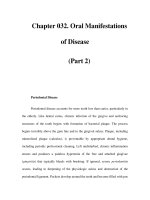
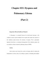
The normal ventilatory volumes and rates are summarised in Figures 5.1 and 5.2.
The
normal resting respiratory rate is 15 (range 14–20) breaths per minute. The
amount of air inspired per breath is called the
tidal volume and is equivalent to
Key components in regulating ventilation
Brain stem medullary respiratory centre
Receptors pulmonary stretch chemoreceptors for CO
2
, O
2
, H
+
Vagus and phrenic nerves increased ventilation
Respiratory muscles chest wall and diaphragm
Mechanics air passages
compliant lungs and chest wall
ACUTE MEDICAL EMERGENCIES: THE PRACTICAL APPROACH
40
05-AcuteMed-5-cpp 28/9/2000 3:38 pm Page 40
7–8 ml/kg body weight (or 500 ml for the 70 kg patient). Therefore the amount of air
inspired each minute, the
minute volume, can be calculated by multiplying the respi-
ratory rate
by the tidal volume (15 × 500 ml) to produce a value of 7·5 l/min.
The tidal volume (500 ml) is distributed throughout the respiratory system but only
350 ml (70%) mixes with alveolar air. The remainder (150 ml) occupies the airways that
are not involved in gas transfer. This volume is referred to as the
anatomical dead
space
. In addition, there are certain areas within the lungs which are also not involved
with gas transfer because they are ventilated but not perfused. The volume produced by
the combination of these areas and the anatomical dead space is called the
total or
physiological dead space
. In healthy individuals these two dead spaces are virtually
identical because ventilation and perfusion are well matched.
BREATHING ASSESSMENT
41
Figure 5.1 Normal ventilatory volumes as measured by spirometry
Tidal volume
500 ml
Minute volume
7.5 I/min
Alveolar ventilation
5250 ml/min
V/Q = 0
Pulmonary
blood flow
Anatomical dead
space 150 ml
Rate / breaths 15/min
Alveolar gas
3000 ml
Pulmonary
capillary blood
70 ml
5500 ml/min
Volumes Flows
Figure 5.2 Normal volumes and flows
05-AcuteMed-5-cpp 28/9/2000 3:38 pm Page 41
It follows that the amount of air reaching the alveoli, i.e. the alveolar ventilation, can
be calculated from:
respiratory rate × (tidal volume – anatomical dead space)
Using data from Figure 5.2, this corresponds to 15
× (500 – 150) = 5250 ml/min.
However rapid shallow respiration causes a marked reduction in alveolar ventilation
because the anatomical dead space is fixed i.e. 30
× (200 – 150) = 1500. This is demon-
strated further in Table 5.1 where the effect of different respiratory rates can be seen.
Table 5.1 The effect of respiratory rate on alveolar ventilation
Finally it is important to be aware of a crucial volume known as the functional resid-
ual capacity
(FRC) (2·5–3·0 l). This is the amount of air remaining in the lungs at the
end of a normal expiration. As 350 ml of each tidal volume is available for gas transfer,
fresh alveolar air will only replace 12–14% of the functional residual capacity. The FRC
therefore acts as a large reservoir, preventing sudden changes in blood oxygen and car-
bon dioxide concentration.
Pulmonary perfusion
At rest the cardiac output from the right ventricle is delivered to the pulmonary circulation
at approximately 5·5 l/min. As alveolar ventilation is 5·25 l/min, the ventilation:pulmonary
perfusion ratio is equal to 0·95 (5·25/5·5).
The pressures in the pulmonary vascular bed are low (around 20/9 mm Hg) and there-
fore affected by posture. As a result there are differences in blood flow to different lung
regions, contributing to the physiological dead space. In the upright position, basal
alveoli are well perfused but poorly ventilated. Consequently, in these areas, venous
blood comes into contact with alveoli filled with low concentrations of oxygen and so less
oxygen can be taken up. This effect is minimised in healthy individuals by pulmonary
vasoconstriction which diverts blood to areas of the lungs that have better ventilation.
There are also direct links between the right and left side of the heart. These normally
allow 2% of the right ventricle’s output to bypass the lungs completely and are collec-
tively known as the
physiological shunt. As the blood in this shunt has had no contact
with the alveoli, its oxygen and carbon dioxide concentrations will remain the same as
those found in the right ventricle.
Pulmonary gas exchange
Oxygen continuously diffuses out of the alveolar gas into the pulmonary capillaries with
carbon dioxide going in the opposite direction. The rate of diffusion is governed by the
following factors:
● partial pressure gradient of the gas
● solubility of the gas
● alveolar surface area
● alveolar capillary wall thickness.
Respiratory rate (/min) 10 20 30
Tidal volume (ml) 600 400 200
Anatomical dead space (ml) 150 150 150
Alveolar ventilation (ml/min) 4500 5000 1500
ACUTE MEDICAL EMERGENCIES: THE PRACTICAL APPROACH
42
05-AcuteMed-5-cpp 28/9/2000 3:38 pm Page 42
The lungs are ideally suited for diffusion as they have both a large alveolar surface area
(approximately 50 m
2
) and a thin alveolar capillary wall. It is also easy to understand why
gas exchange would be compromised by a reduction in the former (e.g. pneumothorax)
or an increase in the latter (e.g. interstitial pulmonary oedema).
Gases move passively down gradients from areas of high to low partial pressure. The
partial pressure of oxygen in the alveoli (
PAO
2
) is approximately (13·4kPa),100mmHg
whereas that in the pulmonary artery is (5·3 kPa) 40 mm Hg. In contrast the gradient for
carbon dioxide is only small, with the alveolar partial pressure being (5·3 kPa) 40 mm Hg
compared with (6·0 kPa) 46 mm Hg in the pulmonary artery. However, carbon dioxide
passes through biological membranes 20 times more easily than oxygen.The net effect is
that, in health, the time taken for exchange of oxygen and carbon dioxide is virtually
identical.
Although alveolar ventilation, diffusion and pulmonary perfusion will all affect the
alveolar
PO
2
(PAO
2
) and hence the arterial PO
2
(PaO
2
), the most important factor in
determining the
PaO
2
is the ratio of ventilation to perfusion.
Ventilation:perfusion ratio
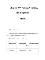
To understand this concept it is helpful to divide each lung into three functional areas:
apical, middle, and basal (Figure 5.3).
Remember that the overall ratio of ventilation to perfusion is nearly one (0·95).
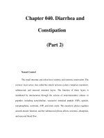
Figure 5.3 Three different ventilation (V) perfusion (Q) ratios (a) normal ventilation with reduced perfu-
sion; (b) normal ventilation with normal perfusion; (c) reduced ventilation with normal perfusion
The apical segment is well ventilated, but unfortunately poorly perfused. Therefore,
not enough blood is available to accept all the alveolar oxygen, however, the red cells that
are available are fully laden (saturated) with oxygen. Thus, the unused oxygen is simply
dissolved in the plasma.
V/Q < normal
High CO
2
content
High CO
2
content
Low O
2
content
Low O
2
content
V/Q = normal
High CO
2
content
High O
2
content
Low O
2
content
Low CO
2
content
V/Q > normal
High CO
2
content
Low O
2
content
Slightly > normal O
2
content
Very low CO
2
content
BREATHING ASSESSMENT
43
(a)
(c)
(b)
05-AcuteMed-5-cpp 28/9/2000 3:38 pm Page 43
The middle segment has ventilation and perfusion perfectly matched. Alveolar oxygen
diffuses into, and is correctly balanced by, the pulmonary capillary blood ensuring that
the red cells are fully saturated. The remaining small amount of oxygen is dissolved in
plasma.
The basal segment alveoli are well perfused, but poorly ventilated. All the available
oxygen is bound to red cells but they are not fully saturated, i.e. there is spare oxygen car-
rying capacity.This is similar to the physiological shunt as described earlier.
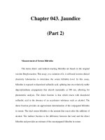
The oxygen content of blood at point X (Figure 5.4) depends on the mixture of blood
coming from all three parts of the lung. The final value is not simply the mid point
between a and c. This is because the
small amount of additional oxygen dissolved in the
plasma cannot offset the
massive decrease in oxygen content produced by the incom-
pletely saturated haemoglobin molecules in part c.Therefore the oxygen content is much
lower than half way between the values of a and c.
Figure 5.4 Mixed blood returning from three sites at point X
V/Q = normal (from (b))
V/Q > normal (from (a))
Low CO
2
content
Slightly increased O
2
content
V/Q < normal (from (c))
High CO
2
content
Very low O
2
content
Normal CO
2
content
Low O
2
content
X
Key point
An area of lung with a high V:Q ratio cannot offset the fall in oxygen content produced by an area
of lung with a low V:Q ratio
Tip
In the basal segment ventilation is reduced when compared to perfusion, i.e., the V:Q ratio < 1
Tip
In the middle segment ventilation and perfusion are matched, i.e., the V:Q ratio = 1
Tip
In the apical segment there is more ventilation than perfusion, i.e., the V:Q ratio > 1
ACUTE MEDICAL EMERGENCIES: THE PRACTICAL APPROACH
44
05-AcuteMed-5-cpp 28/9/2000 3:38 pm Page 44
Oxygen content of arterial blood
The oxygen content of haemoglobin (Hb) going to tissues depends on the:
● saturation of haemoglobin with oxygen
● haemoglobin concentration
● oxygen carrying capacity
● oxygen dissolved in plasma.
Haemoglobin is a protein comprising four subunits, each of which contains a haem
molecule attached to a polypeptide chain. The haem molecule contains iron which
reversibly binds oxygen; hence it is oxygenated but
not oxidised. Each haemoglobin
molecule can carry up to four oxygen molecules. Blood has a haemoglobin concentration
of approximately 15 g/100 ml, and normally each gram of haemoglobin can carry
1·34 ml of oxygen if it is fully saturated. Therefore the
oxygen carrying capacity of
blood is:
Hb × 1·34 × 1
15
× 1·34 × 1 = 20·1 ml O
2
/100 ml of blood
(A value of one indicates that Hb is fully saturated.)
This is approximately 60 times greater than the amount of oxygen dissolved in plasma.
The relationship between the
PaO
2
and oxygen uptake by haemoglobin is not linear,
because the addition of each O
2
molecule facilitates the uptake of the next O
2
molecule.
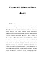
This produces a sigmoid shaped oxygen dissociation curve (Figure 5.5). Furthermore,
because haemoglobin is 97·5% saturated at a
PaO
2
of 100 mm Hg (13·4 kPa) (i.e. that
found in the normal healthy state), increasing the
PaO
2
further has little effect on oxygen
transport.
Figure 5.5 Percentage of oxygen saturation of haemoglobin
Percentage O
2
oxygen saturation
of haemoglobin
97.5
83.5
10
0
0 1.3 6.6 13
PaO
2
(kPa)
Key point
Nearly all of the oxygen carried in the blood is taken up by haemoglobin with only a small amount
dissolved in the plasma
BREATHING ASSESSMENT
45
05-AcuteMed-5-cpp 28/9/2000 3:38 pm Page 45
The affinity of haemoglobin for oxygen at a particular PO
2
(commonly known as the
O
2
–Hb association) is also affected by other factors. A decreased affinity means that oxy-
gen is more readily released. Thus the oxygen dissociation curve is shifted to the right.
This is caused by:
● ↑ hydrogen ion concentration (fall in pH)
● ↑ PaCO
2
● ↑ concentration of red cell 2,3-diphosphoglycerate (2,3-DPG)
● ↑ temperature.
(The opposite of these factors increases the affinity and these will be discussed later.)
The normal haemoglobin concentration (as measured by the haematocrit) is usually
just above the point at which the oxygen transportation is optimal. Consequently a slight
fall in haemoglobin concentration will actually increase oxygen transportation by
decreasing blood viscosity.
In addition to the oxygen combined with haemoglobin, there is also a smaller amount
dissolved in plasma. This amount is directly proportional to the
PaO
2
and is approxi-
mately 0·003 ml/100 ml blood/mm Hg of
PaO
2
.
It follows from the description above that the total content of oxygen in blood is equal
to the oxygen associated with haemoglobin and that dissolved in plasma.
Oxygen blood concentration = (Hb
× 1·34 × saturation) + (0·003 × PaO
2
)
For example, in arterial blood with a haemoglobin content of 15 g and a
PaO
2
of
100 mm Hg the oxygen content would be:
(15
× 1·34 × 97·5%) + (0·003 × 100) = 19·8 ml/100 ml
Alternatively, in venous blood with a haemoglobin content of 15 g and a
PaO
2
of
40 mm Hg the oxygen content would be:
(15
× 1·34 × 75%) + (0·003 × 40) = 15·2 ml/100 ml
Airway
This has been described in detail in Chapter 4.The following summary contains the rele-
vant facts relating to the breathless patient.
Assessment
Most breathless patients will have a patent airway. The number of words said with each
breath is a useful indicator of illness severity and the effects of treatment. If the patient
can count to 10 in one breath, then the underlying condition is unlikely to warrant
immediate intervention. Occasionally, however, the patient will be severely distressed
with stridor, possibly coughing and making enormous but ineffectual respiratory efforts.
Stridor is a sinister sign and should be regarded as indicating impending airway obstruc-
tion.
Resuscitation
High flow oxygen (FiO
2
= 0·85) may relieve some of the patient’s distress. If airway
obstruction is suspected, immediate review by an anaesthetist is required. If, however,
ACUTE MEDICAL EMERGENCIES: THE PRACTICAL APPROACH
46
05-AcuteMed-5-cpp 28/9/2000 3:38 pm Page 46
further history is forthcoming that a foreign body has been inhaled then a Heimlich or
modified Heimlich manoeuvre should be attempted. In contrast, if the patient has a res-
piratory arrest then examine the larynx with a laryngoscope and remove any identifiable
foreign body. If this is impossible proceed to needle cricothyroidotomy and jet insuffla-
tion followed by formal cricothyroidotomy.
Breathing
Assessment
This is summarised in the box.
The immediately life threatening conditions were identified in the primary assessment of
breathing earlier.
Specific clinical features
By the time “B” is assessed all breathless patients should have received high flow oxygen
(FiO
2
= 0·85 at 15 l/min). Do not be concerned about patients who retain CO
2
.
Providing that FiO
2
equals 0·85, a rise in PaCO
2
will not increase mortality – but
untreated hypoxaema will! After the primary assessment has been completed then the
FiO
2
can be titrated according to the arterial blood gas results or the pulse oximeter
reading.
A hyperinflated chest is indicative of asthma or chronic airflow limitation (COPD). In
an acute exacerbation of these conditions the trachea moves downwards during inspira-
tion.This is referred to as tracheal tug and implies airway obstruction or increased respi-
ratory effort. In addition, the internal jugular pressure may be elevated and accessory
muscle use will be prominent, as will intercostal recession over the lower part of the chest
during inspiration. Patients often adopt a seated or standing posture to facilitate respira-
tion.
Although bronchospasm is common to both asthma and COPD, in acute asthma the
inspiratory phase is snatched and expiration is prolonged. With chronic airflow limita-
tion, however, the clinical picture ranges widely from a patient with preserved respiratory
drive with pursed-lip breathing to one who is cyanosed, lethargic, and mildly dyspnoeic.
Wheezes may be heard on inspiration, but especially on expiration.
Acute pulmonary oedema can mimic or coexist with either of these conditions. The
commonest cause of pulmonary oedema is left ventricular failure associated with
ischaemic heart disease. Although these are many other causes, these will be seen only
occasionally in most hospitals.
Summary of breathing assessment
● Look colour, sweating
posture
respiratory rate, effort
symmetry
● Feel tracheal position
tracheal tug
chest expansion
● Percuss
● Listen
BREATHING ASSESSMENT
47
05-AcuteMed-5-cpp 28/9/2000 3:38 pm Page 47
An idea of the “chance” of meeting such a condition is displayed on an arbitrary scale
in the box.
* Patients with some of the more common causes of pulmonary oedema may also
feature in examinations.
However, features that would support a diagnosis of pulmonary oedema include
absence of both neck vein distension and chest hyperexpansion. In addition, the percus-
sion note is often dull, particularly at the lung bases, and there are usually fine inspira-
tory crackles on auscultation. Occasionally, signs of a pleural effusion may also be
evident.
A deviated trachea (a very late sign) should alert the clinician to the possibility of a
tension pneumothorax. Other signs, in particular raised neck veins, a hyperresonant per-
cussion note and absent breath sounds, should be sought on the opposite side to the tra-
cheal deviation.
Resuscitation
Irrespective of the underlying cause of the bronchospasm, treat the patients with nebu-
lised bronchodilators whilst clues to the underlying diagnosis are sought.The clinical fea-
tures described above will have helped distinguish bronchospasm due to asthma, COPD
or pulmonary oedema.
Immediate management of a tension pneumothorax is needle thoracentesis followed
by intravenous access and then chest drain insertion.
TIME OUT 5.1
aDefine (i)i tidal volume
(ii) minute volume
b(i) How does the respiratory rate affect alveolar ventilation?
b(ii) Sketch a graph showing the relationship between PaO
2
and % SaO
2
.
c List the immediately life threatening conditions that affect “B”.
Causes of acute pulmonary oedema and “chance” of meeting the condition*
Cause Chance
Ischaemic heart disease Daily
Myocardial infarction
Cardiac dysrhythmias
Fluid overload Weekly
Severe hypertension
Aortic stenosis/regurgitation Monthly
Mitral stenosis/regurgitation
Cardiomyopathy
Non-cardiac
Left ventricular aneurysm Annually
Infective endocarditis
Cardiac tamponade
Left atrial myxoma Only in examinations
ACUTE MEDICAL EMERGENCIES: THE PRACTICAL APPROACH
48
05-AcuteMed-5-cpp 28/9/2000 3:38 pm Page 48
SUMMARY
Breathing is rapidly assessed using the look, feel, percuss, and listen sequence to identify
life threatening:
● bronchospasm
● pulmonary oedema
● tension pneumothorax.
BREATHING ASSESSMENT
49
05-AcuteMed-5-cpp 28/9/2000 3:38 pm Page 49
This Page Intentionally Left Blank
CHAPTER
6
Circulation assessment
OBJECTIVES
After reading this chapter you will be able to:
● understand the physiology of tissue perfusion
● describe a structured approach to circulatory assessment
● identify the immediately life threatening causes of shock
● identify the anatomy for peripheral and central venous cannulation.
INTRODUCTION
The immediately life threatening ones are shown in the box. Clinical features are used to
assess circulation that can be affected by a variety of conditions.
Therefore it is important to understand the mechanisms that maintain tissue perfusion
in health before considering the effects of disrupting the circulation.
Immediately life threatening conditions
Airway
● Obstruction
Breathing
● Acute severe asthma
● Acute exacerbation of chronic obstructive pulmonary disease (COPD)
● Pulmonary pneumothorax
● Tension oedema
Circulation
● Shock
51
Reading: 30 minutes
06-AcuteMed-6-cpp 28/9/2000 3:41 pm Page 51
Relevant physiology
Blood flow to the tissues
The amount of blood reaching a particular organ depends on several factors:
● venous system
● cardiac output
● arterial system
● organ autoregulation.
Venous system
This is capable of acting as a reservoir for over 70% of the circulating blood volume and
is therefore often referred to as a
capacitance system. The amount of blood stored at
any one time depends on the size of the vessel lumen. This is controlled by sympathetic
innervation and local factors (see later) which can alter the tone of the vessel walls. If the
veins dilate, more blood remains in the venous system and less returns to the heart.
Should there be a requirement to increase venous return, sympathetic stimulation
reduces the diameter of the veins and hence the capacity of the venous system. A change
from minimal to maximal tone can increase the venous return by approximately one litre
in an adult.
Cardiac output
This is defined as the volume of blood ejected by each ventricle per minute. Clearly, over
a period of time, the output of the two ventricles must be the same (or else all the circu-
lating volume would eventually end up in either the systemic or pulmonary circulation).
Thus, the cardiac output equates to the volume of blood ejected with each beat (stroke
volume in ml) multiplied by the heart rate (beats per minute) and is expressed in litres
per minute.
Cardiac output = stroke volume
× heart rate = 4–6 l/min (normal adult)
To allow a comparison between patients of different sizes, the cardiac index (CI) is
used. This is the cardiac output divided by the surface area of the person and hence is
measured in litres per square metre.
Cardiac index = cardiac output/body surface area = 2·8–4·2 l/min/m
2
(normal adult)
The cardiac output can be affected by:
● preload
● myocardial contractility
● afterload
● heart rate.
Preload This is the volume of blood in the ventricle at the end of diastole.The left ven-
tricular end diastolic volume (LVEDV) is about 140 ml and the stroke volume (SV) is
90 ml. Therefore the end systolic volume is approximately 50 ml and the left ventricular
ejection fraction (SV/EDV) ranges from 50 to 70%.
During diastole, the cardiac muscle fibres are progressively stretched as the ventricu-
lar volume increases in proportion to the venous return. Remember that
the more the
myocardial fibres are stretched during diastole, the more forcibly they contract
during systole; hence more blood will be expelled
(Starling’s Law). Therefore, the
ACUTE MEDICAL EMERGENCIES: THE PRACTICAL APPROACH
52
06-AcuteMed-6-cpp 28/9/2000 3:41 pm Page 52
greater the preload, the greater the stroke volume. However, this phenomenon has an
upper limit (due to the internal molecular structure of muscle cells) so that if the muscle
is stretched beyond this point then a smaller contraction is produced.
Thus, the end diastolic fibre length is proportional to the end diastolic volume or force
distending the ventricle. A clinical estimate of this volume, or force, is the end diastolic
pressure (EDP). As the ventricular end diastolic pressure (LVEDP) increases so does the
stroke volume. If the end diastolic pressure exceeds a critical level then the force of con-
traction declines and eventually ventricular failure ensues (Figure 6.1).
Current haemodynamic monitoring is based upon measurements from a pulmonary
artery flotation catheter (PAFC). A commonly used recording is the pulmonary artery
occlusion pressure (PAOP) because it is considered a useful estimate of the left ventric-
ular end diastolic pressure.
Myocardial contractility This is the rate at which the myocardial fibres contract for a
given degree of stretch. Substances affecting myocardial contractility are termed
inotropes, and they can be positive or negative in their actions. A positive inotrope pro-
duces a greater contraction for a given length (or EDP clinically) (Figure 6.1).
Adrenaline, noradrenaline and dopamine are naturally produced substances which have
this effect. Dobutamine is a synthetic catecholamine with positive inotropic activity.
Therefore, depending on where you work, you may find some (or all) of these agents are
used to treat cardiogenic and septic shock.
Figure 6.1 Ventricular performance
Negative inotropes reduce contractility for a given muscle length (Figure 6.1). These
substances are often drugs, for example, antiarrhythmics and anaesthetic agents. Many
of the physiological states produced by shock will also depress contractility, for example,
hypoxia, acidosis, and sepsis. Myocardial damage also has a negative inotropic effect.
Afterload As the left and right ventricular muscle contracts, pressures within the cham-
bers increase until they exceed those in the aorta and pulmonary artery, respectively.The
Left ventricular end diastolic pressure
Ventricular
performance
Positive inotropic effect
Normal curve
Negative
inotropic effect
CIRCULATION ASSESSMENT
53
06-AcuteMed-6-cpp 28/9/2000 3:41 pm Page 53
aortic and pulmonary valves open and the blood is ejected. The resistance faced by the
ventricular myocardium during ejection is termed the afterload. In the left ventricle this
is mainly due to the resistance offered by the aortic valve and the compliance (stiffness)
of the arterial blood vessels. Usually this latter component is the most important and is
estimated by measuring the
systemic vascular resistance (SVR).
Using Ohm’s law where resistance equals pressure divided by flow, the systemic vas-
cular resistance is defined as the mean arterial and venous pressure difference divided by
the cardiac output.
(mean arterial pressure – central venous pressure)
× 80/cardiac output =
770 – 1500 dyn/s/cm
5
(normal adult)
(The value of 80 comes from converting mm Hg to SI units.)
Heart rate An increase in heart rate is mediated via β
1
adrenoreceptors. These can be
stimulated by the sympathetic nervous system (SNS), the release of catecholamines from
the adrenal medulla and drugs (e.g. Isoprenaline). This is termed a
positive
chronotropic effect
. Conversely, the parasympathetic nervous system (PSNS) supplies
the sino-atrial node and atrioventricular node via the vagus nerve. Stimulation of the
PSNS decreases heart rate, i.e. it has a
negative chronotropic effect. This effect can
also be produced by drugs that inhibit the sympathetic nervous system such as
β block-
ers. In contrast, an increased heart rate may follow inhibition of the parasympathetic ner-
vous system muscarinic (M) receptors.
An increase in heart rate can lead to an increase in cardiac output (see equation
earlier). However, ventricular filling occurs during diastole and this phase of the cardiac
cycle is predominantly shortened as the heart rate increases. A sinus tachycardia, above
160 beats/minute in the young adult, drastically reduces the time for ventricular filling.
This leads to a progressively smaller stroke volume and a fall in cardiac output.The crit-
ical heart rate when this occurs is also dependent on the age of the patient and the con-
dition of the heart; for example, rates over 120 beats/minute may cause inadequate filling
in the elderly.
The main factors affecting the cardiac output of the left ventricle are summarised in
the box.
Main factors affecting cardiac output
● Preload, or left ventricular end diastolic volume
● Myocardial contractility
● Afterload, or systemic vascular resistance
● Heart rate
Key point
Increasing the heart rate will only lead to rise in the cardiac output if the rate is below a critical level
Key point
Reducing the afterload for a given preload will allow the myocardial fibres to shorten more quickly
and by a greater amount. It therefore increases the stroke volume and cardiac output
ACUTE MEDICAL EMERGENCIES: THE PRACTICAL APPROACH
54
06-AcuteMed-6-cpp 28/9/2000 3:41 pm Page 54
The arterial system
The walls of the aorta and other large arteries contain relatively large amounts of elastic
tissue that stretches during systole and recoils during diastole. In contrast, the walls of
arterioles contain relatively more smooth muscle. This is innervated by the sympathetic
nervous system that maintains vasomotor tone to a large extent. Stimulation of
α
adrenoreceptors causes vasoconstriction. Therefore, a total loss of arterial tone would
increase the capacity of the circulatory system so much that the total blood volume
would be insufficient to fill it. As a consequence, the blood pressure would fall and the
flow through organs would depend upon their resistance. Some organs would receive
more than normal amounts of oxygenated blood (e.g. skin) at the expense of others
which would receive less (e.g. brain). To prevent this, the arterial system is under con-
stant control by sympathetic innervation and local factors to ensure that blood goes
where it is needed most. This is exemplified in the shocked patient where differential
vasoconstriction maintains supply to the vital organs (e.g. heart) at the expense of others
(e.g. skin). Hence the skin is cold and pale.
Systemic arterial blood pressure
This is the pressure exerted on the walls of the arterial blood vessels. Systolic pressure is
the maximal pressure generated in the large arteries during each cardiac cycle. In con-
trast the diastolic pressure is the minimum. The difference between them is the pulse
pressure. The
mean arterial pressure is the average pressure during the cardiac cycle
and is approximately equal to the diastolic pressure plus one third of the pulse pressure.
As the mean arterial pressure is the product of the cardiac output and the systemic vas-
cular resistance, it is affected by all the factors already discussed.
Autoregulation
Organs have a limited ability to regulate their own blood supply so that perfusion is
maintained as blood pressure varies. This process is known as autoregulation and is
brought about by the presence of smooth muscle in the arteriolar walls. By altering the
calibre of the vessels, flow to the organs is maintained. Furthermore, other local factors,
such as products of anaerobic metabolism, acidosis and a rise in temperature, all cause
the local vascular tree to dilate. Such effects enable active tissues to receive increased
quantities of nutrients and oxygenated blood.
PRIMARY ASSESSMENT AND RESUSCITATION
A summary of the circulatory assessment is shown in the box.
Blood volume
● Adult male = 70 ml per kilogram ideal body weight
● Adult female = 60 ml per kilogram ideal body weight
CIRCULATION ASSESSMENT
55
06-AcuteMed-6-cpp 28/9/2000 3:41 pm Page 55
The aim of this brief assessment is to identify the patient who is shocked.This is a clinical
syndrome resulting from inadequate delivery, or use, of essential substrates (e.g. oxygen)
by vital organs. The causes of shock are described in detail in Chapter 11 and sum-
marised in the next box.
Specific clinical features
All patients with respiratory distress will have a tachycardia as described in the previous
chapter. With severe airways obstruction, however, pulsus paradoxus may be present.
Normally there is a reduction in systolic blood pressure of up to 10 mm Hg on inspira-
tion.This is attributed to a fall in intrathoracic pressure (i.e. it becomes more negative on
inspiration) which enlarges the pulmonary vascular bed and reduces return of blood to
the left ventricle. There is partial compensation by a simultaneous increase in right ven-
tricular output. In severe asthma and COPD there is a more substantial fall in intra-
thoracic pressure on inspiration. This greatly increases the capacity of the pulmonary
vascular bed that in turn reduces output from the left ventricle, resulting in pulsus para-
doxus. This is an exaggeration of the
normal systolic fall on inspiration and not a
paradoxical change in the pulse as the name would imply.The abnormality is the extent
by which the arterial pressure falls. If severe, the pulse may disappear on inspiration and
this can easily be palpated at the radial artery. In contrast, if the fall in systolic pressure
is not so marked, it can be detected using the sphygmomanometer. This physical sign
indicates critical circulatory embarrassment and can also occur in patients with cardiac
tamponade.
Another pulse abnormality is pulsus alternans where evenly spaced beats (in time) are
alternately large and small in volume. As this can indicate left ventricular failure the clin-
ician should check for corroborative signs such as a displaced apex beat, a third heart
sound and a pansystolic murmur of mitral regurgitation. A third heart sound in patients
over 40 years usually indicates elevated ventricular end diastolic pressure. With increas-
ing age, the myocardium and associated valvular structures become less compliant, i.e.
stiffer. Thus, an increase in end diastolic pressure is needed to ensure adequate ventric-
Causes of shock
Preload reduction – hypovolaemia – haemorrhage
– diarrhoea
– impaired return – pregnancy
– severe asthma
Pump failure – endocardial – acute valve lesion
– myocardial – infarction
– inflammation
– epicardial – tamponade
Post (after) load reduction – vasodilation – sepsis
– anaphylaxis
Summary of circulatory assessment
● Look: pallor, sweating, venous pressure
● Feel: pulse – rate, rhythm, and character
capillary refill time
blood pressure
apex beat
● Listen: heart sounds, extra sounds
ACUTE MEDICAL EMERGENCIES: THE PRACTICAL APPROACH
56
06-AcuteMed-6-cpp 28/9/2000 3:41 pm Page 56
ular filling during which sudden tension in these structures generates vibrations which
correspond to the third heart sound.
Shock associated with a dysrhythmia is due to either pulmonary oedema or
hypotension or a combination of these conditions. Under these circumstances tachy-
dysrhythmias, irrespective of the QRS complex width, will require cardioversion (see
Chapter 31). Unfortunately, atrial fibrillation may either fail to cardiovert (especially
when chronic) or only transiently return to sinus rhythm. The remaining options
include:
● chemical cardioversion. Of the many potential drugs available, intravenous amio-
darone is well tolerated. Flecainide is an excellent alternative, but has been shown to
increase mortality in patients with ischaemic heart disease.
● control the ventricular response with intravenous digoxin.
In contrast, a patient with a bradydysrhythmia may require temporary support with
either atropine and an inotrope (e.g. isoprenaline) or external pacing whilst preparations
are made for transvenous pacing (see Chapter 9 on Shock for further details).
Hypovolaemia, commonly due to blood loss, can present with tachypnoea and a
variety of other physical signs including tachycardia, hypotension and reduced urine
output.
The remaining two immediately life threatening causes of breathlessness are pul-
monary embolus and cardiac tamponade. The size and position of the embolus will
determine the haemodynamic effects. Non-fatal emboli blocking the major branches of
the pulmonary artery (PA) provoke a rise in PA pressure due to hypoxia and vasocon-
striction. In addition, tachypnoea follows stimulation of alveolar and capillary receptors.
An acute increase in pulmonary vascular resistance and hence right ventricular afterload
causes a sudden rise in end diastolic pressure and dilatation of the right ventricle. This
produces a raised jugular venous pressure, a fall in systemic arterial pressure and a com-
pensatory tachycardia.
The signs of cardiac tamponade include pulsus paradoxus, raised internal jugular
venous pressure that increases on inspiration (the opposite of normal; Kussmaul’s sign),
and an impalpable apex beat. As fluid accumulates, the elevated pressure in the pericar-
dial sac is raised further during inspiration (this may be related to the downward dis-
placement of the diaphragm). A corresponding increase is seen in the right atrial and
central venous pressures. In contrast, pressures on the left side of the heart may be lower
than that in the pericardium. As a consequence, filling of the left ventricle is compro-
mised, stroke volume is reduced and the interventricular septum bulges into the left ven-
tricular cavity.Thus, the stroke volume of the right ventricle is maintained at the expense
of the left ventricle which collapses on inspiration. With further increases in pericardial
pressure there is diastolic collapse of the right atrium and ventricle.The venous pressure
is always raised and is due to abnormal right heart filling. Kussmaul’s sign can also be
seen in right ventricular disease and pulmonary hypertension.
Treatment
All patients should receive high flow oxygen and have their oxygen saturation, pulse,
blood pressure, and cardiac rhythm monitored. Intravenous access is needed and at least
one large venflon (12–14 gauge) is required in the antecubital fossa.
Hypovolaemia
In acute hypovolaemia a fluid challenge can then be given whilst the cause, usually haem-
orrhage, is sought (see Chapter 9). In contrast, chronic fluid depletion often presents as
dehydration with features of acute renal impairment. Oxygen and careful fluid replace-
ment are required, especially in patients with preexisting cardiac conditions. Diuretics
CIRCULATION ASSESSMENT
57
06-AcuteMed-6-cpp 28/9/2000 3:41 pm Page 57
and angiotensin converting enzyme (ACE) inhibitors are a common cause of this prob-
lem in patients with a history of left ventricular failure. These drugs should be stopped
and fluid replacement titrated against the patient’s clinical condition and central venous
pressure.
Acute severe left ventricular failure
The blood pressure is probably the most important feature in determining treatment.
The combination of acute pulmonary oedema and hypotension demands inotropic
support. Any patient who has a systolic pressure of
less than 90 mm Hg should not be
given diuretics, nitrates or opiates as their immediate action is to cause venodilatation.
This, in turn, will reduce the cardiac preload and potentially exacerbate hypotension.
Dysrhythmia – tachycardia
The presence of a tachydysrhythmia in the shocked or compromised patient necessitates
electrical cardioversion (see Chapter 28). If this fails then drug treatment according to
UK and European resuscitation committee guidelines is advocated (Figure 6.2).
Remember that a sinus tachycardia can be the response of a failing ventricle. However, if
the patient has another baseline rhythm such as atrial fibrillation, the increased sympa-
thetic drive would result in atrial fibrillation with a rapid ventricular response. It can be
difficult to decide whether a dysrhythmia is the cause of heart failure or vice versa.
Previous ECGs are invaluable in this circumstance. If no such information is available
the treatment is dictated by the clinician’s judgement.The following key points can help
in this dilemma.
● A supraventricular tachycardia with a ventricular response of less than 150 is unlikely
to cause failure.
● A broad complex tachycardia is almost always ventricular in a patient with ischaemic
heart disease.
Dysrhythmia – bradycardia
This is treated according to UK and European resuscitation committee guidelines
(Figure 6.3).
TIME OUT 6.2
In a patient with a bradycardia, what are the risk factors for asystole?
If you cannot answer this question copy the bradycardia management flow diagram
(Figure 6.3) to reinforce your knowledge.
TIME OUT 6.1
Ensure that you have a sound understanding of this protocol (Figure 6.2). If neces-
sary take five minutes and copy the tachydysrhythmia management flow diagram to
reinforce your knowledge.
ACUTE MEDICAL EMERGENCIES: THE PRACTICAL APPROACH
58
06-AcuteMed-6-cpp 28/9/2000 3:41 pm Page 58
Pulmonary embolus
The minimum immediate management comprises high flow oxygen and anticoagulation.
More comprehensive treatment details are provided in Chapter 10.
Cardiac tamponade
If this diagnosis is suspected clinically, then intravenous fluid should be administered to
raise the end diastolic pressure and volume in order to maintain the cardiac output.This
is only a temporising procedure and immediate cardiological referral is required for
echocardiography and pericardiocentesis.
CIRCULATION ASSESSMENT
59
Figure 6.2 Management of a tachydysrhythmia (UK and European guidelines)
06-AcuteMed-6-cpp 28/9/2000 3:41 pm Page 59
Investigations
Appropriate investigations at this stage include:
● a full blood count to exclude anaemia (possibly exacerbating left ventricular failure)
● urea and electrolytes for baseline values particularly in patients who are being treated
with vasodilators, diuretics or inotropes
● cardiac enzymes
● arterial blood gases
● 12 lead ECG
● portable chest X-ray.
Key point
If the patient is still breathless and the cause remains in doubt a rapid reevaluation A, B and C is
required
It is important to remember that hypovolaemia is an important cause of breathlessness
ACUTE MEDICAL EMERGENCIES: THE PRACTICAL APPROACH
60
Figure 6.3 Management of a bradydysrhythmia (UK and European guidelines)
06-AcuteMed-6-cpp 28/9/2000 3:41 pm Page 60
Once the patient’s condition is stabilised then further information can be obtained
from the secondary assessment.
Summary
In the primary assessment, the immediately life threatening problems are:
● airway obstruction
● breathing acute severe asthma
acute exacerbation of COPD
pulmonary oedema
tension pneumothorax
● circulation shock
These conditions can be identified and differentiated clinically.
All patients require oxygen and intravenous access.
PERIPHERAL VENOUS CANNULATION
The antecubital fossa is the commonest site for peripheral venous cannulation.
The cephalic vein passes through the antecubital fossa on the lateral side and the
basilic vein enters very medially just in front of the medial epicondyle of the elbow.These
two large veins are joined by the
median cubital or antecubital vein.The median vein
of the forearm also drains into the basilic vein (Figure 6.4).
Although the veins in this area are prominent and easily cannulated, there are many
other adjacent vital structures which can be easily damaged.
The most popular device for peripheral intravenous access is the cannula over needle,
available in a wide variety of sizes, 12–27 gauge (g). It consists of a plastic (PTFE or
similar material) cannula which is mounted on a smaller diameter metal needle, the bevel
of which protrudes from the cannula. The other end of the needle is attached to a trans-
parent “flashback chamber”, which fills with blood indicating that the
needle bevel lies
within the vein. Some devices have flanges or “wings” to facilitate attachment to the skin.
All cannulae have a standard luer-lock fitting to attach a giving set and some have a
valved injection port attached through which drugs can be given.
Complications
● Failed cannulation is the most common, usually as a result of pushing the needle
completely through the vein. It is inversely related to experience.
● Haematomata are usually secondary to the above with inadequate pressure applied to
prevent blood leaking from the vein after the cannula is removed. They are made
worse by forgetting to remove the tourniquet!
● Extravasation of fluid or drugs is commonly a result of failing to recognise that the
cannula is not in the vein before use. Placing a cannula over a joint or prolonged use
to infuse fluids under pressure also predisposes to leakage. The faulty cannula must
be removed. Damage to the surrounding tissues will depend primarily on the nature
of the extravasated fluid.
TIME OUT 6.3
List the major causes of shock.
CIRCULATION ASSESSMENT
61
06-AcuteMed-6-cpp 28/9/2000 3:41 pm Page 61