Basic medical endocrinology - part 1 ppsx
Bạn đang xem bản rút gọn của tài liệu. Xem và tải ngay bản đầy đủ của tài liệu tại đây (708.57 KB, 48 trang )
xvii
PREFACE
Nearly a decade elapsed between publication of the second and third editions
of Basic Medical Endocrinology due in large part to the turmoil in the publishing
industry brought on by massive consolidation.Although this edition is new and the
publisher is new, the aims of earlier editions of this work are unchanged. Its focus
remains human endocrinology with an emphasis on cellular and molecular
mechanisms presented in the context of integration of body functions.The intent
is to provide a sufficient level of understanding of normal endocrine physiology
to prepare students to study not only endocrine diseases, but also the cellular and
molecular alterations that disrupt normal function. Such understanding is a
prerequisite for institution of rational diagnostic procedures, therapeutic inter-
ventions, and research strategies. It is further hoped that this text provides the
necessary background to facilitate continuing self-education in endocrinology.
A decade is a long time in this remarkable era of modern biology. Whole
new vistas of inquiry have been opened since the previous edition of this text
appeared, and new discoveries have mandated reinterpretation of many areas that
were once thought to be solidly understood. Much of the progress of the past
decade must be credited to ingenious application of rapidly evolving technology
in molecular biology. Studies of gene expression and the charting of the genomes
of several species, including our own, has provided a deluge of new information
and new insights. The exquisite sensitivity and versatility of this technology has
uncovered both hormone production and hormone receptors in unexpected
places and revealed hitherto unappreciated roles for classical hormones. Classical
techniques of organ ablation and extract injection have been reapplied using the
once unthinkable technology of gene ablation or overexpression to explore the
functions of individual proteins instead of individual glands. The decade has
also witnessed the discovery of new hormones and expanded our appreciation of
the physiological importance of extraglandular metabolism of hormones.
The understanding of hormone actions has grown enormously and spawned the
quest for “designer drugs” that target particular, critical, biochemical reactions in
combating disease.
In light of these and many other developments, every chapter of this text has
been extensively revised to present the well-established factual basis of endocrinol-
ogy enriched by exciting, rapidly unfolding new information and insights. The
challenge has been to digest and reduce the massive literature to illuminate the
regulatory and integrative roles of the endocrine system without overloading
the text with arcane detail. However, the text is designed to provide somewhat
more than the minimum acceptable level of understanding and attempts to antic-
ipate and answer some of the next level of questions that might occur to the
thoughtful student.
Looking back over 40 years of teaching endocrine physiology, one cannot
fail but to marvel at how far we have come and how resourceful is the human mind
in probing the mysteries of life. As has always been true of scientific inquiry,
obtaining answers to long-standing questions inevitably raises a host of new
questions to challenge a new generation of endocrinologists. It is my hope that this
text will provide a foundation for students to meet that challenge both in the clinic
and in the laboratory.
H. Maurice Goodman
2002
xviii Preface
xix
PREFACE TO THE
FIRST EDITION
This volume is the product of more than 25 years of teaching endocrine physiol-
ogy to first-year medical students. Its focus is human endocrinology with an
emphasis on cellular and molecular mechanisms. In presenting this material, I have
tried to capture some of the excitement of a dynamic, expanding discipline that is
now in its golden age. It is hoped that this text provides sufficient understanding
of normal endocrine physiology to prepare the student to study not only
endocrine diseases but the cellular and molecular derangements that disrupt
normal function and must therefore be reversed or circumvented by rational
therapy. It is further hoped that this text provides the necessary background to
facilitate continuing self-education in endocrinology.
Endocrinology encompasses a vast amount of information relating to at least
some aspect of virtually every body function. Unfortunately, much of the infor-
mation is descriptive and cannot be derived from first principles.Thorough, ency-
clopedic coverage is neither appropriate for a volume such as this one nor possible
at the current explosive rate of expansion. On the other hand, limiting the text to
the bare minimum of unadorned facts might facilitate memorization of what
appear to be the essentials this year but would preclude acquisition of real under-
standing and offer little preparation for assimilating the essentials as they may
appear a decade hence. I therefore sought the middle ground and present basic facts
within enough of a physiological framework to foster understanding of both the
current status of the field and those areas where new developments are likely to
occur while hopefully avoiding the pitfall of burying key points in details and
qualifications.
The text is organized into three sections. The first section provides basic
information about organization of the endocrine system and the role of individual
endocrine glands. Subsequent sections deal with complex hormonal interactions
that govern maintenance of the internal environment (Part II) and growth and
reproduction (Part III). Neuroendocrinology is integrated into discussions of specific
glands or regulatory systems throughout the text rather than being treated as a
separate subject. Although modern endocrinology has its roots in gastrointestinal
(GI) physiology, the gut hormones are usually covered in texts of GI physiology
rather than endocrinology; therefore, there is no chapter on intestinal hormones.
In the interests of space and the reader’s endurance, a good deal of fascinating
material was omitted because it seemed either irrelevant to human biology or
insufficiently understood at this time. For example, the pineal gland has intrigued
generations of scientists and philosophers since Descartes, but it still has no clearly
established role in human physiology and is therefore ignored in this text.
Human endocrinology has its foundation in clinical practice and research,
both of which rely heavily on laboratory findings.Where possible, points are illus-
trated in the text with original data from the rich endocrine literature to give the
reader a feeling for the kind of information on which theoretical and diagnostic
conclusions are based. Original literature is not cited in the text, in part because
such citations are distracting in an introductory text, and in part because proper
citation might well double the length of this volume. For the reader who wishes
to gain entrée to the endocrine literature or desires more comprehensive coverage
of specific topics, review articles are listed at the end of each chapter.
H. Maurice Goodman
1988
xx Preface to the First Edition
xxi
PREFACE TO THE
SECOND EDITION
In the five years that have passed since the first edition of this text, the informa-
tion explosion in endocrinology has continued unabated and may have even accel-
erated.Application of the powerful tools of molecular biology has made it possible
to ask questions about hormone production and action that were only dreamed
about a decade earlier. The receptor molecules that initiate responses to virtually
all of the hormones have been characterized and significant progress has been
made in unraveling the events that lead to the final cellular expression of
hormonal stimulation. As more details of intracellular signaling emerge, the com-
plexities of parallel and intersecting pathways of transduction have become more
evident.We are beginning to understand how cells regulate the expression of genes
and how hormones intervene in regulatory processes to adjust the expression of
individual genes. Great strides have been made in understanding how individual
cells talk to each other through locally released factors to coordinate growth,
differentiation, secretion, and other responses within a tissue. In these regards,
endocrinology and immunology share common themes and have contributed to
each other’s advancement.
In revising the text for this second edition of Basic Medical Endocrinology,
I have tried to incorporate many of the exciting advances in our understanding of
cellular and molecular processes into the discourse on integrated whole body
function. I have tried to be selective, however, and include only those bits of
information that deepen understanding of well-established principles or processes
or that relate to emerging themes. Every chapter has been updated, but not sur-
prisingly, progress has been uneven, and some have been revised more extensively
than others. After reviewing the past five years of literature in as broad an area as
encompassed by endocrinology, one cannot help but be humbled by the seemingly
limitless capacity of the human mind to develop new knowledge, to assimilate new
information into an already vast knowledge base, and to apply that knowledge to
advancement of human welfare.
H. Maurice Goodman
1993
Introduction
Overview and Definitions
Goals and Objectives
Biosynthesis of Hormones
Storage and Secretion of Hormones
Hormones in Blood
Hormone Degradation
Mechanisms of Hormone Action
Specificity
Characteristics of Receptors
Hormonal Actions Mediated by
Intracellular Receptors
Hormonal Actions Mediated by Surface Receptors
The G-Protein Coupled Receptors
The Second Messenger Concept
The Cyclic AMP System
Desensitization and Down-Regulation
The Calcium: Calmodulin System
The DAG and IP
3
System
Cyclic GMP
Receptors That Signal through Tyrosine Kinase
Integration of Hormonal Signals
Regulation of Hormone Secretion
Negative Feedback
Positive Feedback
Feed Forward
Measurement of Hormones
Immunoassays
Radioimmunoassay
Immunometric Assays
Hormone Levels in Blood
Suggested Reading
CHAPTER 1
1
OVERVIEW AND DEFINITIONS
As animals evolved from single cells to multicellular organisms, single cells took
on specialized functions and became mutually dependent in order to satisfy individ-
ual cellular needs and the needs of the whole organism. Survival thus hinged on inte-
gration and coordination of individual specialized functions among all cells.Increased
specialization of cellular functions was accompanied by decreased tolerance for vari-
ations in the cellular environment. Control systems evolved that allowed more and
more precise regulation of the cellular environment, which in turn permitted the
development of even more highly specialized cells, such as those of higher brain cen-
ters; the continued function of highly specialized cells requires that the internal envi-
ronment be maintained constant within narrow limits—no matter what conditions
prevail in the external environment. Survival of an individual requires a capacity to
adjust and adapt to hostile external environmental conditions,and survival of a species
requires coordination of reproductive function with those internal and external
environmental factors that are most conducive to survival of offspring. Crucial to
meeting these needs for survival as a multicellular organism is the capacity of special-
ized cells to coordinate cellular activities through some sort of communication.
Cells communicate with each other by means of chemical signals.These sig-
nals may be simple molecules such as modified amino or fatty acids, or they may be
more complex peptides, proteins, or steroids. Communication takes place locally
between cells within a tissue or organ, and at a distance in order to integrate the
activities of cells or tissues in separate organs. For communication between cells
whose surfaces are in direct contact, signals may be substances that form part of the
cell surface, or they may be molecules that pass from the cytosol of one cell to
another through gap junctions. For communication with nearby cells and also
between contiguous cells, chemical signals are released into the extracellular fluid and
reach their destinations by simple diffusion through extracellular fluid. Such com-
munication is said to occur by paracrine, or local, secretion. Sometimes cells respond
to their own secretions, and this is called autocrine secretion. For cells that are too far
apart for the slow process of diffusion to permit meaningful communication, the
chemical signal may enter the circulation and be transported in blood to all parts of
the body. Release of chemical signals into the bloodstream is referred to as endocrine,
or internal, secretion, and the signal secreted is called a hormone.We may define a hor-
mone as a chemical substance that is released into the blood in small amounts and
that, after delivery by the circulation, elicits a typical physiological response in other
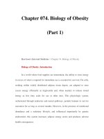
cells, which are often called target cells (Figure 1). Often these modalities are used in
combination such that paracrine and autocrine secretions provide local fine tuning
for events that are evoked by a hormonal signal that arrives from a distant source.
Because hormones are diluted in a huge volume of blood and extracellular
fluid, achieving meaningful concentrations (10
−10
to 10
−7
M) usually requires coor-
dinated secretion by a mass of cells, an endocrine gland. The secretory products of
endocrine glands are released into the extracellular space and diffuse across the
2 Chapter 1. Introduction
capillary endothelium into the bloodstream, thus giving rise to the terms “ductless
glands” and “internal secretion.” In contrast, exocrine glands deliver their products
through ducts to the outside of the body or to the lumen of the gastrointestinal
tract. Classical endocrine glands include the pituitary, thyroid, adrenals, parathy-
roids, gonads, and islets of Langerhans. It has become apparent, however, that this
Overview and Definitions 3
A. Autocrine/Paracrine
B. Endocrine
general circulation
C. Neural
nerve cell effector cell
target cellsendocrine cells
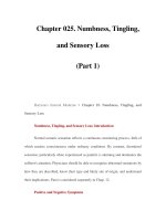
Figure 1 Chemical communication between cells. (A) Autocrine/paracrine. Secretory product,
shown as black dots, reaches nearby target cell by diffusion through extracellular fluid. (B) Endocrine.
Secretory product reaches distant cells by transport via the circulatory system. (C) Neural secretory
product released from terminals of long cell processes reaches target cells distant from the nerve cell
body by diffusion across the synaptic cleft.
list is far too short.Virtually every organ, including brain, kidney, heart, and even
fat, has an endocrine function in addition to its more commonly recognized role.
Many aspects of gastrointestinal function are governed by hormones produced by
cells of the gastric and intestinal mucosae. In fact, the word “hormone” was coined
to describe a duodenal product, secretin, that regulates pancreatic fluid secretion.
However, the gastrointestinal hormones are traditionally considered in textbooks
of gastrointestinal physiology rather than endocrinology and hence will not be
considered here.
It is only recently that endocrinologists have embraced the large number of
locally produced hormonelike agents, called growth factors and cytokines, that regu-
late cell division, differentiation, function, and even programmed cell death, which
is called apoptosis.These agents act locally in a paracrine or autocrine manner, but
may also enter the circulation and affect the functions of distant cells, and hence
function as hormones. Many of these secretions produce effects that impinge on
actions of the classical hormones. Rapidly accumulating information about protein
and gene structure has revealed relationships among these compounds, which can
now be grouped into families or superfamilies. Some of the classical hormones,
such as growth hormone and prolactin, belong to the same superfamily of proteins
as some of the cytokines, whereas the insulin-like growth factors are closely related
to insulin. At the molecular level, production, secretion, and actions of cytokines
and growth factors are no different from those of the classical hormones.
Another mechanism has also evolved to breach the distance between cells
and allow rapid communication. Some cells developed the ability to release their
signals locally from the tips of long processes that extend great distances to nearly
touch their target cells.This mechanism, of course, is how nerve cells communi-
cate with each other or with effector cells. By releasing their signals (neurotrans-
mitters) so close to receptive cells, nerve cells achieve both exquisite specificity and
economy in the quantity of transmitter needed to provide a meaningful concen-
tration within a highly localized space, the synapse. Although use of the action
potential to trigger secretion is not unique for nerve cells, the electrical wave that
travels along the axons enables these cells to transmit information rapidly over great
distances between the perikarya and the nerve terminals. Despite these specialized
features of nerve cells, it is important to note that the same cellular mechanisms are
used for signal production and release as well as for reception and response during
neural, endocrine, and paracrine communication.
Distinctions between the various modes of communication are limited only
to the means of signal delivery to target cells, and even these distinctions are
blurred in some cases. Neurotransmitters act in a paracrine fashion on postsynap-
tic cells and in some cases may diffuse beyond the synaptic cleft to affect other
nearby cells or may even enter the blood and act as hormones, in which case they
are called neurohormones. Moreover, the same chemical signals may be secreted by
both endocrine and nerve cells and even in very small amounts by other cells
4 Chapter 1. Introduction
that use them to communicate with neighboring cells in a paracrine or autocrine
manner. Nature is parsimonious in this regard. Many peptides that have classically
been regarded as hormones or neurohormones may also serve as paracrine regula-
tors in a variety of tissues. Although adequate to cause localized responses, the
minute quantities of these substances produced extraglandularly are usually too
small to enter the blood and interfere with endocrine relationships.
Clearly, the boundaries between endocrinology and other fields of modern
biology are both artificial and imprecisely drawn. Endocrinology has benefitted
enormously from recent advances in other fields, particularly immunology,
biochemistry, cell biology, and molecular biology. Early insights into endocrine
function were gained from “experiments of nature,” i.e., injury or inborn errors
produced pathological conditions that were traced to defects in hormone secretion
or action. Conversely, hormone-secreting tumors or deranged regulatory mecha-
nisms produced early insights into the consequences of excess hormone produc-
tion. Early endocrinologists were able to create similar experiments by excising a
gland or administering glandular extracts and observing the consequences. Progress
in biochemistry made it possible to study pure hormones, and application of
immunological techniques allowed identification and measurement of various
molecular species. The introduction of techniques of molecular biology brought
breakthroughs in the understanding of hormone actions, and curiously brought us
full circle back to the early approaches of studying the consequences of eliminat-
ing the source of a signaling molecule or administering an excess to gain insight
into function. It is now possible to overexpress a hormone or other molecule by
inserting its gene into developing mice to make them “transgenic.” Conversely, it
is possible to disrupt or “knock out” a particular gene and study the consequences
of the lack of its protein product(s) in otherwise intact mice. It is even possible to
limit expression of transgenes to particular organs and evoke their expression at
desired stages of life. Similarly, it is now possible to knock out genes in particular
organs and at particular times of life. In discussing hormone actions in subsequent
chapters it will be necessary to refer to all of these experimental techniques and
many others.
In this text we concentrate on the integrating function of the endocrine sys-
tem and focus our discussion principally on that aspect of cellular communication
that is carried out by the classical endocrine glands and their hormones (Table 1).
Chapters 1 through 5 deal with basic information about various endocrine glands
and their hormones. In the remaining chapters we consider the interaction of
hormones and the integration of endocrine function to produce homeostatic
regulation. Such regulation throughout the body is achieved by regulation of
cellular functions, which, in turn, is achieved by actions of hormones on molecules
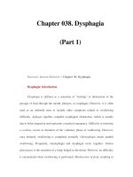
within those cells.We therefore consider the actions of hormones on three levels
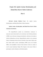
(Figure 2).Throughout this text, emphasis is on normal function, and reference to
disease is limited to those aspects that are logical extensions of normal physiology
Overview and Definitions 5
or that facilitate understanding of normal physiology.Endocrine disease is not sim-
ply a matter of too much or too little hormone; rather, disease occurs when there
is an inappropriate amount of hormone for the prevailing physiological situation
or when there is an inappropriate response by target tissues to a perfectly appro-
priate amount of hormone. Some aspects of endocrine disease are too poorly
understood to be put in the context of normal physiology and are best left for a
more detailed text of pathology or medicine.
Endocrinology is a subject that unfortunately involves a sometimes bewil-
dering array of facts, not all of which can be derived from basic principles.To help
organize and digest this necessarily large volume of material, the student might find
the following outline of goals and objectives helpful.
GOALS AND OBJECTIVES
A. The student should be familiar with
1. Essential features of feedback regulation
2. Essentials of competitive binding assays
6 Chapter 1. Introduction
Table 1
Chemical Nature of the Classic Hormones
Ty r osine Steroids Peptides Proteins
derivatives (<20 amino acids) (>20 amino acids)
Epinephrine Testosterone Oxytocin Insulin
Norepinephrine Estradiol Vasopressin Glucagon
Dopamine Progesterone Angiotensin Adrenocorticotropic hormone
Tr iiodothyronine Cortisol Melanocyte-stimulating Thyroid-stimulating hormone
hormone
Thyroxine Aldosterone Thyrotropin-releasing hormone
Vitamin D Somatostatin Follicle-stimulating hormone
Luteinizing hormone
Gonadotropin-releasing hormone
Growth hormone
Prolactin
Corticotropin-releasing hormone
Growth hormone-releasing
hormone
Parathyroid hormone
Calcitonin
Chorionic gonadotropin
Choriosomatomammotropin
B. For each hormone, the student should know
1. Its cell of origin
2. Its chemical nature, including
a. Distinctive features of its chemical
composition
b. Biosynthesis
c. Whether it circulates free or bound to
plasma proteins
d. How it is degraded and removed from the
body
3. Its principal physiological actions
a. At the whole body level
b. At the tissue level
c. At the cellular level
d. At the molecular level
e. Consequences of inadequate or excess
secretion
4. What signals or perturbations in the internal
or external environment evoke or suppress its
secretion
a. How those signals are transmitted
b. How that secretion is controlled
c. What factors modulate the secretory
response
d. How rapidly the hormone acts
e. How long it acts
f. What factors modulate its action
BIOSYNTHESIS OF HORMONES
The classical hormones fall into three categories:
• Derivatives of the amino acid tyrosine
• Steroids, which are derivatives of cholesterol
•Peptides/proteins, which comprise the largest and most diverse class of
hormones
Table 1 lists some examples of each category. A large number of other
small molecules, including derivatives of amino acids and fatty acids, function as
neurotransmitters or paracrine signals but fall outside the scope of the classical
hormones. In most aspects of their synthesis, secretion, and molecular actions,
these substances are indistinguishable from hormones. Relevant details of hormone
Biosynthesis of Hormones 7
synthesis and storage, particularly for the amino acid and steroid hormones, are
presented with the discussion of their glands of origin, but steps in biosynthesis,
storage, and secretion common to all protein and peptide hormones are sufficiently
general for this largest class of hormones to warrant some discussion here. A brief
review of these steps also provides an opportunity for a general consideration
of gene expression and protein synthesis and provides some background for
understanding hormone actions. In-depth consideration of these complex
8 Chapter 1. Introduction
• ionic and fluid balance
• energy balance (metabolism)
• coping with the environment
• growth and development
• reproduction
WHOLE BODY LEVEL
Regulation and integration of:
HORMONE
ACTIONS
• cell division
• differentiation
• death (apoptosis)
• motility
• secretion
• nutrient uptake
CELLULAR LEVEL
Regulation of:
• gene transcription
• protein synthesis
and degradation
• enzyme activity
• protein conformation
and protein : protein
interactions
MOLECULAR LEVEL
Regulation of:
Figure 2 Levels at which hormone actions are considered.
processes is beyond the scope of this text, and is best left for the many excellent
texts of cellular and molecular biology.
Protein and peptide hormones are encoded in genes, with each hormone
usually represented only once in the genome. Information determining the amino
acid sequence of proteins is encoded in the nucleotide sequence of deoxyribonu-
cleic acid (DNA) (Figure 3). Nucleotides in DNA consist of a five-carbon sugar,
deoxyribose, in ester linkage with a phosphate group and attached in N-glycosidic
linkage to one of four organic bases: adenine (A), guanine (G), thymidine (T), or
cytidine (C).The ability of the purine bases A and G to form complementary pairs
with the pyrimidine bases T and C (Figure 4), respectively, on an adjacent strand
of DNA is the fundamental property that permits accurate replication of DNA and
transmission of stored information from generation to generation. A single strand
of DNA consists of a chain of millions of nucleotides linked by phosphate groups
that form ester bonds with hydroxyl groups at carbon 3 of one deoxyribose and
carbon 5 of the next deoxyribose.The DNA in each chromosome is present as a
pair of long strands oriented in opposite directions and is organized into nucleo-
somes, each of which consists of a stretch of about 180 nucleotides tightly wound
around a complex of eight histone molecules. The nucleosomes are linked by
stretches of about 30 nucleotides, and the whole double strand of nucleoproteins
is tightly coiled in a higher order of organization to form the chromosomes.
Instructions for protein structure are transmitted from the DNA to cyto-
plasmic sites of protein synthesis, the ribosomes, in the messenger ribonucleic acid
(mRNA) template. RNA differs in structure from DNA only in having ribose
instead of deoxyribose as its sugar and uridine (U) instead of thymidine as one of
its pyrimidine bases.The nucleotide sequence of the mRNA precursor is comple-
mentary to the nucleotide sequence of DNA. Messenger RNA synthesis proceeds
linearly from an upstream “start site” designated by a particular sequence of
nucleotides in DNA in a process called transcription.The start site is located down-
stream from the promoter region, which contains sequences to which regulatory
proteins can bind, and a short sequence where RNA polymerase II and a large
complex of proteins, the general transcription complex, bind. The DNA that is
transcribed is composed of segments, exons, that encode structural and regulatory
information; the exons are separated by intervening sequences of DNA, introns,
which have no coding function (Figure 5). Transcription is regulated by nuclear
proteins called transcription factors or transactivating factors, which bind to regulatory
sites that are usually upstream from the promoter and stimulate or repress gene
transcription. These proteins form complexes with multiple other transcription
factors and proteins called coactivators or corepressors, which not only govern attach-
ment and activity of the general transcription complex, but control the “tightness”
of the DNA coil and hence the accessibility of genes to the transcription appara-
tus. Transcription proceeds from the start site through the introns and exons and a
downstream flanking sequence, where a long polyadenine (polyA) tail is added.
Biosynthesis of Hormones 9
10 Chapter 1. Introduction
O
CH
2
O - P=O
O
O
CH
3
O
HN
N
O
Thymine
O
CH
2
O - P=O
O
O
O
O
CH
2
O - P=O
O
O
O
O
CH
2
O - P=O
O
O
O
O
CH
2
O - P=O
O
O
O
O
O
N
N
N
N
NH
2
Adenine
O
HN
N
N
N
H
2
N
Guanine
N
NH
2
O
N
Cytosine
O - P=O
O
Deoxyribose
5'
3'
3
5
2
4
1
Figure 3 Composition of DNA. DNA is a polymer of the five-carbon sugar, deoxyribose, in diester
linkage, with phosphate forming ester bonds with hydroxyl groups on carbons 3 and 5 on adjacent
Biosynthesis of Hormones 11
CH3
O
O
H
N N
N
N
N
Deoxyribose
Thymine
Deoxyribose
N
H
H
Adenine
N
O
H
N
N
N
N
N
Deoxyribose
O
H
N
H
H
Deoxyribose
Cytosine
Guanine
N
.
H
N
Figure 4 Complementary base pairing by the formation of hydrogen bonds between thymine and
adenine and between cytosine and guanine. RNA contains uracil in place of the thymine found in
DNA. Uracil and thymine differ in structure only by the presence of the methyl group (CH
3
) found
in thymine.
sugar molecules.The purine and pyrimidine bases are linked to carbon 1 of each sugar.The number-
ing system for the five carbons of deoxyribose is shown in blue at the top of the figure.The chemical
bonds forming the backbone of the DNA chain are shown in blue.The 5′ and 3′ ends refer to the
carbons in deoxyribose.
A special “cap” structure containing methylated guanosine added to the opposite
end of the RNA transcript permits its export from the nucleus after it is further
modified by removal of the introns and attachment of the exons to each other in
a process called splicing. Under some circumstances the splicing reactions may
bypass some exons or parts of exons, which are then omitted from the final
mRNA transcript. Because of such alternate splicing,a single gene can give rise to
more than one mRNA transcript, and hence more than one protein product
(Figure 6).
On export from the nucleus, the mRNA transcripts attach to ribosomes,
where they are translated into protein (Figure 7). Ribosomes are large complexes
of RNA and protein enzymes that “read” the mRNA code in triplets of
nucleotides called codons.The translation initiation site begins with the codon for
methionine. Each codon designates a specific amino acid.Triplets of complemen-
tary nucleotides (anticodons) are found in small RNA molecules called transfer
RNA (tRNA), each of which binds a particular amino acid and delivers it to the
ribosome. Alignment of amino acids in the proper sequence is achieved by the
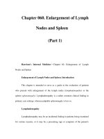
12 Chapter 1. Introduction
transcription
factors and
cofactors
pre-mRNA
splicing
mRNA
introns
start
Pll
terminate
exons
general
transcription
complex
12 34 5
AAAAAAA
12 34 5
AAAAAAA
AAAAAAA
12 34 5
12 34 5
Figure 5 Transcription and RNA processing.The DNA strand contains all of the stored information
for expression of the gene, including the promoter, distant regulatory elements (not shown), binding
sites (response elements) for regulatory proteins, and the coding for the sequence of the protein (exons)
interrupted by intervening sequences of DNA (introns). Exons are numbered 1–5.The primary RNA
transcript contains the complementary sequence of bases coupled to a polyA tail at the 3′ end and
a methyl guanosine cap at the 5′ end.Removal of the introns and splicing the remaining exons together
produce the messenger RNA, which contains all of the information needed for translation, including
the codons for the amino acid sequence of the protein and untranslated regulatory sequences at both
ends. PII, RNA polymerase II.
Biosynthesis of Hormones 13
5' 3'
12 3 4 5 12 312 4 5
primary RNA transcript
24 5 2 323 45
N
C
NC
12 3 4 5
alternatively spliced messenger RNAs
NC
primary protein translation products
Figure 6 Alternative splicing of mRNA can give rise to different proteins. Numbers indicate exons.
Exon 1 is untranslated. N, Amino terminus; C, carboxyl terminus.
complementary pairing of anticodons in the tRNA with codons in the mRNA.
The tRNA thus delivers the correct amino acid to the carboxyl terminus of the
growing peptide chain and holds it in position so that ribosomal enzymes can
release it from the tRNA and link it to the peptide. Once the peptide bond is
formed, the empty tRNA is released and the ribosome moves down the mRNA
to the next codon, where the next tRNA molecule charged with its amino acid
waits to bind to its complementary codon. Elongation of the chain continues
until the ribosome reaches a “stop” codon, at which time it dissociates from the
mRNA. As each ribosome moves down the mRNA, other ribosomes attach
behind them to repeat the process. In this way, before it is degraded, a single
mRNA molecule may be translated over and over again to yield many copies of
a protein.
Protein and peptide hormones are initially synthesized as precursor
molecules (prohormones and preprohormones) that are larger than the final secre-
tory product. Proteins destined for secretion have a hydrophobic sequence of
12–25 amino acids at their amino termini (Figure 8).This signal sequence is recog-
nized by a special structure that directs the growing peptide chain through a pro-
tein channel in the endoplasmic reticular membrane and into the cisternae of the
endoplasmic reticulum. Postsynthetic processing begins in the endoplasmic reticu-
lum as the hormone precursors are translocated to the Golgi apparatus for final
processing and packaging for export. Processing includes cleavage to remove the
signal peptide and interaction with other proteins that facilitate proper folding and
formation of disulfide bonds linking cysteine residues. For some hormones, cleav-
age at appropriate loci removes those amino acid sequences that may have func-
tioned to orient folding of the molecule so that disulfide bridges form in the right
places. Clipping the protein by trypsinlike peptidases may yield more than one
biologically active peptide molecule from a single precursor, as seen with the
adrenocorticotropic and glucagon families of hormones (Chapters 2 and 5). For
some secreted peptides, final clipping occurs in secretory granules with the result
that one or more other molecules are released into the circulation along with the
hormone. Other processing of peptide hormones may include glycosylation (addi-
tion of carbohydrate chains to asparagine residues) or coupling of subunits
that are products of different genes, as seen with the pituitary glycoproteins (see
Chapter 2).
Defects in processing of normal precursor molecules cause some rare
inherited diseases. It is common to find precursor molecules (prohormones) in
the circulation, sometimes in large amounts. This situation may be indicative of
14 Chapter 1. Introduction
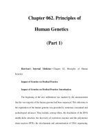
GCU
GU AGUUU
C
GG G A CAUGC
GAA
G
mRNA
ribosom
e
AAC
growing
peptide
chain
arriving
charged
tRNA
departing
empty
tRNA
Gln
Met
Phe
Asp
Leu
ACC
Trp
G
U
A
Figure 7 Translation. A molecule of transfer RNA (tRNA) charged with its specific amino acid,
phenylalanine, and already linked to the growing peptide chain, is positioned on the mRNA by com-
plementary pairing of its triplet of nucleotides with its codon of three nucleotides in the mRNA. A
second molecule of tRNA charged with its specific amino acid, tryptophan, has docked at the adjacent
triplet of nucleotides and awaits the action of ribosomal enzymes to form the peptide bond with
phenylalanine. Linking the amino acid to the peptide chain releases it from its tRNA and allows the
empty tRNA to dissociate from the mRNA.A third molecule of tRNA, which brought the preceding
molecule of leucine, is departing from the left, while a fourth molecule of tRNA, carrying its cargo of
glutamine, arrives from the right and waits to form the complementary bonds with the next codon in
the mRNA that will bring the glycine in position to be joined to tryptophan at the carboxyl terminus
of the peptide chain.The ribosome moves down the mRNA, adding one amino acid at a time until it
reaches a stop codon. (Adapted from Alberts et al.,“Molecular Biology of the Cell.” Garland Publishing,
New York, 1994, copyright Taylor & Francis Group.)
hyperactivity of endocrine cells or even aberrant production of hormone by
nonendocrine tumor cells. Some prohormones have biological activity, and their
effects may be the first manifestation of neoplasia.
Postsynthetic processing to the final biologically active form is not limited to
the peptide hormones. Other hormones may be formed from their precursors after
secretion. Postsecretory transformations to more active forms may occur in liver,
Biosynthesis of Hormones 15
1
2
3
4
mRNA
ribosomes
peptide
endoplasmic reticulum
golgi apparatus
secretory vesicle
5
6
7
S-S
S-S
S-S
Figure 8 Posttranslational processing. The leader sequence, or signal peptide, of proteins destined
for secretion enters the cisternae of the endoplasmic reticulum even as peptide elongation continues.
In the endoplasmic reticulum (1) the leader sequence is removed; (2) the protein is folded with the
assistance of protein chaperones, sulfhydryl bridges (3) may form, and carbohydrate (4) may be added
(glycosylation). The partially processed protein (5) is then entrapped in vesicles that bud off the
endoplasmic reticulum and (6) fuse with the Golgi apparatus, where glycosylation is completed, and
(7) the protein is packaged for export in secretory vesicles in which the final stages of processing
take place.
kidney,fat, or blood, as well as in the target tissues. For example,thyroxine,the major
secretory product of the thyroid gland, is converted extrathyroidally to triiodothy-
ronine, which is the biologically active form of the hormone (see Chapter 3).
Testosterone, the male hormone, is converted to dihydrotestosterone within some
target tissues and may even be converted to the female hormone, estrogen, in other
tissues (Chapter 11).These peripheral transformations, in addition to confounding
the student of endocrinology, are vulnerable to derangement and hence must be
considered as possible causes of endocrine disease.
STORAGE AND SECRETION OF HORMONES
With the notable exception of the steroids, most hormones are stored, often
in large quantities, in their glands of origin, a factor that facilitated their original
isolation and characterization. Protein and peptide hormones, and the tyrosine
derivatives epinephrine and norepinephrine, are stored as dense granules in mem-
brane-bound vesicles and are secreted in response to an external stimulus by the
process of exocytosis. In this process, storage vesicles are translocated to the cell sur-
face,where they dock with specialized membrane proteins. Membranes of the vesi-
cles then fuse with the plasma membrane, causing the vesicle to open and empty
its contents into the extracellular fluid (Figure 9). Movement of the secretory vesi-
cle to the cell surface and membrane fusion usually require transient increases in
cytosolic calcium concentrations, brought about by release of calcium from inter-
nal organelles and from influx of extracellular calcium through activated mem-
brane channels. A detailed description of the complex molecular events that gov-
ern secretion is beyond the scope of this text but can be found in many fine texts
of cell biology. It is obvious that synthesis of hormones must be coupled in some
way with secretion, so that cells can replenish their supply of hormone. In general,
the same cellular events that signal secretion also signal synthesis. In addition, some
cells may be able to monitor how much hormone is stored and to adjust rates of
synthesis or degradation accordingly.
Unlike the peptide hormones, which are encoded in genes, the steroid
hormones are formed enzymatically through a series of modifications of their
common precursor, cholesterol (see Chapter 4). In further contrast to the peptide
hormones, there is little storage of steroid hormones in their cells of origin.
Therefore, synthesis and secretion are aspects of the same process, and the lipid-
soluble steroid hormones apparently diffuse across the plasma membrane as rapidly
as they are formed. The synthetic process proceeds sufficiently rapidly that
increased secretion can be observed as soon as a minute after the secretory
stimulus has been applied, but the maximal rate of secretion is not reached for at
least 10–15 minutes. In contrast, stored peptide and amine hormones may be
released almost instantaneously.
16 Chapter 1. Introduction
Hormones in Blood 17
HORMONES IN BLOOD
Most hormones circulate in blood in free solution at nanomolar (10
−9
M)
or even picomolar (10
−12
M) concentrations. Steroid hormones and thyroid
trans golgi
secretory
granule
1
2
3
4
p
lasma
membrane
secretory
vesicle
Figure 9 Exocytosis. Secretory vesicles (1) bud off the trans-Golgi compartment and (2) move into
the cytosol, where they await a signal for secretion. (3) Secretion is usually accompanied by increased
cellular calcium, which causes elements of the cytoskeleton to translocate secretory granules to the cell
surface (4). The membrane surrounding the granule fuses with the plasma membrane, opening the
secretory vesicle to the extracellular fluid and releasing the processed protein(s) along with enzymes and
peptide fragments.
hormones, which have a limited solubility in water, circulate bound specifically
to large carrier proteins synthesized in the liver. Some protein and peptide
hormones also circulate complexed with specific binding proteins (see Chapter
10). Bound hormones are in equilibrium with a small fraction, sometimes
less than 1%, in free solution in plasma. Generally, only unbound hormones
are thought to cross the capillary endothelium to reach their sites of biological
action or degradation. Protein binding protects against loss of hormone by the
kidney, slows the rate of hormone degradation by decreasing cellular uptake,
and buffers changes in free hormone concentrations. In some instances binding
proteins may affect hormonal responses by facilitating or impeding delivery
of hormones to particular cells. Because biological responses are related to the
concentration of hormone that reaches target cells, rather than the total amount
present in blood, increases in abundance of binding proteins that occur during
pregnancy, for example, or decreases seen with some forms of liver or kidney
disease, may produce changes in total amounts of hormones circulating in
blood, even though free, physiologically important concentrations may be normal.
Most hormones are destroyed rapidly after secretion and have a half-life in
blood of less than 10 minutes.The half-life of a hormone in blood is defined as that
period of time needed for its concentration to be reduced by half and depends on its
rate of degradation and on the rapidity with which it can escape from the circulation
and equilibrate with fluids in extravascular compartments.This process is sometimes
called the metabolic clearance rate. Some hormones, e.g., epinephrine, have half-lives
measured in seconds; others, e.g., thyroid hormones, have half-lives of the order of
days.The half-life of a hormone in blood must be distinguished from the duration of
its hormonal effect. Some hormonal effects are produced virtually instantaneously
and may disappear as rapidly as the hormone is cleared from the blood. Other hor-
monal effects are seen only after a lag time that may last minutes or even hours, and
the time of maximum effect may bear little relation to the time of maximum hor-
mone concentration in the blood. Additionally, the time for decay of a hormonal
effect is also highly variable; it may be only a few seconds, or it may require several
days. Some responses persist well after hormonal concentrations have returned to
basal levels. Understanding the time course of a hormone’s survival in blood as well
as the onset and duration of its action is obviously important for understanding
normal physiology, endocrine disease, and the limitations of hormone therapy.
HORMONE DEGRADATION
Implicit in any regulatory system involving hormones or any other signal is the
necessity for the signal to disappear once the appropriate information has been con-
veyed. Only a small amount of hormone is degraded as an aftermath to the process
of signaling its biological effects. The remainder must therefore be inactivated and
18 Chapter 1. Introduction
excreted.Degradation of hormones and their subsequent excretion are processes that
are just as important as secretion. Inactivation of hormones occurs enzymatically in
blood or intercellular spaces, in liver or kidney cells, as well as in the target cells.
Degradation of peptide and protein hormones often involves uptake into cells by a
mechanism of endocytosis that delivers them to the cellular sites of degradation, the
lysosomes and proteosomes. Inactivation may involve complete metabolism of the
hormone so that no recognizable product appears in urine, or it may be limited
to some simple one- or two-step process such as addition of a methyl group or glu-
curonic acid. In the latter cases recognizable degradation products are found in urine
and can be measured to obtain a crude index of the rate of hormone production.
MECHANISMS OF HORMONE ACTION
The ultimate mission of a hormone is to change the behavior of its target
cells. Cellular behavior is determined by biochemical and molecular events that
transpire within the cell, and these in turn are determined by the genes that
are expressed, the biochemical reactions that carry out cellular functions, and the
conformation and associations of the molecules that comprise the cell’s physical
structure. Hormonal messages must be converted to biochemical events that
influence gene expression, biochemical reaction rates, and structural changes.The
conversion of a hormonal message to cellular responses is called signal transduction
and the series of biochemical changes that are set in motion are described as
signaling pathways, although in reality signaling network might be a more accurate
descriptor, because pathways branch and converge only to branch again. Signal
transduction is a complex topic and the focus of intense investigation in many
laboratories around the world. Detailed consideration is beyond the scope of this
text. Instead, only general patterns of signal transduction are considered in the
following section, but the topic will be revisited where appropriate in subsequent
chapters in discussing individual hormones.
SPECIFICITY
Because all hormones travel in blood from their glands of origin to their
target tissues, all cells must be exposed to all hormones. Yet under normal
circumstances cells respond only to their appropriate hormones. Such specificity of
hormone action resides primarily in the ability of receptors in the target cells to
recognize only their own signal (Figure 10).We may define a hormone receptor as
a molecule or complex of molecules, in or on a cell, that binds its hormone with
great selectivity and in so doing is changed in such a manner that a characteristic
response or group of responses is initiated. Hormone receptors are a subset of
Mechanisms of Hormone Action 19