Endocrinology Basic and Clinical Principles - part 9 pot
Bạn đang xem bản rút gọn của tài liệu. Xem và tải ngay bản đầy đủ của tài liệu tại đây (1013.39 KB, 45 trang )
348 Part IV / Hypothalamic–Pituitary
key to selecting the appropriate patients for laboratory
testing. This includes hypertension in young adults or
teenagers; hypertension unresponsive to three or more
antihypertensives; either sustained hypertension or nor-
motension with paroxysms of hypertension accompa-
nied by symptoms; a hypertensive and symptomatic
response to exercise, abdominal examination, micturi-
tion, or palpation of a neck mass; marked hypertensive
response to induction anesthesia; accelerated or malig-
nant hypertension; paradoxical hypertensive response
to β-blockers; or markedly labile blood pressure with
symptoms. Other conditions for which biochemical test-
ing is appropriate include families with MEN or familial
pheochromocytoma, or the other associated diseases
provided in Table 4. Finally, incidental adrenal tumors
discovered on abdominal computed tomography (CT)
or magnetic resonance imaging (MRI) scans require
screening tests to eliminate the presence of a hormone-
secreting tumor including pheochromocytoma.
The specificity of the most sensitive tests for pheo-
chromocytoma depends, to a large extent, on the proper
selection of a symptomatic, hypertensive patient for
whom other confounding conditions and drugs have
been eliminated. The most sensitive tests for pheochro-
mocytoma are measurement of plasma metanephrines
and/or a 24-h urine collection for metanephrines
(metanephrine and normetanephrine) and/or total uri-
nary catecholamines by high-performance liquid chro-
matography (HPLC). Fluorometric methods remain an
adequate substitute when HPLC methods are not readily
available. If metanephrine or catecholamine levels are
greater than threefold above the upper limit of normal
in a symptomatic and hypertensive patient, then imag-
ing is indicated. If catecholamine levels are <1.5-fold
of the upper limit of normal, then it is unlikely that the
patient has pheochromocytoma. If the levels are mar-
ginally elevated, between 1.5-fold and 3-fold above the
upper limit of normal, then a 12-h, nighttime collection
of urine for catecholamines and metanephrines is indi-
cated. Collection at night eliminates the effects of stress
and upright posture on the production of catechola-
mines that occurs during the day in healthy patients and
will not affect the secretion of catecholamines in pheo-
chromocytoma. If levels remain marginally elevated or
higher, then one should proceed to imaging. If levels
are normal, then one should discontinue testing. If the
patient has only brief paroxysms that occur only a few
times per day or less frequently, then one should obtain
the tests as a timed urine collection (2–4 h) during a
prominent symptomatic hypertensive episode. If val-
ues exceed threefold, then one should proceed to imag-
ing, and if less than threefold, depending on the level of
clinical suspicion, one should either discontinue test-
ing or repeat the test during another episode. This com-
bination of urinary catecholamine and metanephrine
measurement has been reported by most investigators,
for many years, to be a sensitive (98–100%) and spe-
cific (96–98%) biochemical test for pheochromocy-
toma. Plasma metanephrine testing is a recent addition
to the diagnostic tools available. Although it has not
acquired a fraction of the long experience of urinary
studies, it will probably be as reliable (sensitive and
specific) as testing for urinary metanephrine. A major
advantage of obtaining a sample through venopuncture
is that it is far easier than a 24-h urine collection.
A robust biochemical diagnosis is essential before
proceeding to imaging tests. Benign, nonfunctioning
adrenal masses have a much higher incidence than
pheochromocytoma. Performing an unnecessary major
surgical procedure to remove a benign, nonfunction-
ing mass is to be avoided. Alternatively, a mass not
found in the initial examination may result in futile,
expensive, and more invasive attempts to locate a non-
existent tumor.
The purpose of making a diagnosis of pheochromocy-
toma is to enable the surgical excision of the source of
the excessive secretion of catecholamines causing the
patient’s hypertension and symptoms. If significantly
elevated catecholamines cannot be demonstrated dur-
ing a hypertensive, symptomatic episode, then cat-
echolamines are not causing the problem and testing
should not proceed to imaging. If a high degree of sus-
picion remains despite the negative biochemical test-
ing, then the patient should be treated medically and
reevaluated at a later date. Imaging may be indicated in
patients with familial diseases (Table 4) for whom bio-
chemical testing was negative. This has become a more
reasonable option as newer imaging techniques have
become more sensitive and specific.
Pharmacologic tests developed to elicit or inhibit
catecholamine secretion from a pheochromocytoma
bear a significant risk and are generally less specific and
sensitive than urinary collections. Phentolamine
(Regitine), a short-acting α-blocker, administered
intravenously will cause a significant fall in blood
pressure during a hypertensive episode. It may induce
an undesired, profound fall and cause a myocardial or
cerebral infarction. Administration of histamine,
tyramine, and glucagon all cause release of catechola-
mines by different mechanisms and have been used to
elicit either a blood pressure or catecholamine response
from the tumor. An excessive hypertensive response
resulting in a stroke or the development of a significant
arrhythmia could occur during these stimulation tests.
Clonidine is used to exclude false positive plasma cat-
echolamine measurements.
Chapter 22 / Adrenal Medulla 349
Other biochemical testing offers little or no advan-
tage over measurement of urinary catecholamines and
metanephrines. Urinary VMA by colorometric methods
is less specific and by HPLC is equivalent to
metanephrines but is more costly and less readily avail-
able. Measurements of plasma catecholamines produce
more false positives and are more expensive to obtain
and analyze. Theoretically, measurement of plasma
catecholamines would be a more sensitive method of
documenting elevated catecholamine secretion during a
brief hypertensive, symptomatic episode. The logistics
required to obtain such a sample without prolonged
hospitalization is problematic. Chromogranin A and
dopamine β-hydroxylase are released with catechola-
mines during exocytosis. Both are frequently elevated
in pheochromocytoma but are less specific than mea-
surement of urinary catecholamine.
The diagnosis will have been made prior to imaging
based on the history, physical findings, and biochemi-
cal measurements. The purpose of localization (imag-
ing) is to find the tumor and plan the approach for
surgical removal. Finding a mass with characteristics
that are consistent with a pheochromocytoma helps to
confirm but does not make the diagnosis. MRI is the
preferred method of tumor detection. The sensitivity
and specificity of MRI are at least equal to or greater
than of CT, and MRI does not expose the patient to
ionizing radiation. Pheochromocytoma on T
2
-weighted
imaging (MRI) presents an especially bright mass in
comparison to most other tumors. CT provides no simi-
lar distinguishing characteristics of pheochromocy-
toma compared to other masses. MRI of the abdomen
and pelvis is the first examination to be performed,
because 90% of tumors are found below the diaphragm.
If no tumor is found below the diaphragm, then the
chest and neck should be imaged. If no mass is found,
then CT imaging with contrast should be performed of
the same areas and in the same order. If still no mass is
found, then a
131
I-metaiodobenzylguanidine (MIBG)
scan could be considered. Although this scan is highly
specific (100%), it is considerably less sensitive (60–
80%) than either the MRI or CT scans (>98%). The
isotope is specifically concentrated in intra- and
extraadrenal pheochromocytomas. Because it is a
131
I-
based isotope, it has a short half-life (9 d). The MIBG
scan is expensive and not readily available. The
123
I-
based isotope is more sensitive but even more difficult
to obtain. A new imaging technique, 6-[
18
F]-
fluorodopamine ([
18
F]-DA) by positron emission to-
mography, is as specific as MIBG, is more sensitive,
requires no pretreatment to protect the thyroid, and
produces higher resolution images. [
18
F]-DA plus MRI
may be the best combination for the detection of intra-
and extraadrenal tumors, benign or malignant. Unfor-
tunately, [
18
F]-DA is currently available only at the
National Institutes of Health.
There is a high incidence of gallstones in pheochro-
mocytoma, and ultrasound examination of the gallblad-
der and ducts is warranted prior to surgery.
2.5.3.4. Management. The definitive treatment for
pheochromocytoma is surgery. The early, coordinated
team effort of the endocrinologist, anesthesiologist, and
surgeon helps to ensure a successful outcome. The goals
of preoperative medical therapy are to control hyper-
tension; obtain adequate fluid balance; and treat
tachyarrythmias, heart failure, and glucose intolerance.
The nonselective and long-acting α-adrenergic blocker
phenoxybenzamine is the principal drug used to pre-
vent hypertensive episodes. Optimal blockade requires
1 to 2 wk of therapy. Short-acting α
1
-blockers such as
prazosin could be used as well. The effects of the cal-
cium channel blocker nifedipine on the inhibition of
calcium-mediated exocytosis of storage granules are
also moderately effective in controlling hypertension.
Adequate hydration and volume expansion with saline
or plasma is used to reduce the incidence of postopera-
tive hypotension. The addition of α-methyltyrosine
(Demser), a competitive inhibitor of tyrosine hydroxy-
lase and catecholamine biosynthesis, to α-adrenergic
blockade provides several important advantages. Con-
trol of hypertension can be obtained with a lower dose
of α-blocker, which minimizes the duration and sever-
ity of hypotensive episodes. The side effects of α-
methyltyrosine are rarely encountered during the brief
1 to 2-wk preoperative period. β-Adrenergic blockade
is usually not required and should not be given unless
a patient has persistent tachycardia and some supraven-
tricular arrhythmias. β-Blockade should never be insti-
tuted prior to α-blockade. The inability to vasodilate
(β-receptors blocked) and unopposed α-receptor-
stimulated vasoconstriction could precipitate a hyper-
tensive crisis, congestive heart failure, and acute
pulmonary edema. If β-blockade is needed, propranolol
or a more cardioselective β
1
-antagonist, atenolol, can
be used. α-Methyltyrosine may reduce the need for β-
blockers and is the drug of choice to treat catechola-
mine-induced toxic cardiomyopathy. Hyperglycemia
is best treated with a sliding scale of regular insulin in
the immediate preoperative period to maintain blood
glucose between 150 and 200 mg%. Glucose intoler-
ance usually ends abruptly after the tumor’s blood sup-
ply is isolated during surgery. Hypoglycemia during
anesthesia is to be avoided.
The advantages of a coordinated team approach are
most apparent during surgery. All members of the team
will be aware of the patient’s complications and relative
350 Part IV / Hypothalamic–Pituitary
response to the preoperative preparation. Monitoring
of the cardiopulmonary and metabolic status should
become more intense and accurate. The selection of
premedications, induction anesthesia, muscle relaxant,
and general anesthetic to be used in pheochromocytoma
is based on those that do not stimulate catecholamine
release or sensitize the myocardium to catecholamines.
These premedications include diazepam or pentobar-
bital, meperidine, and scopolamine. Thiopental is the
preferred drug for induction and vecuronium for neuro-
muscular blockade. Isofluane and enflurane are excel-
lent volatile general anesthetics, but the newest member
of this family, desflurane, has the distinct advantage of
being very volatile and thus very short acting. Increas-
ing the inhaled concentration of desflurane will rapidly
reduce blood pressure (2 min) during a hypertensive
episode, and hypotensive effects dissipate just as
quickly by reducing the inhaled concentration. The
achievement of rapid, stress-free anesthesia reduces
the risk of complications during surgery. During sur-
gery, tumor manipulation and isolation of the vessels
draining the tumor can result in changes in plasma
catecholamine concentrations of 1000-fold within
minutes. With modest α-receptor blockade, α-methyl-
tyrosine, and desflurane, the need for urgent applica-
tion of nitroprusside or phentolamine to control blood
pressure during surgery may be eliminated. Pheochro-
mocytomas are very vascular by nature and significant
hemorrhage is a potential hazard. Advanced prepara-
tion reduces the impact of these complications. Whole
blood; plasma expanders; nitroprusside; and esmolol,
a short-acting β-blocker, should be immediately avail-
able.
When bilateral adrenalectomy is being performed,
adrenal cortical insufficiency should be treated with
stress doses of hydrocortisone intra- and postoperatively
until stable. Mineralocorticoid should be replaced post-
operatively.
Hypotension is the most common complication
encountered in the recovery room. The loss of the vaso-
constrictive and ion tropic effects of catecholamines,
persistent α-receptor blockade, downregulated adren-
ergic receptors, and perioperative blood loss all con-
tribute. The treatment is aggressive volume expansion.
Sympathomimetic amines are rarely indicated. Hypo-
glycemia may result from administered insulin or be
reactive. Dextrose should be given during the immedi-
ate postoperative period and blood glucose monitored
regularly for several hours.
2.5.3.5. Prognosis. Most patients become normoten-
sive within 1 to 2 wk after surgery. Hypertension per-
sists in about one-third of patients either because they
have an underlying essential hypertension or because
they have residual tumor. Patients with essential hyper-
tension no longer have the symptoms of pheochromocy-
toma, and their blood pressure is usually easily
controlled with conventional therapy. If a patient has a
residual tumor, an unidentified second site, or multiple
metastases, then the signs and symptoms of pheochro-
mocytoma will gradually or abruptly recur in proportion
to the level of catecholamines being secreted.
There are no characteristic histologic changes on
which to base the diagnosis of malignancy. The clini-
cal course showing an aggressive, recurrent tumor or
finding chromaffin cells in nonendocrine tissue such
as lymph nodes, bone, muscle, or liver makes the
diagnosis. Factors have been examined to determine
their potential role in predicting a malignant course.
Extra-adrenal tumors, large size, local tumor invasion,
family history of pheochromocytoma, associated endo-
crine disorders, and young age are significant in pre-
dicting a malignant course. DNA flow cytometry has
been used retrospectively to determine whether the
DNA ploidy pattern could be used in predicting the
clinical course of pheochromocytoma. Although no
pattern has been diagnostic, abnormal patterns (aneu-
ploid, tetraploid) were best correlated with malig-
nancy, and a diploid pattern has been very strongly
correlated with a benign course.
The primary approach to the treatment of malignant
pheochromocytoma is surgical debulking with medi-
cal management similar to that used for preoperative
preparation. All treatment is palliative; there is no cure.
Chemotherapy with a combination of cyclophospha-
mide, vincristine, and dacarbazine produced a 57%
response with a median duration of 21 mo. High doses
of
131
I-MIBG have been used to shrink tumors and
decrease catecholamine secretion in some patients who
demonstrate high-grade uptake of this compound.
Repetitive treatments are needed to obtain a temporary
response over 2 to 3-yr, but the therapy is well toler-
ated. Unlike
131
I-MIBG, [
18
F]-DA used for localiza-
tion would have no beneficial effect in the treatment of
malignant pheochromocytoma.
3. PEPTIDES
3.1. Developmental Origin
The cells of the adrenal medulla have a
pluripotential capacity to secrete a variety of other
peptide hormones that are usually biologically active.
A great deal is known about the development and regu-
lation of the catecholaminergic properties of these
cells, but relatively little is known about the develop-
mental control of their peptidergic properties. Evi-
dence suggests that glucocorticoids derived from an
intact hypothalamic-pituitary-adrenal cortical axis and
Chapter 22 / Adrenal Medulla 351
splanchnic innervation are essential to the develop-
mental expression of these peptides. Peptide neuro-
transmitters have been identified in the neurons
innervating the adrenal as well as the gland itself. The
list of neuropeptides discovered continues to grow
and includes Met-enkephalin, Leu-enkephalin, neuro-
tensin, substance P, vasoactive intestinal peptide
(VIP), neuropeptide Y (NPY), calcitonin-related pep-
tide, orexin-A, adrenomedullin (AM), and proadrenal
medullin N-terminal peptides (PAMPs).
3.2. Potential Physiologic
or Pathophysiologic Roles
Some peptide hormone secretion may be only patho-
physiologic and derived from a neoplastic process, such
as pheochromocytoma. Alternatively, normal physi-
ologic processes can be operative but have yet to be
discovered. VIP, ACTH, and a parathyroid hormone–
like hormone can be produced by pheochromocytoma
and produce symptoms of watery diarrhea, Cushing
syndrome, and hypercalcemia, respectively. NPY is
secreted in sympathetic storage vesicles along with
norepinephrine, chromogranin, dopamine β-hydroxy-
lase, ATP, and AM. Like chromogranin, it is not taken
back up into the neuron after release, and measured
levels may be used as another marker of sympathetic
activity. NPY appears to mediate vasoconstriction
through potentiating noradrenergic stimulation of α-
receptor responses, and secretion is increased in severe
hypertension. VIP and NPY are the most abundant
transmitter peptides in the adrenal. Endothelin-1 is
another potent vasoconstrictor peptide that has been
found along with its mRNA in pheochromocytomas.
Both of these peptides could be involved in normal cir-
culatory regulation, contribute to the pathophysiology
of sympathetically mediated hypertension, or even be
responsible for the unusual hypertensive episodes of
pheochromocytoma that do not correlate well with cat-
echolamine levels.
AM testing was proposed as a diagnostic test for
pheochromocytoma but has not gained popularity. AM
is released by normal adrenals at a low rate and at a
higher rate by pheochromocytoma. PAMP regulates
intracellular signaling pathways that regulate chro-
maffin cells in an autocrine manner, and AM acts on
the vasculature via paracrine mechanisms.
Two peptides linked to obesity have been identified
that affect catecholamine synthesis or release. Orexin-
A, a hypothalamic peptide implicated in the regulation
of feeding behavior and sleep control, has been reported
to stimulate tyrosine hydroxylase activity and catechola-
mine synthesis in bovine adrenal medullary cells
through orexin receptor-1 mRNA. Ghrelin, a peptide
that was initially found in the stomach and that regulates
appetite and growth hormone secretion, has been shown
to inhibit adrenal dopamine release in chromaffin cells.
The relationship between the action of these two pep-
tides on the regulation of adrenal catcholamines and
weight control has not been explored.
Another role suggested for some of the neuropep-
tides—Met-enkephalin (also synthesized in chromaffin
tissue, stored and released in sympathetic granules) and
VIP—is to increase adrenal blood flow in response to
cholinergic stimulation and thus enhance the distribu-
tion of epinephrine into the bloodstream. By contrast,
NPY released by cholinergic stimulation inhibits adre-
nal blood flow and could, therefore, function to inhibit
the distribution of epinephrine.
SELECTED READINGS
Burgoyne RD, Morgan A, Robinson I, Pender N, Cheek TR. Exocy-
tosis in adrenal chromaffin cells. J Anat 1993;183:309.
Evans DB, Lee JE, Merrell RC, Hickey RC. Adrenal medullary dis-
ease in multiple endocrine neoplasia type 2. Appropriate man-
agement. Endocrinol Metab Clin North Amer 1994;23:167.
Graham PE, Smythe GA, Lazarus L. Laboratory diagnosis of pheo-
chromocytoma: which analytes should we measure? Ann Clin
Biochem 1993;30:129.
Ilias I, Yu J, Carrasquillo JA, Chen CC, Eisenhofer G, Whatley M,
McElroy B, Pacak K. Superiority of 6-[
18
F]-fluorodopamine
positron emission tomography versus [
131
I]-metaiodobenzyl-
guanidine scintigraphy in the localization of metastatic pheo-
chromocytoma. J Clin Endocrinol Metab 2003;88:4083.
Kobayashi H, Yanagita T, Yokoo H, Wada A. Pathophysiological
function of adrenomedullin and proadrenomedullin N-terminal
peptides in adrenal chromaffin cells. Hypertens Res 2003;
(Suppl):S71.
Lenders JWM, Pacak K, Walther MM, Linehan WM, Mannelli M,
Friberg P, Keiser HR, Goldstein DS, Eisenhofer G. Biochemical
diagnosis of pheochromocytoma: which test is best? JAMA 2002;
287:1427.
Nagatsu T. Genes for human catecholamine-synthesizing enzymes.
Neurosci Res 1991;12:315.
Nativ O, Grant CS, Sheps SG, O’Fallon JR, Farrow GM, van Heerden
JA, Lieber MM. The clinical significance of nuclear DNA ploidy
pattern in 184 patients with pheochromocytoma. Cancer 1992;
69:2683.
Raum WJ. Pheochromocytoma. In: Bardin CW, ed. Current Therapy
in Endocrinology and Metabolism, 5th Ed. St. Louis, MO: Mosby
1994:172.
Whitworth EJ, Kosti O, Renshaw D, Hinson JP. Adrenal neuropep-
tides: regulation and interaction with ACTH and other adrenal
regulators. Microsc Res Tech 2003;61:259.
Chapter 23 / Hormones of the Kidney 353
353
From: Endocrinology: Basic and Clinical Principles, Second Edition
(S. Melmed and P. M. Conn, eds.) © Humana Press Inc., Totowa, NJ
23
these hormones, angiotensin and aldosterone, both key
products in the axis of the RAS, and the natriuretic pep-
tide family, comprising potent diuretic and vaso-
relaxing hormones secreted from the heart, are regarded
as the most important players. Furthermore, the kidney
is a major organ for the production and action of various
“local hormones,” or autocrine/paracrine regulators,
such as prostaglandins (PGs), adrenomedullin (AM),
and endothelins (ETs). These factors are thought to pro-
vide an integrated mechanism for the fine-tuning of mi-
crocirculation, solute transport, and various cellular
functions in the kidney.
This chapter discusses the roles of the hormones that
are produced or have major actions in the kidney,
focusing on their functional relationships and implica-
tions in physiologic and pathophysiologic conditions.
The roles of vitamin D and the kidney in calcium homeo-
stasis as well as the prostanoid system are detailed in
other chapters.
2. COMPONENTS OF RAS
The RAS is a proteolytic cascade, composed of a
group of proteins and peptides that ultimately produce
Hormones of the Kidney
Masashi Mukoyama, MD, PhD
and Kazuwa Nakao, MD, PhD
CONTENTS
INTRODUCTION
COMPONENTS OF RAS
P
ATHOPHYSIOLOGY OF RAS
C
OMPONENTS OF NATRIURETIC PEPTIDE SYSTEM
PATHOPHYSIOLOGY OF NATRIURETIC PEPTIDE SYSTEM
KALLIKREIN-KININ SYSTEM
ADRENOMEDULLIN AND ENDOTHELINS
ERYTHROPOIETIN
1. INTRODUCTION
The kidney plays an essential role in the mainte-
nance of life in higher organisms, not only through regu-
lating the blood pressure and body fluid homeostasis
and clearing the wastes, but also by acting as a major
endocrine organ. The kidney secretes (1) renin, a key
enzyme of the renin-angiotensin system (RAS) that
leads to the production of a potent pressor hormone
angiotensin, and produces the following hormones and
humoral factors: (2) kallikreins, a group of serine pro-
teases that act on blood proteins to produce a
vasorelaxing peptide bradykinin; (3) erythropoietin
(EPO), a peptide hormone essential for red blood cell
(RBC) formation by the bone marrow; and (4) 1,25-
(OH)
2
vitamin D
3
, the active form of vitamin D essen-
tial for calcium homeostasis, which is produced by the
proximal tubule cells via the enzyme 1α-hydroxylase.
In addition, the kidney serves as an important endo-
crine target organ for a number of hormones, thereby
controlling the extracellular fluid volume, electrolyte
balance, acid-base balance, and blood pressure. Among
354 Part IV / Hypothalamic–Pituitary
a potent octapeptide, angiotensin II (Ang II) (Fig. 1).
Classically, the cascade starts with the proteolytic
enzyme renin, released from the juxtaglomerular cells
of the kidney (Fig. 2). Renin acts on a liver-derived
plasma α
2
-globulin, angiotensinogen, to cleave the N-
terminal decapeptide sequence and produce Ang I. Sub-
sequently, the C-terminal dipeptide His
9
-Leu
10
is
cleaved from Ang I to form Ang II, by angiotensin-
converting enzyme (ACE), primarily within the pul-
monary circulation. Ang II then acts on various target
tissues, resulting in vasoconstriction in the resistance
vessels, increased intraglomerular pressure and sodium
reabsorption in the kidney, and stimulated biosynthesis
and secretion of the mineralocorticoid aldosterone in
the adrenal cortex. In addition to such a well-described
circulating hormonal RAS, it is now recognized that
there are components of the RAS that allow local syn-
thesis of Ang II. Such a system is referred to as the
tissue RAS and may serve local actions of Ang II in an
autocrine/paracrine manner.
The biologic actions of the RAS are mediated by
Ang II via at least two types of the specific membrane
receptors: angiotensin type 1 (AT
1
) and type 2 (AT
2
)
receptors. With the availability of pharmacologic and
genetic tools that inhibit ACE and block Ang II recep-
tors, as well as data from a number of clinical studies,
it is now revealed that the RAS plays a critical role in
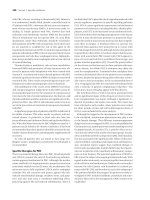
Fig. 2. Juxtaglomerular apparatus. MD = macula densa; JGC =
juxtaglomerular cells; AA = afferent arteriole; EA = efferent
arteriole; N = sympathetic nerve terminal; M = mesangium; GBM
= glomerular basement membrane; E = endothelium; PO =
podocyte; F = foot process; PE = parietal epithelium; B =
Bowman’s space; PT = proximal tubule.
Fig. 1. Biosynthetic cascade of the RAS.
Chapter 23 / Hormones of the Kidney 355
maintaining cardiovascular and renal homeostasis
physiologically, and in developing disease states
pathologically. Accordingly, interruption of the RAS
has become an increasingly important therapeutic
strategy for various cardiovascular disorders such as
hypertension, heart failure, and renal disease.
2.1. Renin
2.1.1. SYNTHESIS AND BIOCHEMISTRY OF RENIN
More than a century ago, Tigerstedt and Bergman
found a potent pressor activity in rabbit kidney extract.
They named a putative substance secreted from the kid-
ney renin, after the Latin word ren (kidney). Forty years
later, Braun-Menéndez et al. and Page et al. showed that
this material was of a protease nature, acting on a plasma
protein to release another pressor substance, which was
later named angiotensin.
Renin (EC 3.4.25.15) is classified as an aspartyl
protease and synthesized as a preproprotein. Renin is
stored and secreted from the renal juxtaglomerular
cells located in the wall of the afferent arteriole, which
is contiguous with the macula densa portion of the same
nephron (Fig. 2). The human renin gene, spanning 12
kb, is located on chromosome 1 (1q32-1q42) and con-
sists of 10 exons and 9 introns. Hormonal-responsive
elements in the 5´-flanking region of the renin gene
include consensus elements for cyclic adenosine
monophosphate (cAMP) and steroids (glucocorticoid,
estrogen, and progesterone). In certain strains of the
mouse, there are two renin genes (Ren-1 and Ren-2),
both located on chromosome 1, and in the rat, the renin
gene is located on chromosome 13. In most mammals,
the kidney is the primary source of circulating renin,
although renin gene expression is found in a number of
extrarenal tissues, including the brain, adrenal, pitu-
itary, submandibular glands, gonads, and heart.
The initial translation product preprorenin, consist-
ing of 406 amino acids, is processed in the endoplasmic
reticulum to a 47-kDa prorenin by removal of a 23-
amino-acid presegment. Prorenin then enters either a
regulated or a constitutive secretory pathway. A sub-
stantial portion of prorenin is further processed, when a
43-amino-acid prosegment is removed, to the active 41-
kDa mature renin, which is a glycosylated single-chain
polypeptide that circulates in human plasma. Prorenin
also circulates in the blood at a concentration several
times higher than active renin. Active renin can be gen-
erated from prorenin by cold storage (cryoactivation);
acidification; or a variety of proteolytic enzymes in-
cluding trypsin, pepsin, and kallikrein. The N- and C-
terminal halves of active renin are similar, and each
domain contains a single aspartic residue in the active
center, which is essential for its catalytic activity.
Angiotensinogen (renin substrate) is the only known
substrate for renin. This reaction appears to be highly
species specific. Human renin does not cleave mouse or
rat angiotensinogen, and human angiotensinogen, in
turn, is a poor substrate for rodent renin.
2.1.2. R
EGULATION OF RENIN RELEASE
Because renin is the rate-limiting enzyme in circulat-
ing Ang II production, control of renin release serves as
a major regulator of the systemic RAS activity. Restric-
tion of salt intake, acute hemorrhage, administration of
diuretics, or acute renal artery clamping results in a
marked increase in renin release. The regulation of renin
release is controlled by four independent factors: renal
baroreceptor, macula densa, renal sympathetic nerves,
and various humoral factors:
1. Mechanical signals, via the baroreceptor or vascular
stretch receptor, of the juxtaglomerular cells sensing the
renal perfusion pressure in the afferent arteriole (Fig. 2):
The renal baroreceptor is perhaps the most powerful regu-
lator of renin release, and reduced renal perfusion pres-
sure strongly stimulates renin release.
2. Tubular signals from the macula densa cells in the distal
convoluted tubule: The cells function as the chemore-
ceptor, monitoring the delivery of sodium chloride to
the distal nephron by sensing the sodium and/or chlo-
ride load through the macula densa cells, and decreased
concentrations within the cells stimulate renin release.
3. The sympathetic nervous system in the afferent arteri-
ole: Juxtaglomerular cells are directly innervated by
sympathetic nerves (Fig. 2), and β-adrenergic activa-
tion stimulates renin release. Renal nerve–mediated
renin secretion constitutes an acute pathway by which
rapid activation of the RAS is provoked by such stimuli
as stress and posture.
4. Circulating humoral factors: Ang II suppresses renin
release (as a negative feedback) independent of alter-
ation of renal perfusion pressure or aldosterone secre-
tion. Atrial natriuretic peptide (ANP) and vasopressin
inhibit renin release, whereas PGE
2
and prostacyclin
(PGI
2
) stimulate renin release.
In addition to the major regulators just described, a
series of other humoral factors is implicated, consider-
ing the finding that the primary stimulatory second
messenger for renin release is intracellular cAMP
whereas the inhibitory signal is increased intracellular
calcium and increased cyclic guanosine monophasphate
(cGMP). For example, local paracrine regulators, such
as adenosine and nitric oxide (NO), may have signifi-
cant influences on renin release, perhaps more impor-
tantly in certain pathologic conditions.
2.2. Angiotensinogen
Angiotensinogen is the only known substrate for
renin capable of producing the family of angiotensin
356 Part IV / Hypothalamic–Pituitary
peptides. In most species, angiotensinogen circulates at
a concentration close to the K
m
for its cleavage by
renin, and, therefore, varying the concentration of
plasma angiotensinogen can affect the rate of Ang I
production. Because angiotensinogen levels in plasma
are relatively constant, plasma concentrations of active
renin, not angiotensinogen, would be the limiting fac-
tor for the rate of plasma Ang I formation in normal
conditions, as determined by the plasma renin activity.
However, in certain conditions such as pregnancy and
administration of steroids, when angiotensinogen pro-
duction is enhanced, circulating angiotensinogen would
have a major effect on the activity of the systemic RAS.
Furthermore, recent studies on the linkage analysis
between angiotensinogen gene and human essential
hypertension suggest that the alterations in plasma
angiotensinogen levels may have a significant impact
on the total RAS activity, affecting blood pressure.
Angiotensinogen shares sequence homology with
α
1
-antitrypsin and belongs to the serpin (for serine pro-
tease inhibitor) superfamily of proteins. The human
angiotensinogen gene (~12 kb long) is located on chro-
mosome 1 (1q42.3) close to the renin gene locus. The
angiotensinogen gene consists of five exons and four
introns, and cDNA codes for 485 amino acids, of which
33 appear to be a presegment. The first 10 amino acids
of the mature protein correspond to Ang I. The 5´-flank-
ing region of the human angiotensinogen gene contains
several consensus sequences for glucocorticoid, estro-
gen, thyroid hormone, cAMP, and an acute phase–
responsive element.
The liver is the primary site of angiotensinogen syn-
thesis and secretion. However, angiotensinogen mRNA
is expressed in a variety of other tissues, including brain,
large arteries, kidney, adipose tissues, reproductive tis-
sues, and heart, which constitutes an important part of
the tissue RAS.
2.3. Angiotensin-Converting Enzyme
ACE, or kininase II (EC 3.4.15.1), is a dipeptidyl
carboxypeptidase, which is a membrane-bound
ectoenzyme with its catalytic sites exposed to the extra-
cellular surface. It is a zinc metallopeptidase that is
required for the final enzymatic step of Ang II produc-
tion from Ang I (Fig. 1). ACE also plays an important
role in the kallikrein-kinin system, by inactivating the
vasodilator hormone bradykinin. In vascular beds,
ACE is present on the plasma membrane of endothelial
cells, where it cleaves circulating peptides; vessels in
the lung, as well as in the brain and retina, are espe-
cially rich in ACE. ACE is also abundantly present in
the proximal tubule brush border of the kidney.
There are primarily two molecular forms of ACE
(somatic and testicular) that are derived from a single
gene by different utilization of two different promot-
ers. Although the majority of ACE is membrane bound,
somatic ACE can be cleaved near the C-terminus, lead-
ing to the release of ACE into the circulation. This
results in three main isoforms of ACE: somatic ACE,
testicular (or germinal) ACE, and soluble (or plasma)
ACE (Fig. 3). The human ACE gene consisting of 26
exons and 25 introns, is located on chromosome 17q23.
The somatic promoter is located in the 5´-flanking
region of the gene upstream of exon 1, whereas the
testicular promoter is present within intron 12. Somatic
ACE is a 170-kDa protein consisting of 1306 amino
acids encoded by a 4.3-kb mRNA, which is transcribed
from exons 1 to 26 except exon 13. It is an extensively
glycosylated protein, containing two highly homolo-
gous domains with an active site in each domain. Tes-
ticular ACE is an approx 90-kDa protein consisting of
732 amino acids, harboring only one C-terminal active
site. This isoform is found only in the testes. Testicular
ACE is encoded by a 3-kb mRNA, transcribed from
Fig. 3. Schematic representation of three isoforms of ACE.
Chapter 23 / Hormones of the Kidney 357
exons 13 to 26, with exon 13 encoding the unique N-
terminus of the testicular isoform.
Somatic ACE is distributed in a wide variety of
tissues, including blood vessels, kidney, heart, brain,
adrenal, small intestine, and uterus, where it is expressed
in the epithelial, neuroepithelial, and nonepithelial cells
as well as in endothelial cells. Somatic ACE in these
tissues (tissue ACE) is postulated to play a crucial role
in the rate-limiting step of the tissue RAS activity. In
addition, studies on the human ACE gene revealed the
presence of a 287-bp insertion (I)/deletion (D) polymor-
phism within intron 16, which may account for the high
degree of individual variability of ACE levels. The D
allele is associated with high plasma and tissue ACE
activity and has been linked to cardiovascular diseases
such as acute myocardial infarction.
In addition to ACE, it is now known that there are
other ACE-independent pathways of Ang II generation
from Ang I (Fig. 1). Among them, chymase, which is
present abundantly in the human heart, is thought to be
most important. The relative importance of such alter-
native pathways in physiologic and pathophysiologic
states, however, is the subject of continuing debate and
awaits further clarification.
2.4. Angiotensin Receptors
For many years, it was thought that Ang II exerts its
effects via only one receptor subtype that mediates vaso-
constriction, aldosterone release, salt-water retention,
and tissue remodeling effects such as cell proliferation
and hypertrophy. This receptor subtype is now termed
the AT
1
receptor. In the late 1980s, it became clear that
there was another Ang II–binding site that was not
blocked by the AT
1
receptor antagonists. This receptor
subtype is now known as the AT
2
receptor. Pharmaco-
logic examinations may suggest the presence of other
receptor subtypes, but to date, no other receptors have
been isolated or cloned.
Most known biologic effects of Ang II are mediated
by the AT
1
receptor. The AT
1
receptor consists of 359
amino acids, with a relative molecular mass of 41 kDa,
and belongs to the G protein–coupled, seven-transmem-
brane receptor superfamily. The principal signaling
mechanism of the AT
1
receptor is through a G
q
-medi-
ated activation of phospholipase C (PLC) with a release
of inositol 1,4,5-trisphosphate and calcium mobiliza-
tion. Activation of the protein tyrosine kinase pathway
may also be involved. In humans, there is a single gene
for this receptor, located on chromosome 3. The human
AT
1
receptor gene consists of five exons and four in-
trons, with the coding region contained within exon 5.
The promoter region contains putative elements for
cAMP, glucocorticoid, and activating protein-1 sites for
immediate early gene products. In rodents, there are
two isoforms of this receptor, named AT
1A
and AT
1B
,
encoded by different genes. These isoforms show a very
high sequence homology (94%) and AT
1A
is considered
to be a major subtype, although the functional signifi-
cance of each isoform is not fully clarified. AT
1
receptor
mRNA is expressed primarily in the adrenals, vascular
smooth muscle, kidney, heart, and specific areas of the
brain implicated in dipsogenic and pressor actions of
Ang II, and it is also abundantly present in the liver,
uterus, ovary, lung, and spleen.
The AT
2
receptor consists of 363 amino acids, with
a relative molecular mass of 41 kDa. This receptor also
exhibits a seven-transmembrane domain topology but
shares only 32% overall sequence identity with the AT
1
receptor. It is likely coupled to a G protein, although it
may also be coupled to a phosphotyrosine phosphatase.
The AT
2
receptor gene, located on chromosome X, is
composed of three exons and two introns, with the entire
coding region contained within exon 3. Expression of
the AT
2
receptor is developmentally regulated. It is
abundantly expressed in various fetal tissues, especially
in mesenchyme and connective tissues; it gets down-
regulated on birth and is not expressed at significant
levels in adult tissues including the cardiovascular
system at normal conditions, being limited to adrenal
medulla, brain, and reproductive tissues. Interestingly,
however, the AT
2
receptor is reexpressed under cer-
tain pathologic conditions, such as on tissue injury and
remodeling, especially in the cardiovascular system.
The signaling mechanism and functional role of the AT
2
receptor have not been fully elucidated, but recent
studies have shown that stimulation of the AT
2
receptor
induces apoptosis and exerts cardioprotective actions
by mediating vasodilatation, probably via activation of
NO and cGMP production. Furthermore, the AT
2
recep-
tor exerts an antiproliferative action on vascular smooth
muscle cells, fibroblasts, and mesangial cells. Thus, it is
now recognized that the AT
2
receptor should act to coun-
terbalance the effects of the AT
1
receptor.
2.5. Angiotensins
A family of angiotensin peptides is derived from Ang
I through the action of ACE, chymase, aminopeptidases,
and tissue endopeptidases. There are at least four bio-
logically active angiotensin peptides (Table 1). Ang I,
decapeptide cleaved from angiotensinogen, is biologi-
cally inactive. Ang II acts on AT
1
and AT
2
receptors,
with equally high affinities. Ang II can be processed by
aminopeptidase A or angiotensinase, to form Ang III.
Like Ang II, Ang III circulates in the blood and shows
somewhat less vasoconstrictor activity but exerts an
almost equipotent activity on aldosterone secretion.
358 Part IV / Hypothalamic–Pituitary
Ang III can be further converted by aminopeptidase B
into Ang 3–8, or Ang IV. In addition, Ang 1–7 can be
produced from Ang I or Ang II by endopeptidases. It is
reported that the fragments Ang IV and Ang 1–7 have
pharmacologic and biochemical properties different
from those mediated by the AT
1
or AT
2
receptors, per-
haps exerting an opposite effect of Ang II such as
vasodilatation. The functional significance and recep-
tors of these peptides, however, still remain elusive.
3. PATHOPHYSIOLOGY OF RAS
3.1. Biological Actions of Ang II
Ang II has short-term actions related to maintaining
normal extracellular fluid volume and blood pressure
homeostasis as well as long-term actions related to car-
diovascular remodeling, most of which are mediated via
the AT
1
receptor. Six primary short-term actions are as
follows:
1. Increasing aldosterone secretion.
2. Constricting vascular smooth muscle, thereby increas-
ing blood pressure and reducing renal blood flow.
3. Increasing the intraglomerular pressure by constric-
tion of the efferent arteriole, contracting the mesangium,
and enhancing sodium reabsorption from the proximal
tubule.
4. Increasing cardiac contractility.
5. Enhancing the sympathetic nervous activity by increas-
ing central sympathetic outflow, and releasing norepi-
nephrine and epinephrine from the adrenal medulla.
6. Promoting the release of vasopressin.
Long-term actions of Ang II include the following:
1. Increasing vascular smooth muscle hypertrophy and
hyperplasia.
2. Promoting cardiac hypertrophy.
3. Enhancing extracellular matrix synthesis, thereby caus-
ing tissue fibrosis.
4. Promoting inflammatory reactions by stimulating the
migration and adhesion of monocytes to the vessel wall.
These actions are closely associated with the cardio-
vascular structural manifestations, or cardiovascular
remodeling, in both human and experimental hyperten-
sion. Ang II also acts on the central nervous system,
increasing thirst and sodium craving. In addition, Ang II
may have potential actions in regulating ovarian and
placental function.
3.2. Tissue RAS
Many tissues and organs can synthesize Ang II inde-
pendent of the classic circulating RAS, and locally
formed Ang II can exert multiple effects acting as an
autocrine and paracrine regulator. Ang II levels may
be much higher in tissues than in plasma. A variety of
tissues express angiotensinogen, renin, ACE, and other
Ang II–generating enzymes, as well as angiotensin
receptors. These additional enzyme systems are referred
to as the tissue RAS.
The effects of locally generated Ang II are long term,
i.e., not just vasoconstriction or salt-water retention,
but the induction of tissue remodeling, modulation of
cell growth, and inflammation. These effects could be
mediated by alternative pathways; thus, these multiple
pathways in tissues allow more ways to synthesize Ang
II, particularly in the areas of inflammation where mast
cells release chymase, monocytes release ACE, and
neutrophils secrete cathepsin G. With the presence
of such non-ACE pathways of Ang II generation, the
inhibition of ACE alone is not theoretically sufficient
to completely inhibit Ang II production. Although the
importance of the tissue RAS has been suggested and
tissue Ang II should be a target for antihypertensive,
antihypertrophic, and antiinflammatory effects, it is
recognized that many of the data available so far are
experimental and there is no definitive proof in humans.
The availability of and analysis with several AT
1
recep-
tor blockers in clinical settings should provide an
answer to this issue.
3.3. Transgenic and Knockout Approaches
Several types of transgenic and knockout animals
have been established to study the functional signifi-
cance of the RAS in vivo. Transgenic lines of mice and
rats harboring both the human renin and angiotensino-
gen genes develop severe hypertension. Hypertension
in the mice likely represents pathologic conditions
brought about by the inappropriate secretion of renin
from outside the kidneys, including pregnancy-associ-
ated hypertension (preeclampsia). Transgenic rats har-
boring the mouse Ren-2 gene exhibited fulminant
hypertension, which overexpressed the transgene in the
adrenal gland. Cardiac-specific overexpression of the
AT
1
receptor resulted in hypertrophy and arrhythmia,
whereas overexpression of the AT
2
receptor in the heart
and vessels showed reduction in hypertrophy and tissue
damage. These models may indicate the functional sig-
nificance of the tissue RAS in cardiovascular control.
Table 1
Angiotensin Peptides
Peptide Sequence
Ang I Asp-Arg-Val-Tyr-Ile-His-Pro-Phe-His-Leu
Ang II Asp-Arg-Val-Tyr-Ile-His-Pro-Phe
Ang III Arg-Val-Tyr-Ile-His-Pro-Phe
Ang IV Val-Tyr-Ile-His-Pro-Phe
Ang 1-7 Asp-Arg-Val-Tyr-Ile-His-Pro
Chapter 23 / Hormones of the Kidney 359
Knockout studies of the components of the RAS
reveal that each component of the cascade (angioten-
sinogen, renin, ACE, and AT
1A
receptor) is indis-
pensible to the maintenance of normal blood pressure.
These knockout animal models invariably show low
blood pressure by ~30 mmHg. Moreover, mice defi-
cient in any component exhibit severe abnormality in
kidney development, characterized by cortical atro-
phy and hypoplasia. ACE-null male mice show greatly
reduced fertility. The AT
2
receptor–knockout mice
reveal enhanced pressor response to Ang II and exagger
-
ated cardiovascular remodeling in response to noxious
stimuli, again suggesting a potential cardioprotective
role of this receptor.
3.4. Genetic Studies
and Clinical Implication
Linkage and association studies have been performed
using polymorphic markers of ACE, angiotensinogen,
renin, and Ang II receptors. In rats, significant linkage
has been demonstrated between the ACE locus and
blood pressure. In humans, on the other hand, no rela-
tion was found between the ACE gene and hyperten-
sion. However, affected sib-pair analysis has found a
strong linkage between the human angiotensinogen gene
and hypertension. Among the polymorphic markers of
the angiotensinogen gene, amino acid conversion at
codon 235 from methionine to threonine (M235T) was
significantly associated with hypertension. 235T sub-
jects also have higher angiotensinogen levels in plasma.
In addition, M235T polymorphism was found to be
linked with several polymorphisms in the 5´-promoter
region of the human angiotensinogen gene, such as
A(-20)C, C(-18)T, and A(-6)G.
The human ACE gene contains an I/D polymorphism
(ACE I/D), characterized by the presence/absence of a
287-bp fragment in intron 16. A significant linkage has
been shown between a deletion polymorphism of the
human ACE gene (ACE DD) and myocardial infarc-
tion. The deletion allele is associated with significantly
increased ACE levels in the tissue and circulation. In
addition, several reports have shown an association
between the ACE DD polymorphism and an increased
risk of cardiovascular events such as restenosis after
coronary intervention, and progression of renal disease
such as IgA nephropathy and diabetic nephropathy.
Multiple lines of evidence have shown that ACE inhibi-
tors and AT
1
receptor blockers are particularly effec-
tive in reducing morbidity and mortality in heart failure,
and in retarding the progression of diabetic and nondia-
betic nephropathies. Therefore, the presence of the
ACE DD polymorphism should provide more compel-
ling indications of these antihypertensive agents.
4. COMPONENTS OF NATRIURETIC
PEPTIDE SYSTEM
Following the discovery of atrial natriuretic peptide
(ANP) from human and rat atrial tissues, two endo-
genous congeners, brain natriuretic peptide (BNP) and
C-type natriuretic peptide (CNP), were isolated from
the porcine brain. These natriuretic peptides share a
common ring structure of 17 amino acids formed by a
disulfide linkage (Fig. 4), which is the essential part of
their biologic actions. The natriuretic peptide system is
a potent natriuretic, diuretic, and vasorelaxing hormone
system, comprising at least three endogenous ligands
and three receptors (natriuretic peptide receptor A
[NPR-A], NPR-B, and the clearance receptor) (Fig. 4).
The accumulated evidence indicates that this system
plays an essential role in the control of blood pressure
and body fluid homeostasis by acting on the kidney and
vasculature as cardiac hormones, as well as by regulat-
ing cardiovascular and renal remodeling, neural con-
trol, and bone metabolism as local regulators.
Furthermore, the importance of this system in the clini-
cal setting has now been established not only as an
excellent diagnostic marker but also as a useful thera-
peutic agent for cardiovascular diseases.
4.1. Natriuretic Peptide Family
4.1.1. ANP AND BNP AS CARDIAC HORMONES
ANP (28-amino-acid peptide) and BNP (32-amino-
acid peptide in humans) act as cardiac hormones. ANP
is predominantly synthesized in the cardiac atrium as
pro-ANP (also called γ-ANP, with 126 amino acids) in
healthy subjects, whereas BNP (from pro-BNP, with
108 amino acids) is mainly produced in the ventricle.
Active peptides reside at the C-terminus of the
prohormones and are cleaved during storage or in a pro-
cess of secretion. Plasma ANP levels are well correlated
with atrial pressure, thereby providing a good marker of
blood volume status. Although BNP was first isolated
from the brain, only small amounts of BNP are detected
in the brain in humans and rodents.
Synthesis and secretion of ANP and BNP are mark-
edly augmented in animal models of ventricular hyper-
trophy and in patients with congestive heart failure
(CHF) in accordance with the severity, in which ven-
tricular production of ANP as well as BNP is signifi-
cantly enhanced. In humans, elevation of BNP becomes
more prominent than ANP in relation to the severity of
heart failure. Therefore, the plasma BNP level is now
the most reliable biochemical marker for left ventricular
dysfunction. In addition, plasma BNP levels are mark-
edly increased in the early phase of acute myocardial
infarction, when plasma ANP is increased only slightly.
360 Part IV / Hypothalamic–Pituitary
It is also shown that a sustained increase in plasma BNP
is associated with decreased ventricular contractility,
increased stiffness, and poor prognosis. These observa-
tions suggest that BNP plays an important role in ven-
tricular remodeling.
ANP and BNP activate a common guanylyl cyclase
(GC)–coupled receptor subtype, NPR-A or GC-A, that
is expressed in a wide variety of tissues. The main
distribution of GC-A includes the kidney, blood ves-
sels, heart, lung, adrenal, and brain. Human urine con-
tains another peptide called urodilatin, an N-terminally
extended form of ANP by four amino acids, which is
synthesized in the kidney and secreted into the tubular
lumen. A functional significance of urodilatin is still
unclear, but it may act as a local regulator of tubular
reabsorption in the distal nephron.
4.1.2. CNP
AS A LOCAL HORMONE
CNP, a 22-amino-acid peptide, is the third member
of the natriuretic peptide family with a highly con-
served ring structure, but uniquely it lacks the C-termi-
nal extension. The precursor structure of CNP is well
preserved among species, and the concentrations of
CNP are much higher than those of ANP and BNP in
the brain, indicating the significance of CNP as a neu-
ropeptide. CNP is found in the cerebral cortex, brain
stem, cerebellum, basal ganglia, and hypothalamus.
Furthermore, CNP is expressed in a variety of periph-
eral tissues, including vascular endothelium, kidney
tubules and glomeruli, adrenal gland, thymus, uterus,
and macrophages. Endothelial production of CNP rep-
resents a potent peptide-type endothelium-derived
relaxing factor. Vascular CNP expression may be
induced in pathologic states such as septic shock and in
injured tissues during vascular remodeling. Notably,
CNP and its receptor, NPR-B or GC-B, are abundantly
expressed in the chondrocytes in the growth plate of
the bone. Transgenic and knockout approaches now
reveal that the CNP/GC-B system is an essential regu-
lator of endochondral bone growth.
4.2. Natriuretic Peptide Receptors
The natriuretic peptide family elicits most of its bio-
logic actions by the activation of particulate GC. Three
classes of NPRs have been identified (Fig. 4), two of
which are the monomeric 130-kDa protein initially des-
ignated as the biologically active receptor, containing
GC-A and GC-B. The other type of receptor not coupled
Fig. 4. Natriuretic peptide system.
Chapter 23 / Hormones of the Kidney 361
to GC, the clearance receptor (C receptor), forms a
homodimer of a 70-kDa protein and is thought to be
involved in the clearance of natriuretic peptides from
the circulation. The rank order of ligand selectivity of
GC-A is ANP Ն BNP >> CNP, whereas that of GC-B is
CNP >> ANP Ն BNP. Thus, GC-A is a receptor for
ANP and BNP, whereas GC-B is selective to CNP. The
rank order of affinity for the clearance receptor is ANP
> CNP > BNP, which is consistent with the lower clear-
ance of BNP than ANP from circulation.
The cDNA sequences of GC-A and GC-B predict
the presence of a single transmembrane domain. The
extracellular putative ligand-binding domains of these
two receptors are 43% identical at the amino acid level
and ~30% identical to that of the clearance receptor.
Just within the plasma membrane lies a protein kinase–
like domain, which may function as a negative regula-
tory element of GC. A cyclase catalytic domain is
present at the C-terminus. The gene for the rat GC-A
spans approx 17.5 kb and is organized into 22 exons
and 21 introns. Exon 7 encodes the transmembrane
domain, and the protein kinase–like and cyclase cata-
lytic domains are encoded by exons 8–15 and 16–22,
respectively. The clearance receptor sequence consists
of 496 amino acids, with a large extracellular domain
and a 37-amino-acid cytoplasmic domain. The bovine
gene for the clearance receptor spans more than 85 kb
and comprises eight exons and seven introns. Exon 1
contains a coding sequence for the large portion of the
extracellular domain, and exons 7 and 8 encode the
transmembrane and cytoplasmic domains, respec-
tively.
Genes of three subtypes of NPRs are widely expressed
with different tissue and cell specificity. GC-A is
expressed in the renal glomeruli, lung, adrenal zona
glomerulosa, heart, and adipose tissue. GC-B exists
in the brain, lung, kidney (mainly in the tubule), pla-
centa, heart, and bone. The clearance receptor is abun-
dantly present in the renal glomeruli, lung, placenta,
and heart.
5. PATHOPHYSIOLOGY
OF NATRIURETIC PEPTIDE SYSTEM
5.1. Biologic Actions of Natriuretic Peptides
Natriuretic peptides exert their actions by activat-
ing GC-A or GC-B, thereby leading to an increase in
intracellular cGMP concentrations. The sites of actions
of ANP and BNP are paralleled with the distribution
of GC-A, whereas CNP actions are dependent on the
expression of GC-B. The effects of natriuretic peptides
can be viewed as a “mirror image” of the RAS, by
generally antagonizing the actions of the RAS both
systemically and locally.
Actions of natriuretic peptides include peripheral and
central effects (Table 2). Renal effects of natriuretic
peptides involve (1) increased glomerular filtration rate,
by potent afferent arteriolar dilation with the modest
efferent arteriolar constriction plus mesangial relax-
ation; (2) increased renal perfusion and medullary
blood flow; and (3) inhibited reabsorption of water
and sodium in the collecting duct and proximal tubule.
Together with the inhibited secretion of renin and aldos-
terone and potent vasodilatation, these effects partici-
pate in their diuretic and antihypertensive effects. The
potent antiproliferative effects on vascular and
mesangial cells may also play important roles in various
pathologic conditions.
5.2. Transgenic and Knockout Approaches
Transgenic and knockout animal models have been
established to study the functional roles of the natri-
uretic peptide system in vivo. Transgenic mice of ANP
or BNP with high circulating levels of these peptides
showed significantly low blood pressure. Moreover,
BNP-transgenic mice appeared to be quite resistant
against various nephropathies and cardiovascular dis-
ease states, suggesting the potential renal and cardio-
vascular protective effects brought about by chronic
excess of circulating natriuretic peptides. Activation of
the CNP/GC-B system in transgenic mice resulted in
skeletal overgrowth.
Knockout studies of the components of the natri-
uretic peptide system have elucidated their distinct
roles. ANP-null mice showed salt-sensitive hyperten-
sion. BNP-null mice, by contrast, were normotensive
but revealed enhanced cardiac fibrosis in response to
pressure overload. Mice lacking GC-A resulted in severe
salt-resistant hypertension, cardiac hypertrophy and
Table 2
Biological Actions of Natriuretic Peptides
Peripheral actions
• Diuresis, natriuresis
• Vasodilatation, reduction in blood pressure
• Inhibition of hormone release: renin, aldosterone
• Inhibition of cell proliferation and hypertrophy:
vascular smooth muscle, mesangium, cardiomyocytes
• Antifibrosis
• Angiogenesis, endothelial regeneration
• Stimulation of endochondral ossification
Central actions
• Inhibition of drinking
• Inhibition of salt appetite
• Reduction in blood pressure
• Inhibition of hormone release: vasopressin, ACTH
362 Part IV / Hypothalamic–Pituitary
fibrosis, and increased sudden death. Therefore, the
ANP/GC-A system is important in regulating blood
pressure and sodium handling, whereas the BNP/GC-A
system plays a role in antifibrosis as a local regulator of
ventricular remodeling. CNP-null mice exhibited
dwarfism owing to impaired endochondral ossification,
indicating that the CNP/GC-B system is essential dur-
ing skeletal development. These studies will provide
plausible evidence for applications of natriuretic pep-
tides to various disease states in clinical settings.
5.3. Clinical Implications
ANP and BNP are elevated in CHF, renal failure, and
hypertension, but their levels appear inappropriately low
for cardiomyocyte stretch caused by chronic volume
and pressure overload. Thus, these disease states may
represent relative natriuretic peptide deficiency. There-
fore, therapeutic strategies are emerging that amplify
the actions of ANP and BNP. One strategy is to admin-
ister these peptides directly, and another is to retard their
metabolic clearance. The latter includes blockade of the
clearance receptor, and inhibition of their degradation
by neutral endopeptidase 24.11 (NEP). Recently, NEP
inhibition has been combined with ACE inhibition in a
series of new antihypertensive agents called vasopep-
tidase inhibitors.
Administration of ANP and BNP has been demon-
strated to exert fairly beneficial effects in patients with
CHF, and this is now considered to be one of the stan-
dard therapeutic strategies in heart failure. Clinical tri-
als with vasopeptidase inhibitors in hypertension are
now ongoing. ANP has also been shown to exert poten-
tial beneficial effects in experimental and clinical acute
renal failure. Clinical efficacy of ANP and vasopep-
tidase inhibitors in chronic renal dysfunction should
await further clarification.
6. KALLIKREIN-KININ SYSTEM
The kallikrein-kinin system consists of four major
components: kininogen, kallikreins, kinins, and kin-
inases (Fig. 5). The kallikrein gene family is a subset of
closely related serine proteases with a narrow range of
substrate specificity. The main function of kallikrein is
the cleavage of a plasma α
2
-globulin known as kinino-
gen to generate kinins, of which bradykinin and Lys-
bradykinin (kallidin) are the main peptides. Kinins are
potent vasodilators with natriuretic, diuretic, and
proinflammatory properties, stimulating the release of
NO, PGs and other mediators. Kinins are short-lived in
vivo because of the presence of kininases (I and II),
which degrade kinins into inactive fragments. Kininase
II is identical to ACE. The multiple roles of the kal-
likrein-kinin system still remain elusive, but recent phar-
macologic and genetic studies suggest the potential
significance of this system in regulating renal salt and
water handling as well as in mediating part of the cardio-
vascular and renal protective effects of ACE inhibitors.
6.1. Synthesis of Kinins
There are two main forms of kininogen: high molecu-
lar weight and low molecular weight. They are encoded
by a single gene and generated by alternative mRNA
splicing. Kininogens are synthesized primarily in the
liver and are present at high concentrations in plasma.
Kininogen is cleaved to release kinins by kallikreins.
Two classes of kallikreins have been identified: plasma
and tissue (glandular). These are separate enzymes that
are derived from different genes and differ in function.
Plasma kallikrein (100 kDa) releases bradykinin only
from high molecular weight kininogen and does not
cleave low molecular weight kininogen. Plasma kal-
likrein is involved in coagulation, fibrinolysis, and pos-
sibly activation of the complement system. By contrast,
Fig. 5. Biosynthetic pathway of kallikrein-kinin system.
Chapter 23 / Hormones of the Kidney 363
tissue kallikrein (24–44 kDa), found principally in the
kidney and in the exocrine and endocrine glands such as
salivary gland and pancreas, cleaves both low molecu-
lar weight and high molecular weight kininogens to
release Lys-bradykinin (Fig. 5). The tissue kallikrein
gene family comprises a large number of closely related
genes. The sizes of this gene family vary among spe-
cies, up to 20 genes in the rat, 24 in the mouse, and 3
in the human. These members exhibit high sequence
homology, suggesting that they share a common ances-
tral gene.
6.2. Kinin Receptors and Their Function
Kinins act on two receptors, B
1
and B
2
receptors,
which differ in tissue distribution, regulation, phar-
macologic properties, and biologic activities. The B
2
receptor has a high affinity to bradykinin and Lys-
bradykinin, whereas the B
1
receptor is selectively acti-
vated by des-Arg
9
-bradykinin or des-Arg
10
-kallidin.
These receptors belong to a seven-transmembrane-
domain, G protein–coupled receptor superfamily. On
stimulation, both B
1
and B
2
receptors lead to activation
of PLC with inositol phosphate generation and calcium
mobilization. The B
2
receptor gene contains three
exons and two introns; the third exon encodes a whole
receptor protein of 364 amino acids, which shows 36%
amino acid identity with the B
1
receptor. The promoter
region of the B
2
receptor gene contains consensus
interleukin-6 (IL-6) and cAMP-responsive elements.
The B
1
receptor is generally not expressed in normal
conditions but appears in pathologic states such as
administration of lipopolysaccharide, inflammation,
and injury. The B
2
receptor, on the other hand, is widely
distributed in many tissues including the kidney, heart,
lung, brain, and testis. Therefore, in normal conditions,
most of the physiologic effects of kinins are mediated
by the B
2
receptor.
Kinins have prominent effects in the cardiovascular,
pulmonary, gastrointestinal (GI), and reproductive
systems. Kinins, via the B
2
receptor, appear to play an
important role in the regulation of local blood flow. In
the vasculature, kinins induce vasodilatation with
release of various mediators, such as NO, PGs, platelet-
activating factor, leukotrienes, and cytokines, and may
be involved in vasodilatation and edema formation
observed during inflammation. Kinins induce smooth
muscle contraction in the GI tract, uterus and bronchi-
oles. The B
2
receptor is also likely to be involved in
renal salt handling and in blood pressure regulation
in individuals consuming a high-sodium diet. The B
1
receptor may be implicated in the chronic inflammatory
and pain-producing responses to kinins, but studies are
still needed to clarify their functional significance.
6.3. Renal Kallikrein-Kinin System
Tissue kallikrein is synthesized in the kidney and
excreted in urine. Filtered kinins, which are active on
the glomerular vasculature, would not be found down-
stream in the nephron because of the high activity of
kininases in the proximal tubule. Renal kallikrein has
been localized by immunohistochemical techniques to
the distal nephron segments, mostly in the connecting
tubule. Kinin receptors are present in the collecting
duct. Therefore, a paracrine role for the renal kal-
likrein-kinin system near the site of action has been
proposed to explain the importance of this system. In
addition, kinins generated in the cortical distal neph-
ron segments may act on the glomerular vasculature,
because the sites are in close association with the glom-
erular tuft.
Pharmacologic evidence shows that kinins play an
important role in the regulation of renal microcircula-
tion and water and sodium excretion. Renal actions of
kinins involve glomerular and tubular actions. Bradyki-
nin dilates both afferent and efferent arterioles and can
increase renal blood flow without significant changes
in glomerular filtration rate, but with a marked increase
in fluid delivery to the distal nephron. It appears that
natriuresis and diuresis are the result of an effect of
kinins on renal papillary blood flow, which inhibits
sodium reabsorption. Kinins also inhibit vasopressin-
stimulated water permeability and sodium transport in
the cortical collecting duct. Because the effect of brady-
kinin is greatly attenuated by cyclooxygenase inhibi-
tion, the natriuretic and diuretic actions of kinins may
be mediated mostly, or at least partly, by PGs.
6.4. Pathophysiology
of the Kallikrein-Kinin System
Decreased activity of the kallikrein-kinin system
may play a role in hypertension. The urinary excretion
of kallikrein is significantly reduced in patients with
hypertension or in children with a family history of
essential hypertension, and the urinary kallikrein levels
are inversely correlated with blood pressure. Reduced
urinary kallikrein excretion has also been described in
various models of genetic hypertension. A restriction
fragment length polymorphism for the kallikrein gene
family in spontaneously hypertensive rats has been
linked to high blood pressure. Collectively, these find-
ings suggest that genetic factors causing a decrease in
renal kallikrein activity might contribute to the patho-
genesis of hypertension.
Endogenous kinins clearly affect renal hemodynam-
ics and excretory function. This notion is supported by
studies using kininogen-deficient Brown Norway rats,
which show a brisk hypertensive response to a high-
364 Part IV / Hypothalamic–Pituitary
sodium diet. Furthermore, B
2
receptor knockout mice
have provided more definitive data supporting the con-
clusion that kinins can play an important role in prevent-
ing salt-sensitive hypertension.
Increased tissue concentrations of kinins and poten-
tiation of their effect may be involved in the therapeutic
effects of ACE inhibitors. This hypothesis is supported
by the finding that a kinin antagonist partially blocks
the acute hypotensive effects of ACE inhibitors. More-
over, beneficial effects on the heart and kidney by ACE
inhibition are significantly attenuated or reversed by
treating with the kinin antagonist, or in mice lacking the
B
2
receptor and kininogen-deficient rats. These data
strongly suggest a potential role of kinins in mediating
part of the cardioprotective and renoprotective effects
exerted by treatment with ACE inhibitors.
7. ADRENOMEDULLIN
AND ENDOTHELINS
7.1. Adrenomedullin
AM is a potent vasorelaxing peptide with 52 amino
acids that is isolated from the adrenal medulla and shares
structural homology with calcitonin gene–related pep-
tide. The preproadrenomedullin gene encodes two
active peptides, AM and proadrenomedullin N-termi-
nal 20 peptide (PAMP), which are generated by post-
translational processing of the same gene. AM is
produced primarily in the vasculature; is released as
an endothelium-derived relaxing factor; and is also
expressed in the adrenal medulla, brain, heart, and kid-
ney. AM exerts its effects via activation of cAMP pro-
duction and nitric oxide synthesis. PAMP, on the other
hand, does not activate cAMP or NO synthesis and
exerts its vasodilatory effects via presynaptic inhibition
of sympathetic nerves innervating blood vessels. AM
receptors are composed of two components, a seven-
transmembrane calcitonin receptor-like receptor and a
single-transmembrane receptor–activity-modifying
protein, whereas PAMP receptors remain elusive and
are yet to be cloned. AM has potent diuretic and natri-
uretic actions, and AM and PAMP also inhibit aldoster-
one secretion. Thus, the AM gene encodes two distinct
peptides with shared biologic activity, but unique
mechanisms of action.
AM increases renal blood flow and has tubular
effects to stimulate sodium and water excretion. AM
also has a potent inhibitory effect on proliferation of
fibroblasts, mesangial cells, and vascular smooth muscle
cells. In addition, experiments of AM infusion and AM
gene delivery have shown that it has a potent vaso-
dilatory and antifibrotic property, resulting in cardio-
vascular and renal protective effects. Furthermore, AM
exerts a potent angiogenic activity, as demonstrated by
AM-deficient mice that exhibit a profound defect in fetal
and placental vascular development, leading to embry-
onic death. In humans, plasma concentrations of AM are
elevated in various cardiovascular disorders including
CHF, hypertension, and renal failure, which may repre-
sent a compensatory role of AM in these disorders.
Furthermore, preliminary clinical studies have revealed
that the administration of AM causes beneficial effects
on CHF and pulmonary hypertension, suggesting the
possibility of potential clinical usefulness of AM in such
diseases.
7.2. ET Family
The vascular endothelium is able to modulate the
vascular tone in response to various mechanical and
chemical stimuli, and such modulation is achieved, at
least partly, by endothelium-derived humoral factors,
relaxing factors and constricting factors. ET was iso-
lated as an endothelium-derived constricting peptide
with 21 amino acids that is the most potent endogenous
vasoconstrictor yet identified. The first peptide identi-
fied is called ET-1, and the ET family now consists of
three isoforms, ET-1, ET-2, and ET-3, acting on two
receptors, ET
A
and ET
B
. ET-1 is the primary peptide
secreted from the endothelium and detected in plasma,
and its mRNA is also expressed in the brain, kidney,
lung, uterus, and placenta. Endothelial ET-1 production
is stimulated by shear stress, hypoxia, Ang II, vaso-
pressin, thrombin, catecholamines, and growth factors
and inhibited by CNP and AM. ET-2 is produced in the
kidney and jejunum, and ET-3 is identified in the intes-
tine, adrenal, brain, and kidney. The ET
A
receptor is
relatively specific to ET-1, whereas the ET
B
receptor
has an equal affinity to three isoforms. Both receptors
are coupled to G proteins, leading to activation of PLC
with inositol phosphate generation and calcium mobili-
zation.
Plasma ET-1 concentrations are elevated in renal
failure, acute myocardial infarction, atypical angina,
essential hypertension, and subarachnoid hemorrhage.
ET-1 exerts a positive inotropic action and potent vaso-
constriction (coronary, pulmonary, renal, and systemic
vasculature) as well as vascular and cardiac hypertro-
phy. An important synergism exists between ET-1 and
Ang II, especially in the heart during cardiac hypertro-
phy, which is counteracted by ANP and BNP. Pharma-
cologic blockade of ET receptors has been effective in
some forms of experimental hypertension and heart
failure, and the nonselective antagonist bosentan has
been approved for treatment of primary pulmonary
hypertension. In the kidney, the receptors are mainly
present in the blood vessels and mesangial cells.
Chapter 23 / Hormones of the Kidney 365
Although these are predominantly the ET
A
subtype,
the ET
B
receptor may have pathophysiologic signifi-
cance, particularly in the distal tubules, where ET
B
receptor activation causes sodium excretion. Involve-
ment of renal ET
B
receptor in sodium-sensitive hyper-
tension remains to be clarified. Furthermore, gene
knockout approaches have revealed that the ET system
plays an essential role during development; the ET-1/
ET
A
system is crucial in branchial arch development
and cardiac septum formation, whose mutation causes
mandibulofacial and cardiac abnormalities. By con-
trast, the ET-3/ET
B
system is essential for migration
of neural crest cells (melancytes and neurons of the
myenteric plexus), whose mutation results in agangli-
onic megacolon (Hirschsprung disease) and vitiligo.
8. ERYTHROPOIETIN
The kidney is the primary organ responsible for
regulating the production of the protein hormone EPO,
in response to perceived changes in oxygen pressure.
A number of experimental and clinical studies have
demonstrated an essential role of the kidney in eryth-
ropoiesis, including the development of severe anemia
by renal ablation, and in renal failure patients. EPO is
a glycosylated protein composed of 165 amino acids
with a relative molecular mass of 34 kDa. Plasma con-
centrations of EPO normally range from 8 to 18 mU/
mL and may increase 100- to 1000-fold in anemia.
EPO mRNA levels are highly sensitive to changes in
tissue oxygenation, and, therefore, its synthesis is regu-
lated primarily at the level of gene transcription.
The site of EPO production in the kidney is now
shown to be the interstitial cells of the renal cortex,
around the base of the proximal tubule. Oxygen defi-
ciency is sensed effectively by the “oxygen sensor”
in these cells. Reduced capillary blood flow may also
induce the increased production of EPO. Studies on the
EPO gene have shown that its production in response to
hypoxia is induced by a transcription factor, hypoxia-
inducible factor-1.
Erythropoiesis begins when the pluripotent stem
cells in the bone marrow are stimulated by nonspecific
cytokines, such as IL-3 and granulocyte-macrophage
colony-stimulating factor, to proliferate and transform
into the erythroid-committed progenitor cells. EPO
then acts on these early progenitor cells bearing its
receptor to expand and differentiate into colony-form-
ing unit-erythroid (CFU-E). EPO further continues to
stimulate CFU-E to erythroid precursors, which even-
tually reach the stage of mature RBCs. CFU-E is the
key target cell for EPO, which indeed regulates RBC
production.
Anemia can develop relatively early in the course of
chronic renal failure, which is referred to as renal ane-
mia. The impairment of EPO production appears to
parallel the progressive reduction of functional neph-
ron mass, and plasma EPO levels are relatively very
low for the degree of severity of anemia in these
patients. Recombinant human EPO can potently reverse
anemia in such states, and its administration has now
widely been performed routinely for correcting anemia
in hemodialysis and peritoneal dialysis patients, as well
as in patients with moderate renal impairment.
SELECTED READINGS
Cambien F, Poirier O, Lacerf L, et al. Deletion polymorphism in the
gene for angiotensin-converting enzyme is a potent risk factor
for myocardial infarction. Nature 1992;359:641–644.
Candido R, Burrell LM, Jandeleit-Dahm KA, et al Vasoactive pep-
tides and the kidney. In: Brenner BM, ed. The Kidney, 7th Ed.,
vol. 1. Philadelphia, PA: W. B. Saunders 2004:663–726.
Chao J, Chao L. New experimental evidence for a role of tissue
kallikrein in hypertension. Nephrol Dial Transplant 1997;12:
1569– 1574.
Chusho H, Tamura N, Ogawa Y, et al. Dwarfism and early death in
mice lacking C-type natriuretic peptide. Proc Natl Acad Sci USA
2001;98:4016–4021.
Drewett JG, Garbers DL. The family of guanylyl cyclase receptors
and their ligands. Endocr Rev 1994;15:135–162.
Dzau VJ. Circulating versus local renin-angiotensin system in car-
diovascular homeostasis. Circulation 1988;77:I-4–I-13.
Horiuchi M, Akishita M, Dzau VJ. Recent progress in angiotensin
II type 2 receptor research in the cardiovascular system. Hyper-
tension 1999;33:613–621.
Inagami T. Molecular biology and signaling of angiotensin recep-
tors: an overview. J Am Soc Nephrol 1999;10(Suppl 11):S2–S7.
John SW, Krege JH, Oliver PM, et al. Genetic decreases in atrial
natriuretic peptide and salt-sensitive hypertension. Science 1995;
267:679–681.
Kitamura K, Kangawa K, Kawamoto M, et al. Adrenomedullin: a
novel hypotensive peptide isolated from human pheochromocy-
toma. Biochem Biophys Res Commun 1993;192:553–560.
Lifton RP, Gharavi AG, Geller DS. Molecular mechanisms of human
hypertension. Cell 2001;104:545–556.
Lopez MJ, Wong SKF, Kishimoto I, et al. Salt-resistant hyperten-
sion in mice lacking the guanylyl cyclase-A receptor for atrial
natriuretic peptide. Nature 1995;378:65–68.
Mukoyama M, Nakao K, Hosoda K, et al. Brain natriuretic peptide
as a novel cardiac hormone in humans: evidence for an exquis-
ite dual natriuretic peptide system, atrial natriuretic peptide and
brain natriuretic peptide. J Clin Invest 1991;87:1402–1412.
Nakao K, Ogawa Y, Suga S, Imura H. Molecular biology and bio-
chemistry of the natriuretic peptide system. I: Natriuretic pep-
tides. J Hypertens 1992;10:907–912.
Suganami T, Mukoyama M, Sugawara A, et al. Overexpression of
brain natriuretic peptide in mice ameliorates immune-mediated
renal injury. J Am Soc Nephrol 2001;12:2652–2663.
Takahashi N, Smithies O. Gene targeting approaches to analyzing
hypertension. J Am Soc Nephrol 1999;10:1598–1605.
Tamura N, Ogawa Y, Chusho H, et al. Cardiac fibrosis in mice
lacking brain natriuretic peptide. Proc Natl Acad Sci USA 2000;
97:4239–4244.
Yanagisawa M, Kurihara H, Kimura S, et al. A novel potent vasocon-
strictor peptide produced by vascular endothelial cells. Nature
1988;332:411–415.
Chapter 24 / Reproduction and Fertility 367
367
From: Endocrinology: Basic and Clinical Principles, Second Edition
(S. Melmed and P. M. Conn, eds.) © Humana Press Inc., Totowa, NJ
24
Reproduction and Fertility
Neena B. Schwartz, PhD
CONTENTS
INTRODUCTION
GONADS AND ACCESSORIES
BRAIN AND PITUITARY
ONTOGENETIC DEVELOPMENT OF REPRODUCTIVE ABILITY
ENVIRONMENT
CYCLICITY: WHAT MAKES THE SYSTEM CYCLE IN FEMALES?
C
ONTRACEPTION
INFERTILITY
NEW FRONTIERS IN REPRODUCTION
If mating does not occur, this cycle is repeated and
mature corpora lutea do not form. Primates run a 28-d
menstrual cycle, which includes an active progesterone-
secreting luteal phase.
Figure 1 is an illustration summarizing the current
understanding of the organs and hormones involved in
regulating reproduction in male and female mammals.
Numbers in the figure are cited in the text in parenthesis.
The left side of Fig. 1 represents the components of the
system in the female mammal; the right side shows the
analogous components in the male.
2. GONADS AND ACCESSORIES
The gonads are characterized by the presence of the
germ cells, their accompanying “nurse cells,” and cells
that secrete sex-specific steroids into the circulation (see
Table 1). Steroid receptors are intracellular, and when
the steroid ligand binds the specific receptor in the tar-
get organ, within either the cytosol or nucleus, the com-
bined entity (transcription factor) binds to specific
nuclear DNA and causes transcription of target genes.
Both the ovaries and testes are totally dependent on two
peptide hormones secreted by the gonadotrope cells
1. INTRODUCTION
The crucial participation of hormones in reproduc-
tion and fertility is the most complicated story in endo-
crinology, because it involves several organ systems;
gametes as well as hormones; two classes of receptors
and intracellular signals; and a myriad of environmental
factors such as seasonal signals and, of course, the
nearby presence of a conspecific carrier of the opposite
gamete type. As complicated as this system is in mam-
mals, being quite different among major classes, it is
even more complex when one deals with the vast num-
ber of nonmammalian vertebrate species. In a marvel-
ous recent review, Rothchild discussed the evolution of
placental mammals from other vertebrates. This chapter
is limited to two mammals: the rat, which has been the
species of choice for elucidating basic science, and the
primate, which is obviously of major interest in dealing
with clinical issues. The rat runs a 4- or 5-day estrous
cycle, from the onset of follicular growth under the
influence of follicle-stimulating hormone (FSH), to
ovulation following an luteinizing hormone (LH) surge.
368 Part IV / Hypothalamic–Pituitary
within the anterior pituitary gland: LH (1-1) and FSH
(1-2). Specific receptors for these hormones are found
within gonadal cell membranes; these receptors are of
the seven-transmembrane loop variety, requiring intra-
cellular second messengers to transmit signals to the
cell nucleus.
2.1. Testis (1-3)
In the adult male, spermatogenesis is continuous,
except in seasonal breeders, with a dividing population
of spermatogonia. It takes 40 d in the rat and 70 d in
humans for a diploid spermatogonium to become four
mature haploid spermatozoa, ready to leave the tubule
and move into the epididymis, where they are stored and
become mature (1-4). Sertoli cells, the “nurse” cells for
future sperm, possess FSH receptors and can aromatize
testosterone to estradiol. Testosterone is synthesized and
secreted by the interstitial cells of the testis (1-5), which
are found outside the spermatic tubules in close proxim-
ity to blood vessels, which empty into the spermatic
veins, then carrying blood back to the heart. FSH is
necessary for the normal functioning of Sertoli cells; in
the absence of FSH, even if LH is present, spermatoge-
nesis does not proceed normally. Spermatogenesis also
depends on local high levels of testosterone diffusing
from the interstitial cells (1-6). Interstitial cells have LH
receptors on their cell membranes and secrete testoster-
one only when LH is present. Sertoli cells also synthe-
size and secrete a peptide hormone called inhibin (1-7),
which can downregulate FSH synthesis and secretion
by pituitary gonadotropes. Testosterone (1-8), acting at
the hypothalamus and in the gonadotrope, can suppress
LH secretion and, in some cases, increase FSH synthe-
sis and secretion.
Table 1
Gonadal Cell Types
Testis Ovary
Nurse cells Sertoli cells Granulosa cells
Gamete Sperm; renewing Ova; maximum number fixed at birth
Steroid-secreting cells Interstitial cells (hormone: testosterone) Granulosa cells (hormone: estradiol)
Sertoli cells (hormone: estradiol) Thecal cells (hormone: testosterone)
Corpus luteum cells (hormone: progesterone)
Fig. 1. Illustration summarizing reproductive system in male and female mammals. E = estrogen; P = progesterone; Test = testoster-
one; LH = luteinizing hormone; FSH = follicle-stimulating hormone; GnRH = gonadotropin-releasing hormone; GC = granulosa
cells; TC = thecal cells; SC = Sertoli cells; IC = interstitial cells; In = inhibin.
Chapter 24 / Reproduction and Fertility 369
2.2. Ovary (1-9)
Oogenesis stops in mammals before birth, when the
oocytes enter the first phase of meiosis. Most oocytes
undergo apoptosis (“atresia”) and die between the pre-
natal meiotic event and adulthood. The oocytes are
encased within the follicles, where they are surrounded
by granulosa cells; the outer layer of the follicle con-
sists of thecal cells (1-10). Granulosa cells initially
express only FSH receptors and the thecal cells express
LH receptors. Meiosis resumes in surviving mature oo-
cytes only after the LH preovulatory surge occurs dur-
ing adult cycles.
Within a given cycle in adults, follicular maturation
occurs in a stepwise fashion. Once cycles begin at
puberty, a surviving follicle (or follicles, in multiovu-
latory species such as the rat) starts to grow, as the
granulosa cells divide under the influence of FSH. The
thecal cells, under the influence of LH, start to synthe-
size and secrete testosterone locally (1-10). The test-
osterone diffuses into the granulose cell layers and is
converted into estradiol by the aromatase enzyme in
the granulosa cell. As the granulosa cells continue to
divide, estradiol secretion into the bloodstream occurs,
and estradiol (1-11) begins to act on target tissues and
to exert negative feedback on the pituitary and hypo-
thalamus (1-12). Inhibin secretion from granulosa cells
also occurs (1-13), and FSH levels fall. The granulosa
cells gradually develop LH receptors. Rising levels of
estradiol abruptly initiate a rapid rise in gonadotropin-
releasing hormone (GnRH) secretion from the hypo-
thalamus (1-14), which causes the preovulatory surges
of LH and FSH. After the preovulatory surge of LH
occurs, a series of molecular events ensue in the ovary
that lead to suppression of estradiol and inhibin secre-
tion, stimulation of progesterone secretion (1-15), and
dispersal of the granulosa cells surrounding the ovum.
The ovum then completes the first stage of meiosis,
throws off the first polar body, and is extruded from the
follicle (1-16) into the oviduct. If sperm are present
fertilization may occur (1-17) and the second polar
body is cast off, leaving the fertilized diploid egg; if
the uterine environment is favorable, owing to proper
action of estradiol and progesterone, implantation of
the growing blastocyst occurs in the uterine lining (1-18)
after about 4 to 5 d in the oviduct.
3. BRAIN AND PITUITARY
The brain and the anterior pituitary gland are linked,
with respect to reproduction, by the secretion of a pep-
tide, GnRH, from the hypothalamus (1-14, 1-21) and
the presence of GnRH receptors on the cell membranes
of the gonadotrope cells. LH and FSH are dimeric pro-
teins, which share a common α-subunit but have dif-
ferent β-subunits, and the entire molecules are
recognized by different specific receptors on the cell
membranes of the gonads. A pulsatile secretion of
GnRH is necessary for continuation of secretion by the
gonadotrope cells. Cell lines of GnRH neurons (Gt1
cells) in culture show spontaneous pulses at a frequency
of about one per hour. Although GnRH pulses cause
secretion of both LH and FSH, differing ratios of the
two hormones can be secreted under the influence of
alterations in GnRH receptor levels, pulse frequency,
and amplitude (Table 2). The GnRH-secreting neurons
are found in the arcuate nucleus of the hypothalamus,
and GnRH is secreted directly into a portal system that
bathes the anterior pituitary cells. LH is more depen-
dent on GnRH than FSH is: increases in GnRH ampli-
tude or frequency enhance LH secretion more than
FSH, and GnRH antagonists lower LH more than FSH.
The greater the number of GnRH receptors on gonad-
otropes, the more LH secretion is favored over FSH.
GnRH receptors (two kinds) are of the seven trans-
membrane domains and are found in the membranes of
the gonadotropes. Most gonadotrope cells synthesize
and contain both LH and FSH, although the ratio of the
Table 2
Factors Altering Relative LH and FSH Secretion
Increase FSH/LH Increase LH/FSH
Low-frequency GnRH High-frequency GnRH
Low number of GnRH receptors High number of GnRH receptors
GnRH antagonists Removal of ovaries
Low inhibin Removal of testes
High activin
Low follistatin
Increased progesterone
Increased glucocorticoids
Increased testosterone
370 Part IV / Hypothalamic–Pituitary
hormones within the cells and their distribution across
the pituitary varies during the cycle, with maximal lev-
els found just before the LH surge. The second-messen-
ger system transducing the GnRH signal to the
gonadotrope is highly complex; it involves the mitogen-
activated protein kinase pathway and calcium mobiliza-
tion. Targets of GnRH activation of its receptor are the
genes for the α- and β-subunits of the gonadotropins and
the GnRH receptor gene itself.
Progesterone (1-15), testosterone (1-5), and gluco-
corticoids enhance FSH synthesis when applied directly
to pituitaries in culture or in vivo (Table 2). There are
also three peptides that are of crucial importance in regu-
lating FSH synthesis and secretion by the pituitary
(Table 2). Inhibin is a heterodimer related to the trans-
forming growth factor-β (TGF-β) family and is secreted
by the ovarian follicles or Sertoli cells, specifically in-
hibiting FSH synthesis and secretion directly (1-7, 1-
13) in the gonadotrope. Activin is a dimer of the inhibin
α-subunit. Activin stimulates FSH synthesis and secre-
tion; activin is made locally in the pituitary gland and is
probably as important as GnRH, if not more important,
in stimulation of FSH secretion (1-19). The third pep-
tide is follistatin (1-20), which is not homologous to the
TGF-β family. It is made in both the ovary and the pitu-
itary gland and acts as an inhibitory binding protein for
activin. Follistatin blocks the stimulation of FSH syn-
thesis and secretion from activin, and also from proges-
terone, testosterone, and glucocorticoids. This suggests
that the steroids act on FSH production via stimulation
of activin.
Hormonal feedback from gonadal steroids acts at
both hypothalamic and pituitary levels (1-8, 1-12) and is
primarily negative in nature because removal of the
gonads increases GnRH and LH and FSH secretion. The
stimulation by estrogen of the preovulatory surges in
female mammals is usually labeled “positive” feedback,
although if ovulation occurs, ovarian secretion is low-
ered as the follicle switches to a corpus luteum, thereby
dropping estrogen secretion and favoring progesterone
secretion.
GnRH secretion (1-14, 1-21) is regulated by many
stimulatory and inhibitory neuromodulators released
from interneuron synapses acting at the GnRH neu-
rons. Excitatory inputs include norepinephrine and
glutamate (
L-glutamic acid), which is the major stimu-
latory agonist in the hypothalamus. Neuropeptide Y
stimulates GnRH release, as well as the action of GnRH
in releasing LH from the pituitary, in animals previ-
ously exposed to estrogen. γ-Amino- butyric acid is
probably the major inhibitory input to the GnRH neu-
rons. Opioids are also inhibitors of GnRH release.
Because GnRH neurons in situ do not contain steroid
receptors, steroids probably act on GnRH release fre-
quency and amplitude by acting on interneurons.
4. ONTOGENETIC DEVELOPMENT
OF REPRODUCTIVE ABILITY
In the early embryo, a set of primordial germ cells
formed in the placental membranes migrates to the uro-
genital ridge, inducing gonad formation with a somatic
contribution from the local epithelium. The Y chromo-
some contains a gene, sry (sex-determining region of the
Y chromosome), which codes for a transcription factor
that induces formation of Sertoli cells. In the absence
of the sry gene, the somatic supporting cell precursors
become follicle cells and proceed toward ovarian devel-
opment. A number of other genes must also be expressed
in order for the formation of normal testis and ovary
to take place. Müllerian-inhibiting substance (MIS) is
secreted by the testis early and diffuses to the locally
forming duct systems; MIS kills the cells of the Müllerian
duct, which would have formed into oviduct/uterus. In
female embryos, this duct system survives but the Wolf-
fian duct system, which is destined to develop into
the male accessory ducts, does not survive because it
depends on testosterone secretion. Testosterone secre-
tion from the developing testis induces masculinization
of the external genitalia. These incipient genitalia cells
must convert the testosterone into dihydrotestosterone
by means of the enzyme α-5-reductase for this mascu-
linizing of the genitalia to take place. For normal female
ovarian, duct, and genitalia differentiation to take place,
both X chromosomes must be present, and a number of
ovarian genes must be expressed.
Normal anterior pituitary gland and hypothalamic dif-
ferentiation is also necessary for reproduction to develop
normally in both male and female genotypes. The cells
that secrete GnRH in the mature individual actually are
derived embryologically from cells in the olfactory region
and migrate during brain development to the hypothala-
mus. Two “orphan” nuclear receptors (SF1 and DAX1)
that appear throughout the hypothalamus, gonadotropes,
gonads, and adrenals are heavily involved with normal
gonadal differentiation and function in both sexes.
Newborn animals are sexually immature. Puberty
occurs when the central drive for GnRH secretion kicks
in, and the threshold for negative feedback of gonadal
hormones increases. Premature puberty in human males
is usually associated with central nervous system mal-
function. Reproductive menopause occurs in females
when the supply of oocytes remaining in the ovaries
becomes inadequate to secrete sufficient estrogen to
trigger LH surges. In males, testosterone levels drop
with aging, but generally reproduction is attenuated later
than in females.
Chapter 24 / Reproduction and Fertility 371
5. ENVIRONMENT
The connection of the reproductive system to the brain
via GnRH provides the conduit whereby the environment
provides input to the system. For mammals, there is a
value for birth to occur in the spring, when food supplies
are most plentiful. Sheep ovulate and mate in the fall, as
the ratio of dark to light increases, and with the long
gestation period give birth in the spring. Small rodents,
by contrast, with a 20-d gestation period, mate and ovu-
late in the spring, when the light/dark ratio is increasing.
For some species near the equator, because the hours of
light and dark remain equal throughout the year, rainfall
rather than light may serve as a seasonal signal.
Other species, such as the domestic cat, the rabbit,
and the camel, are coitus-induced ovulators. In these
species, estradiol stimulates mating behavior, and the
cervical stimulus received during coitus triggers a neu-
ral reflex that causes a large release of GnRH. This trig-
gers a preovulatory surge of LH that causes ovulation.
6. CYCLICITY: WHAT MAKES
THE SYSTEM CYCLE IN FEMALES?
Figure 2 illustrates the changes in the pituitary and
ovarian hormones during the nonpregnant rat and pri-
mate cycles. The female rat (Fig. 2A) and primate
(Fig. 2B) manifest repetitive cycles because the rising
levels of estrogen stimulated by background levels of LH
and FSH cause the release of a burst of GnRH, which
causes the abrupt increase in LH (and FSH) release. These
preovulatory surges of LH and of FSH not only cause
resumption of meiosis in the most mature follicle(s), but
also a cascade of enzymatic changes within the granulosa
cells, which terminate estradiol secretion.The resultant
corpus luteum begins secreting progesterone. (By con-
trast, in the male mammal, testosterone and LH levels are
maintained at steady levels from day to day, except for
the oscillations in both that track GnRH pulses.)
There are two principal operational differences
between
the rodent and primate cycles. The first is that
Fig. 2. Time course of changes in estrogen, progesterone, LH, and FSH during rat and primate nonpregnant cycle: (A) rat estrous
cycle. (B) Primate menstrual cycle.
372 Part IV / Hypothalamic–Pituitary
the rodent cycle is tightly tied to the daily light-dark
timing. Not only is the rising estrogen level in the blood
a necessary signal for the GnRH release that precedes
the LH surge, but there is also a daily circadian signal
that occurs between 2:00
PM and 4:00 PM in rats kept in
a room lighted from 5:00
AM to 7:00 PM. This neural
signal acts in conjunction with the estrogen to closely
time the LH/FSH release (Fig. 2A). There is no evi-
dence that such a circadian signal operates with estro-
gen in the primate. The second difference between the
rodent and the primate has to do with the luteal phase.
In the rat, mouse, and hamster, there is no spontaneous
luteal phase analogous to that in the primate following
ovulation. In the primate, the LH surge that triggers
ovulation is also adequate to maintain progesterone
secretion from the corpus luteum, until placental secre-
tion takes over. In the rat, if pregnancy does not occur,
blood levels of both estradiol and progesterone remain
low; the resulting absence of steroidal negative feed-
back (1-12) permits FSH and LH to rise. The prolonged
FSH secretion in the rodent cycle (“secondary FSH
surge”) (Fig. 2A) occurs because inhibin secretion by
the ovary is terminated by the LH surge; this elevated
FSH initiates the growth of the next crop of follicles.
However, in the presence of a male, the precedent estro-
gen secretion followed by the brief proestrous proges-
terone surge induces sexual receptivity in the female
late in the afternoon and mating occurs. The stimulation
of the cervix by mating turns on twice daily surges of
prolactin in the female, which maintain the progester-
one secretion for 12 d or so, permitting implantation.
Pregnancy occurs when a developing embryo implants
into the lining of the uterus, prepared by the preceding
estrogen and progesterone secretion. In the primate, the
corpus luteum formed after the LH surge secretes
progesterone spontaneously for about 12 d. For preg-
nancy to continue, the corpus luteum needs to continue
secreting progesterone for about 14 d in the rat and
2 mo in the primate. This steroid is critical for suppress-
ing uterine contractions. Once the embryo is securely
implanted in the uterine lining (1-18), the placenta (part
maternal, part embryonic) secretes the chorionic gona-
dotropin necessary to maintain the corpus luteum and
eventually also secretes the steroids necessary for main-
tenance of the pregnancy and the onset of lactation.
7. CONTRACEPTION
The population of our planet continues to grow ex-
ponentially, threatening the environment and outpac-
ing food and water supplies. An understanding of the
linkages in Fig. 1 is crucial to the design of contracep-
tives. The oral contraceptive pill, used by vast numbers
of females worldwide, is predominantly progesterone-
like,and suppresses GnRH, FSH, and LH secretion (1-
8), so that ovulation does not occur (1-12). Depopro-
vera is a progestin implant that frees females from
having to ingest pills on a daily basis. Testosterone
implants have been tested in males as a contraceptive;
the high levels of testosterone suppress GnRH, LH, and
FSH secretion, thus suppressing spermatogenesis,
without depriving the recipient of testosterone neces-
sary for libido and potency (1-8). Condoms block the
gametes from meeting and have the advantage of
detering the spread of sexually transmitted diseases. In
the age of acquired immunodeficiency syndrome and
relative sexual freedom, this advantage is extremely
important. Tying of the oviducts (1-16) in females or of
the vas deferens (1-4) in males, obviously, prevents the
gametes from meeting. These simple methods have the
advantage of a brief surgery and no drugs with possibly
harmful side effects. They are popular in older, stable
couples who have completed their families. However,
they must be regarded as irreversible, at present. The
intrauterine device is a loop that is inserted into the
uterus. It alters the uterine luminal environment (1-18)
such that implantation cannot take place normally.
Antisera to LH or FSH (1-1, 1-2) have been tested in ani-
mals and in some human studies; questions of reversi-
bility, side effects, and efficacy are still unsettled.
GnRH antagonists have been tested in men as a contra-
ceptive. Because they reduce LH and FSH (1-21), they
also reduce testosterone secretion so potency falls; if
they are to be useful they must be accompanied by tes-
tosterone.
In females, the “morning after pill,” an estrogen ana-
log, diethylstilbestrol, can prevent pregnancy from
occurring after unprotected sex, apparently by render-
ing the oviductal environment unsuitable for fertiliza-
tion or survival of the fertilized egg. RU486, an
antagonist of the progesterone receptor, can be ingested
within a couple of weeks of the onset of pregnancy to
cause loosening of the implanted embryo. Following
RU486, a prostaglandin-like drug is taken, which ini-
tiates uterine contraction, thus expelling the embryo.
8. INFERTILITY
If differentiation of the gonads or the tracts does not
occur normally, as in the absence of the sry gene respon-
sible for testicular development, or any of the cascade
of genes responsible for steroid synthesis, irreversible
infertility results. If chromosomal abnormalities such
as XO or XXY occur, the resultant inadequate ovary
(XO: Turner syndrome) or inadequate testis (XXY:
Klinefelter syndrome) will result in infertility. Muta-
tion of steroid receptors or of peptide receptors in tar-
get tissue can cause infertility, by preventing gamete