Endocrinology Basic and Clinical Principles - part 10 ppsx
Bạn đang xem bản rút gọn của tài liệu. Xem và tải ngay bản đầy đủ của tài liệu tại đây (1.69 MB, 45 trang )
Chapter 26 / Endocrinology of the Ovary 393
mRNA and protein synthesis in the oocyte, and it begins
to increase in size.
The signal for follicle recruitment is unknown. It is
known that recruitment can occur in hypophysecto-
mized animals, indicating that recruitment is not depen-
dent on luteinizing hormone (LH) or FSH. There is
evidence that the rate of recruitment can be modulated
by intraovarian and environmental factors. The rate of
recruitment is related to the total number of primordial
follicles in the ovaries, indicating that intraovarian
mechanisms are important for regulating recruitment.
Evidence from experiments in rodents indicates that
recruitment can be attenuated by neonatal thymectomy,
starvation, or administration of exogenous opioid pep-
tides, suggesting that there may be endocrine signals
capable of modulating the rate of recruitment.
2.2. Selection of Dominant Follicle
The selection of the dominant follicle is one of the
final steps in the year-long program of follicle develop-
ment. In women, the follicle that will ovulate is selected
in the early follicular phase of the menstrual cycle. At
that time, each ovary contains a cohort of rapidly grow-
ing follicles 2–5 mm in diameter. These small antral
follicles contain a fully grown oocyte, approx 1 million
granulosa cells, and several layers of theca cells. From
this cohort, the follicle most advanced in the develop-
mental program is selected to become dominant. Once
it reaches a size of 6–8 mm in the early follicular phase,
changes occur, possibly in the structure of the basal
lamina, that permit FSH to enter the follicle and begin
to stimulate the granulosa cells. The granulosa cells and
theca cells of the selected follicle show a high rate of cell
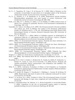
Fig. 2. Morphology of ovarian follicle.
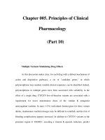
Fig. 3. Ultrastructure of ovarian steroid-secreting cells. The spe-
cialized ultrastructure of steroid-producing cells includes mito-
chondria (M) with vesicular cristae, abundant agranular ER, and
numerous lipid vesicles (L) containing cholesteryl esters. N =
nucleus (magnification: ×21,000)
394 Part IV / Hypothalamic–Pituitary
proliferation, whereas mitosis stops in the cells of other
cohort follicles. The ability to sustain a high capacity for
rapid cell division is a characteristic feature seen only in
dominant follicles. The smaller follicles in the cohort
with slower growth inevitably undergo atresia.
When biologically active FSH first enters the fol-
licle at about 6 to 7 mm, the granulosa cells begin to
express the aromatase enzyme and to secrete estradiol.
In addition, the granulosa cells begin to secrete increas-
ing amounts of inhibin B. Together, these hormones
cause a small but significant and progressive decre-
ment in the circulating FSH concentration owing to
their inhibitory effects on pituitary secretion. The lack
of FSH support to the cohort follicles causes develop-
mental failure and certain atresia. Counteracting the
FSH withdrawal by administration of exogenous FSH
is the basis for ovulation induction protocols that are
used clinically to develop multiple preovulatory fol-
licles for assisted reproduction techniques.
In contrast to the cohort follicles, the dominant fol-
licle preferentially sequesters FSH in the follicular
fluid, thus enabling it to maintain adequate FSH sup-
port even though circulating FSH concentrations
decline. Another important mechanism that confers a
developmental advantage to the dominant follicle is
sensitization of the follicle cells to FSH. The granulosa
cells of the dominant follicle produce growth and dif-
ferentiation factors, such as insulin-like growth factors
(IGFs) and inhibin, that augment the stimulatory
effects of FSH. By virtue of these mechanisms, the
dominant follicle can continue to grow and thrive
while the cohort follicles die. By simply changing
the concentration of FSH during the follicular phase
of the cycle, the number of preovulatory follicles can
be determined.
The theca cells do not respond to FSH but are regu-
lated by LH. The mean circulating concentrations of
LH do not change appreciably during the follicular
phase of the menstrual cycle. At the time theca cells
first appear in secondary follicles, they have ste-
roidogenic capacity, but the stimulatory effects of
LH are attenuated by granulosa cell–secreted factors.
Because estradiol is a key mediator of follicle selection
and theca cell steroidogenesis is essential for the fol-
licle to secrete estradiol, it is important for thecal ste-
roidogenesis to increase in dominant follicles. It is
likely that the same factors that sensitize the granulosa
cells to FSH also augment the stimulatory effects of LH
on theca cell steroidogenesis. Thus, theca cell steroido-
genesis is enhanced only when the granulosa cells have
expressed the aromatase enzyme. In women, the capac-
ity to secrete large amounts of estrogen is the exclusive
property of dominant follicles.
2.3. Atresia
Greater than 99% of the follicles present in the ova-
ries die by atresia. Atresia occurs in both preantral and
antral follicles and is not exclusively related to the fail-
ure of a follicle to become dominant. Indeed, approx
95% of the follicles become atretic prior to the first
ovulation.
The process of follicle atresia occurs by apoptosis.
The granulosa cells undergo nuclear and cytoplasmic
condensation, plasma membrane blebbing, and the
release of apoptotic bodies containing cellular orga-
nelles. The nuclear DNA undergoes internucleosomal
cleavage, and the cellular fragments are removed from
the ovary by phagocytosis. It is clear that removal of
FSH support from follicles in the gonadotropin-depen-
dent stages of follicle development will trigger atresia,
but the causes of apoptosis in preantral follicles are less
certain.
3. STEROID HORMONE PRODUCTION
3.1. Two-Cell, Two-Gonadotropin Concept
of Follicle Estrogen Production
The production of large quantities of estradiol is one
of the most important endocrine functions of the domi-
nant follicle. It is through estradiol concentrations that
the state of follicle development is communicated to the
hypothalamus and pituitary such that the midcycle ovu-
latory surge of LH is timed appropriately. Another key
function of estradiol is to prepare the endometrium for
implantation of the embryo.
Experiments conducted during the 1950s demon-
strated that both the theca interna and the granulosa com-
partments of the ovarian follicle are required for
estradiol production. In addition, both LH and FSH
stimulation are required for estradiol production to
occur. These observations have been confirmed many
times in a variety of mammalian species, and the molec-
ular basis for the two-cell, two-gonadotropin concept
for follicle estrogen biosynthesis has been established
(Fig. 4).
From the time the theca cells first differentiate into
endocrine cells, they contain LH receptors and the key
steroidogenic enzymes required for androgen biosyn-
thesis from cholesterol: cholesterol side-chain cleav-
age cytochrome P450 (CYP11A), 3β-hydroxysteroid
dehydrogenase (3β-HSD), and 17α-hydroxylase/C
17–
20
lyase cytochrome P450 (CYP17). Thus, the theca
cells are endowed with the capacity to synthesize
androgens from cholesterol de novo under the control
of LH. Although the principal androgen secreted by
the theca cells is androstenedione, the human CYP17
e
nzyme is extremely inefficient at converting 17β-
Chapter 26 / Endocrinology of the Ovary 395
hydroxyprogesterone to androstenedione. Consequently,
steroidogenesis proceeds via the delta 5 pathway, where
17β-hydroxypregnenolone is metabolized into dehydro-
epiandrosterone (DHEA) by the CYP17 enzyme, and
then DHEA is converted into androstenedione by the
3β-HSD. The theca cells in women do not express
aromatase CYP19 and, hence, cannot produce estra-
diol. In certain species, notably the horse and pig,
theca cells do express low levels of CYP19 and can
produce small amounts of estrogen; however, coop-
eration with the granulosa cells is still required to
secrete high concentrations of estradiol.
In contrast to the theca cells, the granulosa cells are
incapable of de novo steroidogenesis in the follicular
phase of the menstrual cycle. It is not until the peri-
ovulatory period that the granulosa cells express LH
receptors and CYP11A as they begin to luteinize. There-
fore, in the follicular phase of the cycle, the granulosa
cells cannot produce the androgen substrate required
by the CYP19 enzyme. When a follicle is selected to
become dominant, the granulosa cells express high lev-
els of CYP19 and 17β-hydroxysteroid dehydrogenase
(17β-HSD) under the control of FSH. This enables the
granulosa cells to metabolize the androstenedione pro-
duced by the theca cells to estradiol. Thus, it takes two
cells, theca and granulosa, and two gonadotropins, LH
and FSH, for the ovarian follicle to produce estradiol.
3.2. Intracellular Compartmentalization
of Steroidogenic Enzymes
The regulation of steroid hormone production occurs
in two ways. Acute regulation of the rate of steroidogen-
esis takes place by controlling the rate of cholesterol
access to the CYP11A enzyme. This is possible because
the CYP11A is localized in the inner leaflet of the inner
mitochondrial membrane (Fig. 5). Because cholesterol
is sparingly soluble in water, diffusion from the outer to
the inner mitochondrial membrane is very slow. Acute
stimulation with LH causes production of the steroido-
genesis acute regulatory protein (StAR) that facilitates
the transport of cholesterol across the mitochondrial
membranes. StAR is thought to function by bringing the
outer and inner mitochondrial membranes into contact
at focal points, thus facilitating the movement of choles-
terol from the outer to the inner membrane. The activity
of the StAR protein is rapidly terminated by proteolytic
cleavage. When cholesterol is present in the inner mito-
chondrial membrane, the CYP11A enzyme readily
metabolizes it to pregnenolone. Pregnenolone is able to
diffuse out of the mitochondria, where it is metabolized
to other steroids that, in the ovary, are localized in the
microsomes.
3.3. Hormonal Regulation
of Cellular Differentiation
The second means for regulating steroid hormone
biosynthesis is to control cellular differentiation by
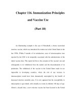
Fig. 4. Two-cell, two-gonadotropin concept of follicle estrogen
production. LH stimulates the theca cells to differentiate and
produce androstenedione from cholesterol. FSH stimulates the
differentiation of the granulosa cells. The androstenedione dif-
fuses across the basal lamina and is metabolized to estradiol in
the granulosa cells. Gs = stimulatory G-protein; AC = adenylate
cyclase, ATP = adenosine triphosphate; cAMP = cyclic adenos-
ine monophosphate.
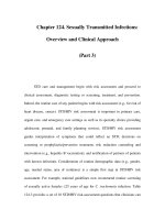
Fig. 5. Compartmentalization of steroidogenic enzymes. Diffu-
sion of cholesterol across the mitochondrial membranes is facili-
tated by StAR (S). The function of StAR is terminated by
proteolysis. Cholesterol in the mitochondria is converted into
pregnenolone by CYP11A. Pregnenolone diffuses out of the
mitochondria and is metabolized to other steroids in the mi-
crosomes. In the ovary, depending on the cell type, the final
product may be progesterone or androgen.
396 Part IV / Hypothalamic–Pituitary
altering the concentrations of the various steroidogenic
enzymes expressed in the cells. Changes in the concen-
trations of steroidogenic enzymes occur over more pro-
longed and developmentally regulated time frames on
the order of days or longer, whereas the acute regulation
of steroidogenesis occurs on the order of minutes.
The signal initiating granulosa cell growth and dif-
ferentiation has not been fully defined. It is clear that
gonadotropins are not involved because the granulosa
cells in primordial follicles do not express FSH or LH
receptors. Evidence is beginning to emerge indicating
that proteins secreted by the oocyte such as growth dif-
ferentiation factor-9, a member of the transforming
growth factor-β (TGF-β) superfamily, play an essential
role in initiating follicle development. Prior to the selec-
tion of the dominant follicle, the granulosa cells do not
express CYP19 and therefore do not contribute to estra-
diol production.
When preantral follicles contain approximately two
layers of granulosa cells, the granulosa cells secrete
proteins into the stroma that cause undifferentiated
mesenchymal cells to differentiate into theca cells. The
signals have not been fully defined, but it appears that
several small molecular weight proteins potentially
including IGF-1 and stem cell factor or kit ligand may
be components of the differentiation signal. When the
theca cells first differentiate, they contain LH receptors,
StAR, CYP11A, 3β-HSD, and CYP17. Thus, they are
capable of producing androstenedione at the preantral
stage of follicle development.
Excessive androgens can have detrimental effects on
ovarian function; therefore, it is beneficial to ensure that
androgens do not accumulate before CYP19 is expressed
in the granulosa cells. The granulosa cells secrete sev-
eral factors that inhibit the stimulatory actions of LH on
theca cell steroidogenic enzyme gene expression and
androgen production including activin and TGF-β
(Table 1).
If a follicle becomes selected, the inhibitory signal
from the granulosa cells changes to one in which the
stimulatory effects of LH are enhanced. Many of the
same molecules both enhance the effects of LH on theca
cell differentiation and sensitize the granulosa cells to
the stimulatory effects of FSH. It is through the enhance-
ment of LH and FSH action by factors such as IGF-1 and
inhibin family members (Table 1) that expression of
steroidogenic enzymes in the theca cells and CYP19 in
the granulosa cells is increased even though the concen-
trations of LH and FSH do not increase in the circula-
tion. Although the nature of the signals is not fully
understood, it is clear that there is a detailed system of
communication among the oocyte, granulosa cells, and
theca cells that ensures that the differentiation and func-
tion of the follicle cells are coordinated. Successful
completion of this developmental program results in a
preovulatory follicle ready to ovulate.
4. OVULATION
Ovulation is the end process of a series of events
initiated by the gonadotropin surge and resulting in the
Table 1
Autocrine/Paracrine Factors Regulating Ovarian Steroid Hormone Production
Cellular Effect on LH-dependent Effect on FSH-dependent
Factor
a
origin androgen production in vitro
b
estrogen production in vitro
b
Growth factors • IGF-I GC + +
• Activin GC – +
• Inhibin GC + –
• TGF-β GC/TC – +
• TGF-α TC – –
• bFGF GC – –
• NGF TC + ?
• GDF-9 Oocyte + –
• HGF TC – –
• KGF TC – ?
Cytokines • TNF-α Oocyte/GC/resident – –
ovarian macrophages
• IL-1β GC/resident ovarian macrophages – –
a
IGF-1 = insulin-like growth factor-1; TGF-β = transforming growth factor-β; bFGF = basic fibroblast growth factor; NGF = nerve
growth factor; GDF-4 = growth differentiation factor-9; HGF = hepatocyte growth factor; KGF = keratinocyte growth factor; TNF-α = tumor
necrosis factor-α; IL-1β = interleukin-1β.
b
+, augments; –, inhibits; ?, unknown.
Chapter 26 / Endocrinology of the Ovary 397
release of a mature fertilizable oocyte from a Graafian
follicle. During the second half of the follicular phase
and as follicles grow, plasma estradiol concentrations
begin to rise. About 24–48 h after plasma estradiol lev-
els reach a peak, the midcycle LH surge takes place.
This preovulatory LH surge occurs at around d 14 of a
28-d cycle, with a total duration of approx 48 h. Ovula-
tion occurs 36 h after the onset of the LH surge. Proges-
terone and FSH levels remain low in the follicular phase
until just before ovulation. At this time, a small FSH
surge accompanies the greater LH surge, and progester-
one levels rise slightly just before ovulation.
The precise hormonal regulation mechanisms oper-
ating during ovulation are not fully elucidated. How-
ever, it is well known that the gonadotropin surge at the
end of the follicular phase is essential for ovulation.
The midcycle LH surge results from activation of posi-
tive estradiol feedback at the level of both the pituitary
and hypothalamus. The increasing amounts of estra-
diol secreted by the dominant follicle trigger the hypo-
thalamic gonadotropin-releasing hormone (GnRH)
surge. The administration of a GnRH antagonist in
women prevents the surge or interrupts it if it has
already started. This suggests that GnRH is necessary
not only for the surge to occur but also for the mainte-
nance of the surge. Additionally, the pituitary LH surge
is facilitated by an increased responsiveness of gonad-
otrope cells to GnRH observed following exposure to
rising estradiol and by an increase in GnRH receptor
number. The feedback signal to terminate the LH surge
is unknown. The decline in LH may be owing to the
loss of the positive feedback effect of estrogen, result-
ing from the increasing inhibitory feedback effect of
progesterone, or owing to a depletion of LH content of
the pituitary from downregulation of GnRH receptors.
The rise in progesterone concentrations may lead to a
negative feedback loop and inhibit pituitary LH secre-
tion by decreasing GnRH pulse frequency. Moreover,
LH downregulates its own receptors just before ovula-
tion, resulting in decreased estrogen production.
The LH surge stimulates resumption of meiosis I
in the oocyte with release of the first polar body. The
oocyte nucleus or germinal vesicle undergoes a series of
changes that involve germinal vesicle breakdown and
the progression of meiosis to the second meiotic
metaphase or first polar body stage. It has been sug-
gested that the LH surge overcomes the arrest of meiosis
by inhibiting the oocyte maturation inhibitor (OMI)
secretion. This inhibitor is produced by granulosa cells
and leads to the arrest of meiosis during folliculogenesis.
It appears that OMI exerts its inhibitory action on meio-
sis, not directly on the oocyte, but acts to increase the
concentrations of cAMP in the cumulus cells, which
then passes via gap junctions into oocyte and halts mei-
otic maturation. The LH surge, by inhibiting OMI secre-
tion and thereby decreasing cAMP, allows the
resumption of meiosis. The second meiotic division is
completed at the time of fertilization, if it occurs, yield-
ing the ovum with the haploid number of chromosomes
and the second polar body that is released.
With the LH surge, the production of antral fluid in
the dominant follicle increases, and the follicle enlarges
markedly. This results in a relatively thin peripheral
rim of granulosa cells and regressing thecal cells to
which the oocyte, with its associated cumulus cells, is
attached only by a tenuous and thinning stalk of granu-
losa cells. The increasing size of the follicle and its
position in the cortex of the ovarian stroma cause it to
bulge out from the ovarian surface, leaving only a thin
layer of epithelial cells between the follicular wall and
the peritoneal cavity. At one site on its surface, the
follicle wall becomes even thinner and avascular; the
cells in this area dissociate and then appear to degen-
erate and the wall balloons outward. The follicle then
ruptures at this site, the stigma, causing the fluid to
flow out on the surface of the ovary, carrying with it
the oocyte and its surrounding mass of cumulus cells.
Follicle rupture and oocyte extrusion are evoked by
LH and progesterone-induced expression of pro-
teolytic enzymes such as collagenases. Enzymatic deg-
radation of the follicle wall is a primary hypothesis to
explain the rupture. Increased prostaglandin (PG) syn-
thesis also appears to play a role in the extrusion of the
oocyte. PGs probably contribute to the process of ovu-
lation through various pathways, such as affecting the
contractility of the smooth muscle cells on the ovary
and activating proteolytic enzymes, especially those
associated with collagen degradation.
5. LUTEINIZATION
Luteinization is the process that transforms the granu-
losa and theca cells into luteal cells. This process is
triggered by the surge of LH at midcycle, once the granu-
losa cells have acquired receptors for LH, and does not
necessarily signify that ovulation has occurred. The LH
surge causes profound morphologic changes in the fol-
licle that becomes corpus luteum. These include acqui-
sition by the granulosa cells of the capacity of de novo
synthesis of steroids (mainly progesterone and estro-
gen) and invasion of the previously avascular granulosa
cell layer by a vascular supply.
After ovulation and expulsion of the unfertilized egg,
the granulosa cells continue to enlarge, become vacu-
olated in appearance, and begin to accumulate a yellow
pigment called lutein, and they are now called as granu-
losa lutein cells. Luteinization of granulosa cell involves
398 Part IV / Hypothalamic–Pituitary
the appearance of lipid droplets in the cytoplasm, devel-
opment by the mitochondria of a dense matrix with
tubular cristae, hypertrophy of the ER and enlargement
of the granulosa cell into the “large luteal cell.” Thecal
cells are also luteinized (theca-lutein cells) and make up
the outer portion of the corpus luteum. These “small
luteal cells” are much less active in steroidogenesis and
have no secretory granules. The basal lamina of the fol-
licle dissolves, and capillaries invade into the granulosa
layer of cells in response to secretion of angiogenic fac-
tors such as vascular endothelial growth factor by the
granulosa and thecal cells.
The corpus luteum is a transient endocrine organ that
predominately secretes progesterone, and its primary
function is to prepare the estrogen-primed endometrium
for implantation of the fertilized ovum. The granulosa-
lutein cells express cholesterol side-chain cleavage
enzyme and 3β-HSD, and, accordingly, they have a
high capacity to produce progesterone and estradiol.
Blood vessels penetrating the follicle basal lamina pro-
vide these cells with low-density lipoproteins, the main
source of cholesterol as a substrate for progesterone
and estradiol synthesis in luteal cells. Seven days after
ovulation, approximately around the time of expected
implantation, peak vascularization is achieved. This
time also corresponds to peak serum levels of proges-
terone and estradiol. The secretion of progesterone and
estradiol is episodic and correlates with the LH pulses.
During the process of luteinization, LH is required to
maintain steroidogenesis by granulosa-lutein cells. The
role of other luteotropic factors such as prolactin (PRL),
oxytocin, inhibin, and relaxin remains unclear. Theca-
lutein cells that express the enzymes in the androgen
biosynthetic pathway and produce androstenedione
are also involved in steroid biosynthesis.
The life-span of the corpus luteum is 14 days after
ovulation and depends on continued LH support. The
mechanism involved in maintaining the function of the
corpus luteum for 14 d and in precipitating the process
of luteolysis (programmed cell death) at the end of this
period is incompletely understood. It is clear, however,
that LH maintains the functional and morphologic
integrity of the corpus luteum, yet it is insufficient to
prevent luteolysis. Corpus luteum function declines by
the end of the luteal phase unless human chorionic gona-
dotropin (hCG) is produced by a pregnancy. Luteolysis
can be viewed as a default response to lack of stimula-
tion by hCG. If pregnancy does not occur, the corpus
luteum undergoes luteolysis under the influence of
luteolytic factors. These factors include estradiol, oxy-
tocin, and PGs. The luteolytic effect of both estrogen
and oxytocinappears to be mediated, at least in part, by
local formation of PGF
2a
. PGF
2a
exerts its effects via
the synthesis of endothelin-1 (ET-1), which inhibits
steroidogenesis and stimulates the release of a cytokine,
tumor necrosis factor-α (TNF-α), which induces cell
apoptosis.
Corpus luteum starts to undergo luteolysis approx 8
d after ovulation. Luteolysis involves fibrosis of the
luteinized cells, a dramatic decrease in the number of
secretory granules with a parallel increase in lipid
droplets and cytoplasmic vacuoles, and a decrease in
vascularization. The luteal cells become necrotic,
progesterone secretion ceases, and the corpus luteum is
invaded by macrophages and then by fibroblasts. Endo-
crine function is rapidly lost, and the corpus luteum is
replaced by a scarlike tissue, the corpus albicans.
6. DEFECTS IN OVULATORY FUNCTION
Ovulatory defects can be classified into three groups
based on the World Health Organization (WHO) defini-
tion. These classes suggest different etiologies and,
consequently, different optimal treatment approaches.
1. Group I: hypogonadotropic hypogonadism: Patients with
hypogonadotropic hypogonadism comprise 5–10% of
anovulatory women. These patients have low serum FSH
and estradiol levels. This category includes women with
hypothalamic amenorrhea (HA), stress-related amenor-
rhea, anorexia nervosa, and Kallman syndrome. These
women will respond to gonadotropin therapy for ovula-
tion induction.
2. Group II: eugonadotropic hypogonadism: Patients are
eugonadotropic, normoestrogenic, but anovulatory and
constitute the majority of anovulatory women evaluated
(60–85%). They exhibit normal FSH and estradiol lev-
els. This category includes women with polycystic ovary
syndrome (PCOS), among other disorders. These women
respond to most ovulatory agents.
3. Group III: hypergonadotropic hypogonadism: Patients
with hypergonadotropic hypogonadism account for 10–
30% of women evaluated for anovulation. These patients
tend to be amenorrheic and hypoestrogenic, a category
that includes all variants of premature ovarian failure
(POF) and ovarian resistance syndromes. These patients
will not respond to ovulation induction but are candi-
dates for oocyte donation.
Hyperprolactinemia accounts for 5–10% of women
with anovulation, and these patients respond well to
medications that lower PRL secretion. Although many
of these women have normal estrogen levels (i.e., are
euestrogenic) and therefore can be categorized as hav-
ing a WHO Group II ovulatory defect, some of these
women may be hypoestrogenic and be more similar to
Group I patients. Consequently, these patients are of-
ten considered separately from those women meeting
the standard WHO classification of ovulatory disor-
ders.
Chapter 26 / Endocrinology of the Ovary 399
Following we discuss in some detail the pathophysi-
ology and clinical presentation of patients with HA
(WHO Group I), PCOS (WHO Group II), and POF
(WHO Group III).
6.1. Hypothalamic Amenorrhea
Amenorrhea with signs or symptoms of hypoestro-
genism and low gonadotropin levels with exclusion of
related disorders confirms a diagnosis of HA. WHO
classifies HA as Group I anovulation. Hypothalamic or
pituitary dysfunction may involve the amount of prod-
ucts (e.g., GnRH, FSH) secreted or the pulse frequency
of the products.
A thorough history and physical examination can
help elucidate potential etiologies. Hyperprolactine-
mia and hypo/hyperthyroidism should be ruled out in
all women with amenorrhea. An imaging study of the
hypothalamus and pituitary is imperative to evaluate
for tumors. The accuracy of the assays used for FSH
and LH is poor in the lower ranges. Therefore, the “lab
results” for FSH and LH in patients with HA may be
“low” or “low normal.”
Anatomic or developmental lesions of the hypo-
thalamus or pituitary gland can lead to hypothalamic
amenorrhea. Patients with Kallman syndrome have a
failure of migration of the GnRH neurons from the
nasal placode to the hypothalamus. They present with
amenorrhea and anosmia. Patients with idiopathic
hypogonadotropic hypogonadism present similar to
those with Kallman syndrome, but without anosmia.
Treatment options include the GnRH pump or gonado-
tropin ovulation for infertility, and HRT for osteoporo-
sis prevention and estrogen replacement.
Hypothalamic lesions, tumors, or space-occupying
lesions (e.g., sarcoidosis) can lead to HA. Craniophar-
yngiomas are the most common tumor affecting the
reproductive function of the hypothalamus. They are
treated surgically in combination with radiotherapy. Ia-
trogenic HA may result from damage during surgery or
irradiation of the hypothalamus. These patients should
be tested for insufficiency of all pituitary secretagogues
and replaced as indicated.
HA from pituitary lesions can be the result of tumors
(micro/macroadenomas), infarction (e.g., Sheehan syn-
drome), empty sella syndrome, trauma (with transec-
tion of the pituitary stalk), space-occupying lesions (e.g.,
sarcoidosis), and lymphocytic hypophysitis.
Prolactinomas are the most common type of adeno-
mas found in the pituitary. Initial treatment of PRL-
secreting micro- or macroadenomas is with dopamine
agonists, e.g., bromocriptine or cabergoline. The effects
of dopamine analogs on PRL levels can be detected
within weeks. Surgery is reserved for refractory cases.
Other secretory products of adenomas include GH,
adrenocorticotropic hormone, and FSH.
Patients with empty sella syndrome may present with
normal, low, or elevated levels of pituitary hormones.
Those with trauma or infarction may have aberrations of
various pituitary hormones, and assessment of patients
should include the adrenal, thyroid, ovary, and GH.
These women need to be treated on an individualized
basis as indicated.
Nonanatomic defects of the pituitary are indistin-
guishable from hypothalamic lesions. A GnRH stimu-
lation test is not routinely used in clinical practice to
differentiate between pituitary and hypothalamic dys-
function because of difficulties in interpretation of test
results and little effect on patient management.
Functional lesions disrupting the hypothalamic pitu-
itary axis can result from a variety of stressors. Physical
stressors such as anorexia nervosa or excessive exercise
lead to HA. Diagnosis is based on history or findings of
severe weight loss or cachexia with laboratory findings
consistent with HA. Treatment includes resolution of
the stressor and HRT to prevent osteoporosis.
6.2 Polycystic Ovary Syndrome
Androgens are C19 steroids secreted by the zona
reticularis of the adrenal cortex and the theca and stroma
of the ovaries, produced through de novo synthesis from
cholesterol. The ovarian theca is responsible for secret-
ing approx 25% of circulating testosterone, and for 50%
of all androstenedione, the most important precursor of
dihydrotestosterone and testosterone. Androgen excess
Table 2
Etiologies of POF
Decrease in initial pool of oocytes Increase rate of loss of oocytes
Gonadal dysgenesis X-chromosome defects
Thymic aplasia Autoimmune
Iatrogenic (surgical/chemotherapy)
Enzymatic abnormalities
Infection/toxins
400 Part IV / Hypothalamic–Pituitary
or hyperandrogenism affects 5–10% of reproductive-
age women.
A common feature of androgen excess disorders is
ovulatory dysfunction, which may arise from a disrup-
tion of gonadotropin secretion or from direct ovarian
effects. Androgens may directly alter the secretion of
gonadotropins in women. However, the effect of andro-
gens on the hypothalamic-pituitary-ovarian axis appears
to be primarily dependent on their aromatization to es-
trogens. Excessive androgen levels may also directly
inhibit follicle development at the ovarian level, which
may result in the accumulation of multiple small cysts
within the ovarian cortex, the so-called polycystic ovary
(Fig. 6).
By far the most common cause of androgen excess is
the PCOS, accounting for approx 80–85% of patients
with androgen excess, and 4–6% of reproductive-age
women. Although there is continuing debate regarding
the definition of PCOS, useful diagnostic criteria arose
from a 1990 National Institutes of Health (NIH) confer-
ence on the subject. These criteria note that PCOS should
include, in order of importance, (1) clinical and/or bio-
chemical evidence of hyperandrogenism; (2) ovulatory
dysfunction; and (3) the exclusion of other causes of
androgen excess or ovulatory dysfunction, including
adrenal hyperplasia, hyperprolactinemia, thyroid dys-
function, and androgen-secreting neoplasms (ASNs).
The presence of polycystic ovaries on ultrasound was
not included as part of the definition arising from the
1990 NIH conference. However, in approx 70% of pa-
tients with PCOS, the ovaries contain intermediate and
atretic follicles measuring 2–5 mm in diameter, result-
ing in a “polycystic” appearance at sonography (Fig. 7).
Diagnostic criteria for PCOS using ovarian morphologic
features have been suggested. However, note that “poly-
cystic ovaries” on sonography or at pathology might
simply be a sign of dysfunctional folliclar development.
For example, this ovarian morphology is frequently
seen in patients with other androgen excess disorders,
including nonclassic and classic adrenal hyperplasia. It
is also frequently observed in patients with hyper-
prolactinemia, type 2 diabetes mellitus, and bulimia
nervosa, independent of the presence of hyperandro-
genism. Up to 25% of unselected women have poly-
cystic ovaries on ultrasound, many of whom are
normoandrogenic regularly cycling. Hence, we consider
the presence of polycystic ovaries to be only a sign,
albeit nondiagnostic, of androgen excess or PCOS. A
recent expert conference has suggested including the
presence of polycystic ovaries as part of the diagnostic
scheme for PCOS (Rotterdam, 2004)
Classically, pathologic features of the ovaries in
PCOS includes thickening and collagenization of the
tunica albuginea, a paucity of corpus luteum, basal
membrane thickening, an increased number of follicles
in various stages of development and atresia, and
stromal/thecal hyperplasia (hyperthecosis) (Fig. 8A).
Although the number of cysts measuring 4–6 mm is
greater than normal, the fact that most of these are in
various stages of atresia leads to a relative deficiency in
granulosa cells and/or predominance of theca/stromal
cells. Although we and others have reported that the
theca/stromal cells in PCOS frequently demonstrate
“luteinization” (Fig. 8B), Green and Goldzieher (1965)
did not observe any abnormality of the follicular or th-
eca cells on light or electron microscopy.
The presence of multiple follicular cysts typically
results in “polycystic”-appearing ovaries, which give
the syndrome its name. However, note that patients with
PCOS demonstrate a spectrum of histologic findings.
Givens (1984) has described ovaries with an increased
number of follicular cysts and minimal stromal hyper-
plasia, classified as type I. Alternatively, type IV ova-
ries demonstrated a small number of follicular cysts,
with marked stromal hyperplasia and “hyperthecosis.”
Types I and IV ovaries appear to represent the two
extremes of a continuum. Kim et al. (1979) studied nine
patients with clinical evidence of androgen excess. Four
of these patients demonstrated “polycystic” ovaries, and
the remaining five had histologically normal ovaries. In
these patients, adrenocortical suppression with dexam-
ethasone (2 mg daily for 3 d) minimally suppressed
androgen levels in all patients, whereas an oral contra-
ceptive administered for 21 d normalized androgens in
both groups of patients with androgen excess. Thus,
ovarian hyperandrogenism was present in patients with
and without polycystic-appearing ovaries.
Fig. 6. Polycystic ovary bivalved during ovarian wedge resec-
tion. Note the multiple follicular cysts measuring 2–6 mm in
diameter, and the increased stromal volume.
Chapter 26 / Endocrinology of the Ovary 401
In addition to the direct effects of androgens on ova-
rian function, hyperinsulinism and excess LH levels ap-
pear to contribute to the ovarian androgen excess present
in PCOS. Many women with PCOS appear to be
uniquely insulin resistant, with compensatory
hyperinsulinemia, independent of body weight. The
compensatory hyperinsulinemia, resulting from the un-
derlying insulin resistance, augments the stimulatory
action of LH on the growth and androgen secretion of
ovarian thecal cells, while inhibiting the hepatic pro-
duction of sex hormone–binding globulin. Overall, in-
sulin resistance and secondary hyperinsulinemia affect
a large fraction of patients with PCOS and may cause or
augment ovarian androgen excess in these patients.
The LH/FSH ratio is also elevated in 35–95% of
patients with PCOS, although recent ovulation ap-
pears to be associated with a transient normalization
in the ratio. The use of insulin sensitizers to treat
patients with PCOS may result in lower circulating
levels of LH, suggesting that insulin resistance or,
more likely, hyperinsulinemia is in part responsible
for the gonadotropic abnormalities observed in many
women with PCOS although not all researchers agree.
The excess LH present contributes to the stimulation
Fig. 7. Transvaginal ultrasound visualization of a polycystic ovary. Note the string of subcapsular follicles measuring 3–6 mm in
diameter, with increased central stroma mass.
Fig. 8. Section of polycystic ovary. Note (A) the markedly thickened ovarian capsule with multiple subcapsular Graafian follicles
(hematoxylin adn eosin [H&E] stain, ×2.5) and (B) the islands of luteinized stromal cells, characteristic of hyperthecosis (H&E
stain, ×10).
402 Part IV / Hypothalamic–Pituitary
of theca cell biosynthesis, further leading to the
excess ovarian secretion of androgens.
Ovulatory dysfunction in PCOS frequently results
in oligoovulatory infertility. As a general rule, women
with PCOS require ovulation induction with either clo-
miphene citrate or gonadotropins. In this context,
women with PCOS are at especially increased risk of
developing the hyperstimulation syndrome, a syn-
drome of massive enlargement of the ovaries; develop-
ment of rapid and symptomatic ascites, intravascular
contraction, hypercoagulability, and systemic organ
dysfunction; and multiple gestations. These complica-
tions occur generally following treatment with gona-
dotropins, although ovarian hyperstimulation has even
been reported in women with PCOS conceiving a
singleton pregnancy spontaneously or after the use of
clomiphene or pulsatile GnRH.
6.3. Premature Ovarian Failure
Menopause occurring prior to 40 yr of age is termed
POF. The diagnosis is based on findings of amenorrhea,
hypoestrogenism, and elevated gonadotropins. In a
study of 15,253 women attending menopause clinics in
Italy, the Progetto Menopausa Italia Study Group found
that 1.8% of the women reported POF. Coulam et al.
(1986) reported a 1% risk incidence of POF in a group
of 1858 women in Rochester, Minnesota. The preva-
lence of POF in women with primary amenorrhea is
estimated at 10–28%. Women with secondary amenor-
rhea have a lower prevalence, at approx 4–18%. Risk
factors for POF include nulliparity and lifelong irregu-
lar menses; however, age at menarche, oral contracep-
tive use, and smoking were not associated with the
condition.
These findings may not imply irrevocable quies-
cence of follicular activity, because there are numerous
reports of reinitiation of menses in women previously
diagnosed with POF. Because of the multiple possible
etiologies and the unresolved nature of the damage,
there are no predictors of remission. Little is known
about these periods other than that they do occur spon-
taneously and can result in viable pregnancy. Failure of
oocytes secondary to depletion or inhibition of their
function results in POF. Histologic examination of the
ovaries reveals minimal follicular activity; dense con-
nective tissue and/or lymphocytic infiltrates may be
seen. Nelson et al. (1994) reported on follicular activity
in women with POF with normal karyotypes. They
found that although almost half of the patients had
estradiol levels consistent with follicular activity, the
follicles were not functioning normally.
Approximately 50% of POF is of an idiopathic eti-
ology. Other causes include chromosomal anomalies
leading to gonadal dysgenesis, autoimmunity, chemo-
therapeutics, ovarian surgery, inherited enzymatic
defects, and infections. In these settings, POF may
result from a reduction in the initial pool of follicles, or
an accelerated loss of oocytes (Table 2). Pure gonadal
dysgenesis (46, XX) results in women born with ova-
ries lacking oocytes. More common is the increased
destruction of oocytes. Women with X-chromosome
defects (Turner syndrome, Turner mosaic, trans-
locations, deletions, and heterozygote fragile X) show
accelerated loss of oocytes, which is clearly associated
with POF. Specifically, a critical region on Xq appears
to play a key role in ovarian function, and a disruption
of this region leads to premature activation of follicu-
lar apoptosis. The thymus is essential to ensure appro-
priate number of oocytes at birth; therefore, thymic
aplasia can also lead to POF.
The association between autoimmunity and POF is
clear. POF was associated with other endocrine autoim-
mune conditions including those of the adrenal (Addison
disease: 2.5%) and thyroid glands (hypothyroidism:
27%), as well as diabetes mellitus, in a prospective
analysis of 120 women with POF and normal karyotype.
Women with POF have increased prevalence of other
autoimmune diseases such as hypoparathyroidism,
myasthenia gravis, pernicious anemia, and systemic
lupus erythematosus. Antibodies against various ova-
rian antigens have been isolated in higher frequency in
women with POF. Antiovarian antibodies have been
targeted against oocytes, theca, granulosa, and gonado-
tropin receptors. Several investigators have found anti-
bodies against steroid-producing antibodies and
antibodies toward steroidogenic enzymes (CYP21,
CYP17, CYP19, and 3β-HSD) in women with Addison
disease and POF.
Surgical and chemotherapeutic treatments can lead
to POF. Women undergo oophorectomy and ovarian
cystectomy for a variety of reasons including dermoids,
endometriosis, persistent cysts, and cancer. Women
with multiple ovarian surgeries are at increased risk of
POF. Alkylating agents used in chemotherapy are asso-
ciated with oocyte damage and POF. The use of GnRH
analogs and ovarian autografts may prevent oocyte
damage.
Several enzymatic defects have been implicated as
the etiology for POF. Women with defects in galactose-
1-phosphate uridyl-transferase have a high prevalence
of POF. Women with this disorder appear to have an
adequate number of follicles but show evidence of
accelerated loss prior to menarche. 17α-Hydroxylase
deficiency has also been associated with POF.
Infections with varicella, shigella, or malaria were
found in 3.5% of women with POF. Exposure to envi-
Chapter 26 / Endocrinology of the Ovary 403
ronmental toxins has been associated with POF.
Polcyclic aromatic hydrocarbons, from combustion of
fossil fuels or in cigarette smoke, can stimulate apoptosis
in oocytes leading to POF. POF was found to be second-
ary to exposure to 2-bromopropane in a study of 16
women exposed to the cleaning solvent.
The patient’s history and physical examination
should include assessment for the possible etiologies
just discussed. Laboratory evaluation should include
levels of FSH, LH, estradiol, thyroid-stimulating hor-
mone, prolactin, fasting glucose, calcium, phosphate,
and electrolytes. A chromosomal analysis should be
done on those under 35 yr of age.
Treatment for POF should focus on supporting and
educating the patient, treating symptoms of hypo-
estrogenemia, and preventing osteoporosis. Infertility
should be addressed by educating the patient regarding
possible remission with resumption of menses and fer-
tility, ovum donation, and adoption. The patient may be
started on any regimen of hormone replacement therapy
(HRT) with estrogen and progesterone as indicated, with
appropriate counseling regarding the risks of thrombo-
sis and breast cancer. Patients with POF should be fol-
lowed closely and evaluated for other endocrinopathies,
especially adrenal insufficiency, on an annual basis.
There is little prospective evidence to support the use of
glucocorticoids in the treatment of POF, and this man-
agement has significant risks such as avascular necrosis
of the femoral head, and knee, as well as iatrogenic
Cushing syndrome. Numerous case reports offer the
promise of potential therapies for POF to restore ova-
rian function, but these should be avoided until proven
with appropriate studies.
REFERENCES
Coulam CB, Adamson SC, Annegers JF. Incidence of premature
ovarian failure. Obstet Gynecol 1986;67:604–606.
Givens JR. Polycystic ovaries—a sign, not a diagnosis. Semin
Reprod Endocrinol 1984;2:271–280.
Green JA, Goldzieher JW. The polycystic ovary. IV. Light and elec-
tron microscope studies. Am J Obstet Gynecol 1965;91:173–181.
Kim MH, Rosenfield RL, Hosseinian AH, Schneir HG. Ovarian
hyperandrogenism with normal and abnormal histologic find-
ings of the ovaries. Am J Obstet Gynecol 1979;134:445–452.
Nelson LM, Anasti JN, Kimzey LM, et al. Development of lutein-
ized graafian follicles in patients with karyotypically normal
spontaneous premature ovarian failure. J Clin Endocrinol Metab
1994;79:1470–1475.
The Rotterdam ESHRE/ASRM-Sponsored PCOS Consensus Work-
shop Group. Revised 2003 consensus on diagnostic criteria and
long-term health risks related to polycystic ovary syndrome.
Fertil Steril 2004;81:19–25.
SUGGESTED READINGS
Chabbert-Buffet N, Bouchard P. The normal human menstrual cycle.
Rev Endocr Metab Disord 2002;3:173–183.
Clayton RN, Ogden V, Hodgkinson J, Worswick L, Rodin DA, Dyer
S, Meade TW. How common are polycystic ovaries in normal
women and what is their significance for the fertility of the popu-
lation. Clin Endocrinol 1992;37:127–134.
Goldzieher JW, Green JA.The polycystic ovary. I. Clinical and his-
tologic features. J Clin Endocrinol Metab 1962;22:325–338.
Hillier SG. Gonadotropic control of ovarian follicular growth and
development. Mol Cell Endocrinol 2001;179:39–46.
Knochenhauer ES, Key TJ, Kahsar-Miller M, Waggoner W, Boots
LR, Azziz R. Prevalence of the polycystic ovarian syndrome in
unselected Black and White women of the Southeastern United
States: A prospective study. J Clin Endocrinol Metab 1998;83:
3078–3082.
LaBarbera AR, Miller MM, Ober C, Rebar RW. Autoimmune etiol-
ogy in premature ovarian failure. Am J Reprod Immunol Micro-
biol 1988;16:115–122.
Laml T, Preyer O, Umek W, Hengstschlager M, Hanzal H. Genetic
disorders in premature ovarian failure. Hum Reprod Update
2002;8:483–491.
Marshall JC, Eagleson CA, McCartney CR. Hypothalamic dysfunc-
tion. Mol Cell Endocrinol 2001;183:29–32.
Polson DW, Wadsworth J, Adams J, Franks S. Polycystic ovaries—
a common finding in normal women. Lancet 1988;1:870–872.
Progetto Menopausa Italia Study Group. Premature ovarian failure:
frequency and risk factors among women attending a network of
menopause clinics in Italy. Br J Obstet Gynaecol 2003;110:
59–63.
Rebar RW, Connoly HV. Clinical features of young women with
hypergoandotropic amenorrhea. Fertil Steril 1990:53:804–810.
Richards JS, Russell DL, Robker RL, Dajee M, Alliston TN.
Molecular mechanisms of ovulation and luteinization. Mol Cell
Endocrinol 1998;145:47–54.
Yen SS, Rebar R, Vandenberg G, Judd H. Hypothalamic amenor-
rhea and hypogonadotropinism: responses to synthetic LRF. J
Clin Endocrinol Metab 1973;36:811–816.
Zawadzki JK, Dunaif A. Diagnostic criteria for polycystic ovary
syndrome: towards a rational approach. In: Dunaif A, Givens JR,
Haseltine F, Merriam GR, eds. Polycystic Ovary Syndrome.
Boston, MA: Blackwell Scientific 1992;377–384.
Chapter 27 / The Testis 405
405
From: Endocrinology: Basic and Clinical Principles, Second Edition
(S. Melmed and P. M. Conn, eds.) © Humana Press Inc., Totowa, NJ
27
lar steroidogenesis. The detailed description of steroid
hormone action is discussed in another chapter.
2.1. Regulation of Leydig Cell Function
2.1.1. GONADOTROPIN-RELEASING HORMONE
The regulation of testicular function depends on
gonadstropin-releasing hormone (GnRH) secretion by
the small numbers of GnRH neurons scattered in the
anterior hypothalamus (Fig. 2). GnRH is then trans-
ported through axons to the median eminence, where it
enters the capillaries of the hypothalamic portal blood
to the anterior pituitary. GnRH secretion is affected by
many neurotransmitters including glutamate acting via
nitric oxide, dopamine, γ-aminobutyric acid, neuropep-
tide Y, opiates, galanin, and galanin-like peptide.
GnRH is released into the portal blood in pulses, and
the pulse frequency is regulated by a pulse generator in
the mediobasal hypothalamus. Changes in cell mem-
brane potential may predispose the GnRH neurons to
bursts of GnRH release that are in synchrony with LH
secretory bursts.
GnRH binds and activates a G protein cell membrane
receptor on the gonadotropes in the anterior pituitary.
Binding of GnRH to its receptor activates the mem-
brane-associated phospholipase C and increases intrac-
ellular inositol phosphate. Inositol triphosphate
mobilizes intracellular calcium and opens the voltage-
gated calcium channels, resulting in increases in intra-
The Testis
Amiya Sinha Hikim, PhD, Ronald S. Swerdloff, MD,
and Christina Wang, MD
CONTENTS
INTRODUCTION
LEYDIG CELLS AND STEROIDOGENESIS
SPERMATOGENESIS AND SERTOLI CELL FUNCTION
1. INTRODUCTION
The mammalian testis has two basic compartments:
the interstitial (intertubular) compartment and the semi-
niferous tubule compartment (Fig. 1A). The interstitial
compartment is highly vascularized and contains Leydig
cells clustered near or around the vessels. These cells
are responsive to luteinizing hormone (LH) and secrete
testosterone, which subsequently accumulates in the
interstitium and the seminiferous tubules at relatively
high concentrations. The Leydig cell possesses abun-
dant smooth endoplasmic reticulum (SER) and mito-
chondria, both of which contain the enzymes associated
with steroid biosynthesis (Fig. 1B). The seminiferous
tubule compartment contains Sertoli cells and develop-
ing and mature germ cells. The formation of spermato-
zoa from stem spermatogonia (spermatogenesis)
includes mitotic and meiotic division, followed by cel-
lular differentiation (spermiogenesis). Thus, the two
major areas of activity within the testis center on ste-
roidogenesis and spermatogenesis. A large body of lit-
erature provides evidence that LH (via stimulation of
testosterone) and follicle-stimulating hormone (FSH)
are the key regulators of spermatogenesis.
2. LEYDIG CELLS AND STEROIDOGENESIS
In this section, we briefly review the endocrine and
paracrine regulation of Leydig cell function and testicu-
406 Part IV / Hypothalamic–Pituitary
cellular calcium. Rises in intracellular calcium result in
the release of both LH and FSH. GnRH also increases
the transcription of genes for the gonadotropins via
diacylglycerol, phosphokinase C, and mitogen-acti-
vated protein kinase/JNK pathways. GnRH receptors
are upregulated by pulsatile GnRH. On the other hand,
continuous GnRH results in desensitization of the GnRH
receptors followed by suppression of LH and FSH and
disturbance of gonadal function.
2.1.2. G
ONADOTROPINS
Both LH and FSH production are dependent on
GnRH. Both gonadotropins are glycoproteins consist-
ing of a common α-subunit and a hormone-specific β-
subunit. GnRH increases gene transcription of both the
LH and FSH β-subunit gene via specific transcription
factors (e.g., LH via SF-1, EGR-1, and SP1 and FSH via
fos and jun as well as androgen response elements). The
α-subunit is less rigorously regulated and both pulsatile
and continuous GnRH increase gene expression. Stud-
ies in monkeys and humans have shown that bursts of
GnRH secretion are necessary for the pulsatile release
of LH. In the human, LH pulses occur every 60–120
min. In pubertal boys, there is increased LH pulsatile
secretion during sleep. With aging there are decreases in
the pulse amplitude of LH secretion. The synthesis of
testosterone by the testis is under the regulation of LH
through a G protein–associated transmembrane recep-
tor. LH binds to the receptors to initiate signaling
through activation of G
s
protein, adenylate cyclase,
cycxlic adenosine monophosphate (cAMP), and protein
kinase A (PKA), stimulating testicular steroidogenesis.
GnRH and LH/FSH secretion is regulated by negative
feedback mechanisms. In primates including humans,
testosterone suppresses LH synthesis and secretion pri-
marily through its action on the GnRH neurons and
pulse generator. FSH secretion is also under the nega-
tive feedback of testosterone. In humans, it has been
shown that the nonaromatizable androgen 5α-dihy-
drotestosterone decreases LH pulse frequency, sug-
gesting that androgens act via the androgen receptor
Fig. 1 (A) Light micrograph showing interstitial (intertubular)
and seminiferous tubular (ST) compartments of mouse testis.
The interstitial compartment contains Leydig cells (L) clustered
around the blood vessels (V). (B) Electron micrograph showing
interstitial (IT) and seminiferous tubular (ST) compartments of
mouse testis. Leydig cells (L) are seen in the IT compartment. A
portion of a Sertoli cell (S) with distinct nucleus is seen in the
seminiferous tubular compartment.
Fig. 2. Regulation of hypothalamic-pituitary-testis. The solid
lines represent stimulating effects and the dashed lines negative
feedback actions.
Chapter 27 / The Testis 407
(AR) to regulate GnRH secretion. Testosterone also
decreases LH secretion by gonadotropes in rodent pitu-
itary, but its role in the negative feedback of human
pituitary is less clear. Estradiol (E
2
), acting predomi-
nantly through the estrogen receptor α (ERα), also
plays a role in the negative feedback of GnRH and
gonadotropin secretion. When administered to men
estrogen antagonists (clomiphene) and aromatase
inhibitors (testolactone) result in elevation of both
LH and FSH. Men with ERα gene mutation and
aromatase deficiency also have elevated FSH and LH.
These pharmacologic manipulations and models in
nature indicate that E
2
plays a role in the negative regu-
lation of GnRH and gonadotropin secretion.
2.1.3. A
CTIVINS, INHIBINS, AND FOLLISTATIN
Although FSH secretion is primarily regulated by
GnRH and gonadal steroids, there are other paracrine
and endocrine factors such as pituitary activin and
follistatin and testicular inhibin β that only regulate
FSH without affecting LH secretion and synthesis.
Activin stimulates FSH β gene transcription through
activation of the Smad family of proteins. Follistatin
binds to activin and inhibits its biologic activity. The
testicular protein inhibin β, secreted by Sertoli cells,
competes with activin for binding to the activin recep-
tor, preventing the initiation of signaling of Smads and
thus decreasing FSH gene transcription.
2.1.4. C
LINICAL IMPLICATIONS
Serum FSH, LH, and inhibin β levels are useful in the
diagnosis of hypogonadism and infertility. In men with
hypothalamic-pituitary dysfunction, serum LH and FSH
levels are low (hypogonadotropic hypogonadism),
whereas in men with primary testicular dysfunction,
serum LH levels are elevated if Leydig cell function is
compromised, and serum FSH is also elevated if Sertoli
cell function or seminiferous tubule damage occurs
(hypergonadotropic hypogonadism). Because inhibin
selectively suppresses FSH secretion, circulating
inhibin β and FSH are inversely related in healthy men
and men with primary or testicular disease. Circulating
inhibin β reflects Sertoli cell function and is decreased
in men with seminiferous tubule dysfunction.
2.2. Leydig Cell Function
2.2.1. LEYDIG CELLS
The structure of the adult Leydig cell is shown in
Fig. 1B. The predominant cytoplasmic organelle is the
SER, which is characteristically more abundant in ste-
roidogenic cells. Mitochondria and lipid droplets are
also numerous in Leydig cells, playing important roles
in steroidogenesis. Leydig cells are believed to be
mesenchymal in origin though recent evidence sug-
gests that there may be a neural crest component. In the
human, fetal Leydig cells become apparent at 8 wk and
multiply to reach a maximum at 15 wk of gestation,
coinciding with a rise in androgen concentration in tes-
tis and blood, and then remain inactive for the rest of
gestation. The number of Leydig cells increases at 2 to
3 mo after birth, which is associated with the surge of
serum testosterone at this early age. Leydig cells then
enter into a period of quiescence until puberty. During
puberty the number of adult Leydig cells increases fur-
ther and reaches a maximum of 500 million at about the
age of 20 yr. The increased number of cells and their
stimulation by increasing LH levels results in a peak of
serum testosterone in early adulthood. The number of
Leydig cells remains stable between age 20 and 60, and
then gradually decreases after the age of 60. The
decrease in the number of Leydig cells and function
may be responsible for the androgen deficiency asso-
ciated with aging in men.
2.2.2. T
ESTICULAR STEROIDOGENESIS (FIG. 3)
The biosynthetic pathway of testosterone produc-
tion is shown schematically in Fig. 3. The conversion
of cholesterol into pregnenolone occurs within the
mitochondria via the enzyme cytochrome P450 side-
chain cleavage (P450
scc
). Pregnenolone then diffuses
into the cytoplasm and can be converted via the ∆5
(Fig. 3, right) or ∆4 pathway (Fig. 3, left) into the end
product, testosterone. In the ∆5 pathway, pregnenolone
is converted by P450
c17
/C17 hydroxylase into 17α-
pregnenolone and then into dehydroepiandrosterone
(DHEA) by P450
C17
/C17, 20 lyase/desmolase. This ∆5
pregnenolone pathway is predominant over the ∆4
progesterone pathway in the human testis. Although
DHEA can then be converted by the 17β-hydroxy-
steroid dehydrogenase (17β-HSD) via androstenediol
into testosterone, the dominant pathway in the human
testis is for DHEA to be converted into androstenedi-
one by 3β-hydroxysteroid dehydrogenase (3β−HSD)
and then into testosterone. In the ∆4 pathway, predomi-
nant in rodents, pregnenolone is converted into proges-
terone by 3β−HSD. Progesterone is then converted into
17α-hydroxyprogesterone and androstenedione via
P450
C17
. Androstenedione is converted into testoster-
one by 17β-HSD.
2.2.3. M
ECHANISMS OF TESTOSTERONE ACTIONS
Testosterone can act on the androgen receptor (AR)
directly or as a prohormone. (Discussion of the bind-
ing of testosterone to the AR and the complexity of
regulation of the AR action is beyond the scope of this
chapter.) Mutations of AR transcription events result
408 Part IV / Hypothalamic–Pituitary
in the syndrome of androgen insensitivity that spans
the clinical manifestations from male infertility to a
female phenotype. The 5α-reductase enzyme converts
testosterone into 5α-dihydrotestosterone (5α-DHT),
an irreversible reaction. 5α-DHT is then metabolized
to 5α-androstane-3α (and -3β), 17β-diols, and the tri-
ols. 5α-DHT binds to the AR to exert its action. There
are two 5α-reductase enzymes: 1 and 2. 5α-Reductase
2 enzyme is expressed in male reproductive tissues
(prostate, testis, epididymis, seminal vesicles) from
the embryogenesis to adulthood, genital skin, hair fol-
licles, and liver. 5α-Reductase 1 enzyme is present in
nongenital skin, sebaceous gland, and liver, and more
recently, it was found to be expressed in bone and brain.
Genetic mutations of 5α-reductase enzyme 2 result in
males with ambiguous genitalia, small hypospadiac
phallus, and blind vagina. These males may achieve
partial virilization at puberty owing to the surge of
serum testosterone allowing some conversion to 5α-
DHT. Males with 5α-reductase 2 deficiency have small
prostates, decreased facial and body hair and relatively
normal bone mineral density (BMD).
Testosterone is also converted by the aromatase
enzyme into E
2
. This conversion occurs in the Leydig
cells and accounts for <10% of E
2
produced in the adult
male. The majority of E
2
in males is derived from the
peripheral conversion of testosterone into E
2
or andros-
tenedione into estrone and then into E
2
. E
2
acts via ERα
and ER-β. The action of E
2
in various tissues depends
on the balance of expression and transcription between
ERα and ERβ. In the human, aromatization of test-
osterone to E
2
appears to be important for achieving
and maintaining bone mass and BMD. In rodents, con-
version of androgens into estrogens is important for
male aggressive and sexual behavior. In primates and
in humans, the requirement of conversion of testoster-
one into estrogens for brain functions is much less clear.
Recent development of knockout mouse models for
ERα and ERβ and reports of aromatase and ERα gene
mutations in human males allows better understanding
Fig. 3. Regulation of testicular steroidogenesis by LH. The black box represents the mitochondria and the heavy solid lines the
predominant steroidogenic pathway in humans. STAR = steriodogenic acute regulatory protein.
Chapter 27 / The Testis 409
of the functions of these receptor subtypes. There is
also recent evidence in mice suggesting that in the pros-
tate, 5αDHT is converted into 5α-androstane 3β, 17β-
diol. This steroid binds and exerts its effect via ERβ
rather than through the AR crosslinking the androgens
and estrogen actions in the male.
2.2.4. R
EGULATION OF LEYDIG CELL STEROIDOGENESIS
LH is the major regulator of Leydig cell function;
however, in the fetus, recent evidence suggests that fac-
tors such as pituitary adenylate cyclase–activating
polypeptide (PACAP) may regulate Leydig cell func-
tion. LH elicits two types of responses in the Leydig
cells: acute or chronic. LH binds to the transmembrane
G protein receptor and signals through the PKA-cAMP
pathway. The acute response results in a rapid produc-
tion of testosterone within minutes and does not require
new transcription of mRNA. In the acute response, car-
rier proteins deliver the substrate cholesterol for P450
scc
enzyme complex in the mitochondria. This mitochon-
drial transport of cholesterol is regulated by the ste-
roidogenic acute regulatory (StAR) protein. Mutations
of the StAR gene result in lipoid congenital adrenal
hyperplasia in which steroidogenesis is absent in both
the adrenals and gonads owing to absence of shuttling of
cholesterol to P450
scc
complex within the mitochon-
dria. Although StAR is important for cholesterol shut-
tling to the mitochondrial, other proteins such as PBR
may also be important for this trafficking.
Chronic stimulation by LH has tropic effects on the
Leydig cells requiring both transcription and increased
translation of the proteins. There is increased expres-
sion of the steroidogenic enzymes P450
scc
, P450
c17
, 3β-
HSD and 17β-HSD. The steroidogenic organelle
including the mitochondrial membrane potential and
SER volume are both supported by LH.
In addition to LH, local cell-to-cell interactions and
paracrine factors produced by Sertoli cells, germ cells,
peritubular cells, and macrophages may affect Leydig
cell function. FSH receptors are located only in Sertoli
cells. FSH can act via Sertoli cell secretory proteins to
regulate Leydig cells. Using FSH β and FSH receptor
knockouts, recent studies showed that LH alone is suf-
ficient for normal postnatal development of Leydig cells
only when FSH receptors are present. In the absence of
LH, FSH stimulates Leydig cell steroidogenesis. Sertoli
cell factors such as insulin-like growth factor-1 increase
whereas transforming growth factor-β (TGF-β) and
interleukin-1 (IL-1) inhibit Leydig cell steroidogenesis.
Other peptide hormones including PACAP, vasoactive
intestinal peptide, and arginine vasopressin have been
shown to regulate Leydig cell steroidogenesis in vitro,
but the significance of these findings is not clear.
2.2.5. C
LINICAL IMPLICATIONS
Decreased Leydig cell function is associated with
decreased production of testosterone and may mani-
fest clinically with symptoms and signs of male hypo-
gonadism. Men with low serum testosterone levels may
complain of decreased libido and erectile dysfunction,
lack of energy, tiredness, mood changes, decreased
muscle mass, and bone pain and fractures. Physical
examination and tests may show loss of body hair and
regression of secondary sex characters, low lean body
mass and low BMD. When intratesticular testoster-
one decreases to a low level, spermatogenesis will be
impaired, resulting in infertility. Except for the infer-
tility, these clinical features ameliorate with testoster-
one treatment. Leydig cell numbers and volume
decrease with aging. In addition the steroidogenic
machinery appears to be impaired with aging. Leydig
cell dysfunction associated with aging may result
in declining serum testosterone levels in older men.
Androgen deficiency is treated by testosterone replace-
ment therapy. However, the benefits and risks must be
considered especially in the treatment of older males
with low serum testosterone levels.
3. SPERMATOGENESIS
AND SERTOLI CELL FUNCTION
Spermatogenesis is an elaborate process of cell dif-
ferentiation in which stem spermatogonia, through a
series of events, become mature spermatozoa and
occurs continuously during the reproductive lifetime of
the individual. Stem spermatogonia undergo mitosis to
produce two types of cells: regenerating stem cells and
differentiating spermatogonia, which undergo rapid and
successive mitotic divisions to form primary spermato-
cytes. The spermatocytes then enter a lengthy meiotic
phase as preleptotene spermatocytes and proceed
through two cell divisions (meiosis I and II) to give rise
to haploid spermatids. These in turn undergo a complex
process of morphologic and functional differentiation
resulting in the production of mature spermatozoa. The
formation of spermatozoa takes place within the semi-
niferous epithelium, consisting of germ cells at various
phases of development and supporting Sertoli cells. The
different generations of germ cells form associations
with fixed composition or stages, which constitute the
cycle of seminiferous epithelium (12 in mouse, 14 in
the rat). When germ cell development is complete, the
mature spermatids are released from the Sertoli cells
into the tubular lumen and proceed through the testicu-
lar excurrent duct system, known as the rete testis, until
they enter the epididymis via ductus efferens. During
passage through the epididymis, the spermatids undergo
410 Part IV / Hypothalamic–Pituitary
a series of biochemical changes to become the motile
spermatozoa capable of fertilization.
This review highlights the hormonal and genetic
control of spermatogenesis. A brief overview of testicu-
lar organization, germ cell development, and cascade of
cell-cell interactions in the testis is also presented.
3.1. Organization of Spermatogenesis
The general organization of spermatogenesis is essen
-
tially the same in all animals and can be divided into
three main phases, each involving a class of germ cells.
3.1.1. S
PERMATOGONIAL PHASE
The initial phase (also known as spermatocytogen-
esis) is the proliferative or spermatogonial phase, dur-
ing which stem spermatogonia undergo mitosis to pro-
duce two types of cells: additional stem cells and
differentiating spermatogonia, which undergo rapid and
successive divisions to form preleptotene spermato-
cytes. In both rat and mouse, there are three types of
spermatogonia: stem cell (A
is
, or A
isolated
), proliferative
(A
pr
, or A
paired
and A
al
, or A
alinged
), and differentiating
[A
1
, A
2
, A
3
, A
4
, In (intermediate), and B] spermatogo-
nia. The stem cells, A
is
, divide sporadically to replicate
themselves as isolated entities and to produce pairs of
A
pr
spermatogonia. The latter engage in a series of syn-
chronous divisions leading to the formation of chains of
A
al
spermatogonia connected to each other by the intra-
cellular bridges. The A
al
spermatogonia do not divide
but, rather, differentiate into A
1
spermatogonia. The type
A1 cells, however, divide to give rise to more differen-
tiating (A
2
, A
3
, A
4
, In, and B) cells. In men, mostly three
different types of spermatogonia (the dark type A [Ad],
pale type A [Ap], and B type) have been identified. The
Ap cells have the capacity to give rise to new Ap cells
as well as to the more differentiated B spermatogonia
and are considered to be the renewing stem cells. The
Ad spermatogonia are reserve stem cells, which nor-
mally divide only rarely. The precise mechanism by
which stem spermatogonia transform into differentiat-
ing spermatogonia and simultaneously renew their own
population is not known.
3.1.2. M
EIOTIC OR SPERMATOCYTE PHASE
The meiotic or spermatocyte phase leads to the for-
mation of haploid spermatids from young primary sper-
matocytes and is traditionally divided into five
sequential stages: leptotene, zygotene, pachytene, diplo-
tene, and diakinesis. The meiotic phase involves DNA
synthesis in the youngest primary spermatocytes
(preleptotene) entering into the long meiotic prophase
and RNA synthesis in the diplotene stage. Elaborate
morphologic changes occur in the chromosomes as they
pair (synapse) and then begin to unpair (desynapse)
during the first meiotic prophase. These changes include
(1) initiation of intimate chromosome synapsis at the
zygotene stage, when the synaptonemal complex begins
to develop between the two sets of sister chromatids in
each bivalent; (2) completion of synapsis with fully
formed synaptonemal complex and occurrence of cross-
ing over at the pachytene stage; and (3) dissipation of
the synaptonemal complex and desynapsing (allowing
the chromosomal pairs to separate except at regions
known as chiasmata) at the diplotene stage. Following
the long meiotic prophase, the primary spermatocytes
rapidly complete their first meiotic division to form two
secondary spermatocytes, each containing duplicated
autosomal chromosomes and either a duplicated X or a
duplicated Y chromosome. These cells undergo a sec-
ond maturation division, after a short interphase with no
DNA synthesis, to produce four spermatids, each with
a haploid number of single chromosomes.
Responding to unknown signals, type B spermatogo-
nia divide to form young primary spermatocytes, the
preleptotene cells. These cells are the last cells of the
spermatogenic sequence to go through the S-phase of
the cell cycle. The morphology of preleptotene cells is
very similar to that of B cells except that the preleptotene
cells are slightly smaller and have less chromatin along
the nuclear envelope (Fig. 4). The presence of leptotene
cells signals the initiation of the meiotic prophase. Dur-
ing the leptotene phase, the chromosome appears as
single, randomly coiled threads, which thicken and
commence pairing during the zygotene phase through
the formation of synaptonemal complex. The long
pachytene phase that occupies over a week in the
mouse commences with the completion of synapses and
is associated with further thickening and shortening of
the chromosome. During this phase, exchange of chro-
mosomal material between maternal and paternal ho-
mologous chromosomes occurs by a “crossing over,”
with the chromosomes linked at such sites by chias-
mata. The pachytene phase is further characterized by
nuclear and cytoplasmic growth, during which the cell
and its nucleus progressively increase in volume. As
desynapsis occurs during the next phase, known as the
diplotene phase, the paired chromosomes partially sepa-
rate but remain joined at their chiasmata. The diplotene
cells are the largest primary spermatocytes and also the
largest of any of the germ cell types. Subsequently, in
the diakinetic phase, further shortening of the chromo-
somes occurs, as they detach from the nuclear mem-
brane. Soon after this phase, the primary spermatocytes
rapidly complete their first meiotic division, or meiosis
I, going through metaphase, anaphase, and telophase,
during which the homologous chromosomes separate
and migrate to the poles of the cell, which then splits to
Chapter 27 / The Testis 411
form two daughter cells called secondary spermato-
cytes.
Thus, at the end of meiosis I, the chromosomal
complement has been reduced from tetraploid to dip-
loid and two secondary spermatocytes have formed
from one primary spermatocyte. An electron micro-
graph of a secondary spermatocyte is shown in Fig. 5.
The mitochondria are round dispersed within the cyto-
plasm and often aggregated in small groups. The Golgi
apparatus is extensive, but no proacrosomal granules
Fig. 6. Portion of mouse stage XI tubule showing elongated
spermatids (ES) embedded deeply in the Sertoli cell (S) cyto-
plasm.
Fig. 5. Mouse secondary spermatocyte. The mitochondria (M) are
round dispersed within the cytoplasm and often aggregated into
small groups. The Golgi apparatus (G) is extensive, but no
proacrosomal granules characteristic of step 1 spermatids are seen.
Fig. 4. A portion of the mouse stage VII tubule shows
preleptotene (PL) and pachytene (P) spermatocytes and step 7
(7) spermatids. Two of the PL spermatocytes are connected by
intercellular bridges.
characteristic of step 1 spermatids are seen. The sec-
ond meiotic division, or meiosis II, quickly follows,
consisting of a transient interphase II with no chromo-
some replication, followed by prophase II, metaphase
II, anaphase II, and telophase II. Thus, at the end of
meiosis II, each secondary spermatocyte gives rise to
two spermatids so that there are a total of four sperma-
tids derived from the individual primary spermatocyte.
3.1.3. S
PERMIOGENESIS
The spermiogenic phase (spermiogenesis) involves
morphologic and functional differentiation of newly
formed spermatids into mature spermatozoa. Early in
this transformation, the Golgi apparatus packages
material that initiates acrosome formation. A flagellum
forms from the centrioles and becomes associated with
the nucleus. The nucleus progressively elongates as its
chromatin condenses. These elongated spermatids are
deeply embedded in the Sertoli cell cytoplasm (Fig. 6).
During spermiogenesis the genome is repackaged with
protamins rather than histones, which is necessary to
reduce the volume of the genetic payload from the rela-
tively bulky round spermatids to the streamlined sper-
matozoa (compare the size of the elongated spermatids
412 Part IV / Hypothalamic–Pituitary
shown in Fig. 6 with that of the round spermatids in
Fig. 4). Late spermatids are released (spermiation)
almost simultaneously through the activity of the Ser-
toli cell. The release of spermatids is associated with
the loss of the residual cytoplasm. The process of sper-
miogenesis occurs without cell divisions, is one of the
most phenomenal cell transformations in the body, and
can be subdivided into many characteristic steps. For
example, this process can be divided into 16 steps in the
mouse and 6 steps in the human.
3.1.4. S
TAGES OF THE SEMINIFEROUS EPITHELIUM
An intriguing feature of spermatogenesis is that the
developing germ cells form associations with fixed
composition or stages (Fig. 7), which constitute the
cycle of the seminiferous epithelium (12 in the mouse
and 14 in the rat). Each stage lasts for a fixed period of
time at the end of which each germ cell type within that
stage will progress into the next stage. For example, in
the Sprague-Dawley rat, the progression of stage VII to
stage VIII will take little more than 2 d. The time inter-
val between the successive appearances of the same
cell association at a given area of the tubule is known
as the cycle of the seminiferous epithelium (which is
about 8.8 d in mice and 12.9 d in Sprague-Dawley rats).
The duration of the seminiferous epithelial cycle in the
human is about 16.0 d. However in humans, unlike
rodents, individual tubular profile almost always con-
tains more than one cell association or stage. Stages in
human tubules may be mapped by drawing stage
boundary lines among individual cell associations in a
cross-sectioned tubule.
3.1.5. S
ERTOLI CELLS
The Sertoli cells provide the fundamental organiza-
tion and integrity of the seminiferous epithelium.
These tall, irregularly columnar cells span the distance
from the base of the tubule into the tubular lumen and
are elaborately equipped to support spermatogenesis.
The Sertoli cell nucleus is large, with its characteristic
Fig. 7. Diagrammatic representation of seminiferous epithelial cycle in mouse. The columns numbered with Roman numerals show
the various types of cells present at each cellular association, which are encountered in the various cross-sections of the seminiferous
tubule. Different types of A spermatogonia are not indicated in the cycle map. mIn = dividing intermediate spermatogonium; B = B
spermatogonium; Pl = preleptotene; L = leptotene; Z = zygotene; P = pachytene; Di = diakinesis; m2
0
m, dividing spermatocytes; 1–
16 = 16 steps of spermiogenesis. (Reproduced from Russell et al., 1990.)
Chapter 27 / The Testis 413
tripartite nucleolus (Fig. 8), and located within the
basal aspect of the cell, which rests on the basement
membrane. Adjacent Sertoli cells form contacts (tight
junctions) with each other at their lateral surfaces and
near their base to effectively compartmentalize and
separate the two populations of germ cells. Thus, at
each stage of the seminiferous epithelial cycle, the
germ cells are in intimate association with Sertoli cells
in a predictable fashion, with the more immature cells
(spermatogonia and young spermatocytes) located
near the basal compartment and the advanced (most
spermatocytes and spermatids) germ cells in the
adluminal compartment.
The elaborate configuration and numerous processes
to encompass developing germ cells result in a much
greater surface of these cells. In comparison to the rat
hepatocyte, e.g., the surface-to-volume ratio of the Ser-
toli cell is about 11 times greater than that of hepato-
cytes. This high surface-to-volume ratio is reflective of
the extremely irregular shape and extensive surface pro-
cess of these cells. Perhaps the most notable feature of
the Sertoli cell of many species is the compartmental-
ization of organelles within its cytoplasm. This is reflec-
tive of regional functioning of the Sertoli cell in
relationship to the physiologic needs of various germ
cell types as well as polarized function/secretion of the
cell. The SER is the most abundant organelle during
active spermatogenesis. The rough endoplasmic reticu-
lum is relatively sparse in the Sertoli cell. The mito-
chondria occupy about 6% of the Sertoli cell
cytoplasmic volume. Compared with normal rats with
active spermatogenesis, Sertoli cells from the regressed
testes of hypophysectomized rats show a significant
reduction in the cell volume and surface area and abso-
lute volumes and surface areas of nearly all of their orga-
nelles.
3.2. Hormonal Regulation
of Spermatogenesis
The hormonal control of spermatogenesis has been
studied for several decades since its dependence on pitu-
itary gonadotropins was first described. This process is
thought to be primarily under the control of pituitary
gonadotropins, FSH, and LH (via the stimulation of tes-
tosterone), and on the interplay between Sertoli and
germ cells. Sertoli cells possess receptors for both FSH
and androgen, and it is likely that these hormones exert
their stimulatory effects on Sertoli cells, which, in turn,
results in stimulation of intratubular factors for the sur-
vival of germ cells through a paracrine mechanism.
However, despite the considerable attention that hor-
monal control of spermatogenesis has received to date,
the specific role and relative contribution of FSH and
testosterone on the regulation of spermatogenesis are
still debatable.
3.2.1. G
ONADOTROPINS AND ANDROGEN
REGULATION OF SPERMATOGENESIS
The hormonal control of spermatogenesis has been
the subject of numerous studies over many years. Previ-
ous studies have shown that quantitatively normal
spermatogenesis (assessed by measurements of homog-
enization-resistant advanced [steps 17–19] spermatids)
can be restored by exogenous administration of test-
osterone alone in adult rats made azoospermic by treat-
ing them with implants of testosterone and estradiol or
by active immunization against either GnRH or LH. A
separate study reported that testosterone alone is
capable of maintaining advanced spermatid numbers in
adult rats actively immunized against GnRH. These
results of quantitative maintenance or restoration of
spermatogenesis by testosterone alone in rats in the
absence of both radioimmunoassayable LH and FSH
suggest that FSH has no effect on the regulation of sper-
matogenesis in the adult rat. Quantitative maintenance
of spermatogenesis has also been achieved in adult
rats in which LH and FSH had been suppressed phar-
macologically by a GnRH antagonist (GnRH-A) with
testosterone alone, when the testosterrone was adminis-
tered at higher doses. However, because testosterone
supplementation increases both the serum concentra-
tions and pituitary content of FSH in GnRH-A-treated
rats, the observed quantitative maintenance of spermato-
genesis in these rats cannot be attributed with certainty
to testosterone. Others have further shown that sper-
matogenesis is not quantitatively restored in GnRH-
immunized rats that received even the same larger
Fig. 8. Electron micrograph showing typical mouse Sertoli cell
nucleus (N) with its characteristic tripartite nucleolus (white N).
414 Part IV / Hypothalamic–Pituitary
amount of testosterone as used in the earlier studies and
further emphasized the need for both FSH and testoster-
one in the restoration of spermatogenesis. In additional
studies, it was shown that cotreatment of testosterone
with an FSH antiserum to prevent T-induced restoration
of serum FSH levels in these GnRH-A-immunized rats
is not effective in restoring spermatogenesis. This
implies the need of FSH for restoration of sperma-
togenesis in adult rats after chronic gonadotropic sup-
pression. Supportive of this implication is the demon-
stration of the failure of quantitative restoration of
spermatogenesis in gonadotropin-deficient (hpg) mice
by androgens alone. Similarly, in most studies of
hypophysectomized rats, spermatogenesis was not
quantitatively maintained or restored by exogenous
administration of testosterone, suggesting that FSH and/
or other pituitary hormones might be required for com-
plete regulation of spermatogenesis in this species.
Clinical studies in men also suggest that both LH and
FSH are required to maintain quantitatively normal
spermatogenesis.
A number of investigators have previously suggested
a definitive role of FSH on the regulation of spermato-
genesis in the adult rats under various experimental situ-
ations. These studies were, however, of limited duration
(1 to 2 wk). Thus, stimulatory effects of FSH on sper-
matogenesis that are obvious after 1 or 2 wk of gonado-
tropin and/or testosterone deprivation might not become
so obvious after long-term treatment. The most defini-
tive evidence, however, comes from an earlier study
that showed that replacement of recombinant human
FSH GnRH-A-treated rats fully attenuated the early
(1 wk) GnRH-A-induced reduction in germ cell num-
bers at stage VII as well as the number of advanced
(steps 17–19) spermatids and effectively prevented the
GnRH-A-induced reduction in the number of pachytene
and step 7 spermatids for 2 wk. In addition, replacement
of FSH in GnRH-A-treated rats was able to increase the
number of B spermatogonia available for entry into
meiosis and maintain the number of preleptotene
spermatocytes throughout the treatment period. The ob-
served beneficial effects of recombinant human FSH in
spermatogenesis in GnRH-A-treated rats are most likely
not owing to the stimulation of Leydig cell function (via
paracrine interaction between Sertoli and Leydig cells),
because the addition of FSH to GnRH-A had no discern-
ible effect on intratesticular or plasma testosterone lev-
els, accessory sex organ weight, and total volume of the
Leydig cells when compared with GnRH-A alone. Mice
deficient in FSH β-subunit exhibited a striking decrease
in testis weight, seminiferous tubule volume, and epid-
idymal sperm number (up to 75%) compared with litter-
mate controls. This 75% reduction in epididymal sperm
number is identical to the reported 76% decrease in the
transformation of round to elongated spermatids fol-
lowing immunoneutralization of FSH in the adult rat.
Thus, the reduction in epididymal sperm number in
FSH-deficient mice is most likely attributed to a
decrease in the number of elongated spermatids during
spermiogenesis. However, the absence of any apparent
fertility defect, despite a 75% reduction in the epididy-
mal sperm number, in these mice suggest that there is far
more sperm produced in the adult mice than is required
to achieve fertility. Moreover, in the FSH receptor
knockout mice, the testes volume may be smaller but the
mice may still be fertile.
The role of FSH in the regulation of spermatogen-
esis in primates and humans has been documented.
For example, administration of exogenous testosterone
implants in adult macaque monkeys for 20 wk induced
azoospermia in some animals and variable degrees of
spermatogenic suppression in others. Interestingly,
such variability in testosterone-induced spermatoge-
nic suppression was not associated with differences in
residual intratesticular androgens, LH, or inhibin B lev-
els but, rather, was associated with differences in the
degree of FSH suppression between azoo- and
nonazoospermic animals. These results suggest that
FSH is a key factor in the maintenance of spermatoge-
nesis in monkeys. Testosterone treatment of healthy
men suppressed gonadotropins, and intratesticular tes-
tosterone also induced azoospermia in some individu-
als and variable degrees of suppression in others. When
these testosterone-treated men were supplemented with
LH or human chorionic gonadotropin (hCG), their sper-
matogenesis recovered qualitatively; the sperm count
remained suppressed (25–50 million/mL) from the pre-
treatment concentrations (75–100 million/mL). FSH
supplementation also stimulated spermatogenesis,
though not quantitatively, in these men. Spermatogen-
esis of these men, however, was restored by the simul-
taneous administration of both LH and FSH. Data
from men with a mutation in the gene encoding either
the FSH-R or the FSH β-subunit further provided an
opportunity to evaluate the role of FSH in human sper-
matogenesis. Men homozygous for an inactivating
mutation of FSH receptor gene experienced variable
suppression of both spermatogenesis and fertility. Men
with markedly impaired secretion of FSH caused by a
homozygous mutation in the gene for FSH β-subunit
had azoospermia and severely reduced testis size. From
these experiments of nature, it appears that FSH may
not be an absolute requirement for spermatogenesis in
men but may be necessary for quantitatively normal
spermatogenesis. Some other conditions/factors in ad-
dition to FSH deficiency may cause the azoospermia,
Chapter 27 / The Testis 415
since one of the men was resistant to FSH therapy, and
the other also had low testosterone and high LH, indi-
cating primary Leydig cell failure. Whatever the final
role of FSH in spermatogenesis, its effects seem to be
mediated through action on the Sertoli cells, given that
the receptors for FSH are limited to these cells.
3.3. Sertoli Cell Regulation
of Spermatogenesis
While circulating hormones clearly play an impor-
tant role in initiating and regulating the process of sper-
matogenesis, the Sertoli cell barrier prevents most
substances from entering the seminiferous tubule com-
partment and directly influencing germ cell develop-
ment. Therefore, the tubules must independently
produce their own regulatory substances. Sertoli cells,
the somatic cells of the seminiferous tubules, play an
especially prominent role in the cell-to-cell interactions
necessary for germ cell development by their physical
association with and the transfer of molecules to the
developing germ cells. Sertoli cells also establish an
important physiologic separation between basal (con-
sisting of primarily spermatogonia) and adluminal (con-
sisting of meiotic and postmeiotic) compartments of
germ cells by the formation of a Sertoli–Sertoli cell
barrier. This compartmentalized organization of the
seminiferous epithelium allows bidirectional movement
of Sertoli cell products to different populations of devel-
oping germ cells. Sertoli cells secrete many products,
including activin, inhibin, Müllerian-inhibiting sub-
stance, stem cell factor (SCF) (c-kit ligand), IL-1, IL-6,
transferrin, ceruloplasmin, cathepsin L, α
2
macroglobu-
lin, TGF-α, TGF-β, androgen-binding protein, retinol-
binding protein, sulfated glycoproteins 1 and 2, and
testibumin. All of these products have been proposed to
have putative actions on germ cells. Evidence is now
beginning to indicate that some of these Sertoli cell
products are also regulated by specific hormones. For
example, SCF gene expression (at both transcriptional
and posttranscriptional levels) in the rat seminiferous
tubule was upregulated by FSH in a stage-specific man-
ner. By contrast, testosterone, estradiol, TGF-α, TGF-β,
and activin had no effect on SCF gene expression. A
paracrine role of glial-derived neurotropic factor
(GDNF), secreted by the Sertoli cells, in modulating the
cell fate of undifferentiated spermatogonia has also been
shown. Gene-targeted mice with one GDNF-null allele
show depletion of stem cell reserves, whereas mice
overexpressing GDNF show accumulation of undiffer-
entiated spermatogonia. Sertoli cells also have typical
ARs that mediate the effects of testosterone on sper-
matogenesis. The fact that testosterone exerts its effects
on somatic cells rather than germ cells was highlighted
by recent germ cell transplantation studies, in which
spermatogonia from AR-deficient mice developed into
spermatozoa in wild-type recipient mice. Likewise, a
Sertoli cell–selective knockout of the AR caused sper-
matogenic arrest in meiosis and complete absence of
elongated spermatids. In summary, Sertoli cells thus
appear to mediate the biologic actions of both circulat-
ing hormones and the paracrine regulatory factors.
3.4. Genes Regulating Spermatogenesis
An exciting advance in the understanding of the ge-
netic regulation of spermatogenesis is the use of geneti-
cally altered mice either overexpressing or harboring
null mutation of specific genes. Studies using these mice
are making an increasing contribution to the understand-
ing of the roles of various genes in regulating spermato-
genesis (Table 1).
There is clear evidence that homozygous disruption
of a number of genes results in spermatogenic disrup-
tion and, in turn, infertility. These findings give a first
glimpse of the mechanisms involved in the regulation of
spermatogenesis. In adult mammals, including human,
germ cell death is conspicuous during normal spermato-
genesis and plays a pivotal role in sperm output. A grow-
ing body of evidence now demonstrates that both
spontaneous (during normal spermatogenesis) germ cell
death and that triggered by various regulatory stimuli
occur via apoptosis. Recent studies in humans have fur-
ther demonstrated that both spontaneous and increased
germ cell death in conditions of abnormal spermatoge-
nesis involve apoptosis and implicate a prominent role
of programmed germ cell death in male fertility. Thus,
it is not surprising that genes, which regulate cell death,
are also required for normal spermatogenesis. For
example, the ablation of the Bax gene (proapoptotic) by
homologous recombination also results in male sterility
owing to accumulation of atypical premeiotic germ cells
but with accelerated apoptosis of mature germ cells lead-
ing to complete cessation of sperm production. Expres-
sion of high levels of Bcl-2 or Bcl-x
L
proteins
(antiapoptotic) in the germ cells results in hyperplasia
within the spermatogonial compartment with subse-
quent disruption of spermatogenesis due to accelerated
apoptosis of the mature germ cells. These results also
suggest that a proper balance between germ cell prolif-
eration and death is critical for normal spermatogenesis.
A deficiency of Bcl-w, another antiapoptotic member of
the Bcl-2 family, has also been reported to cause male
sterility. Mutant animals have a block in the later phases
of spermatogenesis and exhibit progressive depletion of
germ cells through accelerated apoptosis to a Sertoli-
cell-only phenotype by approx 6 mo of age followed by
loss of Sertoli cells. It is pertinent to note here that Bcl-
416 Part IV / Hypothalamic–Pituitary
Table 1
Some Genes Involved in Regulation of Spermatogenesis in Mice
Gene deleted Phenotype
A-myb Arrest at pachytene spermatocyte stage, complete absence of post-meiotic cells such as spermatids or
spermatozoa, infertile
Apaf-1 Spermatogenic disruption and infertility
Atm Complete arrest at pachytene spermatocyte phase, increased germ cell apoptosis, infertile
Bax Accumulation of atypical premeiotic germ cells but no mature spermatozoa, marked increase in germ cell
apoptosis, infertile
Bclw Progressive depletion of germ cells through accelerated apoptosis to a Sertoli-cell-only phenotype by
approx 6 mo followed by a loss of Sertoli cells
BMP 8A Progressive infertility, spermatogenic impairment and epididymal defects
BMP 8B Variable degrees of germ cell deficiency and infertility
Camk4 Infertile with impairment of spermatogenesis involving elongated spermatids
CREM Complete absence of late spermatids and a significant increase in germ cell apoptosis
Cyp 19 (ArKO) Progressive disruption of spermatogenesis
Dazla Complete absence of meiotic and postmeiotic germ cells, infertile
Desert hedgehog Infertile, defects in germ cell development
Egr4 Premature germ cell death with severely impaired spermatogenesis and oligozoospermia
ERKO Disrupted spermatogenesis and infertility
H2AX Male infertility with spermatogenic arrest at pachytene phase, increased apoptosis.
HR6B Severely impaired spermatogenesis with a few predominantly abnormal sperm, increased germ cell
apoptosis
Hsp70-2 Failure of meiosis with a marked increase in spermatocyte apoptosis, infertile
iNOS Increased testis size and sperm numbers owing to less spontaneous germ cell apoptosis, no change in the
rate of germ cell proliferation
MLH-1 Spermatogenic arrest at pachytene phase, accelerated germ cell apoptosis, infertile
Man2a2 Failure of germ cells to adhere to Sertoli cells and prematurely released, infertile
Mili Complete absence of postmeiotic germ cell, infertile
RARα Infertile secondary to severe loss of germ cells
RARβ Male infertility owing to failure of sperm release and oligoasthenoteratozoospermia
SCARKO Spermatogenic arrest in meiosis
Trf2 Sterile owing to severe spermatogenic defects
w is expressed in the elongated spermatids and in Sertoli
cells. It is likely that the death of late spermatids is
owing to the absence of Bcl-w function in those germ
cells, whereas depletion of the entire germ line in adults
reflects the loss of Bcl-w function in the Sertoli cell.
Other regulators of germ cell apoptosis have also been
identified. For example, the ubiquitin system is also
required for spermatogenesis, because inactivation of
the HR6B ubiquitin-conjugating DNA repair enzyme in
mice results in male infertility associated with distur-
bance in chromatin remodeling and accelerated germ
cell apoptosis. Another protein that has recently been
implicated in the regulation of meiosis is the mouse
DMC1, an Escherichia coli RecA homolog that is spe-
cifically expressed in leptotene and zygotene spermato-
cytes. Targeted gene disruption of DMC1 (disrupted
meiotic cDNA) results in failed meiosis, accelerated
spermatocyte apoptosis, and male infertility. Surpris-
ingly, null mutation of some genes, which are merely
overexpressed in the testis, and are also expressed else-
where, can accelerate germ cell apoptosis and cause
specific defects in spermatogenesis. Examples of these
mutations that affect germ cell apoptosis are knockouts
of the Hsp (heat shock protein) 70-2, CREM (cAMP-
responsive element modulator) gene, and Atm (ataxia
telangiectasia mutated) gene.
Consequently, future studies with these and other
genetically manipulated mice will continue to have an
impact on our understanding of how various extrinsic
factors (such as survival factor deprivation, chemothera-
peutic drugs and testicular toxins, DNA damage, heat
stress) through life and death signals in germ cells may
affect spermatogenesis. How precisely these mouse
mutations model human fertility will only become clear
Chapter 27 / The Testis 417
as the molecular basis of human fertility is elucidated.
Nonetheless, these observations in mice clearly define
important genetic principles that may apply to genes
important for human fertility.
3.5. Summary of Regulation
of Spermatogenesis
Spermatogenesis is an elaborate process of cell dif-
ferentiation in which stem spermatogonia, through a
series of events, become mature spermatozoa. The pro-
cess can be divided into three main phases—mitotic,
meiotic, and postmeiotic—each involving a class of
germ cells. The process is primarily under the control of
pituitary gonadotropins, FSH, and LH (via the stimula-
tion of testosterone) and on interplay between Sertoli
and germ cells. Sertoli cells are crucial for providing
essential supports for germ cell proliferation and pro-
gression for various phases of development. The func-
tions of Sertoli cells are also modulated directly and
indirectly by various hormones, particularly FSH and
LH. The striking changes in Sertoli cell morphology
between active and inactive states of spermatogenesis
are structural manifestations of alterations of these cells
in response to concomitant endocrine changes in the tes-
tis. Studies using genetically altered mice either
overexpressing or harboring a null mutation of specific
genes further suggest that the process of spermatogen-
esis is regulated by multiple genes that control sper-
matogonial proliferation, meiosis, and spermiogenesis.
Future efforts toward improved fertility control and clini-
cal management of infertility associated with reduced
sperm production in men are hampered by an incom-
plete understanding of the processes responsible for the
regulation of normal spermatogenesis. Elucidation of
the mechanisms by which these genes control the pro-
cess of germ cell development will fill a major gap in the
knowledge of this fundamental biologic process.
3.6. Clinical Implications
The understanding of the process and regulation of
spermatogenesis has important implications in male
infertility, male contraception and reproductive toxi-
cology.
3.6.1. M
ALE INFERTILITY
Male factor accounts for at least 20% of infertility
and may contribute to another 25% of infertility when
causal factors are identified in both partners. Disorders
of the hypothalamic pituitary account for a small pro-
portion of male infertility. These patients usually have
decreased secretion of both gonadotropins, resulting in
cessation of spermatogenesis. Return of fertility is pos-
sible with treatment by exogenous hCG (or recombi-
nant hLH) and recombinant hFSH. Primary testicular
failure including congenital abnormalities such as
Klinefelter syndrome, cryptorchidism, androgen insen-
sitivity syndrome, and 5α-reductase deficiency and
acquired disorders such as orchitis, trauma, torsion, and
chemotherapeutic agents and, in some cases, severe
damage of the seminiferous tubule epithelium result-
ing in Sertoli-cell-only syndrome and hyalinization of
tubules (as commonly seen in patients with Klinefelter
syndrome) may be associated with varying degrees of
hypospermatogenesis. In many patients with primary
testicular failure, there is depletion of germ cells in the
testes and the infertility cannot be treated.
Recent studies have shown that up to 20% of men
with azoospermia (no spermatozoa in the ejaculate) or
severe oligozoospermia (<1 or 3 million/mL of sperm
cells in the ejaculate) may have microdeletions in the
long arm of the Y chromosome. Many of these mapped
to the Yq6 region known as the AZF (azoospermia
factor) region. Deletions can occur at the AZFa, AZFb,
and AZFc regions, and larger deletions can occur in
two to three regions. Studies of the phenotype-geno-
type relationship suggest that when the entire AZF
region is deleted, the patient is azoospermic. Deletions
in the AZFa and AZFb regions are also frequently
associated with azoospermia rather than oligozoosper-
mia. However, there is no apparent relationship that
exists between the microdeletion region and the tes-
ticular phenotype. Because of the common occurrence
of these Y chromosome abnormalities, patients with
severe oligozoospermia or azoospermia are advised to
be tested in Y chromosome microdeletions.
Obstructive azoospermia may be associated with
obstruction at the epididymis and vas deferens, which
might be associated with past genital tract infections.
Congenital bilateral absence of the vas deferens occurs
in about 30–50% of patients with obstructive azoosper-
mia. These patients are usually heterozygous for a severe
mutation in one allele in combination with another mild
irritation of the cystic fibrosis transmembrane conduc-
tance regulator gene. Examination of the testes shows
normal spermatogenesis.
Despite recent advances, there remains a substantial
proportion of infertile men for whom the etiologic cause
cannot be found. Treatment of male infertility owing to
testicular defects has been significantly changed with
the utilization of assisted reproductive technology and
intracytoplasmic injection of spermatozoa into the eggs
of the female partner.
3.6.2. M
ALE CONTRACEPTION
Currently, there are two male methods of contracep-
tion: condom and vas occlusion. Although the use of