Stem Cells in Endocrinology - part 6 ppsx
Bạn đang xem bản rút gọn của tài liệu. Xem và tải ngay bản đầy đủ của tài liệu tại đây (749.57 KB, 29 trang )
Chapter 6 / Islet Precursor Cells 131
28. Pictet R, Rutter WJ. Development of the embryonic pancreas. In: Steiner DF, Frenkel N, eds.
Handbook of Physiology, Section 7. Washington, DC, American Physiological Society, 1972,
pp 25–66.
29. Bonner-Weir S, Baxter LA, Schuppin GT, Smith FE. A second pathway for regeneration of
adult exocrine and endocrine pancreas: a possible recapitulation of embryonic development.
Diabetes 1993;42:1715–1720.
30. Rooman I, Lardon J, Bowens L. Gastrin stimulates β cell neogenesis and increases islet mass from
transdifferentiated but not from normal exocrine pancreas tissue. Diabetes 2002;51:686–690.
31. Rooman I, Hereman Y, Heimberg H, Bowens L. Modulation of rat pancreatic acinoductal
transdifferentiation and expression of Pdx-1 in vitro. Diabetologia 2000;43:907–914.
32. Bonner-Weir S, Taneja M, Weir G, et al. In vitro cultivation of human islets from expanded
ductal tissue. Proc Natl Acad Sci USA 2000;97:7999–8005.
33. Gao R, Ustinov J, Pulkkinen MA, Lundin K, Korsgren O, Otonkoski T. Characterization of
endocrine progenitor cells and critical factors for their differentiation in human adult pancre-
atic cell culture. Diabetes 2003;52:2007–2015.
34. Zulewski H,Abraham EJ, Gerlach MJ, et al. Multipotential nestin-positive stem cells isolated
from adult pancreatic islets differentiate ex-vivo into pancreatic endocrine, exocrine and
hepatic phenotypes. Diabetes 2001;50:521–533.
35. Alpert S, Hanahan D, Teitelman G. Hybrid insulin genes reveal a developmental lineage for
pancreatic endocrine cells and imply a relationship with neurons. Cell 1988;53:295–308.
36. Gittes GK, Rutter WJ. Onset of cell-specific gene expression in the developing mouse pan-
creas. Proc Natl Acad Sci USA 1992;89:1128–1132.
37. Herrera PL, Huarte J, Sanvito F, Meda P, Orci L, Vassalli JD. Embryogenesis of the murine
endocrine pancreas; early expression of the pancreatic polypeptide gene. Development
1991;113:1257–1265.
38. Golosow N, Grobstein C. Epitheliomesenchymal interaction in pancreatic morphogenesis.
Dev Biol 1962;4:242–255.
39. Wessels NK, Cohen JH. Early pancreas organogenesis: morphogenesis, tissue interactions,
and mass effects. Dev Biol 1967;15:237–270.
40. Upchurch B, Aponte GW, Leiter AB. Expression of peptide YY in all four islet cell types in
the developing mouse pancreas suggests a common peptide YY producing progenitor. Devel-
opment 1994;120:245–252.
41. Gannon M, Wright CVE. Endodermal patterning and organogenesis. In: Mood S, ed. Cell
Lineage and Fate Determination. New York, Academic Press, 1999, pp. 583–615.
42. Guz Y, Montminy MR, Stein R, et al. Expression of murine stf-1, a putative insulin gene
transcription factor, in β cells of pancreas, duodenal epithelium and pancreatic exocrine and
endocrine progenitors during ontogeny. Development 1995;121:11–18.
43. Offield MF, Jetton TL, Labosky PA, et al. PDX-1 is required for pancreatic outgrowth and
differentiation of the rostral duodenum. Development 1996;122:983–995.
44. Naya FJ, Stellrecht CM, Tsai MJ. Tissue-specific regulation of the insulin gene by a novel
basic helix-loop-helix transcription factor. Genes Dev 1995;9:1009–1019.
45. Naya FJ, Huang HP, Qui Y, et al. Diabetes, defective pancreatic morphogenesis, and abnor-
mal enteroendocrine differentiation in BETA2-NeuroD-deficient mice. Genes Dev 1997;11:
2323–2334.
46. Herrera PL. Adult insulin and glucagon-producing cells differentiate from two independent
lineages. Development 2000;127:317–2322.
132 Teitelman and Nasir
47. Pang K, Mukonoweshuro C, Wong GC. Beta cells arise from glucose transporter type 2(Glut-
2)-expressing epithelial cells of the developing rat pancreas. Proc Natl Acad Sci USA
1994;91:9559–9563.
48. Teitelman G, Alpert S, Polak JM, Martinez A, Hanahan D. Precursor cells of mouse endocrine
pancreas coexpress insulin,glucagon,and the neuronal proteins tyrosine hydroxylase and
neuropeptide Y, but not pancreatic polypeptide. Development 1993;118:1031–1039.
49. Vincent M, Guz Y, Rozenberg M, et al. Abrogation of protein convertase 2 (PC-2) activity
results in delayed islet cell differentiation and maturation, increase in alpha cell proliferation
and islet neogenesis. Endocrinology 2003;144:4061–4069.
50. Fernandes A, King LC, Guz Y, Stein R, Wright CVE, Teitelman G. Differentiation of new
insulin producing cells is induced by injury in adult pancreatic islets. Endocrinology
1997;138:1750–1762.
51. Guz Y, Nasir I, Teitelman G. Regeneration of pancreatic β cells from intra-islet precursor cells
in an experimental model of diabetes. Endocrinology 2001;142:4956–4969.
52. Like AA, Rossini AA. Streptozotocin-induced pancreatic insulinitis: new model of diabetes
mellitus. Science 1976;193:415–418.
53. Rodrigues B, Poucheret P, Batell ML, McNeill JH. In: McNeill JH, ed. Streptozotocin-
induced diabetes: induction, mechanisms(s), and dose dependency. Experimental Models
of Diabetes. Boca Raton, FL, CRC Press, 1999, pp. 3–14.
54. Guz Y, Torres A, Teitelman G. Detrimental effect of protracted hyperglycaemia on beta-cell
neogenesis in a mouse murine model of diabetes. Diabetologia 2002;45:1689–1696.
55. Leiter EH, Gerling IC, Flynn JC. Spontaneous insulin-dependent diabetes mellitus (IDDM) in
nonobese diabetic(NOD) mice: comparison with experimentally induced IDDM. In: McNeill
JH, ed. Experimental Models of Diabetes. Boca Raton, FL, CRC Press, 1999, pp. 257–294.
56. Reddy S, Young M, Poole CA, JM Ross. Loss of glucose transporter-2 precedes insulin loss
in the non-obese diabetic and the low-dose streptozotocin mouse models: a comparative
immunohistochemical study by light and confocal microscopy. Gen Comp Endocrinol
1998;111:9–19.
57. Sorenson RL, Brejle TC. Adaptation of islets of Langerhans to pregnancy: β cell growth,
enhanced insulin secretion and the role of lactogenic hormones. Horm Metab Res
1996;29:301–307.
58. Nielsen JH, Galsgaard ED, Moldrup A, et al. Regulation of β cell mass by hormones and
growth factors. Diabetes 2001;50(Suppl. 1):S25–S29.
59. Wang J, Webb G, Cao Y, Steiner DF. Contrasting patterns of expression of transcription
factors in pancreatic alpha and beta cells. Proc Natl Acad Sci USA 2003;100:12660–12665.
Chapter 7 / Transcription Factor-Directed Differentiation of Stem Cells 133
133
From: Contemporary Endocrinology: Stem Cells in Endocrinology
Edited by: L. B. Lester © Humana Press Inc., Totowa, NJ
7
Transcription Factor-Directed
Differentiation of Stem Cells
Along an Endocrine Lineage
William L. Lowe, Jr.
CONTENTS
INTRODUCTION
TRANSCRIPTION FACTOR-DIRECTED DIFFERENTIATION
OF
NONENDOCRINE CELL TYPES
USE OF TRANSCRIPTION FACTORS TO DIRECT DIFFERENTIATION
ALONG AN ENDOCRINE CELL LINEAGE
CONCLUSION
REFERENCES
1. INTRODUCTION
Loss of endocrine gland function from a variety of causes (e.g., autoimmune
destruction, infection, injury) is commonly encountered in clinical endocrinol-
ogy. Although hormone replacement is generally adequate to replace the basic
function of the gland and maintain viability, it typically cannot reproduce the
intricate regulation of hormone secretion. Thus, despite the availability of hor-
mone replacement, those who require it are often at risk for the development of
long-term problems (e.g., microvascular complications or severe hypoglycemia
in diabetes, complications of long-term overreplacement of hydrocortisone or
inadequate hydrocortisone replacement during times of stress). Thus cell replace-
ment therapy capable of restoring endocrine function similar to that of the native
gland would represent a major therapeutic advance. To that end, the differentia-
tion of stem cells to generate new endocrine cells offers great potential.
As described in other chapters, a number of different approaches can be
employed to differentiate embryonic or other stem cells along a specific lin-
eage. One approach that has been employed is forced differentiation. This can
134 Lowe
be accomplished by expressing a gene important for cell lineage determination
to direct stem cell differentiation along a specific pathway. Typically, these
genes initiate a hierarchical cascade of gene expression that ultimately results in
cell differentiation. Beyond providing a means to develop cells capable of being
used for cell replacement therapy, this approach of using transcription factor
expression to direct stem cell differentiation also provides important insight into
the genetic programs directed by different transcription factors and the develop-
mental program of different cell types. This chapter will describe how this ap-
proach has been used to develop partially or fully differentiated cells capable of
replacing cell function.
2. TRANSCRIPTION FACTOR-DIRECTED DIFFERENTIATION
OF NONENDOCRINE CELL TYPES
Multiple approaches have been used to successfully transfer DNA into stem
cells and permit expression of specific genes (e.g., stable transfection of DNA
after electroporation, use of adenoviral or lentiviral vectors). To date, the approach
of directed differentiation via transcription factor expression in stem cells has been
used to greatest effect to generate nonendocrine cells. Thus, a few examples of
directed differentiation of stem cells into nonendocrine cells are described.
2.1. Hematopoietic Cells
Removing embryonic stem (ES) cells from feeder cells or leukemia inhibitory
factor, both of which inhibit ES cell differentiation, and placing them on a
nonadherent surface results in the formation of clusters of cells referred to as
embryoid bodies. Within embryoid bodies, ES cells spontaneously differentiate
and generate cells from all three germ layers (i.e., mesoderm, ectoderm, and
endoderm). Among the cell types formed in embryoid bodies are blood elements.
However, the differentiation of blood elements in embryoid bodies appears to
recapitulate primitive hematopoiesis, which occurs in the yolk sac, and not
definitive hematopoiesis, which is mediated by definitive hematopoietic stem
cells and persists throughout life (reviewed in ref. 1). Given the inability to
generate definitive hematopoietic stem cells from ES cells, long-term stable
engraftment of ES-derived hematopoietic cells in bone marrow after transplan-
tation into irradiated recipients has not been accomplished. To address the prob-
lem of generating definitive hematopoietic stem cells, screens to define factors
important for hematopoietic stem cell development have been undertaken. From
these screens, strategies have been developed to generate transplantable ES cell-
derived hematopoietic stem cells capable of engrafting in the bone marrow of
irradiated mice.
Among the factors identified in these screens was the transcription factor
HoxB4 (1). HoxB4 is a homeobox transcription factor and a member of a family
Chapter 7 / Transcription Factor-Directed Differentiation of Stem Cells 135
of genes that are transcribed from four clusters referred to as HoxA, HoxB, HoxC,
and HoxD (2). Several members of this large family of genes, including HoxB4,
are important for hematopoietic lineage commitment. A second factor identified
in the screens was the transcription factor Stat5 (3). Stat5 is a member of a family
of transcription factors that are present in the cytoplasm and form homo- or
heterodimers following tyrosine phosphorylation (4). The phosphorylated dimers
translocate to the nucleus where they mediate a program of gene expression. The
Stats are activated by a variety of cytokines and other peptides, including those
that are important for hematopoiesis. Stat5 is downstream of the Bcr/Abl
oncogene, which is important in the pathogenesis of chronic myelogenous leu-
kemia and regulates definitive hematopoietic stem cells (3,5).
To determine the impact of either HoxB4 or Stat5 expression on the differen-
tiation of ES cells, ES cells capable of doxycycline-inducible expression of one
of the two transcription factors were developed (3,6). In the case of Stat5, a
mutant form of the protein which is constitutively active was expressed. In both
cases, the transcription factors were expressed from day 4 to day 6 of cell differ-
entiation in embryoid bodies. Expression of both transcription factors enhanced
the formation of hematopoietic colony-forming cells. Importantly, subsequent
culturing of the cells on stromal cells in the presence of cytokines and doxycy-
cline generated hematopoietic blast cells. Transplantation of the HoxB4- and
Stat5-induced ES-derived hematopoietic cells into irradiated syngeneic mice
had different outcomes. HoxB4-induced cells were able to home to the bone
marrow, contribute to myeloid and lymphoid lineages, and be represented in the
hematopoietic stem cell pool (6). Stat5-expressing cells were able to engraft only
in the presence of the continued induced expression of Stat5, and, even under
these conditions, their contribution to hematopoietic lineages was lost after 8
weeks (1). Despite the more limited potential of these cells, Stat5 expression
clearly augmented commitment of ES cells to a hematopoietic pathway. These
studies demonstrate the potential utility of manipulating gene expression as a
means to direct cell differentiation, and, in the case of cells expressing Stat5,
demonstrate that activation of an effector of specific signaling pathways was able
to direct ES cell differentiation.
2.2. Neural Cells
Several different approaches have been employed to direct or augment the
differentiation of ES cells into neural cells. Among the earliest genes to be
expressed in neuroepithelium during differentiation of neural cells are basic
helix–loop–helix transcription factors that are members of the NeuroD/
neurogenin family (7). NeuroD3 is expressed early, followed by expression of
NeuroD1 and NeuroD2. Stable transfection of ES cells with vectors that express
a member of the NeuroD family followed by growth under conditions that pro-
mote ES cell differentiation resulted in differentiation along a neural lineage (8).
136 Lowe
Depending on the transcription factor that was expressed, the phenotype of the
cells varied. Expression of NeuroD3 resulted in primitive-appearing neural cells
that were bipolar with short, branched processes. In contrast, cells expressing
NeuroD2 were unipolar with longer processes.
The SOX proteins are a family of transcription factors that contain an HMG-
box DNA binding domain (9). Members of this family, including SOX1, SOX2,
and SOX3, appear to contribute to cell fate decisions in the developing nervous
system (9). SOX1 expression occurs at the time of neural induction, suggesting
that it may direct cells toward a neural fate (10). Indeed, in embryonal carcinoma
cells, which, as with ES cells, are capable of differentiating into all three germ
layers, treatment with retinoic acid induces neural differentiation and stimulates
SOX1 expression (10). Similarly, expression of a Sox1 cDNA in embryonal
carcinoma cells results in neural differentiation, as reflected by the expression of
neuroepithelial and neuronal markers (10). Importantly, SOX1 was expressed in
an inducible fashion in the embryonal carcinoma cells, and only transient expres-
sion of SOX1 was required to induce neural differentiation. In this example of
using a transcription factor to direct differentiation, SOX1 expression was able to
substitute for a known inductive factor, retinoic acid. In other tissues, the genetic
programs responsible for tissue development and cell differentiation are being
elucidated, but the inductive factors that stimulate them remain more obscure.
This example suggests that expressing genes that initiate and direct genetic pro-
grams stimulated by inductive factors is one approach to direct differentiation
along a specific pathway.
In addition to using transcription factor expression to initiate a genetic pro-
gram that directs stem cell differentiation, transcription factor expression can
also be used to augment the differentiation of ES cells along a specific pathway.
Cells of potential clinical importance are midbrain neurons that secrete dopamine,
because they offer a potential therapy for Parkinson’s disease. The generation of
these cells has been accomplished by modifying a previously devised method for
the differentiation of ES cells into neurons. Specifically, the proportion of neu-
rons capable of producing dopamine was increased by treating cells late in the
differentiation process with fibroblast growth factor 8 and sonic hedgehog (11).
Among the transcription factors induced by treatment with sonic hedgehog and
fibroblast growth factor 8 is nuclear receptor related-1 (Nurr1) (11). To augment
the differentiation of cells into dopamine-secreting neurons, a cDNA-encoding
Nurr1 was stably and constitutively expressed in ES cells, and the cells were then
subjected to the same differentiation protocol. This increased the proportion of
neurons expressing tyrosine hydroxylase, the enzyme responsible for conversion
of tyrosine to dopamine, from approximately 20% to 78% (12). Consistent with
this, these cultures produced greater amounts of dopamine and expressed higher
levels of mRNA encoding proteins important for the development and function
Chapter 7 / Transcription Factor-Directed Differentiation of Stem Cells 137
of dopamine neurons. Most important, the differentiated Nurr1-expressing cells
were more effective in correcting abnormal behaviors when transplanted into
rodents in which a Parkinson’s disease-like syndrome had been induced (12).
2.3 Endoderm Development
Endocrine glands such as the pancreas and thyroid are derived from endo-
derm. To date, differentiation of ES cells into cells of endodermal origin has
proven more challenging than differentiation into cells of mesodermal or ecto-
dermal origin. Among the transcription factors expressed in early endoderm
layers from which the pancreas arises are Foxa1 and Foxa2 (previously referred
to as hepatocyte nuclear factor 3α [HNF3α] and 3b [HNF3β], respectively) (13–
15). Mice with a null mutation of Foxa2 fail to develop foregut and mid-gut
endoderm (16,17). When ES cells overexpressing HNF3β were differentiated in
embryoid bodies, increased expression of genes present in endoderm-derived
tissues, including albumin and the cystic fibrosis transmembrane conductance
regulator, was observed, although genes expressed late in endoderm differentia-
tion (e.g., α1-antitrypsin and phosphoenolpyruvate carboxykinase) were expressed
either at low levels or not all (15). Overexpression of HNF3α markedly increased
cystic fibrosis transmembrane conductance regulator expression, but had only a
small effect on albumin expression (15). These studies demonstrate that expres-
sion of specific transcription factors is able to initiate a series of regulatory events
that directs differentiation along an endoderm lineage. Such an approach may
hold promise for facilitating the differentiation of ES cells into endocrine glands.
3. USE OF TRANSCRIPTION FACTORS TO DIRECT
DIFFERENTIATION ALONG AN ENDOCRINE CELL LINEAGE
Examples of using transcription factors to direct the differentiation of stem
cells along an endocrine lineage are more limited. To date, most effort has been
directed toward the development of insulin-secreting cells, although this
approach has also been used to generate cells capable of steroid hormone
synthesis. These efforts are described in the following sections.
3.1. Insulin-Secreting Cells
Type 1 diabetes occurs secondary to the autoimmune-mediated destruction of
insulin-producing β-cells in pancreatic islets. In contrast, insulin resistance is
important in the development of type 2 diabetes, although β-cell dysfunction
characterized by an inability to secrete adequate amounts of insulin to overcome
insulin resistance also contributes to the pathogenesis of type 2 diabetes. Thus the
development of insulin-secreting cells would provide an effective therapy for
type 1 and, possibly, type 2 diabetes.
138 Lowe
3.1.1. PANCREAS DEVELOPMENT
The molecular mechanisms of pancreatic development provide insight into
the transcription factors needed to initiate the hierarchical cascade of gene
expression that results in differentiation along an islet cell lineage. This knowl-
edge will facilitate developing strategies to generate insulin-secreting cells from
stem cells. The molecular and cellular mechanisms important for pancreatic
development have been the subject of several recent reviews (14,18–20). A brief
overview is presented here.
Pancreatic islet development is a complex process dependent on multiple
factors, including expression of a series of transcription factors important for cell
differentiation and transmission of signals generated from surrounding mesen-
chyme and blood vessels. Differentiation of endoderm precursor cells into islets
is controlled by a cascade of transcriptional events directed by a series of transcrip-
tion factors that are expressed in a temporal and cell-specific pattern (Fig. 1).
Expression of Pdx-1, a homeodomain protein, is important for early pancreatic
development, because mice and humans homozygous for mutations in the Pdx1
gene are apancreatic. Subsequently, neurogenin3 (ngn3) expression is important
for the differentiation of pancreatic endocrine cell types. Null mutations of the
ngn3 gene abrogate islet development in mice (21,22). Additional transcription
factors, including NeuroD1/β 2 and Pax 6, also affect islet cell development,
whereas Pax 4, Nkx2.2, and Nkx6.1 are important for β-cell development, al-
though some of these factors also contribute to the differentiation of α, δ, or
pancreatic polypeptide cells in islets. As indicated in Fig. 1, many of these tran-
scription factors are expressed not only during development but also in differen-
tiated adult islet cells.
3.1.2. I
NSULIN-SECRETING CELLS FROM ES CELLS
As described elsewhere (Chapter 8), protocols to induce the differentiation of
ES cells into insulin-secreting cells have been developed (23–25). To date, the
efficiency of generating insulin-secreting cells using these protocols has been
low, and the cells have, in general, been relatively hypofunctional compared with
native islets. One approach to enhance the differentiation process has been to
express transcription factors important in islet development.
The impact of constitutively expressing either Pdx-1 or Pax4 in ES cells was
recently described (26). Pdx-1 functions at multiple levels of pancreatic devel-
opment. It is important not only for development of the exocrine and endocrine
pancreas, but it is also important for maintaining the differentiated β-cell pheno-
type, as it regulates the expression of several genes important for β-cell function,
including the genes that encode insulin, the glucose transporter GLUT2, and
glucokinase (14,18–20). Pax4 is a paired domain homeobox transcription factor
that is important for committing endocrine precursor cells along the β- and δ-cell
Chapter 7 / Transcription Factor-Directed Differentiation of Stem Cells 139
lineage, because islets from mice with a null mutation of the Pax4 gene lack β and
δ cells (14,18–20). Three different approaches have been used to differentiate
native ES cells and ES cells expressing either Pdx-1 or Pax4: (1) spontaneous
differentiation in embryoid bodies followed by adherent culture in standard
medium, (2) selection of nestin-positive cells and differentiation using a protocol
similar to that described by Lumelsky et al. (24), and (3) use of nestin-positive
cells in histotypic culture that promotes the generation of spheroids (26). In cells
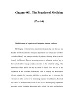
Fig. 1. Model for the role of transcription factors during islet differentiation. The pro-
posed role for different transcription factors in islet differentiation is shown. For simplic-
ity, the association of a single transcription factor with different developmental events is
based on the timing of their expression or the timing of their predominant role in differ-
entiation. Any given factor likely functions at multiple steps during differentiation, and
expression of multiple factors is probably required at each step of differentiation. Also
shown are differentiated adult islet cells. Below each cell is the hormonal product of that
cell type and the transcription factors that are expressed in the differentiated adult δ, β,
α, and pancreatic polypeptide cells.
140 Lowe
undergoing spontaneous differentiation, Pax4- and Pdx-1-expressing cells gen-
erally showed increased expression of genes encoding transcription factors and
other proteins important for or characteristic of differentiated islet cell function.
Moreover, the amount of insulin mRNA and percentage of cells expressing
insulin was increased in the Pdx-1- and Pax4-expressing cells, although the
impact of Pax4 was greater than that of Pdx-1. After the selection and differen-
tiation of nestin-positive cells, approximately 80% of Pax4-expressing cells
produced insulin. Growth of cells in histotypic culture resulted in spheroids
containing cells with insulin-positive granules, albeit at a density lower than that
present in adult β cells. When transplanted into diabetic mice, differentiated
nestin-positive Pax4-expressing and wild-type ES cells were equally efficacious
in restoring euglycemia. Thus expression of transcription factors important for
β-cell development and differentiation augments the in vitro differentiation of
ES cells into insulin-secreting cells, although the functional consequences in
vivo remain unclear. One problem with the approach described previously is that
transcription factor expression during development is dynamic. Indeed, Pax4 is
important for β-cell differentiation during development, but it is essentially
absent in adult murine β cells (27). Pdx-1 expression is relatively uniform early
in development, but is later heterogeneous with high levels in β cells and lower
levels in undifferentiated precursor cells (19). Thus constitutive expression fails
to reproduce the dynamic regulation of transcription factor expression character-
istic of cellular differentiation.
3.1.3. I
NSULIN-SECRETING CELLS FROM TISSUE STEM CELLS
An alternative approach to using ES cells is to redirect the differentiation of
adult stem cells along an islet lineage. One means of accomplishing this has been
to use cells of endodermal origin. This has been attempted using IEC-6 cells,
which are immature rat intestinal stem cells that exhibit an undifferentiated
morphology and limited expression of intestinal-specific genes (28). Various
approaches have been used to direct the differentiation of these cells into insulin-
secreting cells. Stable and constitutive expression of Pdx-1 in IEC-6 cells caused
them to assume an enteroendocrine cell phenotype capable of expressing sero-
tonin, cholecystokinin, gastrin, and somatostatin (29). To direct these cells along
an islet cell lineage, the Pdx-1-expressing cells were subsequently treated with
betacellulin (30,31). Betacellulin is a member of the epidermal growth factor
family of peptides that is expressed in adult and fetal pancreas, signals through
the ErbB family of tyrosine kinase receptors, and stimulates the proliferation of
multiple cell types, including β cells (32,33). Several lines of evidence suggest
that betacellulin plays a key role in islet cell proliferation or differentiation.
Betacellulin enhances pancreatic regeneration after a 90% pancreatectomy by
increasing β-cell proliferation and mass (34). It also increases DNA synthe-
Chapter 7 / Transcription Factor-Directed Differentiation of Stem Cells 141
sis in human fetal pancreatic epithelial cells and enhances β-cell development
in fetal murine pancreatic explant cultures (33,35). Treatment of PDX-1-express-
ing IEC-6 cells with betacellulin resulted in insulin expression and the formation
of secretory granules. However, insulin secretion was neither glucose-dependent
nor stimulated by arginine (30,31). Among the transcription factors induced by
betacellulin treatment was Isl-1. Isl-1 is an LIM homeodomain factor that is
important early in pancreatic development and is expressed in pancreatic epithe-
lium and mesenchyme surrounding the pancreas (36). It is also expressed later
in development in postmitotic endocrine cells and is present in mature islet cells
(36). Its role in islet function is unclear. Overexpression of Isl-1 in Pdx-1-express-
ing cells also resulted in insulin expression (30,31). Transplantation of IEC-6 cells
expressing both Pdx-1 and Isl-1 into diabetic rats transiently decreased the blood
glucose level, although euglycemia was not restored (30). These studies suggest
that expressing specific transcription factors in tissue stem cells can redirect their
differentiation along an islet lineage, but that additional factors will be needed
to fully differentiate the cells.
Liver is a second endoderm-derived tissue that has been used as a source of
cells that can be directed to differentiate into islets. Like pancreas, liver is derived
from ventral endoderm, and both tissues express members of the hepatocyte
nuclear family and exhibit glucose responsiveness (37). Indeed, it has been sug-
gested that there is an endodermal progenitor cell common to liver and pancreas
(38). In vivo expression of transcription factors has been used to differentiate
liver cells into insulin-secreting cells (37). Adenoviral-mediated expression of
Pdx-1 has successfully generated insulin-producing cells in liver (39,40). After
expression of Pdx-1, liver produced not only insulin, but also other islet genes,
including those encoding glucagon, somatostatin, and islet amyloid polypeptide.
Expression of these genes, as well as the Pdx1 gene, was prolonged as Pdx-1,
insulin, and somatostatin expression was present 6–8 months after the initial
infection. Glucagon expression was extinguished after about 4 months. Pro-
longed expression of Pdx-1, and presumably other islet proteins, appeared to be
due to auto-induction of the native Pdx1 gene by Pdx-1 expressed from the
adenoviral vector (40). After Pdx-1 expression, the insulin content of the liver
was increased 10- to 30-fold, but this was still only 1.3–3% of the insulin content
of pancreas (40). Insulin produced by the liver was functional in that it was able
to treat and prevent diabetes induced by streptozotocin, a β-cell toxin (39,40).
The cells producing insulin were distinct from those that produced glucagon and
were localized in proximity to the central vein. Mature hepatocytes reside in this
region of the liver, although, because only a small percentage of infected cells
expressed insulin, only a small subpopulation of cells appears to be capable of
transdifferentiation. The nature of these cells that undergo transdifferentiation is
not clear.
142 Lowe
A similar approach has been used to develop insulin-producing cells from
epithelial progenitor cells derived from fetal liver (41). These cells express
markers of hepatocytes, bile ducts, and oval cells and are capable of differenti-
ating into mature hepatocytes in vivo (42). Oval cells are thought to represent
hepatic stem cells (43). Transduction of these progenitor cells with a lentivirus
that constitutively expresses mRNA encoding Pdx-1 results in partial differen-
tiation along an islet lineage (41). Despite expression of Pdx-1, these cells con-
tinued to express hepatocyte markers, including glycogen, dipeptidyl peptidase
IV, and γ-glutamyl transpeptidase. Autoinduction of the endogenous Pdx1 gene
was again evident, and some transcription factors present in adult β cells (e.g.,
NeuroD1, Nkx6.1) were also expressed, whereas others such as Nkx2.2 and Pax6
were absent (41). Interestingly, neurogenin3, which is present in developing but
not mature islets, was also present. Finally, insulin and the prohormone
convertases PC1/3 and PC2 as well as islet amyloid polypeptide, glucagon,
pancreatic polypeptide, and elastase were expressed. Thus proteins present in
both the endocrine and exocrine pancreas were produced. It has not been estab-
lished whether these different hormones and enzymes are coexpressed by the
same or different cells. Importantly, these cells exhibit glucose-stimulated insu-
lin secretion, albeit with a curve that is shifted to the right compared with native
islets. This may reflect a lack of expression of GLUT2 and glucokinase and
expression of only the Kir6.2 subunit of the ATP-sensitive potassium channel
that is important for insulin secretion. Importantly, these cells appeared to secrete
mature processed insulin and were able to reverse streptozotocin-induced diabetes.
In studies using an adenoviral vector capable of higher and more prolonged
expression, in vivo Pdx-1 expression in the liver had a different effect. In this
circumstance, insulin-producing cells were present, but cells exhibiting charac-
teristics of exocrine cells, including expression of trypsin, were also present
(44,45). Interestingly, insulin and trypsin were coexpressed by the same cells,
and the latter induced a severe hepatitis (44,45). In contrast, use of this same
adenoviral vector to express the transcription factor NeuroD1/Beta2 and
betacellulin resulted in the formation of islet clusters capable of reversing
streptozotocin-induced diabetes (44,45). The islet-like clusters were, in general,
localized immediately underneath the liver capsule. Thus the cells from which
islet-like structures were generated appeared to be distinct from those in the
proximity of the central vein that differentiated into insulin-secreting cells fol-
lowing Pdx-1 expression. After expression of NeuroD1 and betacellulin, gluca-
gon, somatostatin, and pancreatic polypeptide were also present in the islet-like
structures. Unlike native islets, individual cells in the islet-like structures pro-
duced multiple hormones. Other genes characteristic of mature islets were also
expressed, including those encoding the prohormone convertases PC1/3 and
Chapter 7 / Transcription Factor-Directed Differentiation of Stem Cells 143
PC2 and the Kir6.2 and SUR1 subunits of the ATP-sensitive potassium channel
(44,45). Insulin granules were also present in the cells.
3.2. Steroidogenic Cells
Another endocrine gland susceptible to destruction by autoimmunity, infec-
tion, and bleeding is the adrenal gland. Because oral replacement of cortisol does
not accurately reproduce the pattern of cortisol secretion by the native adrenal
gland, the generation of adrenal cells from stem cells would be of therapeutic
benefit.
The only transcription factor that has been expressed in ES cells to help direct
differentiation along a steroidogenic cell lineage is steroidogenic factor 1 (SF-1)
(46). SF-1 is an orphan member of the steroid receptor superfamily (reviewed in
(47). It is expressed in a variety of tissues, including the adrenal cortex, testis
(Sertoli cells), ovary (granulosa and theca cells), the placenta, and the pituitary
and hypothalamus. During development, SF-1 is expressed in the urogenital
ridge as early as embryonic day 9 in mice, and its role in the differentiation of
steroidogenic tissues is demonstrated by the absence of adrenal glands and
gonads in mice with a null mutation of the SF-1 gene (48,49). In humans,
mutations in SF-1 are associated with hypogonadism and hypoadrenalism (47).
Among the targets of SF-1 are the genes that encode the steroidogenic cyto-
chrome P450 enzymes (47).
Given the role of SF-1, it is not surprising that its expression in ES cells directs
their differentiation toward a steroidogenic phenotype (46). The morphology of
ES cells stably transfected with a vector expressing SF-1 changes from bire-
fringent spheres into flat, phase-dull sheets despite the continued presence of
mouse embryo fibroblast feeder cells and leukemia inhibitory factor, both of
which prevent ES cell differentiation. Among the factors known to induce ste-
roidogenesis in steroidogenic cell lines are retinoic acid and cyclic adenosine 5′-
monophosphate, which is the downstream effector of hormones such as
adrenocorticotropic and luteinizing hormones. Treatment of the SF-1–express-
ing ES cells with a cyclic adenosine 5′-monophosphate analogue with or without
retinoic acid markedly increased expression of the rate-limiting steroidogenic
enzyme P450 side-chain cleavage (P450
scc
), an effect not observed in native ES
cells (46). Moreover, in cells provided with 20α-hydroxycholesterol, a substrate
for P450
scc
, progesterone was synthesized in amounts proportional to the expres-
sion of P450
scc
mRNA. It is important to note that this change in cell phenotype
occurred despite the continued presence of mouse embryo fibroblasts and leuke-
mia inhibitory factor. Thus SF-1 expression is capable of initiating a program
that converts ES cells into steroidogenic cells and may serve to augment the
development of steroidogenic tissues from stem cells.
144 Lowe
4. CONCLUSION
The studies described here indicate that transcription factor expression has the
potential to direct or augment stem cell differentiation. As demonstrated, expres-
sion of a specific transcription factor can initiate a genetic program typically
activated by inductive factors elaborated in vivo by surrounding tissues and cells,
thus allowing differentiation to proceed in vitro. One of the problems with this
approach, however, is that the constitutive expression of transcription factors is
not able to reproduce the dynamic expression of transcription factors that is
characteristic of the differentiation process. This may interfere with the final
maturation of cells or alter cell function. Approaches that have been used to
address this concern are using vectors (e.g., adenoviral vectors) in which expres-
sion is time-limited or vectors that allow inducible expression of the gene of
interest. Clearly, expressing transcription factors in differentiating stem or pro-
genitor cells will provide important insight into the genetic programs responsible
for differentiation along specific cell lineages and has the potential to facilitate
ongoing efforts to develop means to differentiate stem cells into specific hor-
mone-secreting cells that will be available for cell replacement therapy.
REFERENCES
1. Kyba M, Daley GQ. Hematopoiesis from embryonic stem cells: lessons from and for ontog-
eny. Exp Hematol 2003;31:994–1006.
2. Payne KJ, Crooks GM. Human hematopoietic lineage commitment. Immunol Rev 2002;187:
48–64.
3. Kyba M, Perlingeiro RC, Hoover RR, Lu CW, Pierce J, Daley GQ. Enhanced hematopoietic
differentiation of embryonic stem cells conditionally expressing Stat5. Proc Natl Acad Sci
USA 2003;100(Suppl. 1):11904–11910.
4. Ihle JN. The Stat family in cytokine signaling. Curr Opin Cell Biol 2001;13:211–117.
5. Shuai K, Halpern J, ten Hoeve J, Rao X, Sawyers CL. Constitutive activation of STAT5 by
the BCR-ABL oncogene in chronic myelogenous leukemia. Oncogene 1996;13:247–254.
6. Kyba M, Perlingeiro RC, Daley GQ. HoxB4 confers definitive lymphoid-myeloid engraft-
ment potential on embryonic stem cell and yolk sac hematopoietic progenitors. Cell
2002;109:29–37.
7. Lee JE. Basic helix-loop-helix genes in neural development. Curr Opin Neurobiol 1997;7:13–20.
8. O’Shea KS. Neuronal differentiation of mouse embryonic stem cells: lineage selection and
forced differentiation paradigms. Blood Cells Mol Dis 2001;27:705–712.
9. Wilson M, Koopman P. Matching SOX: partner proteins and co-factors of the SOX family of
transcriptional regulators. Curr Opin Genet Dev 2002;12:441–446.
10. Pevny LH, Sockanathan S, Placzek M, Lovell-Badge R. A role for SOX1 in neural determi-
nation. Development 1998;125:1967–1978.
11. Lee SH, Lumelsky N, Studer L, Auerbach JM, McKay RD. Efficient generation of midbrain
and hindbrain neurons from mouse embryonic stem cells. Nat Biotechnol 2000;18:675–679.
12. Kim JH, Auerbach JM, Rodriguez-Gomez JA, et al. Dopamine neurons derived from embry-
onic stem cells function in an animal model of Parkinson’s disease. Nature 2002;418:50–56.
Chapter 7 / Transcription Factor-Directed Differentiation of Stem Cells 145
13. Ang SL, Wierda A, Wong D, et al. The formation and maintenance of the definitive endoderm
lineage in the mouse: involvement of HNF3/forkhead proteins. Development 1993;119:1301–
1315.
14. Chakrabarti SK, Mirmira RG. Transcription factors direct the development and function of
pancreatic beta cells. Trends Endocrinol Metab 2003;14:78–84.
15. Levinson-Dushnik M, Benvenisty N. Involvement of hepatocyte nuclear factor 3 in endoderm
differentiation of embryonic stem cells. Mol Cell Biol 1997;17:3817–3822.
16. Ang SL, Rossant J. HNF-3 beta is essential for node and notochord formation in mouse
development. Cell 1994;78:561–574.
17. Weinstein DC, Ruiz i Altaba A, Chen WS, et al. The winged-helix transcription factor HNF-
3 beta is required for notochord development in the mouse embryo. 1994;Cell 78:575–588.
18. Kemp DM, Thomas MK, Habener JF. Developmental aspects of the endocrine pancreas. Rev
Endocr Metab Disord 2003;4:5–17.
19. Murtaugh LC, Melton DA. Genes, signals, and lineages in pancreas development. Annu Rev
Cell Dev Biol 2003;19:71–89.
20. Jensen J. Gene regulatory factors in pancreatic development. Dev Dyn 2004;229:176–200.
21. Gradwohl G, Dierich A, LeMeur M, Guillemot F. neurogenin3 is required for the development
of the four endocrine cell lineages of the pancreas. Proc Natl Acad Sci USA 2000;97:1607–1611.
22. Schwitzgebel VM, Scheel DW, Conners JR, et al. Expression of neurogenin3 reveals an islet
cell precursor population in the pancreas. Development 2000;127:3533–3542.
23. Soria B, Roche E, Berna G, Leon-Quinto T, Reig JA, Martin F. Insulin-secreting cells derived
from embryonic stem cells normalize glycemia in streptozotocin-induced diabetic mice. Dia-
betes 2000;49:157–162.
24. Lumelsky N, Blondel O, Laeng P, Velasco I, Ravin R, McKay R. Differentiation of embryonic stem
cells to insulin-secreting structures similar to pancreatic islets. Science 2001;292:1389–1394.
25. Hori Y, Rulifson IC, Tsai BC, Heit JJ, Cahoy JD, Kim SK. Growth inhibitors promote differ-
entiation of insulin-producing tissue from embryonic stem cells. Proc Natl Acad Sci USA
2002;99:16105–16110.
26. Blyszczuk P, Czyz J, Kania G, et al. Expression of Pax4 in embryonic stem cells promotes
differentiation of nestin-positive progenitor and insulin-producing cells. Proc Natl Acad Sci
USA 2003;100:998–1003.
27. Smith SB, Ee HC, Conners JR, German MS. Paired-homeodomain transcription factor PAX4
acts as a transcriptional repressor in early pancreatic development. Mol Cell Biol 1999;19:
8272–8280.
28. Quaroni A, May RJ. Establishment and characterization of intestinal epithelial cell cultures.
Meth Cell Biol 1980;21B:403–427.
29. Yamada S, Kojima H, Fujimiya M, Nakamura T, Kashiwagi A, Kikkawa R. Differentiation
of immature enterocytes into enteroendocrine cells by Pdx1 overexpression. Am J Physiol
Gastrointest Liver Physiol 2001;281:G229–G236.
30. Kojima H, Nakamura T, Fujita Y, et al. Combined expression of pancreatic duodenal
homeobox 1 and islet factor 1 induces immature enterocytes to produce insulin. Diabetes
2002;51:1398–1408.
31. Yoshida S, Kajimoto Y, Yasuda T, et al. PDX-1 induces differentiation of intestinal epithe-
lioid IEC-6 into insulin-producing cells. Diabetes 2002;51:2505–2513.
32. Dunbar AJ, Goddard C. Structure-function and biological role of betacellulin. Int J Biochem
Cell Biol 2000;32:805–815.
33. Huotari MA, Miettinen PJ, Palgi J, et al. ErbB signaling regulates lineage determination of devel-
oping pancreatic islet cells in embryonic organ culture. Endocrinology 2002;143:4437–4446.
146 Lowe
34. Li L, Seno M, Yamada H, Kojima I. Promotion of beta-cell regeneration by betacellulin in
ninety percent-pancreatectomized rats. Endocrinology 2001;142:5379–5385.
35. Demeterco C, Beattie GM, Dib SA, Lopez AD, Hayek A. A role for activin A and betacellulin
in human fetal pancreatic cell differentiation and growth. J Clin Endocrinol Metab
2000;85:3892–3897.
36. Ahlgren U, Pfaff SL, Jessell TM, Edlund T, Edlund H. Independent requirement for ISL1 in
formation of pancreatic mesenchyme and islet cells. Nature 1997;385:257–260.
37. Meivar-Levy I, Ferber S. New organs from our own tissues: liver-to-pancreas transdifferentiation.
Trends Endocrinol Metab 2003;14:460–466.
38. Grompe M. Pancreatic-hepatic switches in vivo. Mech Dev 2003;120:99–106.
39. Ferber S, Halkin A, Cohen H, et al. Pancreatic and duodenal homeobox gene 1 induces
expression of insulin genes in liver and ameliorates streptozotocin-induced hyperglycemia.
Nat Med 2000;6:568–572.
40. Ber I, Shternhall K, Perl S, et al. Functional, persistent, and extended liver to pancreas
transdifferentiation. J Biol Chem 2003;278:31950–1957.
41. Zalzman M, Gupta S, Giri RK, et al. Reversal of hyperglycemia in mice by using human
expandable insulin-producing cells differentiated from fetal liver progenitor cells. Proc Natl
Acad Sci USA 2003;100:7253–7258.
42. Malhi H, Irani AN, Gagandeep S, Gupta S. Isolation of human progenitor liver epithelial cells
with extensive replication capacity and differentiation into mature hepatocytes. J Cell Sci
2002;115:2679–2688.
43. Petersen BE. Hepatic “stem” cells: coming full circle. Blood Cells Mol Dis 2001;27:590–600.
44. Chan L, Fujimiya M, Kojima H. In vivo gene therapy for diabetes mellitus. Trends Mol Med
2003;9:430–435.
45. Kojima H, Fujimiya M, Matsumura K, et al. NeuroD-betacellulin gene therapy induces islet
neogenesis in the liver and reverses diabetes in mice. Nat Med 2003;9:596–603.
46. Crawford PA, Sadovsky Y, Milbrandt J. Nuclear receptor steroidogenic factor 1 directs em-
bryonic stem cells toward the steroidogenic lineage. Mol Cell Biol 1997;17:3997–4006.
47. Parker KL, Rice DA, Lala DS, et al. Steroidogenic factor 1: an essential mediator of endocrine
development. Recent Prog Horm Res 2002;57:19–36.
48. Sadovsky Y, Crawford PA, Woodson KG, et al. Mice deficient in the orphan receptor ste-
roidogenic factor 1 lack adrenal glands and gonads but express P450 side-chain-cleavage
enzyme in the placenta and have normal embryonic serum levels of corticosteroids. Proc Natl
Acad Sci USA 1995;92:10939–10943.
49. Luo X, Ikeda Y, Parker KL. A cell-specific nuclear receptor is essential for adrenal and
gonadal development and sexual differentiation. Cell 1994;77:481–490.
Chapter 8 / Generation of Islet-Like Structures From ES Cells 147
147
From: Contemporary Endocrinology: Stem Cells in Endocrinology
Edited by: L. B. Lester © Humana Press Inc., Totowa, NJ
8
Generation of Islet-Like Structures
From ES Cells
Nadya Lumelsky
CONTENTS
INTRODUCTION
PANCREATIC ISLET: A MINIORGAN
ES CELLS, UNLIMITED EXPANSION CAPACITY, AND PLURIPOTENCY
ES CELL DIFFERENTIATION: THE ISSUE OF CONTROL
PANCREATIC SPECIFICATION AND DEVELOPMENT
SPONTANEOUS PANCREATIC DIFFERENTIATION OF ES CELLS
INDUCED PANCREATIC DIFFERENTIATION OF ES CELLS
CONCLUSION
REFERENCES
1. INTRODUCTION
Type 1 and type 2 diabetes, though different diseases, both involve inadequate
cell mass of insulin-producing β cells, the most abundant cell type of pancreatic
islets of Langerhans. Insulin injections alleviate hyperglycemia in the majority
of diabetic patients. However, insulin therapy cannot provide the finely tuned
control of glucose homeostasis afforded by native pancreatic islets. As a result,
diabetic patients commonly develop multiple life-threatening complications,
such as cardiovascular and kidney disease, neuropathy, and blindness. Recent
successes in pancreatic islet transplantation (1) fueled new hope that this proce-
dure could significantly improve the quality of life for diabetic patients. Unfor-
tunately, because the islets needed for transplantation are obtained from cadaveric
donors only, few patients can receive this therapy. The shortage of islets could
potentially be overcome by deriving them from alternative sources such as
embryonic stem (ES) cells. This chapter will provide a review of the recent
progress in generating islet-like hormone-producing cell clusters from ES cells.
148 Lumelsky
2. PANCREATIC ISLET: A MINIORGAN
The mammalian pancreas is composed of the exocrine acini, endocrine islets,
and pancreatic ducts. The exocrine pancreas makes digestive enzymes, which are
released into the digestive system through pancreatic ductal system. The islets,
which constitute about 1–2% of the total pancreatic cell mass, are distributed in
the exocrine tissue. They produce endocrine hormones required for utilization of
glucose. An islet is not merely an aggregate of cells, but rather a miniorgan
containing different hormone producing and other types of cells that participate
in functionally important cell–cell interactions (2). The hormone-producing cells
of the islets are α, β, δ, and pancreatic polypeptide cells. They secrete glucagon,
insulin, somatostatin, and pancreatic polypeptide, respectively. Insulin-produc-
ing β cells are the most abundant hormone-producing cell type of the islet.
Among nonhormonal cell types, islets contain peripheral neurons, mesenchy-
mal, and peri-islet Schwann cells as well as endothelial and smooth muscle cells
that compose islet vasculature (3,4). Also residing in the islets may be a popu-
lation of pancreatic stem and progenitor cells (5–7). It has been suggested that
these stem and progenitor cells may have the capacity to generate new islet cells
to compensate for cell loss during normal cell turnover and after islet damage. In
view of the functional importance of islet complexity, it is likely that islet-like
structures approximating this complexity would provide a better alternative to
purified β cells for therapeutic applications.
3. ES CELLS, UNLIMITED EXPANSION CAPACITY,
AND PLURIPOTENCY
Mouse ES cells were derived more than 20 years ago by Evans and Kaufman
from the inner cells mass of the blastocyst stage embryo (8). This pioneering
work and that which followed identified several important properties of ES cells.
It was found, in particular, that when cultured in vitro, these cells could be
propagated indefinitely in the undifferentiated state. Also, they could shift from
proliferation to differentiation mode by simple change of culture medium. The
differentiated progeny of ES cells composes cells of all three germ layers: endo-
derm, mesoderm, and ectoderm. Moreover, it is thought that during in vitro
differentiation, the ES cells may be recapitulating normal embryonic develop-
ment (9). When ES cells are injected into mouse blastocysts in vivo, they colo-
nize all tissues of the developing embryo derived from this blastocyst, including
the germ line (10,11). This property, called pluripotency, has been used exten-
sively for introducing specific mutations into the mouse genome. In line with
their pluripotency, when injected into the immunodeficient nude mice, ES cells
Chapter 8 / Generation of Islet-Like Structures From ES Cells 149
generate heterogeneous tumors called teratomas, which are composed of differ-
ent cell types derived from all three germ layers (12).
The capacity of ES cells for multilineage differentiation in vitro has attracted
considerable interest after recent derivation of ES cells from human blastocysts
(13). It turns out that, similarly to mouse cells, human ES cells can be continually
propagated in vitro in the undifferentiated state, and also induced to differentiate
into multiple cell lineages. It was thus realized that human ES cells could poten-
tially provide an unlimited source of transplantable material for treatment of a
variety of diseases resulting from the loss of differentiated cell mass, including
diabetes. However, before ES cell-based therapies will become practical reality,
several important obstacles will need to be overcome. These are discussed in the
following sections.
4. ES CELL DIFFERENTIATION: THE ISSUE OF CONTROL
One of the main difficulties in introducing ES cell technology into clinical
practice stems from our insufficient knowledge of mechanisms that control the
cell fate determination in ES cell cultures. Although several protocols have been
proposed describing directed differentiation of ES cells into specific lineages,
such as neural (14,15), hematopoietic (16), endothelial, smooth muscle (17), and
cardiac muscle (18), in addition to the cell type of interest, a variety of other cell
types are always generated in a typical ES cell culture. None of the existing
differentiation protocols result in a fully controlled and uniform pattern of dif-
ferentiation. Another complicating issue is a potential tumorigenicity of ES cell-
derived cell populations. Because the undifferentiated ES cells are tumorigenic,
even a small fraction of cells that escape differentiation would create a potential
source of tumors after transplantation in vivo.
Several approaches to improve control over ES cell differentiation have been
proposed. For example, because in vitro differentiation the ES cells is thought to
approximate normal embryonic development, the exposure of ES cell cultures to
growth factors, extracellular matrix components, and cell–cell interactions con-
trolling normal development might promote and streamline the differentiation
process (19,20). Additionally, the enrichment of ES cell cultures with the desired
cell type can be achieved using positive selection to purify the cells of interest,
or using negative selection to remove the heterologous cells (21,22). Such selec-
tion approaches can also aid in purging ES cell cultures from undifferentiated
tumorigenic cells. Still another way to eliminate tumorigenic cells is to geneti-
cally modify the ES cells to express suicide genes: this would render them sen-
sitive to specific pharmacological toxins (23,24). It is likely that a combination
of several approaches will be used in the future ES cell-based clinical protocols.
150 Lumelsky
5. PANCREATIC SPECIFICATION AND DEVELOPMENT
The existing protocols of pancreatic differentiation of ES cells suffer from low
efficiency, high rate of cell death accompanying differentiation, and experiment-
to-experiment variability. It is widely recognized that improvement of these
protocols will be critically dependent on the progress in our understanding of the
mechanisms of pancreatic development. These mechanisms will be discussed
briefly. Several recent reviews are available for in-depth discussion on this topic
(25–27).
The pancreas develops from endoderm, which in the mouse is specified to
pancreatic fate around embryonic day 8.5 (E8.5). Although the exact mecha-
nisms of pancreatic specification are still poorly understood, recent results
obtained in the chicken system suggest that the signals responsible for pattern-
ing of the endoderm to become pancreas are generated by the mesoderm adjacent
to the prospective pancreatic endoderm (28). Moreover, the results of the same
work indicate that several members of the transforming growth factor-β (TGF-β)
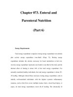
superfamily may be responsible for this inducing activity. After specification, the
pancreas develops in dorsal and ventral portions, which are in close proximity
with two mesodermal tissues: notochord (dorsal pancreas) and cardiac meso-
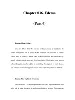
derm (ventral pancreas) (Fig. 1). The notochord and the cardiac mesoderm gov-
ern survival and differentiation of the pancreas by generating permissive signals
produced by fibroblast growth factor (FGF), TGF-β, and the hedgehog families
of growth factors (29–31). In addition to pancreas, the neural tube contacts
notochord during early embryogenesis. Consequently, the dorsal pancreas and
the neural tube are exposed to the same signaling molecules. It is therefore not
surprising that pancreatic and neural development are controlled by similar
mechanisms (25,32). Recently, it was established that, in addition to notochord,
the dorsal aorta, which is another mesodermal derivative (and is juxtaposed with
dorsal pancreas after its separation from the notochord), generates signals essen-
tial for pancreatic development (33–35). Later in embryogenesis, the dorsal and
ventral pancreatic buds fuse and the pancreas becomes embedded in the sur-
rounding pancreatic mesenchyme. During the late stages of development, the
mesenchyme serves as a source of signals for pancreatic growth, differentiation,
and morphogenesis (32).
Pancreatic transcription factors are outlined in Fig. 2A,B. It is noteworthy that
the majority of pancreatic transcription factors are also involved in nervous
system development (25,27). Homeodomain transcription factors, Hb9 (encoded
by Hlxb9 gene), PDX-1 (also called Ipf1), and a helix–loop–helix transcription
factor, neurogenin3 (ngn3), are among the earliest markers of pancreatic devel-
opment. Recently another transcription factor, Ptf1a/P48, was added to the list
of essential transcriptional regulators of early pancreatic development (34,36).
Chapter 8 / Generation of Islet-Like Structures From ES Cells 151
Fig. 1. Scheme of early steps of pancreatic organogenesis. Shown are cross sections through a mouse embryo at the level of deve
loping
pancreas. The 10-somite stage roughly corresponds to E8; the 28-somite stage, to E10 in mouse. D, dorsal; V, ventral. (From ref
.
31.)
152 Lumelsky
152
Chapter 8 / Generation of Islet-Like Structures From ES Cells 153
Advances in microarray technology have allowed generation of global pancre-
atic transcriptional profiles (37,38). Because this analysis allows simultaneous
screening of many genes, it is expected that it will facilitate discovery of new
elements regulating pancreatic development. This information will be essential
for designing novel strategies for pancreatic differentiation of ES cells.
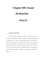
The existing protocols for generating endocrine hormone-producing cells from
ES cells can be divided into two groups (Fig. 3). The first group of protocols takes
advantage of the capacity of ES cells to undergo spontaneous pancreatic differ-
entiation in fetal bovine serum-containing medium. These protocols may or may
not include a genetic selection step to enrich the cultures for hormone-producing
cells. Protocols of the second type attempt to induce pancreatic differentiation
with specific growth factors and extracellular matrix molecules in defined cul-
ture medium.
6. SPONTANEOUS PANCREATIC DIFFERENTIATION
OF ES CELLS
6.1. Differentiation Without Selection
Assady and coworkers (39) have studied a pattern of pancreatic gene expres-
sion during spontaneous differentiation of human ES cells. They carried out
these experiments with two culture techniques: in suspension, where ES cells
form simple cell aggregates called embryoid bodies (EBs), and in adherent cul-
tures grown at high cell density. Insulin expression was examined by immuno-
histochemistry in the 19-day-old EBs. The authors have found insulin-expressing
cells scattered throughout EBs and in small clusters within EBs. They also
found that as the EBs matured, the number of insulin-expressing cells gradu-
ally increased. To characterize the insulin-producing cells further, they mea-
sured insulin secretion from 20- to 22-day-old EBs and 22- and 31-day-old,
high-density adherent cultures in the presence of 5.5 mM and 25 mM glucose.
Insulin secretion into the medium was detected in both types of cultures but this
insulin secretion was not sensitive to increasing glucose concentration. The reverse
transcriptase-polymerase chain reaction (RT-PCR) analysis of a panel of pancre-
Fig. 2. (opposite page) (A) A model depicting the role of islet transcription factors in
endocrine differentiation during development. The proposed position for each transcrip-
tion factor is based on its time of expression, functional role, or both. Although some
transcription factors function at several steps, only single steps are shown for simplicity.
(From ref. 27.) (B) A model depicting the role of the key pancreatic transcription factors
during different steps of pancreatic organogenesis. (From ref. 25.) Ipf1 in (B) and Pdx1
in (A) designate the same transcription factor.
154 Lumelsky
atic endocrine genes was also carried out. Their results showed that insulin,
PDX-1, ngn3, glucokinase, and the β-cell-specific glucose transporter, Glut2,
are all induced in EB and in adherent cultures. During the course of the culture,
the expression of PDX-1 and ngn3 preceded expression of insulin, Glut2, and
glucokinase. These results suggest that, similarly to normal pancreatic develop-
ment, PDX-1 and ngn3 may control expression of insulin, Glut2, and glucoki-
nase in human ES cell cultures (40).
Shiroi et al. have investigated spontaneous pancreatic differentiation of mouse
ES cells (41). After EB formation, they platted the EBs on tissue culture plates
to allow cell outgrowth. The authors used the zinc-chelating agent dithizone,
which selectively stains β cells, to observe emergence of insulin-positive cell
clusters. After 21 days, the first cells faintly stained with dithizone became
visible; the intensity of staining became more apparent by day 28. Dithizone-
positive cell clusters were isolated from the culture dishes and subjected to RT-
PCR analysis for expression of several pancreatic markers; insulin, glucagon,
pancreatic polypeptide, but not somatostatin expression was observed. Also,
expression of Glut2, PDX-1, and a marker of endoderm, hepatocyte nuclear
factor-3β, was detected.
Fig. 3. Summary of current pancreatic endocrine differentiation protocols.
Chapter 8 / Generation of Islet-Like Structures From ES Cells 155
Kahan at al. used a similar nonselective differentiation protocol to analyze the
pattern of gene expression during pancreatic differentiation of mouse ES cells
(42). In agreement with the results of other investigators, they found progressive
accumulation of hormone expressing cells in their cultures. The RT-PCR analy-
sis showed that gene expression of several pancreatic transcription factors was
induced in their cultures. They also found that early in the culture the majority
of hormone positive cells coexpressed different islet hormones. At the end of the
experiment, however, the majority of cells expressed only a single hormone. The
authors argued that this dynamic pattern of gene expression might be a reflection
of normal islet differentiation.
Although the results of these experiments shows that spontaneous pancreatic
differentiation can occur in mouse and human ES cell cultures, the efficiency of
this process is undoubtedly too low to be of practical value for generating signifi-
cant numbers of hormone-producing cells.
6.2. Selection of Insulin-Producing Cells From Spontaneously
Differentiating ES Cell Cultures
It has been shown previously that genetic selection against heterologous cell
types generated during the course of spontaneous ES cell differentiation can
result in enrichment for the cell types of interest. For example, this approach has
been used to obtain purified cardiomyocyte- and neural-like cells from mouse ES
cell cultures (21,22). Soria and coworkers used a similar strategy to select insu-
lin-producing cells from spontaneously differentiating mouse ES cells (43). They
introduced into the ES cells a plasmid conferring resistance to two antibiotics.
The first antibiotic-resistance gene was under control of a constitutive promoter,
and the second gene was under control of an insulin promoter. During the first
stage of the culture the undifferentiated ES cells were selected for resistance to
the first antibiotic. This allowed generation of a stable cell line in which every
cell carried the plasmid. After this step, the ES cells were transferred into differ-
entiation medium containing the second antibiotic. Because the second antibi-
otic resistance gene was under control of insulin promoter, only cells producing
insulin survived this round of selection. The authors report that the insulin con-
tent of the ES cell-derived progeny obtained with this protocol was approxi-
mately 90% of the insulin content of normal mouse islets. When the insulin
release in response to glucose and other agonists was measured in vitro, the cells
showed stimulated release. Moreover, when implanted into diabetic mice, the
insulin-producing cells normalized hyperglycemia. This normalization disap-
peared, however, after 12 weeks in about 40% of transplanted animals. The com-
parison of glucose tolerance of the transplanted animals with that of the nondiabetic
controls showed that in the transplanted animals the plasma glucose levels were
significantly elevated, and the recovery to normal glucose levels was delayed.