The Foot in Diabetes - part 2 ppsx
Bạn đang xem bản rút gọn của tài liệu. Xem và tải ngay bản đầy đủ của tài liệu tại đây (684.16 KB, 37 trang )
Although the UKPDS suggested that tight control of blood glucose and
blood pressure might in¯uence the development of certain cardiovascular
endpoints, such as stroke and sudden death, statistical evidence that these
in¯uence the progression of PVD was not forthcoming
13,14
. However,
educational strategies aimed at the cessation of smoking and control of
dyslipidaemia therefore remain of paramount importance. Moreover, in
view of trends observed in the United Kingdom Prospective Diabetes Study
(UKPDS), optimal glycaemic and blood pressure control should be aimed for.
DIABETIC NEUROPATHY
The diabetic neuropathies are a heterogenous group of conditions that may
be subclassi®ed into various polyneuropathies and mononeuropathies on
clinical grounds
15
. The association between peripheral neuropathy and foot
ulceration has been recognized for many years: Pryce, a surgeon working in
Nottingham over 100 years ago, remarked that ``it is abundantly clear to me
that the actual cause of the perforating ulcer was a peripheral nerve
degeneration'', and ``diabetes itself may play an active part in the causation
of the perforating ulcers''. It is the sensory and the peripheral autonomic
polyneuropathies that play an important role in the pathogenesis of
ulceration, and these will be discussed in some detail.
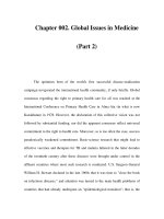
The Pathway to Ulceration 21
Figure 3.1 Pathways to foot ulceration in diabetic patients
Sensory Neuropathy
Chronic sensorimotor neuropathy is by far the commonest of all the diabetic
neuropathies, and occurs in both main types of diabetes. An internationally
agreed de®nition is ``the presence of symptoms and/or signs of peripheral
nerve dysfunction in people with diabetes after exclusion of other causes''
16
.
Although the quantity, and sometimes quality, of epidemiological data on
the prevalence of neuropathy remains low, there have been some studies
published in recent years, as summarized in Table 3.1. It can be seen
however, that neuropathy is very common, and it can be safely assumed
that at least half of older type 2 diabetic patients have signi®cant sensory
loss.
The onset of the chronic neuropathy is gradual and insidious and indeed,
on occasions, the initial symptoms may go unnoticed by the patients.
Typical symptoms include paraesthesiae, hyperaesthesiae, sharp, stabbing,
shooting and burning pain, all of which are prone to nocturnal
exacerbation. Whereas in some patients these uncomfortable symptoms
predominate, others may never experience any symptoms. Clinical
examination usually reveals a sensory de®cit in a glove and stocking
distribution, and signs of motor dysfunction are usually present, with small
wasting and absent ankle re¯exes. A particularly dangerous situation,
originally described by J. D. Ward, is the ``painful±painless leg'' in which
the patient experiences painful or paraesthetic symptoms, but on
examination has severe sensory loss to pain and proprioception: such
patients are at great risk of painless injury to their feet.
It must be realized that there is a spectrum of symptomatic severity in
sensorimotor neuropathy: at one extreme, patients experience severe
symptoms, whereas others experience occasional mild symptoms, or even
none at all. Thus, whereas a history of typical symptoms is strongly
suggestive of a diagnosis of neuropathy, absence of symptoms does not exclude
22 The Foot in Diabetes
Table 3.1 Epidemiological data on diabetic peripheral sensorimotor neuropathy
Reference (Country) Number of
subjects
Type of
Diabetes
Prevalence
(%)
Reference
Population-based studies
17 (UK) 811 2 41.6 17
18 (Finland) 133 2 8.3
a
41.9
b
18
Clinic-based studies
19 (UK) 6487 1,2 28.5 19
20 (Europe) 3250 1 28.0 20
21 (Spain) 2644 1,2 22.7 21
a
At diagnosis.
b
After 10 years.
neuropathy and must never be equated with a lack of foot ulcer risk. Therefore,
assessment of foot ulcer risk must always include a careful foot examination
whatever the history
16
.
The ultimate diagnosis of diabetic sensorimotor neuropathy depends on
the prior exclusion of other causes, such as malignancy, drugs, alcohol
and many other rarer causes
15
. Optimal glycaemic control is important in
the prevention and management of neuropathy
15,22
and, in addition, a
number of drugs can help achieve symptomatic relief
15
. Unfortunately,
none of the drugs available at the time of writing affects the natural
history of this condition, which is one of gradual deterioration of nerve
function. Indeed, amelioration of symptoms may indicate progression of
neuropathy to the insensitive foot at risk of ulceration. Thus, again,
absence of symptoms does not equate with freedom from risk of
ulceration.
Autonomic Neuropathy
Sympathetic autonomic neuropathy affecting the lower limbs leads to
reduced sweating and results in both dry skin that is prone to crack and
®ssure, and also increased blood ¯ow (in the absence of large vessel PVD),
with arteriovenous shunting leading to the warm foot. The complex
interactions of sympathetic neuropathy and other contributory factors in
the causation of foot ulcers is summarized in Figure 3.1.
The warm, insensitive and dry foot that results from a combination of
somatic and autonomic dysfunction often provides the patient with a false
sense of security, as most patients still perceive vascular disease as the main
cause of ulcers (see Chapter 10). It is such patients who may present with
insensitive ulceration as they have truly painless feet. Perhaps the highest-
risk foot is the pulseless insensitive foot, because it indicates somatic and
autonomic neuropathy together with PVD.
NEUROPATHYÐTHE MAJOR CONTRIBUTORY FACTOR
IN ULCERATION
Cross-sectional data from established UK foot clinics in London and
Manchester presented in the second edition of this volume suggested that
neuropathy was present in up to 90% of foot ulcers of patients attending
physician- or podiatrist-led services. Thus, most foot ulcers were
considered to be of neuropathic or neuro-ischaemic aetiology. Con®rmation
of these facts in recent years has come from several European and North
American studies.
The ®rst single-centre study suggested that neuropathic patients had a
seven-fold annual increase in the risk of ulceration in a 3 year
The Pathway to Ulceration 23
prospective study
23
. A larger, multicentre study from Europe and North
America extended these observations and reported a 7% annual risk of
ulceration in neuropathic patients
24
. Other prospective trials have
con®rmed the pivotal role of both large ®bre (e.g. proprioceptive de®cit)
and small ®bre (e.g. loss of pain and temperature sensation) neurological
de®cit in the pathogenesis of ulceration
25
. Considering the above data,
there can be little doubt that neuropathy causes foot ulcers with or
without ischaemia, but it must be remembered that the neuropathic foot
does not spontaneously ulcerate; it is the combination of neuropathy and
some extrinsic factor (such as ill-®tting footwear) or intrinsic factor (such
as high foot pressures; see Chapter 4) that results in ulceration. The
other risk factors that are associated with ulceration will now be
considered.
OTHER RISK FACTORS FOR FOOT ULCERATION
Previous Foot Ulceration
Several studies have con®rmed that foot ulceration is most common in
those patients with a past history of ulceration or amputation, and also in
patients from a poor social background. Indeed, in many diabetic foot
clinics more than 50% of patients with new foot ulcers give a past history of
similar problems.
Other Long-term Complications of Diabetes
It has been recognized for many years that patients with retinopathy
and/or renal impairment are at increased risk of foot ulceration.
However, it is now con®rmed that patients at all stages of diabetic
nephropathy, even microalbuminuria, have an increased risk of neuro-
pathic foot ulceration
26
.
Race
Data from cross-sectional studies suggest that foot ulceration is commoner
in Caucasian subjects when compared to groups of other racial origins,
including Hispanics, Blacks and Indian-subcontinent Asians
27,28
. This may
be related not only to physical factors, including limited joint mobility
(LJM) and foot pressures (see below), but also to better footcare in certain
religious groups, including Muslims. However, there is no suggestion that
this risk is related to any geographical differences: indeed, Veves et al
29
showed no differences in risk factors for ulceration according to location at
centres within Europe.
24 The Foot in Diabetes
Postural Instability
Poor balance and instability are increasingly being recognized as
troublesome symptoms of diabetic neuropathy, presumably secondary to
a proprioceptive de®cit. Studies have recently been published con®rming
the association between postural instability, increased body sway and foot
ulceration
30,31
.
Oedema
The presence of peripheral oedema impairs local blood supply and has been
associated with an increased risk of ulceration
11
.
Callus
The presence of plantar callus, especially in the neuropathic foot, is
associated with an increased risk of ulceration: in one study, the risk was
77-fold in a cross-sectional part, whereas in the prospective follow-up,
ulceration occurred only at sites of callus, representing an in®nite increase
in risk
32
.
Deformity
Any deformity occurring in a diabetic foot, such as prominence of
metatarsal heads, clawed toes, Charcot prominences or hallux valgus,
increases ulcer risk.
Duration of Diabetes
Although it is well-recognized that neuropathy and vascular disease are a
function of diabetes duration, a recent report highlighted the high risk of
amputation (and therefore, ulceration) within the ®rst year of diagnosis of
type 2 diabetes
33
. It must be remembered that patients may present with
long-term complications, and careful screening for risk of ulceration must
be carried out at the time of diagnosis.
THE PATHWAY TO ULCERATION
It is the combination of two or more risk factors that ultimately results in
diabetic foot ulceration. Both Pecoraro et al
10
and later Reiber et al
11
have
taken the Rothman model for causation and applied this to amputation and
foot ulceration in diabetes. The model is based upon the concept that a
component cause (e.g. neuropathy) is not suf®cient in itself to lead to
ulceration, but when component causes act together, they may result in a
The Pathway to Ulceration 25
suf®cient cause, which will inevitably result in ulceration (Figure 3.2). In
their study of amputation, Pecoraro et al
10
describe ®ve component causes
that lead to amputation: neuropathy, minor trauma, ulceration, faulty
healing and gangrene.
Reiber et al
11
applied the model to foot ulceration, and a number of causal
pathways were identi®ed: the commonest triad of component causes,
present in 63% of incident ulcers, was neuropathy, deformity and trauma
(Figure 3.3). Oedema and ischaemia were also common component causes.
Other simple examples of two-component pathways to ulceration are:
neuropathy and mechanical trauma [e.g. standing on a nail (Figure 3.4); ill-
®tting footwear]; neuropathy and thermal trauma; and neuropathy and
chemical trauma, e.g. the inappropriate use of chemical ``corn-cures''.
Similarly, the Rothman model can be applied to neuro-ischaemic
ulceration, where the three-component pathway comprising ischaemia,
trauma and neuropathy is most often seen
10,11
.
MECHANICAL FACTORS AND NEUROPATHIC FOOT
ULCERATION
The insensitive neuropathic foot does not ulcerate spontaneously: traumatic
or extrinsic ulcers result as a consequence of trauma to the insensitive foot, as
in Figure 3.4. In contrast, intrinsic or pressure ulcers occur as a result of
pressure that would not normally cause ulceration, but which, because of
26 The Foot in Diabetes
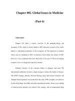
Figure 3.2 Diagram of suf®cient and component causes of diabetic foot ulcers. A±E
represent causes that are not suf®cient in themselves but that are required
components of a suf®cient cause that will inevitably produce the effect. Reproduced
by permission of the American Diabetes Association from reference 11
intrinsic abnormalities in the neuropathic foot, leads to plantar ulceration
when repetitively applied. As stated in the next chapter, abnormalities of
pressures and loads under the diabetic foot are very common. Both
prospective
34
and cross-sectional
28,35
studies have con®rmed that high
plantar pressures are a major aetiological factor in neuropathic foot
ulceration. Veves etal
34
observed a 28% incidence of ulceration inneuropathic
feet with high plantar pressures during a 2.5 year follow-up: in contrast, no
ulcers developed in patients with normal plantar pressures. These ulcers
occur under high-pressure areas such as the metatarsal heads as a result of
repetitive pressure application during walking. Callus tissue that forms in the
dry foot (as a consequence of autonomic neuropathy) may itself further
aggravate the problem. Callus tissue may cause high pressure, whereas its
removal reduces pressure
36
. An example of a foot at high risk of intrinsic
neuropathic ulceration, with insensitivity, prominent metatarsal heads,
clawed toes and resultant high foot pressure, is provided in Figure 3.5.
The component causes for these intrinsic ulcers are greater in number
than those for predominantly traumatic ulcers. Peripheral somatic and
autonomic neuropathy, together with high foot pressures, are each
individual component causes (Figure 3.2), as none in isolation results in
ulceration.
Two additional component causes for intrinsic foot ulcers are callus and
limited joint mobility (LJM). This latter abnormality, originally described in
The Pathway to Ulceration 27
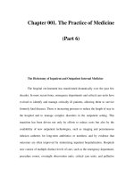
Figure 3.3 The commonest causal pathway to incident diabetic foot ulcers.
Reproduced by permission of the American Diabetes Association from reference 11
the hand, also occurs in the foot. A strong relationship exists between LJM,
insensitivity and high foot pressures
1
.
The ®ve component causes leading to intrinsic foot ulcers are, therefore:
somatic peripheral neuropathy; sympathetic peripheral neuropathy; LJM;
callus; and high foot pressures. There is, therefore, potential for preventing
such ulcers: callus can be removed by the podiatrist; high foot pressures can
be reduced by callus removal, protective insoles and hosiery; the incidence
of neuropathy can be reduced by near-normoglycaemia from the time of
diagnosis of diabetes. Thus, many neuropathic and neuro-ischaemic ulcers
are potentially preventable.
THE PATIENT WITH SENSORY LOSS
It should now be possible to achieve a signi®cant reduction of foot ulcers
and amputations in diabetes. Guidelines now exist for the diagnosis and
28 The Foot in Diabetes
Figure 3.4 Radiograph of patient presenting with a recurrent discharging heel
ulcer. On enquiry, the patient remembered some trauma to the heel but did not
realize he had part of a needle in the subcutaneous tissue under the calcaneumÐan
example of a traumatic ulcer in the insensitive foot which could have been
prevented by wearing appropriate footwear
management of neuropathy
16
and foot problems (see Chapter 21). However,
much work is still required in the assessment and management of
psychosocial factors (Chapter 10) and, as pointed out in an anonymous
audit
4
, guidelines will only be of use if properly implemented.
However, a reduction in neuropathic foot problems will only be achieved
if we remember that patients with insensitive feet have lost their warning
signalÐpainÐthat ordinarily brings the patients to their doctors. It is pain
that leads to many medical consultations: our training in healthcare is
orientated around cause and relief of pain. Thus, the care of the patient with
no pain sensation is a new challenge for which we have no training. It is
dif®cult for us to understand, for example, that an intelligent patient would
buy and wear a pair of shoes three sizes too small, and come to our clinic
with an extensive shoe-induced ulcer. The explanation, however, is simple:
with reduced sensation, a very tight ®t stimulates the remaining pressure
nerve endings and this is interpreted as a normal ®tÐhence the common
complaint when we provide patients with custom-designed shoes is: ``these
are too loose''. We can learn much about management from the treatment of
patients with leprosy (see Chapter 22); if we are to succeed, we must realize
that with loss of pain there is also diminished motivation in the healing of
and the prevention of injury.
REFERENCES
1. Boulton AJM. The diabetic foot. Med Clin N Am 1988; 72: 1513±31.
The Pathway to Ulceration 29
Figure 3.5 The high-risk neuropathic foot. This foot displays a marked prominence
of metatarsal heads with clawing of the toes and is at high risk of pressure-induced
(intrinsic) ulceration
2. Diabetes Care and Research in Europe: the St Vincent Declaration. Diabet Med
1990; 7: 360.
3. Stiegler H, Standl E, Frank S, Mender G. Failure of reducing lower extremity
amputation in diabetic patients: results of two subsequent population-based
surveys 1990 and 1995 in Germany. VASA 1998; 27: 10±14.
4. Anon. An audit of amputations in a rural health district. Pract Diabet Int 1997;
14: 175±8.
5. Larssen J. Lower extremity amputations in diabetic patients. Doctoral thesis,
Lund University, 1994.
6. Ollendorf DA, Cooper T, Kotsanos JG et al. Potential economic bene®ts of
lower extremity amputation prevention strategies in diabetes. Diabet Care 1998;
21: 1240±5.
7. Krentz AJ, Acheson P, Basu A et al. Morbidity and mortality associated with
diabetic foot disease: a 12-month prospective survey of hospital admissions in a
single UK centre. Foot 1997; 7: 144±7.
8. Young MJ, Boulton AJM. Peripheral vascular disease. In Dyck PJ, Thomas PK,
Asbury AK, Winegrad AI, Porte D (Eds), Diabetic Neuropathy. Philadelphia: WB
Saunders, 1999: 105±122.
9. Abbott RD, Brand FN, Kannel WB. Epidemiology of some peripheral arterial
®ndings in diabetic men and women: experiences from the Framingham Study.
Am J Med 1990; 88: 376±81.
10. Pecoraro RE, Reiber GE, Burgess EM. Pathways to diabetic limb amputation:
basis for prevention. Diabet Care 1990; 13: 513±21.
11. Reiber GE, Vileikyte L, Boyko EJ et al. Causal pathways for incident lower
extremity ulcers in patients with diabetes from two settings. Diabet Care 1999; 22:
157±62.
12. Siitonen OI, Niskanen LK, Laakso M, Siitonen JF, Pyorala K. Lower extremity
amputation in diabetic and non-diabetic patients: a population-based study in
Eastern Finland. Diabet Care 1993; 16: 16±20.
13. UKPDS 33. Intensive blood-glucose control with sulphonylurea or insulin
compared with conventional treatment and risk of complications in patients
with Type II diabetes. Lancet 1998; 352: 837±53.
14. UKPDS 38. Tight blood pressure control and risk of macrovascular and
microvascular complications in Type II diabetes. Br Med J 1998; 317: 703±13.
15. Boulton AJM, Malik RA. Diabetic neuropathy. Med Clin N Am 1998; 82:
909±29.
16. Boulton AJM, Gries FA, Jervell JA. Guidelines for the diagnosis and out-
patient management of diabetic peripheral neuropathy. Diabet Med 1998; 15:
508±14.
17. Kumar S, Ashe HA, Parnell L et al. The prevalence of foot ulceration and its
correlates in Type II diabetes: a population-based study. Diabet Med 1994; 11:
480±4.
18. Partanen J, Niskanen L, Lehtinen J et al. Natural history of peripheral
neuropathy in patients with non-insulin dependent diabetes. N Engl J Med
1995; 333: 89±96.
19. Young MJ, Boulton AJM, McLeod AF et al. A multicentre study of the
prevalence of diabetic neuropathy in the UK hospital clinic population.
Diabetologia 1993; 36: 150±6.
20. Tesfaye S, Stevens L, Stephenson J et al. The prevalence of diabetic peripheral
neuropathy and its relation to glycaemic control and potential risk factors: the
Eurodiab IDDM Complications Study. Diabetologia 1996; 39: 1377±84.
30 The Foot in Diabetes
21. Cabezas-Cerrato J. The prevalence of clinical diabetic polyneuropathy in
Spain: a study in primary care and hospital clinic groups. Diabetologia 1998; 41:
1263±9.
22. Adler AI, Boyko EJ, Ahroni JH et al. Risk factors for diabetic peripheral
sensory neuropathy. Diabet Care 1997; 20: 1162±7.
23. Young MJ, Veves A, Breddy JL, Boulton AJM. The prediction of diabetic
neuropathic foot ulceration using vibration perception thresholds. Diabet Care
1994; 17: 557±61.
24. Abbott CA, Vileikyte L, Williamson S, Carrington AL, Boulton AJM. Multi-
center study of the incidence of and predictive risk factors for diabetic
neuropathic foot ulceration. Diabet Care 1998; 21: 1071±4.
25. Litzelman DK, Marriott DJ, Vinicor F. Independent physiological predictors of
foot lesions in patients with NIDDM. Diabet Care 1997; 20: 1273±8.
26. Fernando DJS, Hutchinson A, Veves A, Gokal R, Boulton AJM. Risk factors for
non-ischaemic foot ulceration in diabetic nephropathy. Diabet Med 1991; 8: 223±5.
27. Toledano H, Young MJ, Veves A, Boulton AJM. Why do Asian diabetic
patients have fewer foot ulcers than Caucasians. Diabet Med 1993; 10(suppl 1):
S39.
28. Frykberg RG, Lavery LA, Pham H, Harvey C, Harkless L, Veves A. Role of
neuropathy and high foot pressures in diabetic foot ulceration. Diabet Care 1998;
21: 1714±19.
29. Veves A, Uccioli L, Manes C et al. Comparison of risk factors for foot problems
in diabetic patients attending teaching hospital out-patient clinics in four
different European states. Diabet Med 1996; 11: 709±11.
30. Uccioli L, Giacomini PG, Monticone G et al. Body sway in diabetic neuropathy.
Diabet Care 1995; 18: 339±44.
31. Katoulis EC, Ebdon-Parry M, Hollis S et al. Postural instability in diabetic
neuropathic patients at risk of foot ulceration. Diabet Med 1997; 14: 296±300.
32. Murray HJ, Young MJ, Boulton AJM. The relationship between callus
formation, high pressures and neuropathy in diabetic foot ulceration. Diabet
Med 1996; 13: 979±82.
33. New JP, McDowell D, Burns E, Young RJ. Problem of amputation in patients
with newly diagnosed diabetes. Diabet Med 1998; 15: 760±4.
34. Veves A, Murray HJ, Young MJ, Boulton AJM. The risk of foot ulceration in
diabetic patients with high foot pressure: a prospective study. Diabetologia 1992;
35: 660±3.
35. Lavery LA, Armstrong DG, Vela SA et al. Practical criteria for screening
patients at high risk for diabetic foot ulceration. Arch Int Med 1998; 158: 157±62.
36. Young MJ, Cavanagh PR, Thomas G et al. The effect of callus removal on
dynamic plantar foot pressures in diabetic patients. Diabet Med 1992; 9: 75±7.
The Pathway to Ulceration 31
4
What the Practising
Physician Should Know
about Diabetic Foot
Biomechanics
PETER R. CAVANAGH, JAN S. ULBRECHT
and GREGORY M. CAPUTO
The Center for Locomotion Studies and Pennsylvania State Diabetes Foot
Clinics, Pennsylvania State University, University Park and Hershey, PA, USA
Biomechanics is a branch of the life sciences concerned with the
consequences of forces applied to living tissues. This ®eld is clearly
relevant to diabetic foot disease since the majority of foot ulcers result from
mechanical stress which, because of loss of protective sensation to pain
1
is
not perceived by the patient. The relevance of biomechanics to the
practising physician who is treating diabetic foot problems can be stated
very clearly: many of the recalcitrant diabetic foot ulcers that are seen
failing to heal in a typical practice do so not because of medical issues, in
which the physician is well versed (infection, impaired immunity, vascular
disease, etc.), but because of simple biomechanical issues which were often
not discussed during medical training. Thus, a few minutes spent becoming
familiar with those biomechanical issues will pay considerable dividends in
improved patient care. Biomechanical considerations are important in all
three phases of care of the diabetic foot: primary prevention, healing foot
ulcers, and secondary prevention (prevention of ulcer recurrence).
This chapter discusses several very practical concepts that can be applied
to diabetic feet, and does not address the more quantitative areas of
The Foot in Diabetes, 3rd edn. Edited by A. J. M. Boulton, H. Connor and P. R. Cavanagh.
& 2000 John Wiley & Sons, Ltd.
The Foot in Diabetes. Third Edition.
Edited by A.J.M. Boulton, H. Connor, P.R. Cavanagh
Copyright
2000 John Wiley & Sons, Inc.
ISBNs: 0-471-48974-3 (Hardback); 0-470-84639-9 (Electronic)
biomechanics (such as tissue property characterization and modelling). It
should also be pointed out that there is an entire ®eld of foot biomechanics,
which is concerned with ``balancing'' structural abnormalities in non-
neuropathic feet. The types of interventions that are typically used by
practitioners of that ®eld (such as rigid ``corrective'' orthoses) are not
relevant to our present discussion. Most of this chapter will concern itself
with the most common diabetic foot ulcer, the neuropathic plantar ulcer.
Skin breakdown due to penetrating injuries, burns, and dorsal injuries due
to ill ®tting footwear are all also common, but will be addressed only brie¯y.
STRESS AND STRESS CONCENTRATION
Because force and pressure cannot be seen without the aid of specialized
instruments, it is easy to overlook the dramatic concentrations of load that
can occur at bony prominences on the plantar aspect of the foot. In the single
limb support phase of gait, the total force under the foot will always be
approximately 110% of body weight (the extra 10% comes from the ``inertial''
component as the body decelerates and accelerates throughout the gait
cycle). Since a typical men's size 10 foot has a total area of approximately
130 cm
2
, the average pressure under the foot of a 100 kg person would be
0.77 kg/cm
2
(force/area) or, stated in the more usual units (kilopascals),
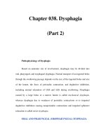
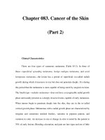
approximately 75 kPa. Figure 4.1 shows an actual pressure distribution
measured during barefoot walking under the foot of a patient who had a
prior ulcer at a prominent metatarsal head. The actual peak pressure is almost
15 times greater than if calculated as above using the simple force/area
argument. In units that might be easier to comprehend, this peak pressure
under this patient's foot is approximately 160 p.s.i. (pounds per square inch)
or 11.2 kg/cm
2
. Pressures under the foot during running and turning while
walking can be 40% greater than those encountered in walking
2
.
NEUROPATHY AND HIGH PRESSUREÐTHE KEY
COMBINATION
As discussed elsewhere in this volume (Chapter 3), peripheral neuropathy
results in what has been called a ``loss of protective sensation'' (LOPS). The
loss of sensation to touch, temperature, pain, and deep pressure can be so
dense that patients, without being aware of it, can allow objects to penetrate
completely through the foot from plantar surface to the dorsum, or they can
burn their feet with hot water, etc.
However, most injuries or ulcers in patients with diabetes or LOPS occur
at site of high plantar pressure. High pressures such as those shown in
Figure 4.1 are not usually found in healthy feet and would result in extreme
pain during ambulation for an individual with adequate sensation. For
34 The Foot in Diabetes
Foot Biomechanics 35
Figure 4.1 (a) Posteromedial view of a peak pressure distribution, measured
during barefoot walking under the foot (b) of a patient with a prior ulcer at a
prominent 2nd metatarsal head
(a)
example, patients with bony deformities from rheumatoid arthritis can
experience such pressures
3
without ulceration because they adjust their gait
to avoid bearing load on a prominent and painful area and/or they choose
footwear that will reduce the pressure (see below).
However, the repetitive application of high pressures to the same soft
tissue overlying a bony prominence in the setting of LOPS is believed to
cause tissue damage which begins deep (close to the bone)
4
. Callus
frequently forms at the surface, and when a patient presents with callus
exhibiting a shadowy dark base to visual examination, this is usually an
indication that there is a deep ulcer causing haemorrhage into callus. This
``pre-ulcer'' will then usually develop into an ulcer with further walking.
Thus, high pressure alone is not suf®cient for plantar ulceration, and neither
is neuropathyÐit is the combination of the two that provides the necessary
and suf®cient conditions for ulceration.
Since most physicians will encounter neuropathic diabetic patients who
are hospitalized for non-foot-related complaints, it is important to
mention here that low pressure applied for long periods of time to feet
with loss of protective sensation can also cause devastating lesions. The
most common manifestation is deep bilateral pressure ulcers, often
penetrating to tendon and bone, on the heels of patients who have been
bedridden for a period of time. A similar result can occur in just a few
hours in patients who have been lying on their backs during a surgical
procedure. In both situations, the ulcers are entirely iatrogenic, caused by
failure of the physician to insist on load relief for neuropathic patients,
and the failure of the nursing staff to either recognize, or act on the
knowledge, that the patient was neuropathic.
THE MECHANISMS FOR ELEVATED PRESSURE
Over time, people with diabetes can develop abnormally high pressure
under the foot during walking, and this can result from a number of
intrinsic, extrinsic and behavioural factors (Table 4.1). According to
Edmonds et al
5
, most neuropathic ulcers occur on the toes (39%), the
hallux (30%), and the metatarsal heads (24%); these areas, therefore, are of
principal concern in understanding both the causes of elevated pressure
and how intervention might be accomplished successfully. There is some
debate about the critical magnitude of plantar pressure that is required for
tissue damage. Veves et al
6
believe that a value of over 1000 kPa during
barefoot walking is required while other studies report ulceration at values
below 500 kPa. Armstrong et al
7
have suggested that a threshold of 700 kPa
is the best compromise between sensitivity and speci®city. It is, however,
likely that each patient's threshold is different and that the more active a
patient is, the less pressure is needed each step to cause ulceration. Also,
36 The Foot in Diabetes
since most studies have measured barefoot pressure, the footwear chosen
by an individual patient can clearly make the difference between ulceration
and no ulceration.
Intrinsic Factors
Certain foot structures predispose an individual to elevated pressures.
Some, like a long second metatarsal (Morton's toe) and a high arch
8,9
, are
not diabetes-related. Callus appears to concentrate pressure rather as if it
were a foreign body under the foot. There are some indications that the
properties of the plantar soft tissue may be adversely affected by
glycosylation end products, although much remains to be explored in
this area. Toe deformities (claw toes, hammer toes, hallux valgus) also
tend to result in higher pressures
9
. Clawing of the toes appears to result
in the plantar fat pads being displaced anteriorly, leaving the condyles of
the metatarsal heads ``exposed'', and this a common ®nding in patients
with diabetic neuropathy. Palpation of the metatarsal heads in a patient
with claw toes often reveals an exquisitely thin layer of soft tissue
overlying the bone, which leads directly to high pressures during
walking unless counter-measures are undertaken (see below). In fact, the
lack of adequate thickness of soft tissue under bony prominences has
been shown to be an extremely important determinant of elevated
pressure
8
. The tips of clawed toes can themselves be locations of ulcers
due to concentrated pressure (Figure 4.2).
The range of motion at many joints has been shown to be decreased
in patients with diabetes
10
. This is not a neuropathic complication, but
probably another effect of glycosylation, whereby the collagen in joint
Foot Biomechanics 37
Table 4.1 Factors that can lead to elevated plantar pressure under the foot during
walking
Intrinsic Extrinsic Behavioural
Foot architecture
Long second toe
High arch
Walking without shoes
Soft tissue alterations
Callus
Glycosylation (presumed)
Migration of tissue
Thin tissue
Poor footwear
Tight or loose shoes
Shoes with hard soles
Poor choice of shoes
Limited joint mobility Accidents and incidents Inadequate callus care
Foot deformity
Claw toes
Hallux valgus
Charcot fracture
Prior surgery Walking patterns
38 The Foot in Diabetes
Figure 4.2 (a) Posteromedial view of a peak pressure distribution, measured
during barefoot walking, showing elevated pressure at the tips of clawed second
toe. Note that the pressure under toe 2 and the hallux are approximately equal. The
foot is shown in (b)
(a)
capsules is stiffened by the glycosylation process. The consequence of
reduced ranges of motion at the major joints of the foot and ankle, such
as the ®rst metatarsophalangeal (MTP), sub-talar and talo-crural joints, is
likely to be increased plantar pressures under the forefoot
4
. The most
frequently problematic joint in this regard is the ®rst MTP
11
. Invariably,
a patient with a neuropathic ulcer under the pad of the hallux will be
found to have reduced capacity for dorsi¯exion at this joint (Figure 4.3).
Despite the above emphasis on the forefoot, a number of conditions
can cause elevated pressure in other regions of the foot. Charcot fractures
of the midfoot
12,13
typically result in a ``rocker bottom'' foot which bears
load principally on the collapsed region of the midfoot (Figure 4.4).
Certain surgical procedures that are intended to reduce loads at primary
areas of ulceration can have the secondary effect of increasing pressure
in other areas. For example, lengthening of the Achilles tendon, which is
sometimes performed following forefoot surgery, can result in what is
known as a ``calcaneus gait'', in which elevated heel pressure occurs
during much of the stance phase (Figure 4.5). Removing metatarsal heads
because of ulceration in that region can lead to higher pressures under
other metatarsal heads.
Extrinsic Factors
In terms of the pressures that the soft tissues are exposed to, footwear is
the single most important extrinsic determinant of elevated pressure.
While appropriate footwear can be of great bene®t in preventing ulcers
(see below), incorrect footwear can actually cause ulceration
14
. The two
major de®ciencies most frequently seen in shoes are incorrect sizing (too
loose or too tight) and inadequate cushioning. Tight shoes can cause
ulceration at a number of locations. Lesions commonly occur over dorsal
deformities, such as a bunion or a dorso-lateral prominence of the ®fth
metatarsal head (MTH5). The tips of the interphalangeal joints on claw or
hammer toes are prime at-risk sites, and ulcers in the interspace between
the toes can be caused by the toes being crushed together in a shoe with
incorrect contours. Loose shoes, which allow the foot to slip, can also
result in ulcers.
As we shall discuss below, ``cushioning'' for the neuropathic foot is
largely de®ned in static terms and can be equated with ``thickness'' of
``soft'' material under the foot. It has been shown that walking in shoes with
leather soles is roughly equivalent to walking barefoot, whereas walking in
simple sports shoes (trainers) can reduce pressure by up to 50% compared
with barefoot walking
15
. Thus, the wrong choice or prescription of shoes
can be devastating for the integrity of the diabetic foot.
Foot Biomechanics 39
40 The Foot in Diabetes
Figure 4.3 (a) Posterolateral view of peak pressure distribution, measured during
barefoot walking, from a patient with a neuropathic ulcer under the pad of the
hallux (b) secondary to a reduced capacity for dorsi¯exion at the ®rst MTP joint.
Note that the MTH1 and hallux pressures are approximately equal, although this
patient has never experienced an ulcer uner MTH2. (b) Reproduced by permission
of W. B. Saunders Company from reference 30
(a)
Foot Biomechanics 41
Figure 4.4 Posteromedial view of a peak pressure distribution under a ``rocker-
bottom'' foot (b) during barefoot walking. Load is principally borne on the collapsed
region of the midfoot and other regions in the rearfoot and forefoot received almost
no load throughout the entire contact phase
(a)
42 The Foot in Diabetes
Figure 4.5 Posteromedial view of peak pressure distribution (a) before and (b) 3
months after surgery, which included an osteotomy of the ®rst metatarsal and a
lengthening of the Achilles tendon. Note the reduction in forefoot pressures and the
increase in the heel peak pressures postsurgically
Behavioural Factors
It is widely believed that barefoot walking is a principal cause of plantar
ulceration that is amenable to behavioural intervention. As mentioned
above, we do not know the number of steps and the magnitude of pressure
that will exceed an individual's threshold for ulceration. However,
experience suggests that there are some patients who protect their feet
adequately in footwear throughout the day, yet ulcerate because of just a
few steps of barefoot walking to urinate during the night. Thus, at least for
some patients, even a few steps of barefoot walking are too many. Taking
showers barefoot is another dangerous behaviour. The provision of padded
slippers, which can be donned easily, is a simple way to intervene in such
cases.
Foot injury also occurs frequently during self-care of nails. Patients with
poor eyesight should not perform self-care of nails and should be
encouraged to have a family member or a chiropodist provide this care.
Total neglect of nail and callus care can also be a cause of ulceration. As
discussed above, callus concentrates pressure on the plantar aspect of the
foot and studies have shown that the presence of callus increases the risk of
ulceration by over 11 times
16
. Thus, callus should be regularly removed.
There are some indications that neuropathic patients have altered gait
patterns, but it is not yet clear whether this results in elevated plantar
pressure. Brand
17
hypothesized that neuropathic gait would be less variable
and that this would result in continued application of stress to the same
plantar location, but this has not been found to be the case
18
. Regardless,
patients with LOPS will not consciously alter their gait, since they will feel
no pain developing in high pressure areas from too much walking. There is
also some evidence that neuropathic patients experience more falls, and
injuries due to falls, than matched non-neuropathic diabetic patients
19,20
.
Balance
21
and limb position sense
22
are also impaired and these two factors
may lead to more frequent traumatic injuries to the feet of neuropathic
patients.
PRIMARY PREVENTION: THE 30 SECOND FOOT
EXAMINATION
We have established above that most foot ulcers that the practising
physician will see result from mechanical insult to tissue that is deprived of
normal sensation. Although we have emphasized neuropathic injury,
diminished pulses identify another group of patients at risk because of
ischaemia. Thus, the most important issues in prevention of foot pathology
are to identify patients who have lost protective sensation and patients with
signi®cant ischaemia. Loss of protective sensation is most simply assessed
Foot Biomechanics 43
with a 10 g mono®lament
23
and we prefer that a forced-choice protocol be
used (``Am I touching you at time A or time B?'') rather than the procedure
described in the International Practical Guidelines in Chapter 21. We
recommend that the examination shown in Table 4.2 be performed
annually. If the examination shows that the patient has protective sensation
and foot pulses and, therefore, is judged not to be at risk, then 30 seconds is
all the time that is needed. During this initial scan, the presence of
signi®cant deformity should also be noted, as this may affect treatment
decisions even in the absence of signi®cant neuropathy.
PRIMARY PREVENTION: THE 2 MINUTE FOOT
EXAMINATION
If the initial examination determines that protective sensation has been lost,
the biomechanics of the foot and shoes become critical issues in the patient's
future. The examination must now be extended to look for the factors
discussed above, and for other non-biomechanical factors discussed
elsewhere in this volume, which could contribute to ulceration. Surpris-
ingly, this need not be a lengthy examinationÐit is remarkable how much
can be achieved in a short time if the clinician has a well-de®ned set of goals
in advance of the foot examination. In approximately 2 minutes, an
examination can cover all of the components shown in Table 4.3 for a
patient who is at risk of foot injury.
The surface examination of the foot is fairly straightforward. The clinician
must identify ulcers, callus, haemorrhage into callus, breaks or cracks in the
skin, skin infection, maceration between toes, and elevated surface
temperature. The latter may be an indication of infection or of an active
Charcot process, as can oedema or erythema. Nail care should be assessed
and the presence of ingrown or long nails, nail fungal infections, and
injuries from self-care of nails should be noted. While some of these are not
biomechanical issues, their importance is self-evident; most of these topics
are covered elsewhere in this volume.
One does not have to be a chiropodist or a foot orthopaedic surgeon to
identify the major deformities that can lead to elevated pressure. Those
shown in Figure 4.6, including prominent metatarsal heads, claw or
hammer toes, excessive callus, hallux valgus and prior amputation can,
in combination with neuropathy, lead to ulceration (also see rocker
bottom deformity in Figure 17.3). The identi®cation of these deformities
will also be important in decisions related to prescription footwear (see
below).
Looking at the patient's footwear is a key component of the examination.
Shoes and socks should be removed. The socks should be examined for
44 The Foot in Diabetes
Foot Biomechanics 45
Table 4.2 The 30-second foot examination. On both feet, the following should be assessed:
Evaluation component Details Action
1. Ascertain if the patient has had any
previous diabetes-related foot lesions
Ask about ulcers or blisters
that were not perceived
as particularly painful; ask about
foot lesions that required vascular
procedures to heal; look for toe or
partial foot amputations
If there is a history of previous foot problems,
the patient is at risk for more
2. Vascular status/pulses Feel for:
Pulse in posterior tibial artery behind
medial maleolus
Pulse in dorsalis pedis artery on the
dorsum of the foot
If both pulses are absent in either foot, consider
further evaluation, particularly if other
symptoms or signs of vascular disease are
present (see Chapter 16)
3. Loss of Protective Sensation:
inability to feel the touch of
a 10 g mono®lament
In a quiet room, test multiple forefoot
plantar sites and in particular toes and
MTHs; avoid callused areas; ask the
patient to tell you every time he/she
can feel the touch (do not ask for a
response only when you are touching
the patient); with the patient's eyes
closed, bend mono®lament against the
skin for 1 second; repeat questionable
sites; apply mono®lament with random
cadence; a relative can observe to
reinforce abnormal ®ndings for the
patient
If the patient cannot feel the mono®lament at
even one site, label patient as ``at risk''. If you
are concerned about questionable or hesitant
responses, repeat the test at each clinic visit
until the result is clearer, or classify the
patient as ``at risk''
4. Look for signi®cant foot deformity See the examples shown in Figure 4.6 Appropriate footwear or referral should be
suggested, even in the absence of neuropathy
If the patient has no history of previous problems, can feel touch at all sites, and has one palpable pulse in each foot, the 30 second examination can end here. Repeat in 1
year, or sooner if ®ndings were questionable.
If the patient has a history of previous problems, has absent pulses or cannot feel touch at even one site, then he/she is at risk for foot injury. Proceed with the
additional ``2 minute examination''.
evidence of drainage from a wound, and the shoe insoles should be studied
to see if they have ``bottomed out'' and no longer provide adequate
cushioning. The size of the shoe should be compared to the size of the foot,
particularly the height and curvature of the forefoot region. At the end of
the examination the patient should be asked to put his/her shoes and socks
on, so that the examiner can assess the patient's mobility during this
46 The Foot in Diabetes
Table 4.3 The 2-minute foot examination. This follows on from the examination in
Table 4.2 if the patient has previous foot problems, has lost protective sensation or
has signi®cant vascular disease. On both feet, the following should be examined:
Evaluation component Details Action
5. Examine all surfaces Look for:
Ulcer
Callus
Haemorrhage into callus
Blister
Maceration between toes
Other breaks in the skin
Skin infection
Oedema, erythema, elevated
temperature
Prescribe unweighting device
to heal ulcer (see Figures 4.8
and 4.9)
Remove callus (sharp debride-
ment and/or dremmel or
emery board)
Treat skin infection or injury
Refer if Charcot fracture
suspected
6. Examine the nails Look for:
Fungal infections
Ingrown toe nails
Evidence of injury
from self-care of nails
Consider treating fungal
infections
Advise against self-care of nails
Suggest chiropody care
7. Identify foot deformity Look for:
Prominent metatarsal heads
Claw or hammer toes
Rocker bottom foot
deformity
Hallux valgus and bunions
Prior amputation
The presence of foot deformity
will dictate footwear speci®-
cations (see text and Table
4.4)
8. Examine the shoes. Have
the patient put their shoes
and socks on as the last
component of the examin-
ation. This will show the
patient's ability to examine
their own feet.
Look for:
Drainage into socks
Worn out (¯attened) insoles
Shoes that are leaning
badly to one side
Poorly ®tting shoes (too
tight, too loose, too short,
not enough room for the
toes)
Gait pattern
Prescribe appropriate footwear
if necessary (see text and
Table 4.4)
Suggest replacement shoes if
necessary
9. Establish need for
education
Ask:
``Why do you think I am
concerned about your
feet?''
Do you walk without shoes
at home?
Who takes care of your nails?
Schedule patient for education
visit with diabetes educator/
nurse if understanding is
lacking or if behaviours are
unacceptable