The Intensive Care Manual - part 6 pot
Bạn đang xem bản rút gọn của tài liệu. Xem và tải ngay bản đầy đủ của tài liệu tại đây (497.65 KB, 42 trang )
8 / Cardiac Arrhythmias 195
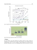
FIGURE 8–4 Twelve-lead ECG from a patient with atrial fibrillation and a controlled ven-
tricular response. Note the chaotic baseline without defined atrial activity. There is a sugges-
tion of a more organized pattern in the V
1
lead, but this is not seen in other leads. The
ventricular response is characteristically “irregularly irregular.”
ch08.qxd 11/7/01 4:14 PM Page 195
Atrial flutter has also been extensively studied electrophysiologically. Unlike
the disorderly atrial activities in fibrillation, it is now well-accepted that for most
instances of clinically encountered atrial flutter, the electrical impulse circulates
around in the right atrium in one large loop. Because atrial flutter is more orga-
nized than atrial fibrillation, it displays more organized atrial activities of larger
amplitude on ECG. Atrial flutter usually has an associated “sawtooth” pattern,
which represents revolving atrial activities and is best appreciated in the inferior
limb leads 2, 3, and aVF (Figure 8–5). In typical atrial flutter, the reentrant circuit
usually has a well-defined cycle length at about 300 beats/min. Often, there is a
2:1 AV conduction pattern during atrial flutter, leading to a consistently regular
ventricular response of 150 beats/min.
Many of the impulses of a SVT can be transmitted down to the ventricle via
the AV junction, especially when AV conduction is enhanced by release of cate-
cholamines. The rapid ventricular rate is usually the main problem associated
with atrial arrhythmias in the ICU. The fast rates are especially troublesome for
patients who have underlying CAD or ventricular hypertrophy, because ischemia
and significant hemodynamic compromise can occur rapidly. The goal of ther-
apy in the care of patients with atrial arrhythmia is stabilization of hemodynam-
ics and ventricular rate control. During sustained atrial arrhythmias in a patient
with stable blood pressure, AV nodal blocking agents, such as beta blockers, cal-
cium channel blockers, and digoxin, are all effective agents in slowing the ven-
tricular response. Diltiazem can be given intravenously as a bolus at a dose of 5 to
20 mg, which may be followed by an infusion of the same drug at rates of 5 to 20
mg/hr. This allows for rapid control of heart rate and subsequent conversion to
oral long-term therapy. Digoxin is also effective, but the onset of action is some-
what longer than that of diltiazem. Digoxin is typically given as a loading dose of
1 mg over the course of 24 hours. We typically give 0.5 mg initially, followed by
another 0.25 mg in 4 to 6 hours and a second 0.25 mg in yet another 4 to 6 hours.
If there is hemodynamic compromise, then urgent restoration of sinus rhythm
with direct-current (DC) energy-synchronized cardioversion is imperative. In
addition, if the rapid ventricular response rate during atrial arrhythmia is making
conditions such as myocardial ischemia, infarction or congestive heart failure
worse, early cardioversion is also indicated.
Pharmacologic antiarrhythmic agents are usually used for chemical cardiover-
sion and maintenance of sinus rhythm, if the patient’s blood pressure permits their
use. Oral antiarrhythmic agents for atrial fibrillation include class 1a drugs, such as
quinidine and procainamide; class 1c drugs, such as propafenone and flecainide;
and class 3 drugs, such as sotalol and amiodarone. Procainamide has been the first-
line intravenous antiarrhythmic that is traditionally used. More recently, intra-
venous amiodarone has also been used with success. Intravenous procainamide is
typically given as a bolus of 10 to 15 mg/kg of body weight over 20 to 30 minutes,
followed by a maintenance infusion at a rate of 1 to 6 mg/min. Care must be taken
when administering procainamide intravenously because it may cause significant
prolongation of the QT interval and the QRS duration; if given rapidly, it may also
196 The Intensive Care Manual
ch08.qxd 11/7/01 4:14 PM Page 196
8 / Cardiac Arrhythmias 197
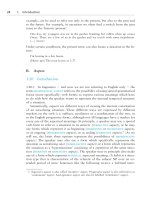
FIGURE 8–5 Twelve-lead ECG from the same patient in Figure 8–4, now showing a charac-
teristic “sawtooth” pattern that is especially apparent in inferior leads. This patient alternates
between atrial fibrillation and “typical” atrial flutter. The rate of the flutter waves is some-
what slower than is usually seen (230/min) as a result of antiarrhythmic therapy.
ch08.qxd 11/7/01 4:14 PM Page 197
cause hypotension. Procainamide should not be given at a rate faster than 50
mg/min. Intravenous amiodarone is usually given in a 150-mg bolus over 10 min-
utes and may be repeated if ineffective. Then a maintenance infusion of 1 g of amio-
darone every 24 hours may be given. A central venous line is recommended with
the use of intravenous amiodarone to avoid phlebitis. Intravenous amiodarone has
not yet been officially approved as a therapy for supraventricular arrhythmias.
Both of these agents can further lower a patient’s blood pressure; therefore, close
monitoring of patients is mandatory when these agents are used. Intravenous ibu-
tilide has also been reported to be an effective agent for cardioversion, although its
conversion rate for atrial flutter is much higher than for atrial fibrillation. Ibutilide
may lead to significant QT prolongation and should be avoided in patients with
electrolyte imbalance or who are already on agents that can prolong QT intervals,
such as phenothiazines. Caution and continuous ECG monitoring must be exer-
cised with the use of ibutilide, because dramatic QT prolongation can lead to tor-
sades de pointes, and potentially convert a nonemergent arrhythmia to one that
causes immediate hemodynamic collapse. Intracardiac thrombi and systemic em-
boli may form in patients with atrial fibrillation or atrial flutter sustained for more
than 48 hours. Therefore, if anticoagulant therapy is not contraindicated by con-
current medical problems, it should be initiated for these patients.
Precipitating factors that may lead to atrial fibrillation and atrial flutter should
be sought if clinical conditions warrant such concerns. For example, it is well-
documented that pulmonary embolism can lead to atrial arrhythmias, especially
atrial fibrillation. This may be important in postoperative patients or patients
with hypercoagulable states. Other factors that can lead to atrial fibrillation or
atrial flutter include hypertensive heart disease, valvular disease, pericarditis,
myocarditis, hyperthyroidism, and even fever.
Another supraventricular rhythm disturbance that is seen frequently in the
critically ill patient is multifocal atrial tachycardia (MAT), which is a rapid irreg-
ular rhythm that is characterized by a rate that exceeds 100 beats/min and has at
least three distinct P-wave morphologies. This is most frequently seen in patients
with severe underlying lung disease, particularly those receiving inhaled bron-
chodilators or theophylline preparations. Treatment is difficult and should be
aimed primarily toward improving the pulmonary condition. There are several
reports on the use of both intravenous metoprolol and intravenous verapamil to
control the rate. Caution must be used when giving beta blockers, such as meto-
prolol, to patients with reactive lung disease; our experience with this agent in
this situation has not been successful.
Reentrant SVTs, including AV nodal reentrant tachycardia and AV reentrant
tachycardia using a bypass tract, are characterized by regular, narrow complex
tachycardia on the surface ECG. It may be possible to identify a retrograde P
wave after the QRS complex, particularly in the case where a bypass tract is in-
volved, but if the retrograde conduction is sufficiently rapid, it may not be visi-
ble. It may also be difficult to detect a P wave in cases of rapid sinus tachycardia.
In these cases, we advise the use of adenosine injections or carotid sinus massage
198 The Intensive Care Manual
ch08.qxd 11/7/01 4:14 PM Page 198
as therapeutic intervention and for diagnostic purposes. The initial dose of
adenosine is 6 mg, given as a rapid intravenous injection. If there is no response,
a dose of 12 mg may be given. In cases of reentrant SVTs or some atrial tachycar-
dias, the response to adenosine is usually prompt termination of the tachycardia.
In the case of sinus tachycardia, however, a brief slowing of the sinus rate is seen,
which usually allows identification of distinct P waves.
WIDE COMPLEX TACHYCARDIA
A wide complex tachycardia may lead to serious consequences or it may be a rel-
atively benign occurrence. The correct diagnosis of such a tachycardia is impera-
tive, especially in the critical care setting. A wide complex tachycardia usually
arises from a ventricular origin; however, an SVT with aberrant conduction can
also manifest as a wide complex tachycardia. Other than ventricular fibrillation,
ventricular tachycardia is the most ominous tachyarrhythmia involved in the
care of patients in the ICU. Because it may lead to rapid hemodynamic collapse,
prompt intervention is necessary. SVT often is better tolerated, although signifi-
cant hemodynamic compromise can occur quickly as well. Hemodynamic stabil-
ity in conjunction with a wide complex tachycardia does not rule out ventricular
tachycardia. Equally important is an understanding of the consequences of both
pharmacologic and nonpharmacologic therapy for wide complex tachycardia to
avoid potentially harmful interventions. Some of the drugs used for the manage-
ment of SVT, such as calcium channel blockers, may lead to adverse conse-
quences in a patient with ventricular tachycardia. Therefore, in the ICU, all wide
complex tachycardia should be assumed to be ventricular in origin until it can be
ruled out with a high degree of certainty, especially in patients with known car-
diac disease.
Distinguishing ventricular tachycardia from SVT with aberrant conduction on
the basis of surface ECGs can be difficult, especially because recordings from only
one or two leads are often all that is available. There are some findings that may
be helpful in diagnosis of the origin of a wide complex tachycardia.
“Atrioventricular dissociation,” or evidence of separate atrial and ventricular
activities, should always be sought in the patient with a wide complex tachycardia
tracing. This is manifested as P waves and QRS complexes that are temporally
unrelated. The P waves, or atrial ECGs, are often difficult to discern and may be
present in any part of the cardiac cycle, including parts of the QRS complex or T
waves. Techniques to amplify the amplitude of the atrial activities, such as
esophageal leads or even placement of a transvenous electrode, may be helpful.
Although the presence of AV dissociation is not completely diagnostic for ven-
tricular tachycardia, it does make a ventricular tachycardia highly likely. The
presence of a 1:1 AV relationship is consistent with either SVT or ventricular
tachycardia and cannot be used to distinguish one from the other.
8 / Cardiac Arrhythmias 199
ch08.qxd 11/7/01 4:14 PM Page 199
Another phenomenon to look for is the presence of a “fusion” beat, i.e., a
combined QRS complex resulting from impulses originating from two different
areas of the heart. A combination, or fused, QRS complex between a beat origi-
nating in the ventricle and one from a supraventricular site is more reliable for
the diagnosis of ventricular tachycardia (Figure 8–6). Typically, this is seen in
ventricular tachycardia with relatively slower rates, allowing time for the supra-
ventricular impulses to conduct down to the ventricle.
When possible, a 12-lead ECG should be obtained for further information in
differentiating the origin of the tachycardia. There are well-tested morphologic
criteria for wide complex tachycardias of both right and left BBB–type patterns in
patients in whom the origins of tachycardia were confirmed by invasive electro-
physiology studies.
If the QRS morphology in a wide complex tachycardia displays a right
BBB–type pattern and, in lead V
1
, the initial R wave (the initial positive deflec-
tion) is dominant, the tachycardia is likely to be of ventricular origin. This can be
seen either as a monophasic R wave in V
1
or as the first initial positive deflection
(R) being taller than the second positive deflection (r′). In a wide complex tachy-
cardia with a right BBB–type pattern, an R wave amplitude of less than the S
wave in lead V
6
suggests ventricular tachycardia. In tachycardias displaying a left
BBB–type pattern delay in the initial forces with a broadened r wave (r > 0.04
sec), notches in the initial QRS downstroke in lead V
1
suggest ventricular tachy-
cardia. Furthermore, during tachycardia with a left BBB–type pattern, a q wave
present in lead V
6
makes it likely that the tachycardia is of ventricular origin.
5
Basic premises for these criteria are that the more fragmented the initial QRS
forces are and the wider the QRS duration is, the more likely there is a ventricular
origin of the tachycardia. This results from muscle-to-muscle conduction during
ventricular tachycardia rather than conduction down to the ventricles through
specialized His and Purkinje tissues during SVT. These criteria were tested in
patients who did not have existing BBBs or Wolff-Parkinson-White syndrome.
Furthermore, these criteria probably cannot be relied on for patients on
antiarrhythmic therapy, because many of these drugs can alter cardiac conductiv-
ity and thereby affect the initial forces of the QRS complex patterns and duration.
Another criterion on 12-lead ECGs that suggests a ventricular origin of a wide
complex tachycardia is concordance of the QRS pattern in the precordial leads
(V
1
through V
6
).
6
Both positive concordance (i.e., all QRS complexes in V
1
though V
6
display monophasic R waves) and negative concordance (i.e., all pre-
cordial QRS complexes display monophasic QS patterns) are suggestive of
ventricular tachycardia. Negative concordance is diagnostic for ventricular tachy-
cardia, but positive concordance may, rarely, result from tachycardia involving
an accessory AV bypass tract. Table 8–3 summarizes the criteria that are useful
for distinguishing the cause of a wide complex tachycardia.
Cycle length variability is not a useful diagnostic criterion for wide complex
tachycardias. While it is true that atrial fibrillation conducted with aberration dis-
plays an irregularly irregular pattern, the rate of a ventricular tachycardia can often
200 The Intensive Care Manual
ch08.qxd 11/7/01 4:14 PM Page 200
8 / Cardiac Arrhythmias 201
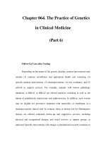
FIGURE 8-6 Twelve-lead ECG demonstrating a wide complex tachycardia. P waves (P) can
be seen dissociated from the QRS in what is termed AV dissociation. In addition, fusion beats
can also be detected (F). The combination of AV dissociation and fusion beats is, in almost all
cases, diagnostic of ventricular tachycardia.
ch08.qxd 11/7/01 4:14 PM Page 201
be irregular as well. Similarly, it has been suggested that alternating cycle length
may be a marker for certain forms of SVT, but alternating cycle length variations
have been well described in patients proven to have ventricular tachycardia.
Always compare a patient’s baseline ECG to the one obtained during wide
complex tachycardia. If a BBB pattern is present during sinus rhythm and the
tachycardia displays a BBB pattern of the alternate bundle, then the tachycardia is
very likely to be ventricular. As mentioned, the wider the QRS duration, the
more likely that the tachycardia is of ventricular origin. Interestingly, a wide
complex tachycardia with QRS duration shorter than the conducted QRS is al-
most always caused by ventricular tachycardia. These tachycardias often are orig-
inating from a septal region, and the left and right ventricles are activated in a
more simultaneous fashion than a supraventricular impulse conducted down to
the ventricle with a bundle branch conduction block.
Other than ECGs, clinical physical examination may also help in distinguish-
ing ventricular tachycardia from SVT with aberrant conduction. The presence of
“cannon A waves,” resulting from atrial contraction against closed AV valves,
during inspection of the jugular pulse suggests the presence of AV dissociation
and, therefore, ventricular origin of the tachycardia. Variations in the intensity of
the first heart sound (S
1
) and splitting of S
1
during auscultation as a result of ven-
tricular dyssynchrony also suggest ventricular tachycardia.
Characteristics of a wide complex tachycardia may provide important clues
about the underlying cardiac pathology. Patients with transmural scars from in-
farctions or cardiomyopathy from various causes have a substrate for reentrant
monomorphic ventricular tachycardia, or a wide complex tachycardia displaying
a consistent QRS morphology from beat to beat. On the other hand, insufficient
myocardial arterial supply or increased myocardial demand may lead to electro-
202 The Intensive Care Manual
TABLE 8–3 Criteria for diagnosis of etiology of wide complex tachycardia based on Qrs
morphology.
8
Aberration VT
RBBB QRS ≤ 0.12 sec QRS ≥ 0.14 sec
Axis: Normal Axis: Superior
V
1
: rsR' or rR' V
1
: R, Rr', RS
V
6
: R/S > 1 V
6
: R/S < 1
LBBB QRS ≤ 0.14 sec QRS ≥ 0.16 sec
Axis: normal or leftward Axis: rightward
Lead V
1
or V
2
: R < 0.04 sec Lead V
1
or V
2
: r ≥ 0.04 sec
Onset to nadir: < 0.07 sec Onset to nadir: ≥ 0.07 sec
Smooth downstroke Notch on downstroke
V
6
: No Q wave V
6
: Q wave
ABBREVIATIONS: VT, ventricular tachycardia; RBBB, right bundle branch block; LBBB, left bundle
branch block.
ch08.qxd 11/7/01 4:14 PM Page 202
physiologic instability within the myocardium, resulting in ventricular fibrilla-
tion or polymorphic ventricular tachycardia, a wide complex tachycardia with
varying QRS morphologies. Therefore, recognition of the different ventricular
arrhythmias as manifestations of the underlying cardiac pathophysiology can
help in choosing the proper therapeutic and management interventions.
Urgent intervention for a wide complex tachycardia is often needed as a result
of the hemodynamic effects. If hemodynamic collapse is evident or if blood pres-
sure is unstable, countershock with DC energy is required. There are other clini-
cal indications for relatively urgent DC cardioversion as well. These include
ischemia or infarction, angina, and severe heart failure. If a patient’s blood pres-
sure is stable, then the various criteria may be applied to distinguish ventricular
and supraventricular origin of the tachycardia and a decision for appropriate
therapy may be applied.
Traditionally, intravenous lidocaine is the first antiarrhythmic used for ven-
tricular tachycardia. Under ischemic conditions, such as during the infarction
period, ventricular arrhythmias often are manifested as polymorphic ventricular
tachycardia (Figure 8–7) or ventricular fibrillation. Under these circumstances,
intravenous lidocaine is reasonably effective and it should be considered as a
first-line agent. For nonacute infarction or non–ischemia-related ventricular ar-
rhythmias, typically manifested as a monomorphic ventricular tachycardia (with
consistent beat-to-beat QRS morphology), several clinical reports have suggested
that intravenous procainamide may be more effective for termination than lido-
caine.
9
Intravenous amiodarone has become widely available over the past few
years. Data are becoming available suggesting its effectiveness in terminating and
suppressing ventricular arrhythmias.
10
Amiodarone probably is superior in com-
parison to lidocaine or procainamide for ventricular arrhythmia management.
However, it may have a profound blood pressure–lowering effect and its use
should be accompanied by cautious hemodynamic monitoring.
8 / Cardiac Arrhythmias 203
FIGURE 8–7 Rhythm strip showing 6-beat run of polymorphic ventricular tachycardia.
There is a variable morphology to the QRS complexes of the tachycardia. This is often seen in
the patients with ischemia.
ch08.qxd 11/7/01 4:14 PM Page 203
The use of adenosine has been advocated as a diagnostic tool for distinguish-
ing ventricular origins from supraventricular origins in a wide complex tachycar-
dia. Adenosine has vasodilator effects and a possible “steal” phenomenon in the
coronary circulation; this may induce myocardial ischemia and lead to further
hemodynamic compromise. Even though the half-life of adenosine is brief, its ef-
fects in patients with severe CAD may trigger a cascade of hemodynamic effects
that may become irreversible. Therefore, we recommend that the use of adeno-
sine as a diagnostic measure for wide complex tachycardia must be taken with
caution, especially in patients with known severe coronary disease. Unless it is
absolutely certain that the diagnosis is SVT, calcium channel blockers, such as
diltiazem or verapamil, should not be used to treat wide complex tachycardias
because there are a multitude of reports detailing hemodynamic collapse in pa-
tients with ventricular tachycardia who were treated with these agents.
7
TORSADES DE POINTES
Torsades de pointes is a subtype of polymorphic ventricular tachycardia that
should be recognized because it has distinct diagnostic and therapeutic implica-
tions that differ from other types of wide complex tachycardia. A French term
meaning “twisting of the points,” torsades de pointes has an appearance similar
to rapid QRS axis shifting. It is usually characterized by prolonged QT intervals,
and it is often initiated with a premature ventricular extrasystole occurring on or
around the T wave of the preceding beat. Known causes of torsade de pointes
typically include conditions that prolong the QT interval, such as congenital long
QT interval syndrome; electrolyte imbalances, such as hypokalemia, hypomag-
nesemia, or hypocalcemia. Drugs that prolong the QT interval are also known to
lead to torsades de pointes; these include class Ia and III antiarrhythmic drugs
and some antihistamines and psychotropic medications. Table 8–4 lists a number
of causes of prolongation of the QT interval and torsades de pointes. Care should
be paid to patients with decreased clearance of any of these suspect medications
as well as any combinations that may compound the prolongation of the QT in-
terval. Remember that bradycardia may prolong the repolarization process, and
thus the QT interval. The effects of these precipitants are more pronounced and
the risk of torsades de pointes is higher in patients with bradycardia.
If sustained, the acute intervention for torsades de pointes, as with all wide com-
plex tachycardia with hemodynamic instability, is countershock with DC energy.
Once a stable rhythm has been restored, the major goal of the therapy is to shorten
the QT interval as much as possible. This obviously includes removal of the of-
fending agent or correcting the underlying conditions. Sometimes cardiac pacing
or the use of an isoproterenol infusion may be necessary to further decrease the
ventricular repolarization time, especially if bradycardia is present. If the episodes
of torsades de pointes are not sustained, then, in addition to the above interven-
tions, empiric intravenous magnesium therapy has been suggested.
204 The Intensive Care Manual
ch08.qxd 11/7/01 4:14 PM Page 204
TOXIC AND METABOLIC CAUSES OF ARRYTHMIAS
The medical ICU often serves as the stabilization site for patients after life-
threatening overdoses and severe metabolic disturbances. These conditions can
result in cardiac rhythm disturbances that require prompt recognition and treat-
ment. Adequate suspicion, proper interpretation of the ECG, and complete
knowledge of the specific emergency treatments are part of the armamentarium
of the ICU physician. Some of the most commonly encountered problems, dis-
cussed here, include hyperkalemia and hypokalemia, hypercalcemia and hypocal-
cemia, and hypothermia; overdoses of a tricyclic agent or digitalis; and acquired
torsades de pointes.
Hyperkalemia
Hyperkalemia may be caused by a number of processes, including acidosis from
any cause, acute renal failure, iatrogenesis, and hemolysis. Life-threatening eleva-
tions in potassium levels can be a complication of the patient’s original problem
or of treatment they received during their admission. Because hyperkalemia
often causes no symptoms in itself, the ECG tracing must be relied on to define
the clinical implications of hyperkalemia and the urgency of treatment.
The ECG changes of hyperkalemia are variable and depend not only on the
severity but also on the chronicity of the elevation in serum potassium level. Al-
though a close correlation exists between the potassium level and ECG changes in
8 / Cardiac Arrhythmias 205
TABLE 8–4 Causes of prolongation of QT interval and torsades de pointes
Drugs Electrolyte Abnormalities Congenital
Quinidine, procainamide, Hypokalemia Jervell and Lange-Nielsen syn-
sotalol, amiodarone drome
Tricyclic and tetracyclic Hypocalcemia Romano-Ward syndrome
antidepressant agents
Phenothiazines Hypomagnesemia
Haloperidol (Haldol)
Antihistamines
Macrolide antibiotics
Pentamidine
Serotonin antagonists
Adenosine
Cocaine
Cisapride
Arsenic poisoning
ch08.qxd 11/7/01 4:14 PM Page 205
animal models, the relation is less clear in clinical cases. Abnormal potassium lev-
els affect P waves, the QRS complex, and T waves. P-wave voltage decreases as a
result of slow intra-atrial conduction with low-amplitude atrial depolarization
and the PR interval lengthens. With severe widening and attenuation of the P
wave, there may be no atrial depolarization seen on the surface ECG, so the erro-
neous diagnosis of a junctional rhythm may be made. Type I or II second-degree
AV block may also occur. As the QRS complex widens, the normally sharp con-
tour of the QRS becomes wider and eventually merges with the T wave, until no
ST segment exists. The T wave becomes symmetrically peaked, the entire QRST
complex can resemble a sine wave, and the QT interval usually remains normal
or short (Figure 8–8).
When any of these abnormalities are present on the ECG tracing, treatment
becomes emergent. Measurement of the serum potassium level should not delay
immediate treatment, which should follow within seconds of the recognition of
the characteristic ECG pattern. The initial treatment of hyperkalemia should
include administration of 1 to 2 amps (10 ml, 10% calcium gluconate) of calcium
gluconate to promote membrane stabilization. Calcium should only be withheld
in cases of digitalis intoxication or critical hyperphosphatemia. After this, intra-
venous insulin and glucose (10 U of regular insulin and at least 50cc of 50% dex-
trose, depending on the serum glucose) plus sodium bicarbonate (8.4%) should
be given to drive potassium into intracellular space. Since these measures do not
reduce whole body potassium level, they should be followed by treatment, such
as dialysis and potassium-binding resins (e.g., sodium polystyrene sulfonate, 30
to 60 g), to drive down whole body potassium levels in situations of whole body
overload.
Hypokalemia
The cardiac and ECG manifestations of hypokalemia can be subtle but the ar-
rhythmias are life-threatening nonetheless. Mild potassium deficiency causes a
prolongation of the QTU interval and increases cardiac electrical instability, pre-
disposing the patient to atrial and ventricular arrhythmias. In patients with se-
vere deficiency of potassium, U waves become prominent, T waves decrease in
amplitude, and torsades de pointes may occur. Concurrent magnesium defi-
ciency worsens the arrhythmic effects of potassium deficiency and creates a re-
fractoriness to potassium replenishment. Replenishment of potassium is the only
therapy for potassium depletion, and details of restoring potassium levels are dis-
cussed elsewhere.
Hypothermia
Severe hypothermia requiring ICU admission can cause characteristic ECG
changes. After the body temperature falls below approximately 30°C to 32°C, pa-
tients often become bradycardic and Osborne waves (also called J waves) occur.
206 The Intensive Care Manual
ch08.qxd 11/7/01 4:14 PM Page 206
8 / Cardiac Arrhythmias 207
FIGURE 8–8 Twelve-lead ECG from a patient with hyperkalemia, demonstrating loss of
atrial activity, prolongation of the QRS duration, and merging of the ST segment with a
prominent, peaked T wave.
ch08.qxd 11/7/01 4:14 PM Page 207
These are best seen as an upward deflection at the onset of the ST segment in
leads II, III, aVF, V
5
and V
6
. The QT interval is often prolonged. These ECG find-
ings require no specific treatment beyond the treatments for severe low body
temperatures.
Hypomagnesemia
Hypomagnesemia cannot be recognized on the ECG but it plays a role in the gen-
esis of arrhythmias. Administration of magnesium may shorten the QT interval,
the PR interval, and the QRS complex and speed intra-atrial conduction. Magne-
sium is administered as MgSo
4
(magnesium sulphate) and the usual dose is 2 to
4 g intravenously over 20 minutes.
Hypocalcemia
Low serum calcium levels prolong the second phase of the action potential and
prolong the ST segment and QT interval. Treatment is repletion of calcium and
this may be done by intravenous infusion of 100 to 200 mg of elemental calcium
over 10 minutes, followed by an infusion of 1 to 2 mg/kg per hour.
Hypercalcemia
Hypercalcemia, on the other hand, shortens the QT interval, sometimes causes
T-wave changes, and rarely causes J waves. Hypercalcemia can be managed
acutely by forced saline diuresis to enhance urinary excretion of calcium.
ELECTRICAL CARDIOVERSION
The technique of electrical cardioversion refers to the controlled administration
of electrical energy to the heart in an attempt to convert abnormal rhythms. De-
fibrillation refers to the administration of electrical energy to terminate ventricu-
lar fibrillation.
Cardioversion and defibrillation are performed using external devices that
deliver a set quantity of energy. The cardiac effects are a direct result of the pas-
sage of electrical current through the heart. The resistance of the chest wall de-
termines the amount of current that reaches the heart. It is imperative that
material be used between the electrodes of the device and the chest wall to
not only reduce the electrical resistance, but also to minimize the risk of chest
wall burns. The electrical shock can be delivered in either a synchronized or un-
synchronized fashion. In unsynchronized mode, the energy will be delivered in-
dependent of the electrical activity of the heart. This is appropriate in situations
208 The Intensive Care Manual
ch08.qxd 11/7/01 4:14 PM Page 208
in which there is no organized cardiac activity, such as ventricular fibrillation,
and when the patient is unstable, but it should be avoided in all other circum-
stances. If the electrical current is delivered to the heart during repolarization
(on the T wave), it may precipitate ventricular fibrillation. In the synchronized
mode the electrical current is delivered simultaneously with the QRS complex.
This mode should be used in all cases except for ventricular fibrillation (in which
there is no QRS complex to be identified) and hemodynamically unstable ven-
tricular tachycardia. In the synchronized mode, there may be a delay between
when the device is activated and when the shock is delivered, because the shock
is delivered only on the QRS configuration. Under most circumstances, the best
positioning for the electrodes is to have one placed anteriorly under the right
clavicle to the right of the sternum and the other at the level of the left nipple in
the midaxillary line. The recommended initial energy for various arrhythmias is
summarized in Table 8–5.
SUMMARY
We have attempted to review some of the most common abnormalities of cardiac
rhythm that are likely to be encountered in the critical care setting. The signifi-
cance of cardiac rhythm disturbances in this setting must be understood because
they may be life-threatening. Careful analysis of the rhythm is essential in making
the correct diagnosis and instituting the correct therapy. While there are excel-
lent pharmacologic agents that are available for the management of rhythm dis-
turbances, all of these agents are potentially toxic and should be used only with
caution and with an understanding of their effects and possible complications.
Table 8–6 lists a number of the commonly used drugs to control cardiac rhythm
in the critical care setting and the usual doses.
8 / Cardiac Arrhythmias 209
TABLE 8–5 Recommended energies for cardioversion/defibrillation of various
arrhythmias
Rhythm Disturbance Electrical Therapy
Ventricular fibrillation Asynchronous shock with initial energy of 200 J, fol-
lowed by 300 J, then 360 J
Rapid or hemodynamically Asynchronous shock at 200 J, followed by 300 J, then
unstable ventricular tachy- 360 J
cardia
Stable ventricular tachycardia Synchronous shock at initial energy of 50 J
Atrial fibrillation Synchronous shock at initial energy of 200 J, followed
by 360 J if unsuccessful
Atrial flutter Synchronous shock at 50 J
Reentrant supraventricular Synchronous shock at 100 J
tachycardia
ch08.qxd 11/7/01 4:14 PM Page 209
REFERENCES
1. Altun A, Kirdar C, Ozbay G. Effect of aminophylline in patients with atropine-
resistant late advanced atrioventricular block during acute inferior myocardial infarc-
tion. Clin Cardiol 1998;21:759–762.
2. Falk RH, Zoll PM, Zoll RH. Safety and efficacy of noninvasive cardiac pacing: A pre-
liminary report. N Engl J Med 1983;309:1166–1168.
3. Lamas GA, Muller JE, Turi ZG, et al. A simplified method to predict occurrence of com-
plete heart block during acute myocardial infarction. Am J Cardiol 1986;57:1213–1219.
4. 1999 update: ACC/AHA guidelines for the management of patients with acute myo-
cardial infarction: Executive summary and recommendations. Circulation 1999;100:
1016–1030.
210 The Intensive Care Manual
TABLE 8–6 Recommended doses for anti-arrhythmic agents commonly used in the
critical care setting
Drug Indication Dosage
Lidocaine Ventricular tachycardia 1.0–1.5 mg/kg as initial dose, followed by
or fibrillation 1–4 mg/min infusion; may give second
bolus of 50–100 mg, 5 min after initial
bolus
Procainamide Ventricular tachycardia, 15 mg/kg, no more than 20 mg/min bolus,
atrial fibrillation, or followed by 1–4 mg/min infusion
supraventricular tachy-
cardia
Ibutilide Conversion of atrial fibril- 1.0 mg over 10 min, may be repeated once,
lation or flutter if there is no effect
Amiodarone Refractory ventricular Bolus of 150 mg over 10 min, followed by
tachycardia or fibril- 1 mg/min for 6 hr, followed by 0.5 mg/
lation min, may repeat bolus as needed
Adenosine Termination of supra- 6 mg as rapid bolus, followed by 12 mg
ventricular tachycardia as rapid bolus, if no response
Diltiazem Atrial fibrillation or 5–20 mg bolus, followed by 5–20 mg/hr
flutter to control ven- continuous infusion
tricular response and
supraventricular tachy-
cardia
Verapamil Termination of supra- 5–10 mg over 5 min
ventricular tachycardia
Esmolol Atrial fibrillation or 500 µg/kg over 1 min followed by infusion
flutter, to control ven- of 50 µg/kg/min (initial infusion rate)
tricular response
Magnesium Torsades de pointes 2 grams of magnesium sulfate over 20 min
Digoxin Atrial fibrillation or flut- 0.5 mg initially, followed by 0.25 every 4–8
ter, to control ventri- hrs to maximum of 1-mg loading dose.
cular response
ch08.qxd 11/7/01 4:14 PM Page 210
5. Kindwall E, Brown J, Josephson ME. Electrocardiographic criteria for ventricular
tachycardia in wide QRS complex left bundle branch morphology tachycardia. Am J
Cardiol 1988;61:1279–1283.
6. Wellens HJJ, Bar FWHM, Lie K. The value of the electrocardiogram in the differential
diagnosis of a tachycardia with a widened QRS complex. Am J Med 1978; 64:27–33.
7. Buxton AE, Marchlinski FE, Doherty JU. Hazards of intravenous verapamil for sus-
tained ventricular tachycardia. Am J Cardiol 1987;59:1107–1110.
8. Miller JM, Hsia HH, Rothman SA, et al. Ventricular tachycardia versus supraventric-
ular tachycardia with aberration: electrocardiographic distinctions. In Zipes DP, Jalife
J, eds. Cardiac electrophysiology: From cell to bedside, 3rd ed. Philadelphia: WB Saun-
ders, 2000:696–705.
9. Gorgels AP, van den Dool A, Hofs A et al. Comparison of procainamide and lidocaine
in terminating sustained monomorphic ventricular tachycardia. Am J Cardiol 1996;
43–46.
10. Helmy R, Herree JM, Gee G et al. Use of intravenous amiodarone for emergency
treatment of life-threatening ventricular arrythmias. J Am Coll Cardiol. 1988;12:
1015–1022.
8 / Cardiac Arrhythmias 211
ch08.qxd 11/7/01 4:14 PM Page 211
This page intentionally left blank.
INTRODUCTION
DIAGNOSIS
TREATMENT
Thrombolytic Agents versus Percutaneous
Transluminal Coronary Angioplasty
Platelet Glycoprotein IIb/IIIa Inhibitors
Aspirin
Heparin
Beta Blockers
Angiotensin-Converting Enzyme Inhibitors
Additional Medical Therapy
213
CHAPTER 9
Approach to Acute
Myocardial Infarction:
Diagnosis
and Management
SETH M. JACOBSON
JOSEPH M. DELEHANTY
COMPLICATIONS OF ACUTE
MYOCARDIAL INFARCTION
CARDIOGENIC SHOCK
PROGNOSIS, RISK STRATIFICATION,
AND SECONDARY PREVENTION
SUMMARY
ch09.qxd 11/7/01 4:15 PM Page 213
Copyright 2001 The McGraw-Hill Companies. Click Here for Terms of Use.
INTRODUCTION
Each year approximately 1.5 million people in the United States experience acute
MI. The mortality rate approaches 30%, with more than half of those deaths
occurring before reaching the hospital.
1
The diagnosis and treatment of acute
MI has evolved considerably in recent years with the advent of new diagnostic
markers and new therapeutic options for early reperfusion. In addition,
evidence-based adjuvant medical therapy has reduced both short-term and long-
term mortality rates and the risk of future coronary events. In the past 25 years, a
47% reduction in age-adjusted coronary mortality rates has been seen. Patient
education, early reporting of symptoms, prompt recognition and medical ther-
apy, and rapid reperfusion therapies will further reduce cardiac mortality in the
coming years. This chapter is a current summary of the diagnosis and treatment
of acute MI.
Acute MI is generally a consequence of coronary atherosclerosis. It occurs
when there is a sudden decrease in coronary blood flow to an area of viable myo-
cardium. In a coronary artery, an atherosclerotic plaque fissures, ruptures, or ul-
cerates and a thrombus forms at the site. This may lead to complete coronary
artery occlusion. Fewer than 5% of MIs occur in the absence of CAD. Instead,
these MIs may be invoked by coronary vasospasm, coronary embolization, or
other unknown causes. Ultimately, myocyte death results within 2 to 4 hours,
unless perfusion is restored. Time and the territory of myocardium supplied by
the occluded vessel determines the degree of myocyte death and the resulting
ventricular dysfunction. Therefore, rapid diagnosis is essential in the manage-
ment of acute MI.
DIAGNOSIS
The triad of diagnosis depends on clinical presentation, ECG analysis, and serum
levels of cardiac markers. In many cases of acute MI, no precipitating factors can
be blamed and many of these events occur at rest. In roughly 40% to 50% of
cases, a precipitating factor may be found, such as vigorous physical activity,
emotional stress, or a medical or surgical illness. The incidence of MI is highest
within a few hours of awakening (6
AM to 12 noon). There also seems to be a sea-
sonal component: more MIs occur in the winter months (even in temperate
climates). Major risk factors for CAD include cigarette smoking, diabetes,
hypercholesterolemia, hypertension, obesity, sedentary lifestyle, age over 50,
male sex, and a family history for premature CAD in a first degree relative.
Chest pain is the most common and most important symptom of acute MI. It
is typically described as a retrosternal heaviness, crushing, or squeezing sensa-
tion, which may radiate to the left shoulder and arm or to the neck and jaw. It is
often accompanied by diaphoresis, nausea, dyspnea, weakness, syncope, or a
sense of “impending doom” and typically lasts more than 20 minutes. Approxi-
214 The Intensive Care Manual
ch09.qxd 11/7/01 4:15 PM Page 214
mately 50% of patients have unstable anginal symptoms hours to days before
their MI. Other less common presentations may be silent (especially in diabetic
patients), or patients may present with pulmonary edema or new arrhythmias
such as ventricular fibrillation, ventricular tachycardia, or atrial fibrillation.
Women often have a more atypical presentation for acute MI which often delays
diagnosis and worsens prognosis.
Physical examination is rarely diagnostic by itself but may help indicate the
severity of the MI. Most patients lie still in bed and appear pale and diaphoretic.
Tachycardia is common in anterior-wall MIs, and bradycardia may be indicative
of an inferior-wall MI with heart block. Hypotension can indicate shock or right
ventricular infarction. A new murmur consistent with a ventricular septal defect
or papillary muscle rupture can be an ominous sign and may require immediate
imaging studies (such as an echocardiogram).
The 12-lead ECG is the initial diagnostic test of choice, since it can be com-
pleted and read within minutes of presentation. The nomenclature of transmural
versus nontransmural MI has a pathologic basis and is rarely used in clinical car-
diology. Even the more common Q-wave versus non–Q-wave MI classification is
beginning to fall out of favor in the rapid reperfusion era. This is because the
ECG’s of many patients with MI do not go on to show Q-waves, and even if they
do, these waves are usually not present at the moment when therapeutic deci-
sions need to be made. A more current differentiation is ST elevation MI versus
non–ST elevation MI, because the former may indicate a need for urgent revas-
cularization with thrombolytics or angioplasty. All patients presenting with ST
elevation MI should be considered for immediate reperfusion therapy.
Classic ECG patterns of acute ST elevation MI include more than than 1-mm ST
elevations in 2 or more contiguous leads or a new onset of BBB. This almost always
indicates a total occlusion of the affected artery. ECG findings present in MI with-
out ST elevation include ST segment depression, T-wave inversions or flattening,
or even a normal ECG. Unfortunately, the ECG analysis is diagnostic in less than
half of patients with acute MI. Reviewing a previous ECG, especially if abnormal, is
important when attempting to evaluate for acute MI. Many times this step is over-
looked or not completed because there is not enough time. This oversight can cause
considerable confusion, misinterpretation, and delay, putting a patient at higher
risk. The ECG abnormalities may evolve over days after an acute MI. Therefore,
daily ECG tracings are indicated for the first 3 days. This is especially helpful after
reperfusion when recurrent chest pain requires reassessment.
Serum cardiac markers (sometimes called “enzymes”) have become the gold
standard for the diagnosis and quantification of acute MI. However, these mark-
ers are less helpful in the triage and management of acute MI in the emergency
department, since they take time for analysis. Levels of these markers do not
begin to rise for 2 to 6 hours after the onset of symptoms.
Troponins I and T levels have virtually replaced creatine kinase–MB (CK-MB)
levels as markers of cardiac injury, because of their higher sensitivity and speci-
ficity for myocardial damage. The initial rise of troponin levels occurs approxi-
9 / Acute Myocardial Infarction: Diagnosis and Management 215
ch09.qxd 11/7/01 4:15 PM Page 215
mately 3 hours after myocardial injury, but it may occur several hours later in
many patients. Therefore, it is essential that the use of troponin levels for the di-
agnosis of acute MI includes at least two measurements with one being 6 to 10
hours after the onset of symptoms. Troponins peak at 12 to 24 hours and are
detectable for up to 7 to 10 days. If troponins are not present 10 hours after
symptoms have resolved, it is extremely unlikely that myocardial damage has
occurred. The role of CK-MB measurement in the acute setting is now limited to
assisting in the timing of a recent MI, to evaluate recurrent chest pain occurring
after MI or cardiac surgery, and to correlate with the extent of myocardial dam-
age. Another rarely used serum cardiac marker is myoglobin levels, which begin
to rise within 2 hours of acute MI and peak at approximately 6 hours after onset,
but the utility of this marker is limited by its low specificity for cardiac injury.
Occasionally, when the diagnosis of acute MI remains in doubt, other diag-
nostic tests may be used. Echocardiography can be performed to evaluate for a
new wall-motion abnormality. Nuclear testing, including pyrophosphate infarct
scintigraphy, Tc-99m sestamibi perfusion imaging, and radiolabeled antimyosin
antibody scans, can also be used to make the diagnosis of acute MI.
TREATMENT
When a patient comes to the emergency department complaining of typical chest
pain, a complete assessment needs to be performed quickly. According to the 1999
American College of Cardiology (ACC) and American Hospital Association (AHA)
guidelines for the management of patients with acute MI, a targeted clinical exam-
ination and interpretation of a 12-lead ECG tracing should be completed in the first
10 minutes.
2
One or more intravenous lines should be established. Supplemental
oxygen and continuous ECG monitoring should be provided to all patients with
acute ischemic chest discomfort. Aspirin, 160 to 325 mg, should be administered
and chewed by the patient. Blood samples for electrolyte levels, CBC count, coagu-
lation times, and serum cardiac markers should be sent for analysis. On the basis of
clinical presentation and the 12-lead ECG results, a decision on whether or not to
perform urgent reperfusion therapy can be made. A flowchart depicting the man-
agement of patients presenting with ischemic chest pain is shown in Figure 9–1.
Thrombolytic Agents versus Percutaneous
Transluminal Coronary Angioplasty
Reperfusion therapy is the cornerstone of treatment for acute MI with ST elevation
and ischemic chest pain of less than 12 hours’ duration. Rapid re-establishment of
flow is the goal. The key to success depends more on the efficiency of delivery than
the choice of reperfusion modality (Tables 9–1 and 9–2). If an institution can pro-
vide both percutaneous transluminal coronary angioplasty (PTCA) and pharma-
ceutical thrombolysis, the PTCA is the preferred approach. Multiple trials have
216 The Intensive Care Manual
ch09.qxd 11/7/01 4:15 PM Page 216
9 / Acute Myocardial Infarction: Diagnosis and Management 217
FIGURE 9–1 Flowchart depicting managment of patients presenting with ischemic chest pain.
ABBREVIATIONS: ASA, aspirin; ECG, electrocardiogram; MI, myocardial infarction; BBB,
bundle-branch block; PTCA, percutaneous transluminal coronary angioplasty; ACE, an-
giotensin-converting enzyme.
TABLE 9–1 Direct Percutaneous Transluminal Coronary Angioplasty
Advantages Disadvantages
• Excellent reperfusion rates; • Requires 24-hour access to catheterization lab
80%–90% TIMI-3 flow for
> 90% of patients
• Facilitates access for placing • Requires skilled personnel in a center with a high
hemodynamic support de- volume of these procedures
vices (e.g., intra-aortic
balloon pump)
• Treats underlying stenosis and • Requires large arterial sheaths
occlusion
• Reperfusion promptly discerned • Requires access to emergent CABG surgery
• Facilitates diagnosis; enables • Costly (initially)
assessment of extent and se-
verity of CAD
• Effective in the setting of • May delay treatment unacceptably
hemodynamic instability
• Low mortality • Restenosis rates fairly high
• Few contraindications • Traumatic (as perceived by patient)
ABBREVIATIONS: CAD, coronary artery disease; CABG, coronary artery bypass graft.
ch09.qxd 11/7/01 4:15 PM Page 217
compared the two methods. Primary PTCA is recommended if it can be performed
quickly (from admission to balloon inflation time in less than 90 minutes) by
skilled interventionists (who perform more than 75 procedures per year) and is
supported by experienced personnel in a center where there is a high volume of
such cases (200 to 300 procedures per year). A major advantage of PTCA over
thrombolysis is apparent in the setting of cardiogenic shock.
Thrombolytic therapy is the primary mode of reperfusion therapy in approxi-
mately 80% to 90% of hospitals in the United States. Contraindications to
thrombolytics are shown in the following lists.
Absolute Contraindications to Thrombolytic Therapy
• Active internal bleeding
• History of hemorrhagic stroke (any time), other stroke (less than 1 year before
MI), intracranial neoplasm, or recent head trauma
• Suspected aortic dissection
• Major surgery or trauma less than 2 weeks before MI
Relative Contraindications to Thrombolytic Therapy
• Blood pressure higher than 180/110 mm Hg on two readings
• Active peptic ulcer disease
• History of stroke
• Known bleeding diathesis (e.g., hemophilia) or current use of anticoagulants
• Prolonged or traumatic cardiopulmonary resuscitation
218 The Intensive Care Manual
TABLE 9–2 Thrombolytic Therapy
Advantages Disadvantages
• Widely available, no catheterization • Given in only 30% to 35% of acute MIs, use
lab or CABG capabilities needed limited by age or contraindications
• Treats the underlying acute problem; • Effectiveness in the setting of hemodynamic
dissolves the occluding thrombus instability is unproven
• Significantly decreases 30-day mor- • Slightly increases overall risk of stroke and
tality rates (large, well-controlled hemorrhagic stroke
trials)
• Significantly decreases 5-year mortal- • Early (90-min) patency in 55% to 80% of
ity rates (large, well-controlled cases; later (3–24 hr) patency in 80%–90% of
trials) cases; some patients fail to reperfuse
• Fast setup; short time to initiate • With standard regimens, early TIMI-3 flow
achieved in only about 50% of patients
• Can be given by nursing or emer- • Reliable assessment of reperfusion involves
gency medical staff extra steps
• Does not alter residual stenosis or plaque
ABBREVIATION: CABG, coronary artery bypass graft.
ch09.qxd 11/7/01 4:15 PM Page 218
• Diabetic hemorrhagic retinopathy
• Pregnancy
• History of chronic severe hypertension
Approved thrombolytic regimens and patency rates, are shown in Table 9–3.
Multiple strategies of reperfusion therapy are being compared in research stud-
ies, including new thrombolytic agents, half-dose thrombolytic agents with
platelet glycoprotein IIb/IIIa inhibitors, and facilitated percutaneous coronary
intervention (FPCI). FPCI is a combination of drugs, angioplasty, and stenting
and may become the intervention of choice in the future.
Platelet Glycoprotein IIb/IIIa Inhibitors
The benefit of platelet glycoprotein IIb/IIIa inhibiting agents in non-ST elevation
MI, acute coronary syndrome, and angioplasty is well described. Briefly, IIb/IIIa in-
hibitors block the final common pathway involved in platelet adhesion, activation,
and aggregation. Contraindications for IIb/IIIa inhibitors are similar to thrombolyt-
ics but also include thrombocytopenia as a relative contraindication. These agents
are now commonly used in the setting of MI without ST segment elevation and as an
adjunct to primary angioplasty. Recommended doses of IIb/IIIa inhibitors are:
• Abciximab (ReoPro), confirmed dose 0.25 mg/kg bolus, then 0.125 µg/kg/
minute (to a maximum of 10 micrograms/min)
• Eptifibatide (Integrilin), 180 µg/kg bolus, followed by an infusion of 2 µg/kg
per minute
• Tirofiban (Agrastat), 0.4 µg/kg bolus, followed by an infusion of 0.1 µg/kg per
minute
9 / Acute Myocardial Infarction: Diagnosis and Management 219
TABLE 9–3 Approved Thrombolytic Regimens, Patency Rates, and Estimated Costs
Patency rate*
Thrombolytic Agent Regimen (at 90 min)
Streptokinase 1.5 million U, infused ~51%
over 30–60 min
Alteplase (t-PA) 15 mg bolus; 0.75 ~84%
mg/kg over 30 min
(max, 50 mg);
0.5 mg/kg over 1 hr
(max, 35 mg)
Anistreplase 30 U injected slowly ~70%
(APSAC) over 2–5 min
Reteplase 10 U injected over
2 min, then ~83%
(r-PA) 10 U injected over
2 min, 30 min later
ch09.qxd 11/7/01 4:15 PM Page 219