The Intensive Care Manual - part 8 pptx
Bạn đang xem bản rút gọn của tài liệu. Xem và tải ngay bản đầy đủ của tài liệu tại đây (355.6 KB, 42 trang )
Antibiotic coverage for SBP should be relatively broad in spectrum, until the
results of cultures and sensitivities become available. Cefotaxime or a similar
third-generation cephalosporin remain the treatment of choice for SBP, since
they cover the most common pathogens, Escherichia coli, Klebsiella pneumoniae,
and pneumococci.
104
Anaerobic organisms are rarely identified as a cause of SBP.
Recently, a randomized, controlled trial has shown that 5 days of antibiotic ther-
apy is as effective as 10 days of such therapy in well-characterized SBP, with or
without bacteremia.
114
A repeated paracentesis in 2 or 3 days is usually not neces-
sary, although it may be useful when a patient fails to improve or secondary bac-
terial peritonitis is a consideration.
Risk factors for developing SBP include low opsonin levels in conjunction
with ascitic total protein levels of less than 1.0 g/dL, recent variceal bleeding (es-
pecially if hypotension occurs), and a previous episode of SBP.
104
The use of nor-
floxacin (400 mg/day orally) has been shown to prevent SBP in patients with low
ascitic total protein levels (i.e., low opsonins) and a previous history of SBP.
115,116
However, oral antibiotics do not prolong survival and can select for resistant gut
flora. In fact, the long-term use of ciprofloxacin was identified in a recent report
as an important risk factor for developing fungal infections.
117
Intermittent doses
of ciprofloxacin (750 mg/week) and using norfloxacin only for inpatients may
prevent SBP without selecting for resistant flora.
118,119
Until randomized trials can document cost savings or survival benefits, the
use of long-term antibiotic prophylaxis should only be considered in those with
risk factors for developing SBP and in those awaiting liver transplantation. Di-
uresis may actually help prevent SBP by increasing ascitic fluid opsonins, com-
plement, and antibody levels, whereas repeated large-volume paracentesis (LVP)
may remove opsonins and thereby increase the risk of developing SBP.
The use of intravenous albumin in addition to antibiotic therapy has been
shown to reduce the incidence of renal impairment and death in patients with
cirrhosis and SBP.
120
This large study was not blinded and used substantial
amounts of albumin. The data suggests that albumin infusion in a subgroup of
patients with more advanced liver disease or more severely impaired renal func-
tion may be beneficial. Whether smaller doses of albumin would be just as effec-
tive should be addressed.
SECONDARY BACTERIAL PERITONITIS Secondary bacterial peritonitis is an
infection of the ascitic fluid caused by a surgically treatable condition. It can ei-
ther result from a perforated viscus (duodenal ulcer) or loculated abscess (per-
inephric abscess). Secondary bacterial peritonitis can masquerade as SBP, and it
is important to differentiate the two, since the latter only requires antibiotic
treatment, whereas the former requires surgical intervention. Typically, signs and
symptoms do not help in differentiating SBP from secondary peritonitis.
One of the best methods is to analyze in detail the initial ascitic fluid and to
carefully monitor the response to therapy. Characteristically, in the setting of free
perforation, the PMN count is considerably more than 250 cells/µL (usually in
11 / Gastrointestinal Problems 279
ch11.qxd 11/7/01 4:18 PM Page 279
the thousands of cells) and multiple organisms are identified on Gram’s stain and
culture. In addition, two or three of the following ascitic fluid criteria are present:
1. Total protein level of 1.0 g/dL or more
2. LDH level of more than the upper limit of normal for serum
3. Glucose level of less than 50 mg/dL
The sensitivity of these criteria is reported to be 100%, but the specificity is
only 45%.
121
Patients with ascitic fluid analysis that fulfill these criteria should undergo up-
right plain films of the abdomen, water-soluble contrast studies of the GI tract,
and an abdominal CT scan to detect evidence of a perforation or abscess forma-
tion. In patients suspected of having secondary peritonitis, anaerobic coverage
should be added to the initial antibiotic regimen and a surgical consultation ob-
tained. With SBP, repeat ascitic PMN count results at 48 hours are invariably
below pretreatment levels when appropriate antibiotics are used, whereas in sec-
ondary peritonitis the PMN count continues to rise despite broad-spectrum an-
tibiotic therapy.
TREATMENT OF UNCOMPLICATED ASCITES
Dietary Sodium Restriction
The initial treatment of uncomplicated cirrhotic ascites is directed at improving
hepatic function by withholding hepatotoxic drugs (especially alcohol) and by
maximizing nutritional status. However, the mainstay of treatment primarily in-
volves the restriction of dietary sodium intake and the use of diuretics to induce a
natriuresis. Dietary sodium intake should be restricted to 2000 mg/day (88
mmol/day). Fluid restriction, although often used, is not necessary unless the
serum sodium concentration drops to less than 120 mmol/L, since natriuresis
usually results in the passive loss of excess body water as well.
Diuretic Therapy
Simply waiting for patients with ascites to develop a natriuresis spontaneously on
sodium restriction alone is not justified, since only 15% of patients lose weight
and note an improvement in their ascites with this form of therapy.
113
Diuretics
are therefore required in most patients. The best approach is to begin with a
combination of spironolactone and furosemide. This also helps to maintain a
stable level of serum potassium, by balancing the effects of a potassium-sparing
diuretic (i.e., spironolactone) with a potassium-losing diuretic (i.e., furosemide).
Therapy is initiated with 100 mg of spironolactone plus 40 mg of furosemide,
given together orally each morning. Close monitoring of serum electrolyte levels,
renal function tests, and blood pressure is necessary during the initiation phase
of diuretic therapy. After 3 to 4 days, if the patient’s body weight and sodium ex-
cretion remain unchanged, the dose of each diuretic should be doubled to 200
280 The Intensive Care Manual
ch11.qxd 11/7/01 4:18 PM Page 280
mg/day and 80 mg/day, respectively. To enhance diuresis further, the doses can
be increased incrementally every 3 to 4 days to a maximum of 400 mg/day of
spironolactone and 160 mg/day of furosemide, maintaining the 100:40 ratio in
doses. Dietary sodium restriction and dual diuretics are effective in well over
90% of patients.
122
A common misconception is that urinary sodium concentrations are of no
use in managing patients on diuretics. Since the main problem with cirrhotic as-
cites is renal sodium retention, determining sodium excretion can prove helpful
in deciding upon the efficacy of medical treatment. The goal is to achieve a
sodium loss in excess of intake. The total daily excretion of sodium via nonuri-
nary mechanisms is about 10 mmol/day in afebrile cirrhotic patients.
104
Thus,
with a maximum dietary sodium intake of 88 mmol/day (i.e., 2,000 mg/day), the
goal of diuretic therapy should be to achieve a urinary sodium of more than 78
mmol/day. Patients who excrete more than 78 mmol/day of sodium but who do
not lose weight are most probably consuming more dietary sodium than the rec-
ommended 88 mmol/day, whereas those with a urinary sodium excretion of less
than 78 mmol/day who do not lose weight should have the dosages of their di-
uretics increased.
There is no clearly defined amount of weight that patients should lose when
they have moderate to severe ascites, as long as peripheral edema is present.
However, once peripheral edema resolves, patients should lose no more than
0.5 kg/day. This usually prevents prerenal azotemia, hyperkalemia, and other re-
lated problems. Indications to withhold diuretics temporarily include a serum
sodium of less than 120 mmol/L despite fluid restriction, a serum creatinine level
of more than 2.0 mg/dL, or the onset of orthostatic symptoms or HE.
Large-Volume Paracentesis
Compared to diuretics, LVP provides a rapid method of removing several liters
of ascitic fluid with a large-bore needle connected to vacuum bottles. This results
in shorter hospital stays and avoids many of the side effects of diuretics. How-
ever, in terms of readmission rates to the hospital, survival rates, or cause
of death, LVP has been found to be no better than diuretics.
123,124
In addition,
LVP does little to correct the underlying cause of ascites, namely renal sodium re-
tention. For this reason, LVP should not be used as first-line therapy for patients
with ascites. However, in patients with tense ascites, a single LVP that removes 4
to 6 L of fluid can be done rapidly and safely without any colloid infusion.
125–127
TREATMENT OF REFRACTORY ASCITES Ascites is defined as “refractory”
when it is unresponsive to a sodium-restricted diet and maximum doses of
spironolactone (400 mg/day) and furosemide (160 mg/day), in the absence of
any potentially reversible factors, such as prostaglandin inhibitors (e.g., NSAID
ingestion).
128
Patients should not be labeled as having refractory ascites unless
they have first been found to be compliant with their diet by measuring 24-hour
11 / Gastrointestinal Problems 281
ch11.qxd 11/7/01 4:18 PM Page 281
urine sodium excretion. In addition, they should have a urine sodium concentra-
tion of less than 78 mmol/day, despite maximum doses of diuretics. The term
“refractory ascites” can also be applied in patients who have developed clinically
significant complications during diuretic therapy. Consequently, fewer than 10%
of patients with cirrhosis and ascites truly fit the definition of being refractory.
104
Further options for these patients include serial LVP, peritoneovenous shunts
(rarely performed nowadays), TIPS, or liver transplantation.
Serial Large-Volume Paracenteses
Serial LVPs, done approximately every 2 weeks, are an effective way of removing
ascites for patient comfort or other reasons. The sodium concentration of ascitic
fluid is close to 130 mmol/L, so the amount of sodium removed with each LVP
can easily be calculated. Runyon
104
states that if a patient is complying with an 88
mmol/day sodium diet and loses 10 mmol/day via nonurinary mechanisms but
excretes no measurable sodium in the urine, a 6-L LVP would remove 780 mmol
of sodium (i.e., 130 mmol/L × 6 L = 780 mmol), which is equivalent to 10 days’
worth of retained sodium (780 mmol/day = 78 mol per 10 days). Patients with
urinary sodium losses can be expected to require serial LVPs even less frequently.
On the other hand, if patients go less than 10 days before needing another LVP,
they are clearly not compliant with their dietary sodium restriction. Serial LVPs
are not without complications, such as iatrogenic SBP and abdominal-wall infec-
tions or hematomas. In addition, frequent LVPs can deplete ascitic total protein
levels and lead to malnutrition and lower opsonin levels, predisposing the patient
to SBP.
Peritoneovenous Shunts
Peritoneovenous (LeVeen or Denver) shunts were once popular surgical options
for refractory ascites. A small-bore catheter was tunneled under the skin from the
peritoneal cavity to the internal jugular vein to permit the return of ascitic fluid
directly to the systemic circulation. Some of these shunts included a single-way
valve and/or pump to maintain unidirectional flow (e.g., Denver shunt). How-
ever, DIC was a common complication of these shunts, and most became oc-
cluded within a few weeks. Furthermore, no survival benefit was shown
compared with medical therapy.
129,130
These shunts may also make liver trans-
plantation more difficult. As a result, peritoneovenous shunts are no longer per-
formed at most centers.
Transjugular Intrahepatic Portacaval Shunt
A procedure recently introduced for selected cases of variceal bleeding, TIPS has
also been shown to be effective for patients with refractory ascites, resulting in
better control of ascites, an increase in lean body mass, and improvements in the
Child-Pugh score.
131
However, prospective studies are needed to determine if
282 The Intensive Care Manual
ch11.qxd 11/7/01 4:18 PM Page 282
these short-term clinical benefits are accompanied by prolonged survival. Fur-
thermore, TIPS may lead to an exacerbation of HE and result in decompensated
liver function, prompting an urgent liver transplant. Moreover, TIPS dysfunction
and frequent revisions are not uncommon.
COLLOID REPLACEMENT DURING LVP The use of colloid replacement to
prevent fluid shifts with LVP remains a controversial issue. Ginés et al
132
have
shown that patients who do not receive intravenous albumin after LVP may de-
velop more perturbations in serum electrolytes, plasma renin, and serum creati-
nine, compared with those given intravenous albumin. However, no patients
developed any symptoms and the changes detected did not appear to be clinically
significant. There were also no differences in morbidity or mortality between the
two groups.
One problem with this and similar studies is that they included patients who
did not have clear-cut refractory ascites. For example, in the Ginés et al study,
40% of patients had tense ascites from “inadequate sodium restriction or insuffi-
cient diuretic dosage (or both)” and 31% did not even receive diuretics before
hospitalization. By contrast, in another study of patients with well-documented
diuretic-resistant ascites, there was no rise in plasma renin activity, central blood
volume, or GFR after a 5-L LVP was performed without giving intravenous albu-
min.
126
This may be because patients with advanced cirrhosis and diuretic-
resistant ascites have some degree of “circulatory hyporeactivity,” whereas
patients with less advanced liver disease and diuretic-sensitive ascites are more
sensitive to intravascular volume depletion with LVP.
133
There are other concerns associated with the routine use of intravenous albu-
min. First of all, no study to date has demonstrated any survival advantage using
colloid replacement for patients undergoing LVP. Furthermore, albumin, when
given exogenously, has been shown to increase its own degradation
134
and to de-
crease its own synthesis in vitro.
135
Albumin is also expensive, at close to $1250
per LVP.
104
Given this, it is difficult to justify its routine use. However, if intra-
venous albumin is used, 10 g should be infused per liter of ascites removed, not
to exceed 50 g. Recent studies recommend giving half the intravenous albumin
infusion immediately after LVP and the other half 6 hours later.
104
Several colloid agents other than albumin are available for plasma expansion
after LVP. Dextran-70 (given in a proportion of 6 g per liter of ascites removed) has
been shown to prevent the hypovolemic changes associated with a 5-L LVP
136
and
to be equivalent to albumin in preventing any hemodynamic complications.
137
However, another study suggests that dextran-70 is not as effective as albumin, al-
though no difference in survival was noted between the two.
138
The main advantage
of using intravenous dextran is that it costs 30 times less than intravenous albumin.
Hemaccel has also shown no significant differences in hemodynamics, complica-
tions, or survival rates compared to albumin in patients with refractory ascites.
139
These plasma expanders may prove to be useful alternatives to albumin. However,
further studies are needed before their widespread use is recommended.
11 / Gastrointestinal Problems 283
ch11.qxd 11/7/01 4:18 PM Page 283
To summarize, an LVP should be avoided in patients with diuretic-sensitive
ascites, unless they present with tense ascites. Instead, better compliance of the
patient with diuretic therapy and strict dietary sodium restriction should be em-
phasized. Serial LVP should be reserved for the 10% of patients with truly refrac-
tory ascites who actually may be less sensitive to LVP-related intravascular
volume changes than diuretic-sensitive patients. Thus, these patients likely do
not require intravenous albumin or other colloid replacement in the first place.
Hepatic Encephalopathy
Hepatic encephalopathy (HE) is a potentially reversible neuropsychiatric syn-
drome that is seen in both acute and chronic liver disease. In chronic liver dis-
ease, HE helps to define a patient’s prognosis as one of the five elements that
constitute the Child-Turcotte-Pugh classification of liver disease severity (Table
11–12). Present in 50% to 70% of patients with cirrhosis,
140
HE may be either
overt or subclinical. Overt HE is characterized by disorientation, lethargy, som-
nolence, asterixis, and hyperflexia. Patients with subclinical HE may present with
irritability, poor short-term memory, problems in concentrating, or altered
sleep-wake cycles. Several grading systems have been developed, which use spe-
cific features, such as the level of consciousness, perturbations in personality and
intellect, neurologic signs, or EEG changes. The most useful is the West Haven
set of criteria (Table 11–9).
The pathogenesis of HE remains unclear, although a variety of mechanisms
have been proposed, including alterations in the blood-brain barrier, changes in
cerebral energy metabolism, the presence of false neurotransmitters, and elevated
gut-derived brain ammonia levels. None of the manifestations of HE are specific
to this disorder, and it is imperative to rule out other causes of altered mental sta-
tus in patients with chronic liver disease (Table 11–14).
TREATMENT
Precipitating Causes
The treatment of acute episodes of HE involves a multifaceted approach. Any
precipitating factors should be identified and corrected (Table 11–15). When
specific precipitating factors cannot be identified, Doppler ultrasonography
should be done to search for large portosystemic shunts, which can be corrected
angiographically or surgically. A nonabsorbable disaccharide, such as lactulose,
should also be administered to clear the gut of ammonia and other substances
that may cause HE.
Dietary Protein Intake
A major goal in the management of HE is to reduce the production and absorption
of ammonia. This can be done by restricting the dietary intake of protein and by in-
hibiting urease-producing colonic bacteria. Patients should initially be placed on a
284 The Intensive Care Manual
ch11.qxd 11/7/01 4:18 PM Page 284
limited protein diet (i.e., less than 20 g/day). When the clinical status improves,
protein intake can be increased by 10 to 20 g/day every 3 to 5 days until the patient’s
protein tolerance has been established. Patients with cirrhosis require a minimal
daily protein intake of 0.8 to 1.0 g/kg to maintain nitrogen balance.
Lactulose
The nonabsorbable disaccharide lactulose acts as a cathartic to remove ammo-
niagenic substrates from the GI tract. In addition, lactulose acidifies the intestinal
contents to create an environment hostile to urease-producing lactobacilli,
thereby further decreasing the luminal production of ammonia. Lactulose also
reduces the absorption of ammonia by nonionic diffusion and results in a net
movement of ammonia from the bloodstream into the GI tract. Initially, patients
should be started on large doses of lactulose (30 to 50 mL every 1 to 2 hours)
until catharsis begins, then the daily dose of lactulose should be titrated (typically
15 to 30 mL, 3 to 4 times a day) to achieve 3 to 4 semi-formed stools daily. Lactu-
lose enemas (300 mL in 1 L of water) may also be used if oral or nasogastric ad-
ministration is not feasible. Lactulose is effective not only in controlling acute
exacerbations of HE but also in maintaining chronic HE in remission.
11 / Gastrointestinal Problems 285
TABLE 11–14 Causes of Abnormal Mental Status in Chronic Liver Disease
Electrolyte disturbances
Hypoglycemia
Hypoxia
Infection
Bleeding (both gsatrointestinal and intracranial)
Alcohol withdrawal
Drug intoxication (narcotics and benzodiazepines)
TABLE 11–15 Precipitating Factors for Hepatic Encephalopathy
Excessive dietary protein
Gastrointestinal bleeding
Exacerbation of underlying liver disease
Infection (including SBP)
Dehydration
Hypoxia
Hypokalemia
Azotemia
Constipation
Portosystemic shunts (spontaneous, surgical, or transjugular intrahepatic)
ABBREVIATION: SBP, spontaneous bacterial peritonitis.
ch11.qxd 11/7/01 4:18 PM Page 285
Antibiotics
Antibiotics directed against urease-producing bacteria have also proven to be ef-
fective in treating HE, but they are rarely used as first-line agents because of their
potential side effects when used in the long term. These agents are usually re-
served for patients who are refractory to lactulose alone. Neomycin in doses of
6 g/day, in divided doses, is similar in efficacy to lactulose.
139
Since small
amounts of neomycin are absorbed, ototoxicity and nephrotoxicity may be a
problem, especially with continuous use. Metronidazole at doses of 800 mg/day
has benefits similar to neomycin.
139
New Treatments
Several innovative treatments for HE have shown promise. One involves in-
creasing the tissue metabolism of ammonia by infusing substrates, such as or-
nithine aspartate
141
or sodium benzoate.
142
These substrates were of some benefit
in small controlled trials, but their role in clinical practice remains unclear. The
use of flumazenil can only be recommended for HE that has been precipitated
by the use of benzodiazepines. Parenteral or enteral formulas enriched with
branched-chain amino acids may also improve HE by reducing brain concentra-
tions of aromatic amino acids, thought to act as false neurotransmitters. Since
most patients with HE tolerate standard synthetic amino-acid preparations rea-
sonably well, branched-chain amino acids should be reserved for those with mal-
nutrition who are intolerant to routine protein supplementation.
143
Zinc may
also play an important role in HE. Two of the five enzymes responsible for the
metabolism of ammonia to urea require zinc as a co-factor. In one study, overt
HE was reversed after zinc supplementation in patients with cirrhosis who were
zinc-deficient.
144
Ultimately, liver transplantation is the only treatment that per-
manently reverses HE by restoring normal liver function and correcting por-
tosystemic shunts.
Hepatorenal Syndrome
PATHOGENESIS Cirrhosis is associated with a wide spectrum of renal abnor-
malities, and the kidney is central to the development of ascites and its complica-
tions. The most severe form of functional renal failure is the hepatorenal
syndrome. Although the exact pathogenesis of hepatorenal syndrome is un-
known, it is characterized by renal hypoperfusion caused by increased vascular
resistance that leads to a low GFR. Anatomically and histologically, the kidneys
are normal and remain capable of proper function if transplanted into an indi-
vidual without liver disease. Furthermore, normal renal function returns rapidly
after liver transplantation is performed for hepatorenal syndrome.
The hepatorenal syndrome has been reported in 7% to 15% of patients with
cirrhosis admitted to the hospital.
145
In a large series of nonazotemic patients
with cirrhosis and ascites who were followed prospectively for 5 years,
146
the
286 The Intensive Care Manual
ch11.qxd 11/7/01 4:18 PM Page 286
probability of developing hepatorenal syndrome was 20% at 1 year and 40% at
5 years. Patients with marked sodium retention who were unable to excrete a
water load had an increased risk of developing hepatorenal syndrome, as were
those with abnormal systemic hemodynamics characterized by low arterial pres-
sure, high plasma renin activity, and increased plasma norepinephrine levels. Fi-
nally, poor nutritional status, the presence of esophageal varices, and the absence
of hepatomegaly all suggested an increased risk of developing hepatorenal syn-
drome. The Child-Turcotte-Pugh classification of liver disease severity did not
correlate with the risk of developing hepatorenal syndrome.
146
DIFFERENTIAL DIAGNOSIS Other causes of acute renal failure in patients with
cirrhosis include nephrotoxicity from drugs (particularly NSAIDs or aminogly-
cosides), acute tubular necrosis from hypotension and radiographic contrast ma-
terial, obstructive uropathy, and prerenal azotemia from bleeding, vomiting,
diarrhea, or renal fluid losses from overly aggressive diuresis. Unfortunately,
there is no specific diagnostic test for hepatorenal syndrome. One must first rule
out other causes of acute renal failure and identify any reversible factors. The In-
ternational Ascites Club has recently proposed specific criteria to help in the di-
agnosis of hepatorenal syndrome (Table 11–16).
128
MANAGEMENT The management of patients with hepatorenal syndrome re-
mains difficult, since the mechanisms responsible for it are poorly defined. There
is no effective treatment, despite several trials assessing drugs intended to reverse
renal vasoconstriction. Thus, much of the treatment for hepatorenal syndrome
involves supportive therapy, especially the identification, removal, and treatment
of any factors known to precipitate acute renal failure. All drugs with potential
renal toxicity should be stopped, low blood pressure from hemorrhage or dehy-
dration returned toward baseline, electrolyte levels corrected, and all infections
identified and treated. Dialysis or continuous hemofiltration should be consid-
ered in patients recovering from ALF or awaiting liver transplantation, with the
hope that renal function will return once liver failure improves. The use of TIPS
has been shown to improve renal function in patients with hepatorenal syn-
drome,
147
although more information is needed before further recommendations
can be made.
11 / Gastrointestinal Problems 287
TABLE 11–16 Diagnostic Criteria of Hepatorenal Syndrome
1. Absence of shock, infection, bleeding or current use of nephrotoxic drugs
2. Serum creatinine > 1.5 mg/dL, or 24-hour creatinine clearance < 40 mL/min
3. No improvement with withdrawal of diuretics and plasma volume expansion with
1.5 L of isotonic saline
4. No evidence of obstruction or renal parenchymal disease on ultrasound
5. Proteinuria of < 500 mg/day
ch11.qxd 11/7/01 4:18 PM Page 287
Liver transplantation is currently the only definitive therapy for hepatorenal
syndrome. Although patients with hepatorenal syndrome who undergo liver
transplantation may develop more complications, the probability of survival
3 years after transplant is 60%, only slightly reduced from the 70% to 80% rate
noted for patients without hepatorenal syndrome.
148
ACUTE COLONIC PSEUDO-OBSTRUCTION
Pathogenesis
Acute colonic pseudo-obstruction is characterized by acute dilation of the large
intestine without any evidence of mechanical obstruction. The pathogenesis of
acute pseudo-obstruction is not known, but a major factor is thought to be an
imbalance in the enteric autonomic nervous system. Acute colonic pseudo-
obstruction usually accompanies serious medical conditions, such as intra-
abdominal inflammation, metabolic derangements (hyponatremia, hypokalemia,
hypermagnesemia, and hypomagnesemia), neurologic disorders, respiratory fail-
ure requiring intubation, MI, sepsis, and the excessive use of narcotics and
sedatives.
Clinical Presentation
Patients usually present with abdominal pain, distention or constipation, or a
combination of these. More often, the patient is already in the ICU as a result of
another serious illness. On examination, the abdomen is distended and tym-
panitic, with reduced or absent bowel sounds. In some cases, a tender dilated
cecum may be palpable. Abdominal radiographs reveal dilation of the colon and
possibly the small bowel as well. The cecum is typically enlarged to a significant
degree. Since acute pseudo-obstruction and mechanical obstruction present with
similar clinical features, a water-soluble enema or colonoscopy may be required
to differentiate the two.
MANAGEMENT In general, the management of acute pseudo-obstruction is
conservative. Patients should be placed on bowel rest and the upper GI tract de-
compressed with a nasogastric tube at intermittent suction. Frequent turning of
the patient may help release intestinal gas, but a rectal tube is of limited benefit.
Electrolyte and fluid abnormalities should be corrected, and drugs that depress
colonic motility should be withdrawn. With treatment of the underlying medical
condition, colonic function usually returns to normal. A few patients who do not
improve with conservative treatment may go on to sustain a cecal perforation.
However, the risk of this does not correlate well with the absolute cecal diameter,
but rather with the duration of cecal distention.
149
288 The Intensive Care Manual
ch11.qxd 11/7/01 4:18 PM Page 288
If the cecal diameter fails to improve after 2 to 3 days of conservative manage-
ment, more aggressive intervention is required. Treatment with neostigmine has
been shown to be an effective way to decompress the colon in patients with acute
pseudo-obstruction.
150
Mechanical obstruction must be ruled out before the use
of neostigmine. Finding air throughout all colonic segments, including the rec-
tosigmoid, on plain radiographs can rule out mechanical obstruction. If air is not
seen in the rectosigmoid colon, a radiocontrast enema must be used to ensure a
mechanical obstruction does not exist. Exclusion criteria for the use of neostig-
mine include a baseline heart rate of less than 60 beats/min or systolic blood
pressure of less than 90 mm Hg; active bronchospasm requiring medication;
treatment with a prokinetic drug, such as metoclopramide, in the preceding 24
hours; history of colon cancer or partial colon resection; active GI bleeding; or a
creatinine level of more than 3 mg/dL. The dose of neostigmine is 2.0 mg, given
intravenously over 3 to 5 minutes. Patients should be monitored by ECG, and
frequent blood pressure recordings should be obtained for at least the first 30
minutes after administration. The patient should remain supine for at least 60
minutes after injection. Atropine, 1.0 mg, should be available at the bedside as
needed for symptomatic bradycardia. If the patient fails to respond, a second
dose can be given similarly 3 hours later.
If conservative measures fail to relieve acute colonic distention, a cecostomy or
other surgical approaches are indicated. Colonoscopy is often used, and success
rates range from 73% to 91%.
151
As the colonoscope is withdrawn, a small de-
compression tube may be left in the cecum, but the benefit of this approach is
unproven.
SUMMARY
Gastrointestinal problems are commonly seen in the intensive care unit either as
the primary reason for admission or the consequence of critical illness. A careful
and systematic approach to these patients, as outlined in this chapter, is of the ut-
most importance. Much of the success in managing these patients has arisen
from improvements in critical care medicine as is covered in this intensive care
manual.
REFERENCES
1. Goggs JS. Gastroesophageal varices: Pathogenesis and therapy of acute bleeding.
Gastroenterol Clin North Am 1993;4:22.
2. Talbot-Stern JK. Gastrointestinal bleeding. Emerg Med Clin North Am 1996;14:173.
3. Steffes C, Fromm D. The current diagnosis and management of upper gastrointesti-
nal bleeding. Adv Surg 1992;25:331.
4. Laine L, Peterson W. Bleeding peptic ulcer. N Eng J Med 1994;331:717.
11 / Gastrointestinal Problems 289
ch11.qxd 11/7/01 4:18 PM Page 289
5. Sugawa C, Steffes CP, Nakamura R, et al. Upper gastrointestinal bleeding in an
urban hospital. Ann Surg 1990;212:521.
6. Rockall TA, Logan RF, Devlin HB, et al. Incidence of and mortality from acute
upper gastrointestinal haemorrhage in the United Kingdom. Br Med J 1995;311:222.
7. Friedman LF, Martin P. The problem of gastrointestinal bleeding. Gastroenterol Clin
North Amer 1993;22:717.
8. Terdiman JP, Ostroff JW. Gastrointestinal bleeding in the hospitalized patient: A
case-control study to assess risk factors, causes, and outcome. Am J Med 1998;
104:349.
9. Cook DJ, Fuller HD, Guyatt GH, et al. Risk factors for gastrointestinal bleeding in
critically ill patients. N Engl J Med 1994;330:377.
10. Bobek BM, Alejandro CA. Stress ulcer prophylaxis: The case for a selective approach.
Cleveland Clin J Med 1997;64:533.
11. Loperfido S, Monica F, Maireni L, et al. Bleeding peptic ulcer occurring in the hospi-
talized patients: Analysis of predictive and risk factors and comparison with out of
hospital onset of hemorrhage. Dig Dis Sci 1994;39:698.
12. Zimmerman J, Meroz Y, Arnon R, et al. Predictors of mortality in hospitalized pa-
tients with secondary upper gastrointestinal haemorrhage. Gastrointest Endosc 1992;
38:235.
13. NIH Consensus Conference. Therapeutic endoscopy and bleeding ulcers. JAMA
1989;262:1369.
14. Chung SS, Lau JY, Sung JJ, et al. Randomised comparison between adrenaline injec-
tion alone and adrenaline injection plus heat probe treatment for actively bleeding
peptic ulcers. Br Med J 1997;314:1307.
15. Jensen DM, Kovacs TOG, Jutabha R, et al. CURE multicenter, randomized, prospec-
tive trial of gold probe vs. injection and gold probe for hemostasis of bleeding peptic
ulcers. Gastrointest Endosc 1997;45:AB92.
16. Woods KL. Acute upper GI bleeding: Pitfalls and pearls in a board review and up-
date in clinical gastroenterology. American College of Gastroenterology, Arlington,
VA, 1998: IB-83.
17. Lau JY, Sung JY, Chan ACW, et al. Repeat endoscopic treatment or surgery in the
management of patients with rebleeding peptic ulcers after initial endoscopic hemo-
stasis: A prospective randomized controlled trial. Gastrointest Endosc 1998;47: AB87.
18. Lau JYW, Sung JJY, Lam Y-H, et al. Endoscopic retreatment compared with surgery
in patients with recurrent bleeding after initial endoscopic control of bleeding ulcers.
N Engl J Med 1999;340:751.
19. Shuman RB, Schuster DP, Zuckerman GR. Prophylactic therapy for stress ulcer
bleeding: A reappraisal. Ann Intern Med 1987;106:562.
20. Wilcox CM, Spenney JG. Stress ulcer prophylaxis in medical patients: Who, what
and how much? Am J Gastroenterol 1988;83:1199.
21. Cook DH, Guyatt GH, Marshall J, et al. A comparison of sucralfate and ranitidine
for the prevention of upper gastrointestinal bleeding in patients requiring mechani-
cal ventilation. N Eng J Med 1998;338:791.
22. Pingleton SK, Hadzima SK. Enteral alimentation and gastrointestinal bleeding in
mechanically ventilated patients. Crit Care Med 1983;11:13.
23. Ben-Menachem T, Fogel R, Patel RV, et al. Prophylaxis for stress-related gastric
hemorrhage in the medical ICU. A randomized, controlled, single-blinded study.
Ann Intern Med 1994;121:568.
290 The Intensive Care Manual
ch11.qxd 11/7/01 4:18 PM Page 290
24. Cello JP. Endoscopic management of esophageal variceal hemorrhage: Injection,
banding, glue, octreotide, or a combination? Semin Gastrointest Dis 1997;8:179.
25. Gostout CJ, Viggiano TR, Balm RK. Acute gastrointestinal bleeding from portal hy-
pertensive gastropathy: Prevalence and clinical features. Am J Gastroenterol 1993;88:
2030.
26. Grace ND. Diagnosis and treatment of gastrointestinal bleeding secondary to portal
hypertension. Am J Gastroenterol 1997;92:1081.
27. Graham DY, Smith JL. The course of patients after variceal hemorrhage. Gastroen-
terology 1981;80:800.
28. Terdiman JP. The importance of accurate diagnosis and vigorous care of the patient
with liver disease and gastrointestinal hemorrhage. Semin Gastrointest Dis 1997;8:166.
29. Make R. A study of octreotide in esophageal varices. Digestion 1990;(suppl) 45:60.
30. Planas R, Quer JC, Boix J, et al. A prospective randomized trial comparing somato-
statin and sclerotherapy in the treatment of acute variceal bleeding. Hepatology
1994;20:370.
31. Shields R, Jenkins SA, Baxter JN, et al. A prospective randomized controlled trial
comparing the efficacy of somatostatin with injection sclerotherapy in the control of
bleeding esophageal varices. J Hepatol 1992;15:128.
32. Sung JJ, Chung SC, Lai CW, et al. Octreotide infusion or emergency sclerotherapy
for variceal hemorrhage. Lancet 1993;342:6307.
33. Besson I, Ingrand P, Person B, et al. Sclerotherapy with or without octreotide for
acute variceal bleeding. N Eng J Med 1995;335:555.
34. Gimson AES, Ramage JK, Panos MZ, et al. Randomized trial of variceal banding
ligation versus injection sclerotherapy for bleeding esophageal varices. Lancet 1993;
342:391.
35. Hou MC, Liu HC, Kuo BIT, et al. Comparison of endoscopic variceal injection scle-
rotherapy and ligation for the treatment of esophageal variceal hemorrhage: A
prospective randomized trial. Hepatology 1995;21:1517.
36. Laine L, Cook D. Endoscopic ligation compared with sclerotherapy for treatment of
esophageal variceal bleeding. A meta-analysis. Ann Int Med 1995;123:280.
37. Laine L, El-Newihi HM, Migikovsky B, et al. Endoscopic ligation compared with
sclerotherapy for the treatment of bleeding esophageal varices. Ann Intern Med
1993;119:1.
38. Lo GH, Lai KH, Cheng JS, et al. A prospective randomized trial of sclerotherapy ver-
sus ligation in the management of bleeding esophageal varices. Hepatology 1995;22:
466.
39. Stiegmann GV, Goff JS, Michaletz-Onody PA, et al. Endoscopic sclerotherapy as
compared with endoscopic ligation for bleeding esophageal varices. N Engl J Med
1992;326:1527.
40. Laine L, Stein C, Sharma V. Randomized comparison of ligation versus ligation plus
sclerotherapy in patients with bleeding esophageal varices. Gastroenterology 1996;
111:529.
41. Sung JJ, Chung SC, Yung MY, et al. Prospective randomized study of effect of oc-
treotide on rebleeding from oesophageal varices after endoscopic ligation. Lancet
1995;346:1666.
42. Lay CS, Tsai YT, Teg CY, et al. Endoscopic variceal ligation in prophylaxis of first
variceal bleeding in cirrhotic patients with high-risk esophageal varices. Hepatology
1997;25:1346.
11 / Gastrointestinal Problems 291
ch11.qxd 11/7/01 4:18 PM Page 291
43. Sarin SK, Lamba GS, Kumar M, et al. Comparison of endoscopic ligation and pro-
pranolol for the primary prevention of variceal bleeding. N Engl J Med 1999;340:
988.
44. Zimmerman HM, Curfman KL. Acute gastrointestinal bleeding. AACN Clinical Is-
sues 1997;8:449.
45. DeMarkles MP, Murphy JR. Acute lower gastrointestinal bleeding. Med Clin North
Am 1993;77:1085.
46. Banks PA. Practice guidelines in acute pancreatitis. Am J Gastroenterol 1997;92:377.
47. Banorjee AK, Kaul A, Bache E, et al. An audit of fatal acute pancreatitis. Postgrad
Med 1995;71:472.
48. Gumaste VV, Roditis N, Mehta D, et al. Serum lipase levels in nonpancreatitic ab-
dominal pain versus acute pancreatitis. Am J Gastroenterol 1993;88:2051.
49. Gorelick FS. Acute pancreatitis. In Yamada T (ed.) Textbook of gastroenterology, 2nd
ed. Philadelphia: Lipincott, 1995:2064.
50. Kemppainen EA, Hendstrom J, Puolakkainen PA, et al. Rapid measurement of uri-
nary trypsinogen-2 as a screening test for acute pancreatitis. N Engl J Med 1997;
336:1788.
51. Tenner S, Dubner H, Steinber W. Predicting gallstone pancreatitis with laboratory
parameters: A meta-analysis. Am J Gastroenterol 1994;89:1863.
52. Liu CL, Lo CM, Fan ST. Acute biliary pancreatitis: Diagnosis and management.
World J Surg 1997;21:149.
53. Toskes PP, Greenberger NJ. Approach to the patient with pancreatic disease. In
Isselbacher KJ, Braunwald E, Wilson JD, et al. (eds.) Harrison’s principles of internal
medicine, 13th ed. New York: McGraw-Hill, 1994:1516.
54. Bradley III EL. A clinically based classification system for acute pancreatitis. Arch
Surg 1993;128:586.
55. Ranson JH, Rifkind KM, Roses DF, et al. Prognostic signs and the role of operative
management in acute pancreatitis. Surg Gynecol Obstet 1974;139:69.
56. Demmy TL, Burch JM, Feliciano DV, et al. Comparison of multiple parameter prog-
nostic systems in acute pancreatitis. Am J Surg 1988;156:492.
57. Agarwal N, Pitchumoni CS. Assessment of severity in acute pancreatitis. Am J Gas-
troenterol 1991;86:1385.
58. Dominguez-Munoz JE, Carballo F, Garcia MJ, et al. Evaluation of the clinical useful-
ness of APACHE II and SAPS systems in the initial prognostic classification of acute
pancreatitis: A multicenter study. Pancreas 1993;8:682.
59. Wilson C, Heath DI, Imrie CW. Prediction of outcome in acute pancreatitis: A com-
parative study of APACHE-II, clinical assessment and multiple factor scoring sys-
tems. Br J Surg 1990;77:1260.
60. Balthazar EJ, Freeny PC, van Sonnenberg E. Imaging and intervention in acute pan-
creatitis. Radiology 1994;193:297.
61. Levant JA, Secrist DM, Resin H, et al. Nasogastric suction in the treatment of alco-
holic pancreatitis: A controlled study. JAMA 1974;51:229.
62. Loiudice TA, Lang J, Mehta H. Treatment of acute alcoholic pancreatitis: The role of
cimetidine and nasogastric suction. Am J Gastroenterol 1984;79:553.
63. Luiten EJT, Hop WCJ, Lange JF, et al. Controlled clinical trials of selective decontam-
ination for treatment of severe pancreatitis. Ann Surg 1995;222:57.
64. D’Amico D, Favia G, Biasiator, et al. The use of somatostatin in acute pancreatitis:
Results of a multicenter trial. Hepatogastroenterol 1990;37:92.
292 The Intensive Care Manual
ch11.qxd 11/7/01 4:18 PM Page 292
65. Neoptolemos JP, Carr-Locke DL, London NJ, et al. Controlled trial of urgent retro-
grade cholangiopancreatography and endoscopic sphincterotomy versus conserva-
tive treatment for acute pancreatitis due to gallstones. Lancet 1988;2:979.
66. Fan ST, Lai ECS, Mok FPT, et al. Early treatment of acute biliary pancreatitis by en-
doscopic papillotomy. N Eng J Med 1993;328:228.
67. Fölsch UR, Nitsche R, Lüdtke R, et al. Early ERCP and papillotomy compared with
conservative treatment for acute biliary pancreatitis. N Eng J Med 1997;336:237.
68. Kamath PS. Clinical approach to the patient with abnormal liver test results. Mayo
Clinic Proc 1996;71:1089.
69. Schaffner JA, Schaffner F. Assessment of the status of liver. In Henry JB (ed.). Clini-
cal diagnosis and management by laboratory methods, 18th ed. Philadelphia: W.B.
Saunders, 1991:229.
70. Yee HF, Lidofsky SD. Fulminant hepatic failure. In Feldman M, Scharschmidt
BF, Sleisenger MH (eds.) Sleisenger and Fordtran’s gastrointestinal and liver disease:
pathophysiology/diagnosis/management, 6th ed. Philadelphia: W.B. Saunders, 1998:
1355.
71. McCashland TM, Shaw BW Jr., Tape E. The American experience with transplanta-
tion for acute liver failure. Semin Liver Dis 1996;16:427.
72. Bismuth H, Samuel D, Castaing D, et al. Liver transplantation in Europe for patients
with acute liver failure. Semin Liver Dis 1996;16:415.
73. Spooner JB, Harvey JG. Paracetamol overdose:Facts not misconceptions. Pharma-
ceut J 1993;252:707.
74. Lee WM. Medical progress: Acute liver failure. N Eng J Med 1993;329:1862.
75. Lee WM. Medical progress: Hepatitis B virus infection. N Eng J Med 1997;337:1733.
76. Saracco G, Macagno S, Rosina F, et al. Serologic markers with fulminant hepatitis in
persons positive for hepatitis B surface antigen; a worldwide epidemiologic and clin-
ical survey. Ann Intern Med 1988;108:380.
77. Bernuau J, Goudeau A, Poynard T, et al. Multivariate analysis of prognostic factors
in fulminant hepatitis B. Hepatology 1986;6:648.
78. Yoshiba M, Dehara K, Inoue K, et al. Contribution of hepatitis C virus to non-A,
non-B fulminant hepatitis in Japan. Hepatology 1994;19:829.
79. Yanagi M, Kaneko S, Unoura M, et al. Hepatitis C virus in fulminant hepatic failure.
N Engl J Med 1995;324:1895.
80. Asher LVSS, Innis BL, Shrestha MP, et al. Virus-like particles in the liver of a patient
with fulminant hepatitis and antibody to hepatitis E virus. J Med Virol 1990;31:229.
81. O’Grady JG, Schalm SW, Williams R. Acute liver failure: redefining the syndromes.
Lancet 1993;342:273.
82. O’Grady JG, Alexander GJM, Hayllar KM, et al. Early indicators of prognosis in ful-
minant hepatic failure. Gastroenterology 1989;97:439.
83. Shakil AO, Kramer D, Mazariegos A et al. Acute liver failure: Clinical features, out-
come, analysis, and applicability of prognostic criteria. Liver Transplantation
2000;6:163.
84. Harrison PM, Keays R, Bray GP, et al. Late N-acetylcysteine administration im-
proves outcome for patients developing paracetamol-induced fulminant hepatic
failure. Lancet 1990;335:1026.
85. Harrison PM, Wendon JA, Gimson AES, et al. Improvement by acetylcysteine of he-
modynamics and oxygen transport in fulminant hepatic failure. N Engl J Med 1991;
324:1852.
11 / Gastrointestinal Problems 293
ch11.qxd 11/7/01 4:18 PM Page 293
86. Baudouin SV, Howdle P, O’Grady JG, et al. Acute lung injury in fulminant hepatic
failure following paracetamol poisoning. Thorax 1995;50:399.
87. Rolando N, Harvey F, Brahm J, et al. Prospective study of bacterial infection in acute
liver failure: An analysis of fifty patients. Hepatology 1990;11:49.
88. Rolando N, Wade JJ, Stangou A, et al. Prospective study comparing the efficacy of
prophylactic parenteral antimicrobials, with or without enteral decontamination, in
patients with acute liver failure. Liver Transplant and Surgery 1996;2:8.
89. Rolando N, Philpott-Howard J, Williams R. Bacterial and fungal infection in acute
liver failure. Semin Liver Dis 1996;16:389.
90. Rolando N, Gimson A, Wade J, et al. Prospective controlled trial of selective parenteral
and enteral antimicrobial regimen in fulminant liver failure. Hepatology 1993;17:196.
91. Gazzard BG, Henderson JM, Williams R. Early changes in coagulation following a
paracetamol overdose and a controlled trial of fresh frozen plasma therapy. Gut
1975;16:617.
92. Caraceni P, Van Thiel DH. Acute liver failure. Lancet 1995;345:163.
93. Daas M, Plevak DJ, Wijdicks EF, et al. Acute liver failure: Results of a 5-year clinical
protocol. Liver Transplant and Surgery 1995;1:210.
94. Davenport A, Will EJ, Davison AM. Effect of posture on intracranial pressure and
cerebral perfusion pressure in patients with fulminant hepatic and renal failure after
acetaminophen self poisoning. Crit Care Med 1990;18:286.
95. Keays TR, Alexander GJM, Williams R. The safety and the value of extradural in-
tracranial pressure monitors in fulminant hepatic failure. J Hepatol 1993;18:205.
96. Canalese J, Gimson AES, Davis C, et al. Controlled trial of dexamethasone and man-
nitol for the cerebral edema of fulminant hepatic failure. Gut 1982;23:625.
97. Forbes A, Alexander GJM, O’Grady JG, et al. Thiopental infusion in the treatment of in-
tracranial hypertension complicating fulminant hepatic failure. Hepatology 1989;10:306.
98. Ede R, Gimson AES, Bihari D, et al. Controlled hyperventilation in the prevention of
cerebral edema in fulminant hepatic failure. J Hepatol 1986;2:43.
99. Maddrey WC. Bioartificial liver in the treatment of hepatic failure. Liver Transplan-
tation 2000;6(Suppl 1):S27.
100. Chen SC, Hewitt WR, Watanabe FD, et al. Clinical experience with procine
hepatocyte-based liver system. Int J Artif Organs 1996;19:664.
101. Watanabe FD, Mullon Claudy J-P, Hewitt WR. Clinical experience with a bioartifi-
cial liver in fulminant hepatic failure? Ann Surg 1997;225:484.
102. Bismuth H, Figuerio J, Samuel D. What should we expect from a bioartificial liver in
fulminant hepatic failure? Artif Organs 1998; 22:26.
103. Ellias AJ, Hughes RD, Wendon JA, et al. Pilot-controlled trial of the extracorporeal
liver assist device in acute liver failure. Hepatology 1996;24:1446.
104. Runyon BA. AASLD practice guidelines. Management of adult patients with ascites
caused by cirrhosis. Hepatology 1999;27:264.
105. Cattau E, Benjamin SB, Knuff TE, et al. The accuracy of the physical exam in the di-
agnosis of suspected ascites. JAMA 1982;247:1164.
106. Guarner C, Runyon BA. Spontaneous bacterial peritonitis: pathogenesis, diagnosis,
and treatment. Gastroenterologist 1995;3:311.
107. Runyon BA. Paracentesis of ascitic fluid: A safe procedure. Arch Intern Med 1986;
14:2259.
108. Runyon BA, Canawati HN, Akriviadis EA. Optimization of ascitic fluid culture tech-
nique. Gastroenterology 1988;95:1351.
294 The Intensive Care Manual
ch11.qxd 11/7/01 4:18 PM Page 294
109. Castellote J, Xiol X, Verdaguer R. Comparison of two ascitic fluid culture methods in cir-
rhotic patients with spontaneous bacterial peritonitis. Am J Gastroenterol 1990; 85:1605.
110. Hoefs JC. Increase in ascites WBC and protein concentration during diuresis in pa-
tients with chronic liver disease. Hepatology 1981;1:249.
111. Runyon BA, Montano AA, Akriviadis EA, et al. The serum-ascites albumin gradient
is superior to the exudate-transudate concept in the differential diagnosis of ascites.
Ann Intern Med 1992;117:215.
112. Runyon BA. Low-protein-concentration ascitic fluid is predisposed to spontaneous
bacterial peritonitis. Gastroenterology 1986;91:1343.
113. Runyon BA. Care of patients with ascites. N Engl J Med 1994;338:337.
114. Runyon BA, McHutchison JG, Antillon MR, et al. Short-course vs. long-course an-
tibiotic treatment of spontaneous bacterial peritonitis: A randomized controlled trial
of 100 patients. Gastroenterology 1991;100:1737.
115. Soriano G, Teixedo M, Guarner C, et al. Selective intestinal decontamination pre-
vents spontaneous bacterial peritonitis. Gastroenterology 1991;100:477.
116. Ginès P, Rimola A, Planas R, et al. Norfloxacin prevents spontaneous bacterial peri-
tonitis recurrence in cirrhosis: results of a double-blind placebo-controlled trial.
Hepatology 1990;12:716.
117. Wade JJ, Rolando N, Hayllar K, et al. Bacterial and fungal infections after liver trans-
plantation. Hepatology 1995;21:1328.
118. Novella M, Sola R, Soriana G, et al. Continuous versus inpatient prophylaxis of the first
episode of spontaneous bacterial peritonitis with norfloxacin. Hepatology 1997;25:532.
119. Rolachon A, Cordier L, Bacq Y, et al. Ciprofloxacin and long-term prevention of
spontaneous bacterial peritonitis: Results of a prospective controlled trial. Hepatol-
ogy 1995;22:1171.
120. Sort P, Navasa M, Arroyo V, et al. Effect of intravenous albumin in renal impair-
ment and mortality in patients with cirrhosis and spontaneous bacterial peritonitis.
N Engl J Med 1999;341:403.
121. Akriviadis EA, Runyon BA. The value of an algorithm in differentiating spontaneous
from secondary bacterial peritonitis. Gastroenterology 1990;98:127.
122. Stanley MM, Ochi S, Lee KK, et al. Peritoneovenous shunting as compared with
medical treatment in patients with alcoholic cirrhosis and massive ascites. N Engl J
Med 1989;321:1632.
123. Ginés P, Arroyo V, Quintero E, et al. Comparison of paracentesis and diuretics in
the treatment of patients with cirrhosis with tense ascites: Results of a randomized
controlled study. Gastroenterology 1987;93:234.
124. Salerno F, Badalamenti S, Incerti P, et al. Repeated paracentesis and IV albumin in-
fusion to treat “tense” ascites in cirrhotic patients: A safe and alternative therapy.
J Hepatol 1987;93:234.
125. Guazzi M, Polese A, Magini F, et al. Negative function of ascites on the cardiac func-
tion of cirrhotic patients. Am J Med 1995;59:165.
126. Peltekian KM, Wong F, Liu PP, et al. Cardiovascular, renal, and neurohumoral
response to single large-volume paracentesis in cirrhotic patients with diuretic-
resistant ascites. Am J Gastroenterol 1997;92:394.
127. Runyon BA. Patient selection is important in studying the impact of large-volume
paracentesis on intravascular volume. Am J Gastroenterol 1997;92:371.
128. Arroyo V, Ginès P, Gerbes AL, et al. Definition and diagnostic criteria of refractory
ascites and hepatorenal syndrome in cirrhosis. Hepatology 1996;23:164.
11 / Gastrointestinal Problems 295
ch11.qxd 11/7/01 4:18 PM Page 295
129. Stanley MM, Ochi S, Lee KK, et al. Peritoneovenous shunting as compared with
medical treatment in patients with alcoholic cirrhosis and massive ascites. N Engl J
Med 1989;321:1632.
130. Ginés P, Arroyo V, Vargas V, et al. Paracentesis with intravenous infusions of albu-
min as compared with peritoneovenous shunting in cirrhosis with refractory ascites.
N Engl J Med 1991;325:829.
131. Trotter JF, Suhocki PV, Rockey DC. Transjugular intrahepatic portosystemic shunt
(TIPS) in patients with refractory ascites: Effect on body weight and Child-Pugh
score. Am J Gastroenterol 1998;92:1891.
132. Ginés P, Tito L, Arroyo V, et al. Randomized study of therapeutic paracentesis with
and without intravenous albumin in cirrhosis. Gastroenterology 1988;94:1493.
133. Moller S, Bendtsen F, Henriksen JH. Effect of volume expansion on systemic hemo-
dynamics and central and arterial blood volume in cirrhosis. Gastroenterology
1995;109:1917.
134. Rothschild M, Oratz M, Evans C, et al. Alterations in albumin metabolism after
serum and albumin infusions. J Clin Invest 1964;43:1874.
135. Pietrangelo A, Panduro A, Chowdury JR, et al. Albumin gene expression is down-
regulated by albumin or macromolecule infusion in the rat. J Clin Invest 1992;
89:1775.
136. Terg R, Berreta J, Abecasis R, et al. Dextran administration avoids hemodynamic
changes following paracentesis in cirrhotic patients: A safe and inexpensive option.
Dig Dis Sci 1992;37:79.
137. Fassio E, Tery R, Landeira G, et al. Paracentesis with dextran 70 vs. paracentesis with
albumin in cirrhosis with tense ascites. J Hepatol 1992;14:310.
138. Planas R, Ginès P, Arroyo V, et al. Dextran-70 versus albumin as plasma expanders
in cirrhotic patients with tense ascites treated with total paracentesis. Results of a
randomized trial. Gastroenterology 1990;90:1736.
139. Salerno F, Badalamenti S, Lorenzano, et al. Randomized comparative study of
hemaccel vs. albumin infusion after total paracentesis in cirrhotic patients with re-
fractory ascites. Hepatology 1991;13:707.
140. Riordan SM, Williams R. Treatment of hepatic encephalopathy. N Engl J Med
1997;337:473.
141. Kircheis G, Nilius R, Held C, et al. Therapeutic efficacy of L-ornithine-L-aspartate
infusions in patients with cirrhosis and hepatic encephalopathy; results of a placebo-
controlled, double-blind study. Hepatology 1997;25:1351.
142. Sushma S, Dasarathy S, Tanden RK, et al. Sodium benzoate in the treatment of
acute hepatic encephalopathy: a double-blind randomized trial. Hepatology 1992;
16:138.
143. Nompleggi DJ, Bonkovsky HL. Nutritional supplementation in chronic liver disease;
an analytical review. Hepatology 1994;19:518.
144. Van der Rijt CC, Schalm SW, Schat H, et al. Overt hepatic encephalopathy precipi-
tated by zinc deficiency. Gastroenterology 1991;100:1114.
145. Bataller R, Ginés P, Guevara M, et al. Hepatorenal syndrome. Semin Liver Dis
1997;17:233.
146. Ginés A, Escorsell A, Ginés P, et al. Incidence, predictive factors, and prognosis
of the hepatorenal syndrome in cirrhosis with ascites. Gastroenterology 1992;105:
229.
296 The Intensive Care Manual
ch11.qxd 11/7/01 4:18 PM Page 296
147. Guevara M, Ginés P, Bandi JC, et al. Transjugular intrahepatic portosystemic shunt
in hepatorenal syndrome: Effects on renal function and vasoactive systems. Hepatol-
ogy 1998;28:416.
148. Bataller R, Ginés P, Guevara M, et al. Hepatorenal syndrome. Semin Liver Dis
1997;17:233.
149. Johnson CD, Rice RP, Kelvin FM, et al. The radiological evaluation of gross cecal
distention: Emphasis on cecal ileus. Am J Radiology 1985;145:1211.
150. Ponec RJ, Saunders MD, Kimmey MB. Neostigmine for the treatment of acute
colonic pseudo-obstruction. N Engl J Med 1999;341:137.
151. Lopez-Kostner F, Hool GR, Lavery IC. Management and causes of acute large-bowel
obstruction. Surg Clin North Am 1997;77:1265.
11 / Gastrointestinal Problems 297
ch11.qxd 11/7/01 4:18 PM Page 297
This page intentionally left blank.
INTRODUCTION
Platelet Abnormality
Coagulation Cascade Abnormality
Fibrinolytic Abnormality
PLATELET DISORDERS
Acquired Thrombocytopenia
Idiopathic Thrombocytopenic Purpura
Post-Transfusion Purpura
Thrombotic Thrombocytopenic Purpura
Heparin-Induced Thrombocytopenia
Extracorporeal Circulation
Platelet Dysfunction
COAGULATION DISORDERS
Disseminated Intravascular Coagulation
Hepatic Insufficiency
Massive Transfusion
Congenital Coagulation Disorders
Vitamin K Deficiency
Thrombolytic Agents
Warfarin
Heparin
299
CHAPTER 12
Approach to Hematologic
Disorders
JANICE L. ZIMMERMAN
BLOOD COMPONENTS
FOR HEMOSTASIS
Fresh Frozen Plasma
Platelets
Cryoprecipitate
ANEMIA
Causes
Consequences
Management
TRANSFUSION THERAPY
FOR ANEMIA
Whole blood
Packed Red Blood Cells
Leukocyte-Reduced
Red Blood Cells
Washed Red Blood Cells
Irradiated Red Blood Cells
Frozen Red Blood Cells
Administration of Blood
Products
RISKS OF TRANSFUSION
SUMMARY
ch12.qxd 11/7/01 4:18 PM Page 299
Copyright 2001 The McGraw-Hill Companies. Click Here for Terms of Use.
INTRODUCTION
An adequate number of functional platelets, a sufficient quantity of clotting fac-
tors, and intact vasculature are necessary to maintain hemostasis. In the critically
ill patient, defects in these components are common and often result in bleeding.
An organized approach to the diagnosis of a bleeding disorder and appropriate
management are necessary to ensure optimal patient outcome. The history and
physical examination along with laboratory tests usually allow the identification
of platelet abnormalities, coagulation cascade abnormalities, and fibrinolytic de-
fects. The most commonly used laboratory tests to evaluate abnormal bleeding
are the prothrombin time (PT), activated partial thromboplastin time (aPTT),
and platelet count. In the appropriate clinical setting, tests of fibrinolysis and fi-
brinogen levels may be indicated. The results of laboratory tests for common
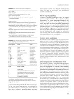
bleeding disorders in critically ill patients are presented in Table 12–1.
Platelet Abnormality
Petechiae on the skin and mucus membranes or spontaneous gingival and nasal
mucosal bleeding suggest an abnormality in platelet number or function. Imme-
diate bleeding after surgery or trauma also suggests a platelet abnormality. In-
formation regarding use of medications such as aspirin or NSAIDs should be
sought. A platelet count should be determined and a low count should be con-
firmed by examination of the peripheral smear to assess platelet size or the pres-
ence of clumping. The bleeding time is used to assess platelet function but is
300 The Intensive Care Manual
TABLE 12–1 Laboratory Studies in Bleeding Disorders
Platelet Bleeding
Abnormality count time PTT PT TT FDP D-Dimer
Thrombocytopenia A A
a
NNNN N
von Willebrand’s N A A N N N N
disease
TTP A A N N N N N
Platelet dysfunction N A N N N N N
DIC A A A A A A A
Hepatic failure N-A N A A A N-A N
b
Hemophilia A or B N N A N N N N
Thrombolytic agent N N A A A A A
Heparin N N A N-A A N N
Coumadin N N N-A A N N N
a
Abnormal if < 100,000/µL.
b
May have mild elevation.
ABBREVIATIONS: PTT, partial thromboplastin time; PT, prothrombin time; FDP, fibrin degradation
products; TTP, Thrombotic thrombocytopenic purpura; DIC, disseminated intravascular coagulopa-
thy; A, abnormal; N-A, normal or abnormal; N, normal.
ch12.qxd 11/7/01 4:18 PM Page 300
infrequently used in critically ill patients. The bleeding time is prolonged if: the
platelet count is less than 100,000/µL (100 × 10
9
/L), aspirin or NSAIDS have been
used, or severe hypofibrinogenemia is present.
Coagulation Cascade Abnormality
A defect in the coagulation cascade (Figure 12–1) is suggested by hemorrhage
into joints, subcutaneous tissue, or muscle; bleeding that responds poorly to local
pressure; and delayed bleeding after trauma or surgery. The primary laboratory
studies used to assess the intrinsic and extrinsic coagulation systems are the PT
and aPTT. Abnormalities of factors II (prothrombin), V, X, or fibrinogen pro-
long the result of both tests. The International Normalized Ratio (INR) adjusts
the PT for differences in sensitivity of test reagent and is used to monitor oral
12 / Hematologic Disorders 301
FIGURE 12–1 The normal coagulation cascade
ch12.qxd 11/7/01 4:18 PM Page 301
anticoagulation. The addition of normal plasma to the test reagents when the PT
or aPTT is abnormal can be used to screen for the presence of inhibitors or factor
deficiencies. In general, correction of the PT or aPTT with normal plasma sug-
gests factor deficiencies while lack of correction indicates the presence of an in-
hibitor. The thrombin time is sensitive to low levels of fibrinogen or abnormal
fibrinogen and inhibitors of thrombin (i.e., heparin, FDPs). Specific factor assays
are also available but should be used selectively, after results of more common
tests are noted to be abnormal.
Fibrinolytic Abnormality
Fibrinolysis is activated by the same factors that activate the coagulation cascade.
Laboratory studies include measurement of fibrin degradation products (FDP),
which are produced from the degradation of fibrin and fibrinogen and D-dimers,
which result from the degradation of cross-linked fibrin, not fibrinogen.
PLATELET DISORDERS
Acquired Thrombocytopenia
Thrombocytopenia exists when the platelet count is less than 150,000/µL (150 ×
10
9
/L). Thrombocytopenia may result from impaired production, enhanced de-
struction, or sequestration of platelets. Increased destruction of platelets may be
caused by immune or nonimmune mechanisms (Table 12–2).
Management of thrombocytopenia in critically ill patients should begin with
confirmation of the platelet count by examination of the peripheral smear. The
presence of large platelets on the smear may suggest increased platelet destruc-
tion. Effective treatment of the underlying disorder is critical to successful
resolution of thrombocytopenia. If thrombocytopenia results from defective
production or nonimmune destruction, intervention relies on supportive
platelet transfusions until the underlying disorder is corrected. Recombinant
human interleukin-11, a thrombopoietic growth factor, may reduce the need
for platelet transfusion after chemotherapy, but experience is limited in other
clinical situations. Immune-mediated thrombocytopenias require specific in-
terventions, but platelet transfusions are generally avoided except in life-
threatening hemorrhage. The decision to transfuse platelets should take into
account the underlying disorder, presence of active bleeding, plans for invasive
procedures, and the risk of spontaneous bleeding. The risk of spontaneous
bleeding increases with platelet counts of less than 10,000/µL (10 × 10
9
/L). How-
ever, invasive procedures or trauma may necessitate the use of platelet transfu-
sions at higher threshold counts. An automatic transfusion trigger for platelets is
not warranted.
302 The Intensive Care Manual
ch12.qxd 11/7/01 4:18 PM Page 302
Idiopathic Thrombocytopenic Purpura
Patients with immune mediated idiopathic thrombocytopenic purpura (ITP)
usually do not have serious bleeding. Treatment is initiated with corticosteroids
(prednisone, 1 to 2 mg/kg daily). In the presence of life-threatening hemorrhage
or planned invasive procedures, intravenous immunoglobulin G (IgG) may be
used (in a dosage of 0.4 to 0.5 g/kg daily for 4 to 5 days) to obtain a transient ele-
vation in platelet count. Patients who are nonresponsive to corticosteroids may
require splenectomy. Other agents, such as vincristine, cyclophosphamide, and
danazol, have been used for ITP refractory to other interventions. In addition,
dexamethasone 40 mg/day for 4 days has been used with some success. Platelets
should be transfused only for severe hemorrhage.
Post-Transfusion Purpura
Post-transfusion purpura is a rare syndrome that develops 5 to 12 days after
transfusion. It occurs in women who lack the platelet antigen PL-A1 who were
previously sensitized during pregnancy. Thrombocytopenia can develop after use
of any blood product containing platelet material in such patients. There is a
rapid decrease in the platelet count to less than 10,000/µL (10 × 10
9
/L) in 12 to 24
12 / Hematologic Disorders 303
TABLE 12–2 Causes of Thrombocytopenia
Impaired production
Drugs or toxins (i.e., chemotherapy, radiation)
Myelophthisis (i.e., neoplasm, infection, fibrosis)
Aplastic disorders
Vitamin B
12
, folate deficiency
Myeloproliferative disorders
Viral illness
Enhanced destruction
Immune-mediated
Autoantibody (idiopathic thrombocytopenic purpura)
Isoantibody (post-transfusion purpura)
Drug-induced (heparin, quinidine, sulfas)
Immune complex disorders
Nonimmune
Disseminated intravascular coagulation
Thrombotic thrombocytopenic purpura or hemolytic
uremic syndrome
Mechanical (i.e., intravascular devices, cardiopulmonary bypass)
Dilutional
Sequestration
Hypersplenism
Hypothermia
ch12.qxd 11/7/01 4:18 PM Page 303