VASCULAR COMPLICATIONS OF DIABETES - PART 10 potx
Bạn đang xem bản rút gọn của tài liệu. Xem và tải ngay bản đầy đủ của tài liệu tại đây (270.57 KB, 21 trang )
SECTION IV • MECHANISMS OF HYPERGLYCAEMIA INDUCED VASCULAR DYSFUNCTION
214
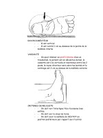
Fig. 24.1 Structure of ruboxistaurin, a macrocyclic bis-indolylmaleimide, which is an
orally active, selective PKC-β inhibitor.
N(CH
3
)
2
H
N
O
O
NN
O
Table 24.1 Tabulated IC
50
values (i.e. concentrations in nM required to achieve 50%
inhibition of enzyme activity) for ruboxistaurin and the non-specific PKC inhibitor,
staurosporine, with respect to each PKC isoform and related intracellular kinases.
Adapted from Science 1996; 472: 728–731.
Tabulated IC
50
values for ruboxistaurin
IC
50
(nM)
Kinase ruboxistaurin Staurosporine
PKC-α 360 45
PKC-β
1
4.7 23
PKC-β
2
5.9 19
PKC-γ 300 110
PKC-δ 250 28
PKC-ε 600 18
PKC-ζ >10
5
>1.5 × 10
3
PKC-η 52 5
Cyclic AMP kinase >10
5
100
Ca
2+
-calmodulin kinase 8 × 10
3
4
Casein kinase >10
5
1.4 × 10
4
Src tyrosine kinase >10
5
1
new vessel formation. Thus, blocking VPF-mediated retinal permeability is a
prime target for therapeutic amelioration of diabetic maculopathy.
Studies in rats have clearly shown that intravitreal administration of VPF
increases vitreous fluorescein leakage, and that pretreatment of these animals
for one week with ruboxistaurin 25 mg/kg/day via oral administrattion ame-
liorated VPF and phorbol ester-induced vitreous fluorescein leakage (Fig.
24.2). Furthermore, whereas control rats showed a two-fold increase in vitre-
ous fluorescein leakage after intravitreal VPF administration, rats pretreated
with the PKC-β inhibitor showed no difference in basal vitreous fluorescein
leakage but there was a 96% reduction in VPF-induced vitreous fluorescein
leakage (Fig. 24.2).
Increased retinal permeability is a hallmark of neovascularization within
the diabetic eye, as well as being a sight-threatening pathological entity even
in the absence of new vessel formation. These experimental data have shown
that oral administration of ruboxistaurin is well tolerated and considerably
CHAPTER 24 • EXPERIMENTAL PHARMACOLOGY USING ISOFORM-SELECTIVE PKC INHIBITORS
215
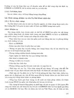
Fig. 24.2 Oral administration of the PKC-β inhibitor, ruboxistaurin, to normal rats
prevents the increase in vitreous fluorescein leakage following intravitreal injection of
VPF. Adapted from Diabetes 1997; 46: 1473–1480.
Vitreous fluorescein leakage
(arbitrary units)
20
10
0
0
0
2
0
0
25
2
25
VPF (ng/eye)
PKC-β inhibitor
(mg/kg rat/day)
P=0.015 P=0.043
SECTION IV • MECHANISMS OF HYPERGLYCAEMIA INDUCED VASCULAR DYSFUNCTION
216
attenuates VPF-mediated retinal permeability. Furthermore, diabetes is char-
acterized by an increase in retinal mean circulation time (MCT), and oral
treatment with ruboxistaurin for two weeks in STZ-diabetic rats reduced reti-
nal MCT, as measured by video fluorescein angiography (Fig. 24.3). This
experimental data has now been confirmed in phase II clinical trials in which
ruboxistaurin administration for one month produced significant improve-
ments in retinal blood flow and MCT among 27 diabetic patients (chapter 25).
Larger, multicentre clinical trials are in progress.
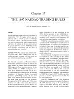
Fig. 24.3 Effect of oral dosing with ruboxistaurin on renal and retinal vascular function in
non-diabetic (●) and STZ-diabetic (●) rats. Untreated diabetic animals show increases in
glomerular filtration rate (GFR), renal filtration fraction (GFR corrected for renal plasma
flow, RPF), urinary albumin excretion rate (AER) and retinal mean circulation time (MCT).
Oral treatment with ruboxistaurin 0.1–10 mg/kg/day ameliorated these renal and retinal
haemodynamic abnormalities. Science 1996; 272: 728–731.
0 0.1 1.0 10 0 0.1 1.0 10
0
Dose of ruboxistaurin (mg/kg)
Dose of ruboxistaurin (mg/kg)
1000 0.1 1.0 10
GFR (ml/min)
Filtration fraction (GFR/RPF)
Urinary AER (mg/day)
MCT (s)
0
20 2
1.5
0.5
0
1
15
10
5
0
2
4
+
+
+
+
+
+
+
+
+
*
*
*
§
§
§
§
§
6
(a) (b)
(c) (d)
0.5
0.4
0.3
0.2
0.1
0
Further experimental studies have shown that diabetes-induced reduc-
tions in Na
+/
K
+
-ATPase and Ca
2±
-ATPase in the retina are mediated, in part,
via PKC-β activation. Oral administration of ruboxistaurin normalizes
Na
+
/K
+
-ATPase activity in retinal microvessels (Fig. 24.4).
PKC-β INHIBITION AND EXPERIMENTAL NEPHROPATHY
The early stages of diabetic renal disease are characterized by glomerular hyper-
filtration, mesangial expansion and microalbuminuria. Hyperglycaemia-
induced de novo synthesis of DAG, coupled with activation of PKC, especially
PKC-β, affects the structural and functional changes in the kidney via several dif-
ferent mechanisms involving various phosphorylation substrates of PKC. For
example, mesangial expansion has been attributed, in part, to PKC-mediated
increases in transforming growth factor-β (TGFβ) gene expression, activation of
cytosolic phospholipase A
2
and inhibition of Na
+
/K
+
-ATPase activity.
217
217
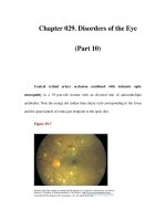
Fig. 24.4 Oral treatment with the PKC-β inhibitor ruboxistaurin, reverses diabetes-
related reductions in Na
+
/K
+
-ATPase activity in retinal microvessels. Adapted from
Diabetes 1998; 47: 464–469.
Na
+
/K
+
-ATPase activity
40
35
30
25
20
15
10
5
0
Diabetes +
ruboxistaurin
*
#
DiabetesNormal
CHAPTER 24 • EXPERIMENTAL PHARMACOLOGY USING ISOFORM-SELECTIVE PKC INHIBITORS
SECTION IV • MECHANISMS OF HYPERGLYCAEMIA INDUCED VASCULAR DYSFUNCTION
218
Experimental studies with ruboxistaurin have shown that, following oral
administration for eight weeks to STZ-diabetic and non-diabetic rats, urinary
albumin excretion rate (AER) and glomerular hyperfiltration were signifi-
cantly reduced (Fig. 24.3). Interestingly, higher doses of the PKC-β inhibitor
(1–10 mg/kg/day) were required to inhibit diabetes-mediated PKC activation
in the kidney compared with the retina (0.1 mg/kg/day). In addition, treat-
ment with ruboxistaurin had no significant effect on GFR and filtration frac-
tion in non-diabetic animals (Fig. 24.3). Among diabetic rats, however, the
dose-response curve for ruboxistaurin in normalizing GFR paralleled its
inhibitory effect on PKC activity.
Renal protection with aminoguanidine and angiotensin-
converting enzyme inhibition (ACE-I) involves normalization of
glomerular PKC activity
In experimental models of diabetic renal disease, e.g. the STZ-diabetic rat, it
is well established that ACE-Is and aminoguanidine retard the structural and
functional abnormalities characteristic of diabetic nephropathy, particularly
with respect to reducing urinary AER. The exact mechanisms by which these
therapeutic interventions work is not entirely clear, but recent work by
George Jerums and colleagues has shown that glomerular PKC activity levels
are normalized in STZ-diabetic rats during experimental treatment with
aminoguanidine and the ACE-I, ramipril. Thus, diabetes-related increases in
glomerular PKC activity may serve as an important common pathway by
which metabolic and haemodynamic factors contribute to the initiation and
progression of diabetic renal disease. Existing renoprotective agents, e.g.
ACE-Is, may slow the progression of nephropathy, in part, by normalizing
diabetes-induced increases in glomerular PKC activity.
EFFECTS OF RUBOXISTAURIN IN EXPERIMENTAL DIABETIC
NEUROPATHY
Various pathways have been implicated in the pathogenesis of diabetic neu-
ropathy, including increased polyol pathway activity, enhanced non-enzy-
matic glycation and PKC activation. In addition, neural ischaemia is thought
to play an important role in diabetic nerve injury, in part via PKC activation
which impairs vasodilation and increases vasoconstrictor pathways in the
endoneurial microvasculature.
In experimental STZ-diabetic rats, motor nerve conduction velocity and
sciatic nerve blood flow are reduced. Treatment with ruboxistaurin amelio-
rated these abnormalities via mechanisms attributable to prevention of neu-
ral ischaemia (Fig. 24.5).
CHAPTER 24 • EXPERIMENTAL PHARMACOLOGY USING ISOFORM-SELECTIVE PKC INHIBITORS
CLINICAL IMPLICATIONS OF AN ORALLY ACTIVE PKC-β
INHIBITOR, RUBOXISTAURIN
Extensive experimental studies have shown that ruboxistaurin selectively
inhibits PKC-β in retinal, neural, renal and vascular tissues following oral
administration without any significant adverse effects. The encouraging tol-
erability profile of ruboxistaurin is no doubt attributable to its pharmaco-
logical specificity for PKC-β
I
and PKC-β
II.
The animal studies have convinc-
ingly shown that, following chronic oral treatment, ruboxistaurin amelio-
rates the early increases in retinal blood flow, glomerular filtration rate and
renal and retinal permeability.
This data opens the possibility of a new and exciting pathway for therapeu-
tic intervention in the earliest stages of diabetic microvascular disease. In par-
ticular, such an approach would be unique in offering protection against the
development and progression of retinopathy and nephropathy via a mecha-
nism that is independent of (and complementary to) glucose or blood pres-
sure reduction. Thus, in clinical practice, PKC-β inhibition would be used as
an adjunct to all existing therapies for the prevention of diabetic vascular com-
plications. Large multicentre clinical trials are on-going not only in diabetic
retinopathy and renal disease but also in patients with other diabetic compli-
cations, e.g. erectile dysfunction and diabetic neuropathy.
219
Fig. 24.5 Ruboxistaurin improves sciatic nerve conduction velocity in experimental
models of peripheral neuropathy. A dose-related effect is illustrated.
Velocity (m/s)
Dose (mg/kg)
60
65
control level
55
0.0
0.1 0.3 1.0 10.0 25.0
SECTION IV • MECHANISMS OF HYPERGLYCAEMIA INDUCED VASCULAR DYSFUNCTION
220
FURTHER READING
Aiello LP, Bursell SE, Clermont A et al. Vascular endothelial growth factor-induced retinal
permeability is mediated by protein kinase C in vivo and suppressed by an orally effective
β-isoform-selective inhibitor. Diabetes 1997; 46: 1473–1480.
Ishii H, Jirousek MR, Koya D et al. Amelioration of vascular dysfunction in diabetic rats by
an oral PKC-β inhibitor. Science 1996; 272: 728–731.
Kowluru RA, Jirousek MR, Stramm L et al. Abnormalities of retinal metabolism in diabetes
or experimental galactosemia: V relationship between protein kinase C and ATPase.
Diabetes 1998; 47: 464–469.
Nakamura J, Kato K, Hamada Y et al. A protein kinase C-β-selective inhibitor ameliorates
neural dysfunction in streptozotocin-induced diabetic rats. Diabetes 1999; 48:
2090–2095.
CURRENT ISSUES
• Ruboxistaurin is a unique orally active PKC inhibitor which is highly
specific for the PKC-β isoforms. Following oral administration to STZ-
diabetic rats, ruboxistaurin prevented diabetes-related increases in retinal
and renal PKC activity in parallel with amelioration of glomerular
hyperfiltration, microalbuminuria and increased retinal blood flow.
• Ruboxistaurin shows an excellent tolerability profile in experimental
diabetic animals, no doubt reflecting its specificity for inhibiting only
two out of twelve PKC isoforms. Furthermore, in non-diabetic animals
(in which there is no augmentation of PKC activity) ruboxistaurin has
no significant effects on retinal or renal haemodynamics. Thus, the
compound seems to be highly specific for PKC-β and only achieves
therapeutic effects in experimental studies in which diabetes-related
increases in PKC are present.
• Large multicentre clinical trials with ruboxistaurin are on-going to assess
its efficacy and safety in patients with diabetic retinopathy and peripheral
neuropathy. In due course further studies will be established to define
the role of this compound in other diabetes complications, including
nephropathy and erectile dysfunction.
INTRODUCTION
Ruboxistaurin is the first molecule in an exciting new class of PKC-β‚ specific
inhibitors which ameliorate the structural and functional vascular abnormali-
ties associated with hyperglycaemia in humans and experimental animals. A
series of detailed molecular and experimental studies were conducted to doc-
ument the effects of ruboxistaurin in retinal, neural and endothelial tissues.
These were followed by a series of multicentre clinical trials to evaluate longer
term efficiency and safety in patients with diabetes related complications. The
design and execution of these trials has posed considerable challenges, and
many of these trials are still ongoing. There are, however, a number of encour-
aging results already in the public domain from phase II studies.
RUBOXISTAURIN IMPROVES ENDOTHELIAL DYSFUNCTION
Diabetes is associated with endothelial dysfunction, and hyperglycaemia
impairs the endothelial-dependent vasodilator response to acetylcholine. In a
placebo controlled, double blind crossover study in healthy volunteers, there
was evidence that ruboxistaurin improved forearm blood flow in response to
incremental arterial infusions of the endothelium-dependent vasodilator
methacholine under hyperglycaemic conditions (Fig. 25.1). Thus, this novel
experimental study has confirmed that inhibition of PKC-β in healthy vol-
unteers prevents the reduction in endothelium-dependent (nitric oxide
mediated) vasodilation induced by acute hyperglycaemia.
CLINICAL TRIALS OF RUBOXISTAURIN IN DIABETIC
RETINOPATHY
Experimental studies have shown that ruboxistaurin inhibits hypergly-
caemia-induced PKC activation in the retina (Fig. 25.2). In addition ruboxis-
taurin prevents neovascularization in a porcine model of retinal vein occlu-
sion (Fig. 25.3). These experimental data provide encouraging evidence that
PKC-β‚ inhibition might have a favourable effect on macular oedema forma-
tion and new vessel formation (two sight threatening complications) in
patients with diabetic retinopathy.
The clinical development of ruboxistaurin began with phase I tolerability
and pharmacokinetic studies in healthy volunteers, followed by phase II effi-
cacy studies in patients with diabetes. In patients with type 1 or type 2 diabetes
and minimal or no evidence of diabetic retinopathy, ruboxistaurin increased
retinal blood flow in a dose-dependent manner, maximal after 32 mg daily for
221
CHAPTER 25
CLINICAL TRIALS WITH RUBOXISTAURIN
Richard Donnelly MD, PhD, FRCP, FRACP
Vascular Complications of Diabetes: Current Issues in Pathogenesis and Treatment, Second Edition
Edited by Richard Donnelly, Edward Horton
Copyright © 2005 by Blackwell Publishing Ltd
SECTION IV • MECHANISMS OF HYPERGLYCAEMIA INDUCED VASCULAR DYSFUNCTION
222
Fig. 25.1 Forearm blood flow in healthy volunteers during euglycaemia and
hyperglycaemia after pretreatment with ruboxistaurin or placebo. The PKC-β‚
inhibitor improved the endothelial-dependent vasodilator response to
methacholine under conditions of high glucose. Adapted from Beckman et al.
Circulation Research 2002; 90: 107–111.
Forearm blood flow
(ml / dl / min)
2
3
1
0
Placebo
Ruboxistaurin
p = 0.08 p = 0.001
Euglycaemia
Hyperglycaemia
Fig. 25.2 Ruboxistaurin attenuates the increase in retinal PKC activity in experimental
rats with diabetes.
PKC activity
(pmo/min/mg of protein)
20
15
10
5
0
0 0.1
Ruboxistaurin (mg/kg/d)
10.0
Nondiabetic
Diabetic
CHAPTER 25 • CLINICAL TRIALS WITH RUBOXISTAURIN
223
Fig. 25.3 Ruboxistaurin prevents neovascularization in a porcine retinal vein
occlusion model of new vessel formation.
Neovascularization score
3
4
2
1
0
Placebo
p = 0.03
Ruboxistaurin 1 mg/kg/d, po
Fig. 25.4 Phase II study of ruboxistaurin in patients with type 1 or type 2 diabetes
and retinopathy. In a double blind, placebo controlled study for four weeks,
ruboxistaurin decreased mean retinal circulation time, i.e. improved retinal blood
flow. Adapted from Aiello et al. Diabetes 1999; 48: A19.
Extent of MCT abnormality
at endpoint
1.0
1.2
0.4
0.2
0.6
0.8
0.0
Placebo 16 mg/d 32 mg/d
SECTION IV • MECHANISMS OF HYPERGLYCAEMIA INDUCED VASCULAR DYSFUNCTION
224
one month (Fig. 25.4).
Having confirmed the basic safety and tolerability of ruboxistaurin, and
demonstrated that it has pharmacodynamic activity on retinal blood flow,
large multi-centre clinical trials were initiated to evaluate the safety and effica-
cy of the treatment in larger patient groups during longer term administration
(two to four years). The PKC-diabetic retinopathy study (DRS) and the PKC-
diabetic macular oedema (DME) study were the first international random-
ized, placebo controlled trials to assess whether oral treatment with ruboxis-
taurin will delay progression in patients with moderate to severe non-prolifer-
ative diabetic retinopathy at base-line, including progression from non-clini-
cally significant to clinically significant macular oedema (CSMO). The results
for approximately 1,000 patients followed for an average of 36–46 months will
be announced soon.
The clinical trials of ruboxistaurin in diabetic retinopathy have two key
objectives. First, to determine if oral treatment with ruboxistaurin over three
years will reduce progression of diabetic retinopathy or the need for laser
photocoagulation in patients with moderately severe to very severe non-pro-
liferative diabetic retinopathy in at least one eye. Second, to determine if all
treatment with ruboxistaurin in patients with mild to moderate non-prolif-
erative diabetic retinopathy and non-visually threatening diabetic macular
oedema will delay development of clinically-significant macular oedema or
the need for laser photocoagulation. The clinical trials with ruboxistaurin
use 7-field stereoscopic fundal photographs and measurements of visual
acuity as markers of drug efficacy.
CLINICAL TRIALS OF RUBOXISTAURIN IN DIABETIC PERIPHER-
AL NEUROPATHY
A number of large phase III clinical trials are in progress to evaluate the effects
of ruboxistaurin on various unpleasant symptoms of diabetic neuropathy and
longer term outcomes in relation to nerve function and neurophysiological
endpoints. These trials use a combination of symptom scores and nerve func-
tion measurements to assess efficacy.
CLINICAL TRIALS OF RUBOXISTAURIN IN OTHER DIABETES-
RELATED COMPLICATIONS
Further clinical trials are in progress to evaluate the effects of ruboxistaurin
on renal function and proteinuria, and on endothelial function in relation to
lower limb ischaemia and macrovascular end-points.
CHAPTER 25 • CLINICAL TRIALS WITH RUBOXISTAURIN
FURTHER READING
Aiello LP, Bursell S, Devries T. Protein kinase C beta selective inhibitor Ruboxistaurin amelio-
rates abnormal retinal haemodynamics in patients with diabetes. Diabetes 1999; 48: A19.
Aiello LP, David MD, Sheetz MJ. The PKC Inhibitor Diabetic Retinopathy Study Group.
Design, baseline patient characteristics and high prevalence of severe to very severe
nonproliferative diabetic retinopathy (NPDR) in the Protein Kinase C Diabetic
Retinopathy Study (PKC-DRS). Diabetes 2002; 51 (suppl2): A209.
Demolle D, de Suray JM, Onkelinx C. Pharmacokinetics and safety of multiple oral doses
of LY333531, a PKC beta inhibitor, in healthy subjects. Clin Pharm Ther 1999; 65: 189.
Demolle D, de Suray JM, Vandenhende F, et al. Ruboxistaurin single escalating oral dose
study in healthy volunteers. Diabetologia 1998; 41: (Suppl 1): A354.
Donnelly R, Idris I, Forrester J. Protein Kinase C inhibition and diabetic retinopathy: a shot
in the dark at translational research. Br J Ophthalmol 2004; 88: 145–151.
225
CURRENT ISSUES
• The results of large multicentre clinical trials of ruboxistaurin in patients
with diabetic retinopathy and neuropathy are eagerly awaited in 2005–7.
• As a completely novel drug, ruboxistaurin has posed unique challenges to
the design and execution of international clinical trials in diabetes
complications.
• The safety and tolerability profile of ruboxistaurin is very encouraging. It is
suitable for once-daily oral administration and has no adverse effects on
immune function.
• There are still some unanswered questions about the optimal dose of
ruboxistaurin for each potential indication.
INDEX
Page numbers in
italic
refer to figures;
those in
bold
refer to tables.
A
ACE inhibitors 28, 40, 49
myocardial infarction 63–4
neuropathy 115, 124
renal protection 218
retinopathy 134
versus beta-blockers 63
acetyl-
L-carnitine 124
acetylcholine 207, 221
acetylcysteine 26
actin 199, 200
acupuncture 118
acute sensory neuropathy 80
adenosine 53, 100, 101
adenosine diphosphate 209
advanced glycated haemoglobin (Hb-AGE) 186
advanced glycation end-products (AGE) 86–7,
114, 179–87, 184
and age-adjusted death rate 180
cataract 172
cross-linking 185
formation 183
harmful effects 185
inhibitors of 114
and oxidative stress 200
receptors 185–6
age factors
cataracts 171
hypertension and proteinuria 28
retinopathy 129, 134
age-adjusted death rate 180
ALADIN-II study 87
albumin-creatinine ratio 25
albuminuria 22, 24
Albustix 24, 25
alcohol dependency 51, 52
aldose reductase 76, 85, 86, 124, 171, 187, 188,
189
aldose reductase inhibitors 85, 113–14, 113,
124
alkaline phosphatase 32, 110
allodynia 80, 91, 116
alpha-lipoic acid 114, 124
alpha-oxoaldehydes 182, 183
alprostadil (prostaglandin E1) 56
ALT-462/486 186
alteplase 47, 69
Amadori rearrangement 179, 182, 183, 185
amblyopia 137
aminoguanidine 86, 114, 124, 186–7
renal protection 218
amitriptyline 116, 125
amputation 17, 35, 70
diabetic foot 105, 106, 107, 125
lower limb 7, 8, 9, 18, 61, 67
smoking 67
anaemia 24, 33, 186
angiography 26, 39, 43, 61, 66
fluorescein 102, 153, 154, 155, 156, 174
angioplasty 40, 42, 44, 62, 66
diabetic foot 107
angiotensin receptor antagonists 30, 70
angiotensin-converting enzyme inhibitors see
ACE inhibitors
ankle reflex 79, 80, 81, 122, 125
anti-arrhythmics 117
anti-oxidants 114
anti-platelet therapy 43
anticoagulation 61
anticonvulsants 116–17,
117, 125
antihypertensive agents 29, 32, 49, 73
erectile dysfunction 54
nephropathy 28–9
antioxidants 30
apomorphine 55
Appropriate Blood Pressure Control in Diabetes
(ABCD) trial 87
arteriography 26
arteriolohyalinosis (Kimmelstiel-Wilson kidney)
26
AS-3201 124
ascorbic acid (vitamin C) 179
ASPECT study 61
aspirin 47, 48
myocardial infarction 43
peripheral vascular disease 62, 68, 70
stroke 47, 48
atenolol 63
atherosclerosis 35
atorvastatin 67
autonomic neuropathy 80, 81
B
balanitis 52
BDA Cohort Study 35, 36
beading 144
Becaplermin 108
Berlin Retinopathy Study 13, 14
beta-blockers 40, 63
biopsy
nerve 101–2
skin 102
226
Vascular Complications of Diabetes: Current Issues in Pathogenesis and Treatment, Second Edition
Edited by Richard Donnelly, Edward Horton
Copyright © 2005 by Blackwell Publishing Ltd
227
biothesiometer 99
bisphosphonates 110
blindness 7, 8, 9, 75
neuropathy 129
retinopathy 129, 137
blood-retinal barrier 156
boat shaped haemorrhage 144
body mass index 5
bradykinin 207
brain-derived neurotrophic factor 88, 115
branch retinal vein occlusion 173, 174
British Diabetic Association 149
British Diabetic Association Cohort study 3
Bruch’s membrane 158, 168
Bypass Angioplasty Revascularization
Investigation (BARI) trial 44, 66
C
C-peptide 89, 124
C-reactive protein 66
calcium 190, 197, 200, 202, 204
caldesmon 200
calphostin 208, 209
capsaicin 118
captopril 29, 63, 73
carbamazepine 117, 125
carbonyl stress 182–3, 184
carboxymethyl-lysine 179
cardiomyopathy 37, 193, 206
carotid stenoses 46
carpal tunnel syndrome 80
cataracts 171–2, 188
treatment 172
central retinal vein occlusion 173
cerebral haemorrhage 43, 45, 48
cerebral infarcts 44
cerebral small-artery disease 46
cerebrovascular disease see stroke
Charcot neuroarthropathy 108–10, 108, 109
management 110
X-rays 109
Charcot neuropathy 79
cholesterol
and coronary heart disease 18, 19, 20, 36,
37, 38, 40, 43, 67
peripheral neuropathy 93
retinopathy 135
Cholesterol And Recurrent Events study 40
chorioretinal scarring 137
chronic inflammatory demyelinating polyneu-
ropathy 83
chronic sensorimotor neuropathy 79–80
cilostazol 68, 69
circinate of exudates 142, 157
citalopram 116, 125
claudication 67–8, 68, 69
clinically significant macular oedema 142, 158
clomethiazole 49
clopidogrel 48, 62, 66, 71
Clopidogrel in Unstable angina to prevent
Recurrent ischaemic Events (CURE)
study 62
Clopidogrel vs. Aspirin in Patients at Risk of
Ischaemic Events (CAPRIE) study 48
collagen 86, 179, 183, 185, 205
compound muscle action potential 100–1
connexin-43 199
CONSENSUS II trial 41, 63
corneal confocal microscopy 102, 103
coronary heart disease 18, 35–44
aetiology 36–7, 37
anti-platelet therapy 43
environmental factors 36
epidemiology 35–6, 36
and ethnicity 38–9
evidence-based practice 38–44
fetal nutrition 36
glycaemic control 41–3, 42
hazard ratios 16
incidence 35
lipid-lowering therapy 43–4
management 40–1
microalbuminuria 38
mortality 19, 35, 36, 37, 38, 43
obesity 35, 36
prevention 61–7
prognosis 37–8, 38
screening 39
statins 43
vascular risk assessment 38–9
see also myocardial infarction
cost of diabetes 7–10, 9, 10
cotton wool spots 139, 141, 142,
143, 173, 174
cranial nerve palsy 174
cranial neuropathy 82
creatinine 26, 27, 31, 32, 73
cross-link formation 185
CT-angiography 26
cyclic GMP 53
cystoid macular oedema 157, 172
D
DECODE study 182
delquamine 57
3-deoxyglucosone 179, 182
desipramine 116
Diabetes Control and Complications Trial
(DCCT) 3, 14, 28, 93, 132
Diabetes in Early Pregnancy Study 136
diabetic amyotrophy 82, 83
INDEX
228
Diabetic Control and Complications Trial
(DCCT) 113
diabetic foot 105–10
amputation 105, 106, 107, 125
angioplasty 107
callus formation 105
causes 105–6, 105
education 107
healing 107, 108
hyperbaric oxygen 107
management guidelines 125
PEDIS classification 106
prevention 107
recombinant platelet derived growth factor
108
revascularization 107
risk factors 80, 105
UTDWCS classification 106
Wagner classification 106
diabetic nephropathy see nephropathy
diabetic neuropathy see neuropathy
Diabetic Retinopathy Study 163
Diabetic Retinopathy Vitrectomy Study 168
diabetic truncal radiculoneuropathy 82–3
diacylglycerol (DAG) 87, 90, 189, 193, 205
DAG-PKC pathway 114, 187, 189
generation of 192
diagnosis 5
dialysis 23, 32
2,3-diaminophenazone 86
diet
coronary heart disease 36, 40
nephropathy 30–1
DIGAMI therapy 48, 64
diplopia 82, 174
dipyridamole 48, 70, 71
disc new vessels 145, 164
dot and blot haemorrhages 141
E
E-selectin 209
Early Treatment Diabetic Retinopathy Study
(ETDRS) 43, 135, 142, 151
EDTA clearance 30
electrophysiology 99–101, 100
enalapril 29, 41, 63, 87
end-stage renal failure 22
endophthalmitis 167, 172, 175
endothelial permeability 197–204, 197, 198,
199, 201
and hyperglycaemia 200
intercellular gaps 198–9
and protein kinase C 200, 203
endothelin 53, 206
entrapment neuropathy 80, 82
environmental factors 4, 35, 136
epalrestat 114, 124
epidemiology 4–6
type 1 diabetes 4
type 2 diabetes 4–6
see also individual conditions
Epidemiology of Diabetes Complications (EDC)
Study 93
erectile dysfunction 51–7
autonomic neuropathy 52, 55
causes 55
clinical presentation 51–2
diagnostic features 54
drug-related 54
evidence-based practice 54–7, 55, 56
incidence 51
management 54–7
pathophysiology 52–3, 53
erythropoietin 33
essential fatty acids 124
ethnicity 21
coronary heart disease 38–9
epidemiology 5
nephropathy 23
EURODIAB Controlled Trial of Lisinopril in
Insulin-Dependent Diabetes Mellitus
(EUCLID) 75, 93, 134
EURODIAB IDDM Complications Study 93
European Stroke Prevention Study 48, 70
external ocular muscle palsies 174
extracellular signal-regulated kinases 86
exudates 141, 142
exudative maculopathy 151
F
F-actin 199
F-waves 101
ferritin 32
fibrates 43
fibrinogen 37, 62, 209
fidarestat 124
flame-shaped haemorrhages 141
fluorescein angiography 102, 153, 154–5, 154,
155, 156, 173, 174
fluoxetine 116
folic acid 67
foot ulceration see diabetic foot
fovea 141
foveola 141
foveolar avascular zone 151
Framingham study 19, 33, 35, 38, 45
free fatty acids 187, 192, 193
fructosamine 13, 182,
183
INDEX
INDEX
229
G
gabapentin 117, 125
gamma-linolenic acid 114, 124
gangrene 24, 35, 47
gastroparesis 33
gemfibrozil 43
gene expression 205
genetics 36
microvascular disease 135
nephropathy 19, 23
retinopathy 135–6
type 1 diabetes 4
type 2 diabetes 4
GF109203X 213
GISSI-3 41, 63
glaucoma 146, 172
ghost-cell 166
haemolytic 166
neovascular 146, 172
glitazones 43
glomerular filtration rate 22, 31
glomerulopathy 209–10
glomerulosclerosis 25, 26, 185
GLUT-1 transporters 187, 189
glycaemic control, and cardiovascular risk
41–2, 42
glycine antagonists 49
glycoprotein IIb/IIIa glycoprotein receptor
inhibitors 62–3
glycosylated haemoglobin (HbA1
C
) 13, 19, 27,
185
and amputation 67
hazard ratios 70
and myocardial infarction 181
retinopathy 14, 15, 16, 136
glyoxal 182, 183
Gothenburg study 45
grid macular treatment 157
growth factors 88–9, 166
gustatory sweating 32, 81
GUSTO-1 study 63
H
haematuria 26
haemodialysis 32
haemoglobin
advanced glycated (Hb-AGE) 186
glycosylated see glycosylated haemoglobin
haemorrhage
boat shaped 144
cerebral 43, 45, 48
dot and blot 141
flame-shaped 141
intracranial 47, 69
intraretinal 141
preretinal 145, 147
retinal 140–1, 144, 168
subarachnoid 45
vitreous 139, 147, 166, 167, 169
hallux valgus 97
Heart Outcomes Prevention Evaluation (HOPE)
study 29–30, 41, 42, 48, 64, 65, 67, 71
heparin 47
stroke 48, 69
surface modified intraocular lenses 172
high density lipoprotein (HDL) 18, 135
histamine 199, 200
homocysteinaemia 67
HOT study 62
hydronephrosis 26
hydroxymethylglutaryl CoA reductase
inhibitors 88
hyperalgaesia 116
hyperbaric oxygen 107
hyperglycaemia 6, 13–19, 14–18
balanitis 52
cataracts 171
diabetic angiopathy 179, 181
endothelial hyperpermeability 200, 201,
202, 206
endothelial permeability 200
macrovascular disease 13, 16
microvascular disease 13, 18, 19, 132
polyol pathway 187
protein kinase C activation 189, 192, 193,
194, 207, 212
retinopathy 132, 133
stroke 46, 47, 48
treatment 64, 115
vascular injury 181
hyperglycaemic neuropathy 80, 85, 89, 93
hyperlipidaemia 5, 19–21, 20
coronary heart disease 36
erectile dysfunction 53
retinopathy 135
hypertension 19–21, 20
coronary heart disease 36, 39
hazard ratios
72
peripheral vascular disease 24
and proteinuria 28
retinopathy 25–6, 134
treatment 28–9, 29, 32, 49, 54, 73
hypertriglyceridaemia 20–1, 67
hypoaesthetic neuropathy 92–3
hypotension, postural 32, 81
I
IgA nephropathy 26
imipramine 116, 125
impaired glucose tolerance 85
impotence see erectile dysfunction
insulin neuritis 80
insulin-like growth factor 89, 115
intercellular adhesion molecule-1 (ICAM-1) 209
International Working Group of the Diabetic
Foot 106
intracavernosal injection therapy 56
intraocular pressure 137, 169
intraretinal haemorrhage 141
intraretinal microvascular abnormalities 143,
165, 165
iris neovascularization 146, 164, 165, 172
ischaemia, lower limb 4
leg ulcers 105, 106, 107
ischaemic maculopathy 151, 153, 154
isoquinolinesulphonamides 213
isosorbide dinitrate 115
spray 118
K
kidneys
asymmetrical 32
biopsy 26
morphological changes 26
see also renal
Kimmelstiel-Wilson kidney (arteriolohyalinosis)
26
L
lactic acidosis 26, 32
lacunar infarcts 45, 46
lamotrigine 117
laser therapy 157–60, 167–8
complications 168
left ventricular hypertrophy
coronary heart disease 38
nephropathy 24
lens proteins, glycation 171
lidorestat 124
LIPID study 49
lipid-lowering therapy 43–4
lisinopril 29, 41, 63
low density lipoproteins (LDL) 18
lubeluzole 49
LY333531 see ruboxistaurin
M
macrovascular disease
evidence-based interventions 61
and hyperglycaemia 13, 16
and hypertension 30
see also coronary heart disease; stroke
macular oedema
classification 147
clinically significant 142, 158
cystoid 157, 172
duration of diabetes 152, 153
and retinopathy 152
maculopathy 139, 141, 151–61
diagnosis 153–6
diffuse oedema 157
epidemiology 151
fluorescein angiography 153, 154–5, 155,
156
focal oedema 157
grid macular treatment 157
ischaemic 151, 153, 154
laser therapy 157–60
micropulsed diode laser 160
optical coherence tomography 155–6
pathogenesis 156
surgery 158
treatment 157–8
see also macular oedema
magnesium 49
magnetic field therapy 110
magnetic resonance imaging 102
Maillard reaction 179, 182
matrix metalloproteinase 86
membrane transport 209–10
metabolic syndrome 5, 19, 20, 53
metformin 11, 32, 42
methylglyoxal 182
metoprolol 63
mexilitine 117
micro-HOPE study 48
microalbuminuria 13, 73
coronary heart disease 38
nephropathy 19, 23–9, 24, 27, 32, 71, 135,
217
retinopathy 14, 134–5
microaneurysms 140
micropulsed diode laser 160
microvascular disease 4
and erectile dysfunction 52
genetic factors 135
and hyperglycaemia 13, 18, 19, 132
and hypertension 20
smoking 21
mitogen-activated protein (MAP) kinases 205
MOLD 184
monocytes 209
INDEX
230
INDEX
231
mortality 3, 38, 39
coronary heart disease 19, 35, 36, 37, 38, 43
myocardial infarction 30, 37, 40
stroke 45
Multiple Risk Factor Intervention Trial study 36
myo-inositol 124
myocardial infarction 20
anticoagulation 61
DIGAMI therapy 48, 64
and glycosylated haemoglobin (HbA1
C
)
181
mortality 30, 37, 40
statins 66–7
thrombolysis 47, 61
myoinositol deficiency 86
myopia 137
myosin light chain kinase 199
N
N-[carboxymethyl]-lysine 179, 183, 184
NADH 113, 114
NADPH 113
NADPH oxidase 206
necrobiosis lipoidica 21
nephropathy 21, 23–34
antihypertensive agents 28–9
clinical presentation 23–4, 24, 25, 26
diagnostic features 24–8, 27, 28
diet adjustment 30–1
and ethnicity 23
evidence-based practice 28–31, 29, 30,
71–3, 72, 73
genetics 19, 23
haematuria 26
IgA 26
left ventricular hypertrophy 24
microalbuminuria 19, 23–9, 24, 27, 32, 71,
135, 217
monitoring renal function 31–3, 33
and retinopathy 134–5
ruboxistaurin treatment 216–17, 218–19
nephrotic syndrome 22
nerve biopsy 101–2
nerve conduction velocity 100
nerve growth factor 124
neuropathy 79–84
acute sensory 80
autonomic 80, 81
blindness 129
Charcot’s 79
cholesterol 93
chronic inflammatory demyelinating
polyneuropathy 83
chronic sensorimotor 79–80
classification 79, 123
clinical screening 97–8
cranial 82
diabetic amyotrophy 82, 83
diabetic truncal radiculoneuropathy 82–3
entrapment 80, 82
evidence-based interventions 75–6
hyperglycaemic 80, 85, 89, 93
hypoaesthetic 92–3
invasive assessment 101–2
management guidelines 121–5, 122–5
non-invasive assessment 102–3, 103
painful 91–2, 91
pathophysiology 85–90
advanced glycation end-products 86–7
growth factors 88–9
hydroxymethylglutaryl CoA reductase
inhibitors 88
immune mechanisms 90
myoinositol deficiency 86
oxidative stress 87
polyol pathway 85–6, 113
protein kinase C-β 87–8
vascular factors 87
peripheral see peripheral neuropathy
quantitative sensory testing 98–101, 99,
122
electrophysiology 99–101, 100
thermal thresholds 99
vibration perception threshold 88, 92,
98–9
staging 80, 81
symptoms 97
Neuropathy Disability Score (NDS) 97, 98
Neuropathy Impairment Score (NIS) 97
neuropeptide-Y 53
neurotrophins 88, 115
new vessels elsewhere 145, 146, 163, 164, 165
nisoldipine 87
nitric oxide synthase 207–9
nitroglycerine 57
non-ketotic hyperosmolar states 47
normoglycaemia 113
O
obesity 5, 11
childhood diabetes 4, 11
coronary heart disease 35, 36
ocular perfusion pressure 137
oculoischaemic syndrome 173, 174
oculomotor nerve palsy 82
OPB-9195 187
ophthalmoscopy 148
opioids 125
optical coherence tomography 155–6
oral glucose tolerance test 6
oxidative stress 87, 200, 207
oxycodone 125
P
painful neuropathy 91–2, 91
pamidronate 110
pancreatic/islet cell transplantation 113
papaverine 56
paroxetine 116, 125
pars plana vitrectomy 168–9
PEDIS classification 106
pentosidine 183, 184
pericytes 140
peripheral neuropathy 91–5
combined assessments 93
negative sensory symptoms 92–3
positive sensory symptoms 91–2, 91
risk factors 94
ruboxistaurin 224
stages of 122
treatment 113–19
peripheral vascular disease
diabetic foot 105–10
evidence-based interventions 67–8, 67
smoking 19–21, 20, 67
peritoneal dialysis 32
Peyronie’s disease 52
phentolamine 56, 57
phimosis 52
phosphodiesterase inhibitors 54
pimagedine 186
plasminogen activatory inhibitor-1 37
platelet derived growth factor, recombinant
108
platelet-mediated vasodilation 209
polycystic kidneys 26
polyol pathway 85–6, 113, 183, 187–8, 187
cataracts 171
ponalrestat 124
population screening 11
pravastatin 67
pregabalin 117, 125
pregnancy, retinopathy in 136
prevalence of diabetes 6
proliferative retinopathy 144–6, 163–70
definitions 163–4, 164, 165
diagnosis/natural history 164–6, 165
disc new vessels 145, 164
epidemiology 163
iris neovascularization 164, 165
laser therapy 167–8
new vessels elsewhere 145, 146, 163, 164,
165
pathogenesis 166
retinal detachment 145–6
treatment 166–7, 167
vascular permeability factor 166
vitrectomy 168–9
vitreous detachment 166
protein kinase C 189–95, 189
and endothelial permeability 200, 203
endothelial and vascular smooth muscle
function 206–10, 207
and gene expression 205
isoforms 191
vascular permeability 197–204
protein kinase C inhibitors 213–20
protein kinase c-β 87–8
activation of 194
inhibitors of 114–15, 119
proteinuria 13, 17, 19, 29
public health impact 3–11
pyridoxamine 86, 187
pyrraline 87
R
ramipril 29–30, 42
myocardial infarction 65
stroke 48, 65
reactive oxygen species 207
Reduction of Cholesterol in Ischaemia and
Function of the Endothelium (RECIFE)
trial 67
RENAAL study 30, 72
retinal detachment 145–6
retinal haemorrhage 144
retinal hypoxia 166
retinal ischaemia 142
retinal mean circulation time 216
retinal neovascularization 146
retinal photography 148
retinal pigment epithelium 156
retinal vascular abnormalities 173–4, 173
retinopathy 14–16, 14, 15, 139–50
ACE inhibitors 134
background 139–42
blindness 129, 137
circinate exudate rings 142
classification 146–7, 147
clinically significant macular oedema
142
cotton wool spots 139, 141, 142, 143, 173,
174
deep retinal haemorrhages 144
diagnosis 147
direct ophthalmoscopy 148
disc new vessels 145, 164
epidemiology 129–37
evidence-based interventions 74–5, 74, 75
exudates 141, 142
familial clustering 135
growth factors 166
haemorrhages 140–1, 140
incidence 132
intraretinal microvascular abnormalities
143, 165, 165
INDEX
232
INDEX
233
iris neovascularization 146, 164, 165
and microalbuminuria 134–5
microaneurysms 140
neovascular glaucoma 146, 172
new vessels elsewhere 145, 146, 163, 164,
165
preproliferative 142–4, 142
prevalence 129–31, 130, 131
proliferative see proliferative retinopathy
retinal detachment 145–6
risk factors 132–7, 133
age 129, 134
blood pressure 25–6, 134
cholesterol 135
duration of diabetes 132
genetics 135–6
glycosylated haemoglobin 15, 16, 136
hyperglycaemia 132, 133
hyperlipidaemia 135
nephropathy 134–5
ocular 137
pregnancy 136
ruboxistaurin 213–17, 215–17, 221–4, 222,
223
screening 148–9
slit-lamp biomicroscopy 148
stages of 139
type 1 diabetes 131
type 2 diabetes 131
UK national screening scheme 149
vascular permeability factor 166
venous abnormalities 144
see also macular oedema; maculopathy
revascularization
angina 66
diabetic foot 107
risk assessment 38–9
risk factors 13–22
rosiglitazone 43
ruboxistaurin 124, 213
clinical trials 221–4
endothelial dysfunction 221, 222
IC
50
values 214
nephropathy 216–17, 218–19
peripheral neuropathy 224
retinopathy 213–17, 215–17, 221–4, 222,
223
structure 214
Rydel-Seiffer tuning fork 97
S
San Luis Valley Diabetes Study 93
Scandinavian Simvastin Survival Study 67
selective serotonin-reuptake inhibitors 116,
125
Semmes-Weinstein monofilament 97
serine/threonine kinases 190
sildenafil 51, 54, 56
simvastatin 41, 68
slit-lamp biomicroscopy 148
smoking 19–21, 20, 67
sorbinil 124
Sorbinil Retinopathy Trial 135
sorbitol 187, 188
spinal cord stimulation 119
Starling forces 198
statins 43–4
coronary heart disease 43
myocardial infarction 40, 66–7
nephropathy 33
Steno-2 study 85
Stockholm Diabetes Intervention Study 113
string-of-sausages appearance 144
stroke 45–50
aetiology 46
clinical presentation 46–7
epidemiology 45
evidence-based practice 47–9, 68–71, 69,
70
hyperglycaemia 46, 47, 48
mortality 45
secondary prevention 48
thrombolysis 47, 61
treatment
aspirin 47, 48
DIGAMI therapy 48
heparin 48, 69
ramipril 48, 65
substance P 118, 206
sugar cataracts 171
superoxide dismutases 87
sweating, gustatory 32, 81
SYDNEY study 87
T
tadalafil 54
talin 199
tenilsetam 86
therapeutic intervention targets 179–95
thermal thresholds 99
thienopyridines 62
thrifty gene hypothesis 5
thrombolysis 47, 61
thrombotic thrombocytopenic purpura 48
ticlopidine 62
timolol 63
tissue plasminogen activator 61
tolrestat 124
tramadol 118, 125
trandalopril 87
transforming growth factor-β 217
transient ischaemic attacks 47
triamcinolone 160
tricyclic antidepressants 116, 125
type 1 diabetes
epidemiology 4
natural history 3–4
retinopathy 131
type 2 diabetes
epidemiology 4–6
natural history 4
retinopathy 131
tyrosine kinases 190
U
UK Prospective Diabetes Study (UKPDS) 4, 30,
42, 54, 59, 132, 181
University Group Diabetes Program 42
urinary albumin excretion rate 197
US Diabetes Control and Complications Trial
(DCCT) 132
UTDWCS classification 106
uveitis 172, 174
V
VA Cooperative Study on type 2 Diabetes
Mellitus (VACSDM) 85
vardenafil 54
vascular adhesion molecule-1 209
vascular endothelial growth factor 89, 115
vascular permeability see endothelial
permeability
vascular permeability factor 166, 200, 202, 213
vascular risk assessment 38–9
vasodilators 124
Viagra (sildenafil) 51, 54, 56
vibration perception threshold 88, 92, 98–9
vimentin 200
vinculin 199
vitamin E 194–5
vitrectomy see pars plana vitrectomy
vitreous detachment 166
Volk Quadraspheric lens 166
Volk Transequator 158
von Willebrand factor 37
W
Wagner classification 106
warfarin 48, 61
WARIS study 61
WHO Multinational Study of Vascular Disease
298
Wisconsin Epidemiologic Study of Diabetic
Retinopathy (WESDR) 15–16, 67, 129,
151, 163
X
xanthine oxidase 206
Y
yohimbe 57
Z
zenarestat 124
zopolrestat 124
INDEX
234