Báo cáo y học: "Histone deacetylases (HDACs) in XPC gene silencing and bladder cancer" pot
Bạn đang xem bản rút gọn của tài liệu. Xem và tải ngay bản đầy đủ của tài liệu tại đây (3.29 MB, 11 trang )
RESEARCH Open Access
Histone deacetylases (HDACs) in XPC gene
silencing and bladder cancer
Xiaoxin S Xu
1
, Le Wang
1
, Judith Abrams
2
and Gan Wang
1*
Abstract
Bladder cancer is one of the most common malignancies and causes hundr eds of thousands of deaths worldwide
each year. Bladder cancer is strongly associated with exposure to environmental carcinogens. It is believed that
DNA damage generated by environ mental carcinogens and their metabolites causes development of bladder
cancer. Nucleotide excision repair (NER) is the major DNA repair pathway for repairing bulk DNA damage
generated by most environmental carcinogens, and XPC is a DNA damage recognition protein required for
initiation of the NER process. Recent studies demonstrate reduced levels of XPC protein in tumors for a majority of
bladder cancer patients. In this work we investigated the role of histone deacetylases (HDACs) in XPC gene
silencing and bladder cancer development. The results of our HDAC inhibition study revealed that the treatment of
HTB4 and HTB9 bladder cancer cells with the HDAC inhibitor valproic acid (VPA) caused an increase in transcription
of the XPC gene in these cells. The results of our chromatin immunoprecipitation (ChIP) studies indicated that the
VPA treatment caused increased binding of both CREB1 and Sp1 transcription factors at the promoter region of
the XPC gene for both HTB4 and HTB9 cells. The results of our immunohistochemistry (IHC) staining studies further
revealed a strong correlation between the over-expression of HDAC4 and increased bladder cancer occurrence (p
< 0.001) as well as a marginal significance of increasing incidence of HDAC4 positivity seen with an increase in
severity of bladder cancer (p = 0.08). In addition, the results of our caspase 3 activation studies demonstrated that
prior treatment with VPA increased the anticancer drug cisplatin-induced activation of caspase 3 in both HTB4 and
HTB9 cells. All of these results suggest that the HDACs negatively regulate transcription of the XPC gene in bladder
cancer cells and contribute to the severity of bladder tumors.
Introduction
Bladder cancer is one of the most common malignan-
cies. Worldwide, more t han 350,000 new cases of blad-
der cancer are diagnosed each year w ith over 145,000
deaths resulting from the disease [1]. Bladder cancer is
strongly associated with exposure to environmental fac-
tors. Cigarette smoking is the single most important
environmental factor in causing bladder cancer [2].
Exposure to other environmental factors, especially poly-
cyclic aromatic amines, such a s aniline, benzidine, and
turoline, is also closely correlated with bladder cancer
risk [2]. The mechanism by which the exposure to
environmental factors causes development of bladder
cancer is unknown. It is believed that the exposure to
the environment makes the bladder tissue more
susceptible to environmental carcinogens and the DNA
damage generated by these carcinogens and/or their
metabolites causes initiation and progression of bladder
cancer.
Nucleotide excision repair (NER) is the major DNA
repair pathway in repairing bulky DNA damage gener-
ated by most environmental carcinogens, including
DNA damage generated by cigar ette smoking [3-5]. The
NER pathway can be further distinguished into the tran-
scription-coupled NER (TCR) and global genome NER
(GGR) sub-pathways. The TCR pathway quickly repairs
DNA damage in highly transcribed DNA sequences,
whereas the GGR pathway repairs DNA damage
throughout the entire genome, but at a dramatically
decreased rate [6,7]. In TCR, DNA damage is recognized
by a stalled transcription event [8,9], whereas in GGR,
DNA damage is recognized by XPC, a DNA damage
recognition protein [1 0,11]. The DNA damage recogni-
tion signal further recruits several important NER
* Correspondence:
1
Institute of Environmental Health Sciences, Wayne State University, 259
Mack Avenue, Detroit, MI 48201, USA
Full list of author information is available at the end of the article
Xu et al. Journal of Hematology & Oncology 2011, 4:17
/>JOURNAL OF HEMATOLOGY
& ONCOLOGY
© 2011 Xu et al; licensee BioMed Central Ltd. This is an Open Access article distributed under the terms of the Creative Commons
Attribution License ( which permits unrestricted use, distribution, and reproduction in
any medium, provided the original work is properly cited.
components, including XPA, RPA, TFIIH, XPG, and
XPF-ERCC1, to t he damage site [4]. The dual incisions
made by XPG [12] and XPF-ERCC1 [13,14] generates a
22-24nt single-stranded gap. The DNA polym erases (pol
δ and ε)fillthegapusingthecomplementaryDNA
strand as a template and DNA ligase seals the flanking
gaps to complete the DNA repair process [15].
Beyond its role in DNA repair, the DNA damage
recognition signal of XPC protein is also required for
many DNA damage-induced cellular responses, includ-
ing cell cycle checkpoint regulation and apoptosis [16].
Activation of p53, a key DNA damage signaling-media-
tor[4],isinvolvedintheXPCproteinDNAdamage
recognition-induced signaling process [16] . The p rotein-
protein interactions of the XPC protein with other NER
components, most notably TFIIH [17-19], seem to play
a critical role in the DNA damage-mediated signal
transduction process. The active p53 protein further
induces transcription of important DNA damage-
responsive genes to result in relevant cellular respons es.
Therefore, the presence of a functional XPC protein is
essential not only for DNA repair, but also for DNA
damage-mediated signal transduction, which results in
restoration of the disrupted cellular functions or elimi-
nation of the severely damaged cells.
Deficiency or attenuation of the XPC protein has
been strongly associated with high incidence of cancer.
The patients of xeroderma pigmentosum (XP), includ-
ing XPC patients, display an over 1000-fold increa se in
skin cancer incidence [5,20,21]. The XPC patients also
display high incidences of lung, liver, and colon cancer
[5]. Transgenic animal studies reveal that XPC gene
knockout mice (XPC
-/-
) develop significantly higher
levels of skin, liver, and lung tumors than their wild
type (XPC
+/+
) or XPC heterozygous (XPC
+/-
)litter-
mates when exposed to chemical carcinogens [22-27].
The results obtained from others and our recent stu-
dies reveal reduced levels of XPC protein in the
tumors for a majority of bladder and lung cancer
patients [27-29]. All of these results suggest that the
presence of a functi onal XPC protein is essential i n
protecting cells against environmental carcinogen-
caused cancer development, and XPC protein attenua-
tion and its deficiency contributes to cancer develop-
ment, especially for canc ers strongly associated with
environmental factors such as lung and bladder cancer.
In addition, reduced levels of XPC protein may also be
a contributing factor in tumor cell resistance to many
commonly used DNA-damaging anticancer drugs
because of the role of the XPC p rotein in initiating
important cellular responses such as apoptosis follow-
ing the treatment with these drugs.
The mechanism that leads to reduced levels of XPC
protein in the tumors of bladder cancer patients is
unknown. The knowledge obtained from recent epige-
netic studies suggests that epigenetic regulation may
play an important role in this aspect [30-35]. The epi-
genetic regulation involves several different mechan-
isms, including DNA methylation, histone acetylatio n/
deacetylation, and microRNA (miRNA). In regards to
histone acetylation/deacetylation, it is widely known
that the acetylation status of histones significantly
affects transcription of target genes [36]. The binding
of acetylated histones at the promoter region of target
genesleadstoamoreopenedchromatinstructure,
which enhances transcription of the target gene. In
contrast, the binding of deacetylated histones at the
promoter region causes a more closed DNA struc ture,
which causes silencing of the target gene. Deacetyla-
tion of the histones occurs through histone deacety-
lases (HDACs), a super family of proteins [37].
Abnormal levels of deacety lases have been reported in
many types of cancer, which suggests a possible role of
HDACs in the disease process [37,38].
In this study, we focused on determining the role of
histone deacetylases (HDACs) in XPC gene silencing
and bladder cancer development. Using HTB4 (T24)
and HTB9 bladder carcinoma cells, the results o f our
HDAC inhibitor studies demonstrated that treatment
with a HDAC inhibitor, valproic acid (VPA), caused
increased transcription of the XPC gene in these cells.
The results obtained from our chromatin immunopreci-
pitation (ChIP) studies revealed that the t reatment of
VPA enhanced the binding of transcription factors
CREB-1 and Sp1 at the promoter region of the XPC
gene in both HTB4 and HTB9 cells. The results
obtained from our immunohistochemistry (IHC) stain-
ing studies further revealed a strong correlation between
the over-expression of HDAC4 and the occurrence of
bladder transitional cell carcinomas (p < 0.001) as well
as a marginal significance between the over-expression
of HDAC4 and the severity of the bladder tumors (p =
0.08). In addition, the results of our caspase 3 activation
studies demonstrated that the prior treatment with VPA
enhanced the anticancer drug cisplatin-induced activa-
tion of caspase 3 in both HTB4 and HTB9 cells. All of
these results suggest that over-expression of the HDAC4
contributes to the XPC gene silencing and the develop-
ment of bladder carcinomas, and inhibiting the HDAC
activities with the HDAC inhibitor VPA sensitizes the
bladder carcinoma cells to anticancer drug cisplatin.
These results provide an important mechanism for the
XPC gene silencing in bladder cancer cells and suggest
an important mechanism in bladder cancer develop-
ment. In addition, the results obtained from this study
also suggest that inhibiting HDAC activity with HDAC
inhibitor may greatly benefit the bladder cancer treat-
ment through its sensitization of bladder cancer cells to
Xu et al. Journal of Hematology & Oncology 2011, 4:17
/>Page 2 of 11
many DNA-damaging anticancer drugs, such as
cisplatin.
Materials and methods
Cell lines and Oligonucleotides
The HTB4 ( T24), HTB9, HTB2, HTB3, HTB5, HT1197,
and HT1376 bladder cancer cells were purchased from
American Type Culture Collection (ATCC) (Rockville,
MD). The G M00637 human fibroblast cells were pur-
chased from the Coriell Institute for Medical Research
(Camden, NJ). The HTB2 and HTB4 cells were cultured
in a McCoy’s 5A med ium supplemented with 10% FBS
at 37°C with 5% CO
2
. The HTB9 cells were cultured in
RPMI1640 medium supplemen ted with 1× non-essential
amino acids (NEAA) and 10% FBS at 37°C with 5%
CO
2
. The HTB3, HTB5, HT1197, and HT1376 bladder
cancer cells were cultured in minimal essential medium
(MEM) supplemented with 10% FBS and 1× NEAA at
37°C with 5% CO
2
. The GM00637 cells were cultured in
MEM supplemented with 10% FBS, 2× essential am ino
acids (EAA), 2x NEAA, and 2x vitamins (Vt) at 37°C
with 5% CO
2
.
The oligonucleotides used in this study are listed in
Table 1 and were synthesized by Retrogen , Inc. (San
Diego, CA). The primers used for determining the level
ofXPCmRNAbyrealtimePCRweredesignedtobind
to the XPC mRNA sequence at exon 5 and exon 6 thus
amplifying a 120 bp DNA f ragment. The primers used
for determining the level of XPA mRNA by real time
PCR were d esigned to bind to the XPA mRNA at exon
3 and e xon 4 in order to amplify a 110 bp DNA frag-
ment. The primers used for detection of the immuno-
precipitation XPC gene promoter sequence were
designed to bind to the XPC gene 5’ regulatory region
sequence at the -95 to -75 region and the +80 to +50
region to amplify a 175 bp DNA fragment.
VPA treatment
The VPA was purchase d from Sigma Corp. (St. Louis,
MO). The HTB4 and HTB9 cells were seeded onto 100
mm cell culture dishes at a density of 1 × 10
6
cells/dish
and incubated at 37°C overnight. The VPA was added
to the cell culture medium to a final concentration of 5
mM. The cells were cultured in the VPA-containing
medium for 48 hours and then used for further studies.
Real time quantitative PCR assay
Total RNA was isolated from both untreated and VPA-
treated HTB4 a nd HTB9 bladder cancer cells using an
RNeasy mini isolation kit (Qiagen). A reverse tran-
scription-based quantitative PCR (real time PCR) was
then performed to determine the mRNA levels of both
xpc and xpa genes from each RNA sample using a
Sybr green-based DNA quantification method (Applied
Biosystems, Foster City, CA). The mRNA level of the
b-actin gene was also determined for each RNA sam-
plebyusingtherealtimePCR.Thereversetranscrip-
tion assay was carried out using 2 μgoftotalRNA
utilizing the protocol suggested by the manufacturer
(Applied Biosystems). The PCR procedure was per-
formed using Taq-Man Universal PCR master mix
with 100 ng cDNA in a total volume of 20 μl. The
PCR assays were completed using the ABI prism 7500
Fast PCR s ystem with the following conditions: 2 min
at94°C,followedby40cyclesof15secondsat95°C,
30 seconds at 56°C, and 60 seconds at 72°C. The real
time PCR data was analyzed using a comparative cycle
threshold (C
t
) method. Relative quantification was per-
formed to determine gene expression between
untreated and VPA-treated cells. The actin gene was
used as an internal control for normalization. Relative
transcriptions of the XPC and XPA mRNAs were cal-
culated as 2
-ΔΔCt
where ΔC
t
was calculated by sub-
tracting the average actin gene C
t
from the average
XPC or XPA gene C
t
value in the same cell line. The
ΔΔC
t
was obtained by the ΔC
t
of the VPA-treated
cells subtracted from the ΔC
t
of the untreated cells.
Western blot hybridization and quantification of the
protein
Cells were harvested and lysed in RIPA cell lysis buffer
(1xPBS, 1% NP40, 0.5% de oxycholic acid, 0.1% SDS).
The cell lysa tes (30 μg total protein) were analyzed by
SDS-PAGE using a 10% gel. The proteins were trans-
ferred to a PVDF membrane and hybridized with the
indicated antibodies for detection of the desired target
proteins. The same membrane was then soaked in a
stripping solution (62.5 mM Tris, pH 6.8, 2% SDS, 0.7%
2-mercaptoethanol) at 50°C for 30 min and then hybri-
dized with a b-actin antibody (Oncogene, Cambridge,
MA) to determine the level of b-actin in each sample.
Quantification of the western results was performed
using a Kodak Image Station 440CF system and the
level of the target protein in each cell lysate was
expressed as a relative level to that of b-actin in the
Table 1 Oligonucleotides used in the study.
Name of oligonucleotide Sequences of the oligonucleotide
1. Primers used for the real time PCR study
XPC primer 1 5’-GTGACCTCAAGAAGGCACAC-3’
XPC primer 2 5’-CTCACGTCACCCAGCACAGG-3’
XPA primer 1 5’-CTGCGGCTACTGGAGGCATGG-3’
XPA primer 2 5’-CCATAACAGGTCCTGGTTGATG-3’
2. Primers used for amplifying the XPC gene 5’ regulatory region in the
IP study
XPC IP primer 1 5’-CGTGGCCAAGCGCACCGCCTC-3’
XPC IP primer 2 5’-GGCCTTGCTCTTGGCCTTG-3’
Xu et al. Journal of Hematology & Oncology 2011, 4:17
/>Page 3 of 11
same cell lysate. The level of XPC protein in the VPA-
treated cells was calculated as a percentage compared to
that of the XPC protein in the untreated cells. The sta-
tistical analysis of the western data was done using
GraphPad PRISM 4.0 software.
Chromatin immunoprecipitation (ChIP)
The cells were harvested and washed in 1xPBS buffer
once. The cells w ere then resuspended into 1xPBS buf-
fer containing 1% formaldehyde and incubated at 37°C
for 15 minutes. The cells were collected and washed
three times with 1xPBS buffer. The cells were then
resuspended into SDS lysis buffer (1 × 10
6
cells/200 μl)
and incubated on ice for 10 minutes. The cells were
sonicated in order to shear the genomic DNA to lengths
of 200-1000 bp. The cell lysates were centrifuged at 4°C
for 10 minutes and the supernatants were collected. For
the ChIP assay, cell lysate (200 μl) was diluted at a ratio
of 1:10 in the ChIP dilution buffer (0.01% SDS, 1.1%
Triton X-100, 1.2 mM EDTA, 16.7 mM Tris-HCl,
pH8.1, 167 mM NaCl) and incubated with either Protein
A-conjugated agarose beads (for Sp1) or Protei n G-con-
jugated agarose beads (for CREB1) at 4°C for 60 min-
utes. The cell lysates were centrifuged at 4°C for 5
minutes to remove the agarose beads. The cell lysate s
were then incubated with 2 μg of CREB1 antibody (X-
12 from Santa Cruz) or Sp1 antibody (H-225 from
Santa Cruz) at 4°C overnight using a rotating mixer.
The Protein A-conjugated agarose beads (for Sp1) or
Protein G-conjugated agarose beads (for CREB1) were
then added and the reactants were incubated at 4°C for
2 hours with a rotating mixer. The beads were collected
and washed three time in 1xPBS buffer and three times
in ChIP washing buffer (0.1%SDS, 1% Triton X-100, 2
mM EDTA, 2 0 mM Tris-HCl, pH8.1, 150 mM NaCl).
Half of the beads were analyzed by western blot to
determinetheamountoftheCREB1orSp1proteins
precipitated by the ChIP protocol. The remainder of the
beads were resuspended into 200 μlofDNAelution
buffer (0.1M Na
2
CO
3
,1%SDS,200mMNaCl)and
incubated at 65°C for 6 hours to reverse the protein-
DNA cross-links. The DNA was recovered by phenol/
chloroform extraction and ethanol precipitation. The
relative level of XPC gene prom oter region DNA co-
precipitated with the beads was determined by a quanti-
tative PCR (qPCR) protocol using the Applied Biosys-
tems’ Fast 7500 Real Time PCR system (Applied
Biosystems, Foster Ci ty, CA). The level of the XPC gene
promoter region DNA co-precipitated with t he CREB1
or Sp1 in the untreated cells was accounted as 100%
and the level of the XPC gene promoter region DNA
co-precipitated with the beads in the VPA-treated cells
was calculated as a fold change relative to that of the
untreated cells.
Immunohistochemistry (IHC) staining
The bladder tumor tissue arrays BL208, BL2081 and
BL2082 were purchased from US BioMax Inc. (Rock-
ville, MD) and were used in the IHC staining study. The
formalin-fixed paraffin-embedded (FFPE) bladder tumor
tissue array slides were first deparaffinized in 100%
xylenes; the slides were then hydrated through a series
of graded alcohols (100%, 95%, 80%, 70%, and 30%) for
5 minutes each. The slides were washed once in H
2
O
for 5 minutes. The slides were then incubated in 10
mM sodium citrate buffer (pH6.0) for 15 minutes at 95°
C to unmask the antigen. The bladder tumor tissue
array slides were then incubated in 1% hydrogen perox-
ide at room temperature for 10 minutes to quench
endogenous peroxid ase activity. The slides were incu-
bated in 1.5% normal blocking serum in 1xPBS for 1
hour and then incubated with the primary antibody at
1:100 dilution in 1xPBS for 30 minutes. The slides were
wash ed in 1xPBS three times and then incubated with a
biotin-conjugated secondary antibody (Santa Cruz) at
room temperature for 30 minutes. The slides were then
washed three times in 1xPBS and incubated with an avi-
din-biotin enzyme reagent (Santa Cruz) for 30 minute s.
The slides were incubated in peroxidase substrate (Santa
Cruz) for 1 to 10 minutes until the desired stain inten-
sity developed. The slides were counterstained in Gill’s
formulation #2 hematoxylin (Santa Cruz) for 10 seconds
and then washed in deionized H
2
O with several H
2
O
changes. The slides were dehydrated through graded
alcohols (30 - 100%) and xylenes and mounted with
glass coverslips using a Clarion permanent mounting
medium (Santa Cruz, CA). The HDAC-positive cells
were determined using light microscopy. Two hundred
cells were counted from each tissue specimen. A
HDAC-negative tissue specimen was established if >20%
of the counted cells were HDAC-positive cells and a
HDAC-positi ve tissue specimen was establ ished if <20%
of the counted cells were HDAC-positive cells.
Caspase-3 assay
The caspase-3 activity was measured using a protocol
described previously [39,40]. Essentially, the cells were
harvested 40 hours after the cisplatin treatment and
lysed in insect cell lysis buffer (BD Biosciences). The
protein concentrations of the cell lysates were deter-
mined. The caspase-3 assay was carried out in a 96-well
plate using fluorogenic Ac-DEVD-AMC as a substrate
(BD Biosciences). Caspase-3 activity was determined by
a spectrafluorometer (Molecular Devices) for detection
of free AMC released from the substrate during a 15-
minute incubation period at 37°C with an excitatio n
wavelength of 380 nm and an emission wavelength of
430-460 nm. Caspase-3 activity was measured as nano-
mole of AMC/min/mg protein
Xu et al. Journal of Hematology & Oncology 2011, 4:17
/>Page 4 of 11
Statistical analysis
Results were expressed as the mean + standard devia-
tion (S.D.). Statistically significant differences were
determined using a one-factor analysis of variance with
p < 0.01. The data was obtained from at least three
independent experiments.
Results
Induced transcription of XPC gene in the VPA-treated
HTB4 and HTB9 bladder cancer cells
In order to determine the role that the HDACs may play
in XPC gene silencing and bladder cancer development,
we first determined the effect of HDAC inhibitor treat-
ment on activation of XPC gene transcription in HTB4
and HTB9 bladder cancer cells. Both HTB4 and HTB9
cancer cells were treated with the HDAC inhibitor of VPA
(5 mM) for 48 hours and the total RNA was isolated.
Total RNA was also isolated from the untreated HTB4
and HTB9 cells. A reverse transcription-based quantitative
PCR (real time PCR) was performed to determine the level
of the XPC mRNA in each RNA sample ( Table 2). The
level of XPA mRNA was also determined f or each RNA
sample in the study. The XPA protein is an important
NER component but t he level of XPA mRNA was not
affected by the DNA damaging treatment [16] . The level
of b-actin mRNA was determined for each RNA sample as
an internal control. The level of the XPC mRNA increased
2.8 ± 0.4 an d 2.4 ± 0.3 fol d in the HTB4 and HTB9 cells
respectively with the VPA treatment (Table 2). In contrast,
the level of the XPA mRNA was not significantly altered
in these cells following the VPA treat ment (Table 2).
These results suggest that the HDACs indeed play an
important role in XPC gene silencing for both HTB4 and
HTB9 bladder cancer cells, and treatment with the VPA
HDAC inhibitor causes activation of the XPC transc rip-
tion in both bladder cancer cell lines.
The VPA treatment caused enhanced binding of the
CREB1 and Sp1 transcription factors at the promoter
region of the endogenous XPC gene in both HTB4 and
HTB9 bladder cancer cells
To determine the mechanism through which the
HDACs inhibit transcription of the XPC gene, we
further performed a chromatin immunoprecipitation
(ChIP)-based transcription factor binding study. We
chose both CREB1 and Sp1 transcription factors for our
ChIP study because the consensus sequences f or both
transcription factors are present at the 5’ promoter
regionoftheXPCgene(Figure1)andarelikelytobe
involved in the transcription regulation of the XPC
gene. Some studies also revealed the overlapping in
binding to D NA targets between the HDAC4 and the
Sp1 [41-46]. The HTB4 and HTB9 cells were treated
with the VPA (5 mM) for 48 hours and fixed in 1% for-
maldehyde. As a control, the untreated HTB4 and
HTB9 cells were also harvested and fixed in 1% fo rmal-
dehyde. The cells were sonicated to shear the chromo-
somal DNA into small fragments. A ChIP protocol was
performed to pull down the CREB1 or the Sp1 tran-
scription factor using antibodies against the individual
transcription factors. Half of the beads obtained from
the ChIP protocol were analyzed by western blots to
determi ne the amount of the transcript ion factor pulled
down by the ChIP protocol (Figure 2). The remainder of
the beads was resuspended into an elution buffer (0.1M
Na
2
CO
3
, 1% SDS, 200 mM NaCl) and the DNA co-pre-
cipitated with the transcription factors was recovered.
The DNA was analyzed by a quantitative PCR (qPCR)
protocol to determine the amount of XPC g ene promo-
ter region DNA co-precipitated with the transcription
factors (Table 3). The results of our western blots
revealed that similar amounts of the CREB1 and Sp1
were pulled down from both untreated and VPA-treated
cells for both HTB4 and HTB9 cells, suggesting a very
successful ChIP protocol (Figure 2). The r esults of our
qPCR studies, however, indicated a very different pattern
of XPC gene promoter region DNA co-precipitation fol-
lowing the VPA treatment. When the CREB1 antibody
was used in the ChIP study, the VPA treatment resulted
in a 4.6 ± 0.4 and 2.2 ± 0.4 fold increase of the co-preci-
pitated XPC gene promoter region DNA in the HTB4
and HTB9 cells respectively (Table 3); when the Sp1
Table 2 The effect of valproic acid (VPA) treatment on
transcription of XPC and XPA genes in both HTB4 and
HTB9 bladder cancer cells.
Genes HTB4 HTB5
No
treatment
VPA
treatment
No
treatment
VPA
treatment
XPC
mRNA
1 2.8 ± 0.4 1 2.4 ± 0.3
XPA
mRNA
1 1.1 ± 0.1 1 0.9 ± 0.1
Figure 1 Diagram of the promoter regi on structure of the XPC
gene. The consensus sequences of transcription factors CREB-1 and
Sp1 were highlighted in the box. The start codon of the XPC gene
is labeled in red.
Xu et al. Journal of Hematology & Oncology 2011, 4:17
/>Page 5 of 11
antibody was used in the ChIP st udy, the VPA treat-
ment caused a 2.2 ± 0.3 and 2.0 ± 0.3 fold increase of
the co-precipitated XPC gene promoter region DNA in
the HTB4 and HTB9 cells respectively. These results
indicate that the VPA treatment enhances the binding
of the CREB1 and Sp1 transcription factors at the pro-
moter region of the endogenous XPC gene in both
HTB4 and HTB9 cells, suggesting that inhibiting the
binding of CREB1 and Sp1 transcription factors to their
consensus sequences plays an important role in the
HDACs-mediated XPC gene silencing.
The correlation between the over-expression of HDAC4
and the development of bladder cancer
To further determine the role of HDACs in XPC gene
silencing and bladder cancer development, we deter-
mined the correlation between the presence of HDACs
and the occurrence of bladder cancer using bladder
tumor tissue arrays with an immunohistochemistry
(IHC) staining procedure (Figure 3 and Table 4). The
bladder tumor tissue arrays were purchased from US
BioMax, Inc. (Rockville, MD) and used in this study.
Both HDAC2 and HDAC4 were chosen for this study
because the work of others has revealed abnormal levels
of these proteins in many types of cancer [47-55]. The
results of our IHC study indicated that the frequency of
theHDAC4-positivetissuespecimenswasmuchhigher
in the bladder tumors than in the normal bladder tissues
(Figure 3 panel and Table 4). The statistical analysis of
the data further reveale d a significant difference in the
frequency of HDAC4-positive tissue specimens between
normal and cancerous bladder tissues (p < 0.001) as
well as a marginal significance between the increasing
incidence of HDAC4 positivity and the increasing sever-
ity of the bladder tumors (p =0.08)(Table4).Thefre-
quency of the HDAC2-positive specimens, however, was
similar between normal and cancerous bladder tissues
(data not shown). These results suggest that over-
expression of the HDAC4 is strongly correlated with the
development of bladder cancer.
The HDAC4 was over-expressed in most of the bladder
cancer cells
The results of our IHC studies revealed strong correla-
tion between over-expression of the HDAC4 and the
occurrence of bladder tumors. To validate the IHC
result, we further determined expression of several
HDACs, including HDAC4, HDAC1, and HDAC2, in
the HTB4, HTB9, HTB2, HTB3, HTB5, HT1197 and
HT1376 blad der cancer cells (Figure 4). The expression
of these HDACs in the GM00637 normal human fibro-
blast cells was also determined in the western blotting
study and used as a control. The results obtained from
our western blots study indicated that the protein levels
of the HDAC1 and HDAC2 were similar in all the
tested cells (Figure 4 middle panels). In contrast, the
expression levels of HDAC4 were greatly increased in
most of the tested bladder cancer cells except the HTB4
bladder cancer cells in comparison to that of the
GM00637 normal human fibroblast cells (Figure 4 top
panel). T his result confirmed our IHC results and sug-
gested the impor tant role of HDAC4 over-expression in
the bladder cancer development.
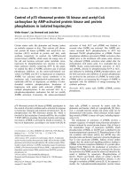
Figure 2 Detection of CREB-1 and Sp1 protein obtained from
the chromatin immunoprecipitation (ChIP). A ChIP protocol was
performed to pull down the CREB-1 and Sp1 proteins from the
individual cell lysates using antibodies against CREB-1 and Sp1
respectively. Half of the agarose beads obtained from the ChIP
study were analyzed by western blots to determine the amount of
the transcription factors precipitated from individual cell lysates. The
remainder of the beads was analyzed by real time PCR to
determine the amount of the XPC gene promoter DNA co-
precipitated with the individual transcription factors.
Table 3 Determination of the level of XPC gene 5’
regulatory region DNA co-precipitated with the
transcription factors CREB1 and Sp1 by IP in both
untreated and VPA-treated HTB4 and HTB9 bladder
cancer cells
a
.
IP
antigen
HTB4 HTB9
No
treatment
VPA
treatment
No
treatment
VPA
treatment
CREB1 1 4.6 ± 0.4 1 2.2 ± 0.2
Sp1 1 2.2 ± 0.3 1 2.0 ± 0.3
a
The level of XPC gene 5’ regulatory region DNA co-precipitated in the
untreated cells was counted as 1 and the level of XPC gene 5’ regulatory
region DNA co-precipitated in the VPA-treated cells was calculated as fold
change to that of the untreated cells for each cell line. The fold change was
expressed as Mean ± S.D. The results were from three independent IP
experiments.
Xu et al. Journal of Hematology & Oncology 2011, 4:17
/>Page 6 of 11
XPC -
XPC +
Bladder tumor Normal bladder tissue
HDAC4 -
HDAC4 +
A B
C D
E F
G H
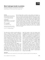
Figure 3 Immunohistochemistry (IHC) stain of XPC and HDAC4 proteins in both normal and cancerous bladder tissue specimens using
bladder tumor tissue arrays. The bladder tumor tissue arrays purchased from US BioMax Inc. were stained with either XPC or HDAC4
antibodies in an immunohistochemistry (IHC) protocol. The presence of XPC or HDAC4 protein was determined by light microscopy and the
image was recorded by a DP Controller software (Olympus Corp., Center Valley, PA).
Table 4 Determination of the presence of the HDAC4 in both normal and cancerous bladder tissues from bladder
tumor tissue arrays.
Type of bladder tissues # of HDAC4(+) # of Total tissues % of HDAC4(+)
Normal bladder tissues 1 23 4.3
Transitional cell carcinomas (Grade 1) 26 58 44.8
Transitional cell carcinomas (Grade 2) 28 59 47.5
Transitional cell carcinomas (Grade 3) 8 25 32.0
P value p
Δ
< 0.001
p
s
= 0.08
Note: p
Δ
value is the comparison between the group of normal bladder tissues and the group of cancerous bladder tissues. p
s
is the comparison among the
groups of normal bladder tissues, Grade 1, Grade 2, and Grade 3 bladder carcinomas.
Xu et al. Journal of Hematology & Oncology 2011, 4:17
/>Page 7 of 11
Prior treatment with the HDAC inhibitor VPA enhanced
cisplatin-induced apoptosis of bladder cancer cells
Extensive studies have demonstrated the cisplatin-
induced apoptosis as major mechanism in cell killing
[16,39,56-58]. Because of the important function of XPC
protein in the cisplatin-caused apopto sis [16] and the
role HDACs in XPC gene silencing, we further investi-
gated the effect of the HDAC inhibitor VPA in cispla-
tin-induced apoptosis of bladder cancer cells. The HTB4
and HTB9 bladder cancer cells were treated with VPA
(5 mM) for 48 hours before they were treated with cis-
platin. The cells were harvested 40 hours after the cis-
platin treatment and the caspase-3 activity was
determined (Figure 5). The caspase-3 activity was also
determined from the HBT4 and HTB9 cells that were
treated with cisplatin but without the prior VPA treat-
ment (Figure 5). The cisplatin treatment itself caused an
increase in caspase-3 activity in both H TB4 and HTB9
bladder cancer cells at high concentrations (20 μMand
40 μM) but not at lower concentrations (5 μMand10
μM) (Figure 5). When these cells were treated with VPA
prior to the cisplatin treatment, however, the caspase-3
activity was significantly increased at lower concentra-
tions as well (Figure 5). For example, when treated only
with cisplatin at 10 μM, the caspase 3 activity was
increased by a 1.5 and 2 fold in the HTB4 and HTB9
cells respectively; when the cells were treated with 5
mM VPA prio r to the cisplatin treatment, however, the
10 μM cisplatin treatment resulted in a 7.3 and 6.6 fold
increase of the caspase-3 activity in the HTB4 and
HTB9 cells respectively (Figure 5). These results suggest
that the prior treatment of HTB4 and HTB9 bladder
cancer cells with the HDAC inhibitor VPA sensitizes
these bladder cancer cells to the anticancer drug
cisplatin.
Discussion
In this work we have determined the role o f HDACs in
XPC gene silencing and bladder cancer development.
The results obtained from our HDAC inhibitor treat-
men t stud ies revealed that the VPA treatment led to an
increase in transcription of the XPC mRNA in both
HTB4 and HTB9 bl adder cancer cells. The results
obtained from our ChIP study demonstrated that the
VPA treatment resulted in an increas e in binding of the
CREB1 and Sp1 transcription factors at the 5’ regulatory
region of the XPC gene in both HTB4 and HTB9 cells.
The results of our IHC studies further indicated a
strong correlation between the over-expression of the
HDAC4 and the occurrence of urinary bladder transi-
tional cell carcinomas. In addition, the results obtained
from our caspase-3 activation studies also demonstrated
that the pre-treatment of HTB4 and HTB9 bladder can-
cer cells with VPA enhanced the anticancer drug cispla-
tin-induced activation of caspase-3, an i mportant
apoptotic caspase indicative of irreversible apoptosis.
Given the important role of the XPC protein in protect-
ing cells against many environmental carcinogen-
induced deleterious effects and the significance of the
HDACsinepigeneticgenetranscription regulation
[31-33], these resu lts suggest that the HDACs play an
HDAC4
HDAC1
HDAC2
-actin
GM00637
HTB4
HTB9
HTB2
HTB3
HTB5
HT1197
HT1376
Figure 4 Detection of expression of HDAC4, HDAC1, and
HDAC2 in various bladder cancer cells. The cell lysates prepared
from the HTB2, HTB3, HTB4, HTB5, HTB9, HT1197, HT1376 bladder
cancer cells and GM00637 normal human fibroblast cells (30 μg
total protein) were analyzed by western blots to determine the
protein levels of HDAC4, HDAC1, HDAC2, and b-actin in each cell
lysate. The antibodies against HDAC4 (A-4), HDAC1 (C-19), HDAC2
(H-54) and b-actin (C-2) were purchased from Santa Cruz
Biotechnology, Inc. (Santa Cruz, CA) and used in the western blots
study.
*
*
*
*
*
*
*
*
*
*
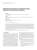
Figure 5 The cisplatin-induced caspase 3 activity in both
untreated and VPA-treated HTB4 and HTB9 bladder cancer
cells. The VPA treatment (5 mM) was done 24 hours prior to the
cisplatin treatment. The cells were treated with cisplatin at the
indicated concentrations for 3 hours and then cultured in the cell
culture incubator for 40 hours before the cells were harvested and
the caspase 3 activity was measured. The caspase 3 activity was
measured as nanomole of AMC/minute/mg of protein. (* statistical
difference to that of the untreated cells with p value < 0.01).
Xu et al. Journal of Hematology & Oncology 2011, 4:17
/>Page 8 of 11
important role in XPC gene silencing and bladder can-
cer development. Therefore, these results provide an
important mechanism of XPC gene silencing and blad-
der cancer development. Because of the essential role of
the XPC protein in initiating DNA damage-induc ed cel-
lular responses [16], these results further suggest that
silencing of the XPC gene may provide a critical early
event for initiation of bladder tumors. In addition, the
results obtained from these studies further suggests that
reactivation of the XPC gene by HDAC inhibitors may
have great benefits for bladder cancer treatment, espe-
cially for DNA-damaging anticancer drugs such as
cisplatin.
The results of our ChIP studies revealed that the VPA
treatment led to an increase in binding of t he CREB1
and Sp1 transcription factors to the 5’ regulatory region
of the XPC gene. These results suggest that inhibiting
the binding of these transcription factors to their con-
sensus sequences plays an important role in the
HDACs-caused XPC gene silencing of b ladder cancer
cells. This provides an important basis for understand-
ing the mechanism of XPC gene silencing in bladder
cancer cells. However, it is widely known that the con-
sensus sequences of many transcription factors are pre-
sent at the promoter region of the XPC gene, whether
or not the bindings of these transcription factors are
also affected by HDACs, and therefore, contribute to
the XPC gene silencing is largely u nknown. It may be
important to determine the effect of HDACs on the
bindings of these individual transcription factors at the
promoter region of the XPC gene in order to provide a
better understanding of the molecular basis by which
the HDACs cause silencing of the XPC gene in bladder
cancer cells.
The results of our IHC studies reveal that the fre-
quency of the HDAC4-positive tissue specimens was sig-
nificantly increased in the urinary bladder transitional
cell carcinomas in comparison to normal bladde r tissues.
However, the results obtained from our IHC study using
a HDAC2 antibody did not show a significant change in
the frequency of HDAC2-positive tissue specimens
between normal and cancerous bladder tissues (data not
shown). Given the similarity between the HDAC2 and
HDAC4 proteins in both their functions, these results
suggest that only certain HDACs are involved in the XPC
gene silencing in the urinary bla dder transitional cell car-
cinomas. Since t he HDACs family proteins also include
several other HDACs, it would be important to deter-
mine the correlation between the presence of the indivi-
dual HDACs and the bladder cancer occurr ence for each
HDAC in order to provide a better understanding of the
role of specific HDACs in XPC gene silencing and blad-
der cancer development.
The work described in this study was mainly focused
on determining the role of HDACs in XPC gene silen-
cing and bladder cancer development. However, it is
known that other epigenetic gene regulation mechan-
isms, including DNA methylation and microRNA
(miRNA), can also lead to silencing of the target genes
[32,33]. In fact, recently reported results suggest that
DNA methylation may play an important role in XPC
gene silencing of lung cancer cells [29]. Therefore,
future studies also need to determine the roles of these
epigenetic regulation mechanisms in XPC gene silencing
and bladder cancer development in order to pro vide a
better understanding of the mechanism of XPC gene
silencing and bladder cancer development.
Attenuated XPC protein has been observed in many
types of cancer, including bladder and lung cancer
[27,59]. Given the strong correlation between environ-
mental carcinogen exposure and cancer occurrence for
both bladder an d lung cancer as well as the similarity of
the lung and bladder organs in exposure to environmen-
tal carcinogens, it is possible that silencing of the XPC
gene may play an important role in cancer development
for many different types of cancer. Therefore, the
knowledge obtained from this study will be important
not only for understanding the mechanism of bladder
cancer development but also for grasping the mechan-
ism of development of these cancers as well. In addition,
the knowledge obtained from this study is also impor-
tant for detection, treatment, and risk assessment of
cancer as well as new anticancer drug design and
development.
Acknowledgements
We thank Mr. Kim Zukowski for his technical help in the
immunohistochemistry staining. Performance of this work was facilitated by
the Cell Culture Core, the Imaging and Flow Cytometry Core, and the
Microarray and Bioinformatic Core of the Environmental Health Sciences
Center in Molecular and Cellular Toxicology with Human Applications at
Wayne State University (P30ES06639). This work was supported in part by
grant R01ES09699 from NIH (G. W.).
Author details
1
Institute of Environmental Health Sciences, Wayne State University, 259
Mack Avenue, Detroit, MI 48201, USA.
2
Karmanos Cancer Institute, Wayne
State University, 4100 John R Street, Detroit, MI 48201 USA.
Authors’ contributions
XX carried out the VPA and IHC studies, and participated in the design and
coordination of the project. LW carried out the cell culture and the
participated in the immunoblotting and immunoprecipitation study. JA
carried out the statistical analysis of the IHC data. GW participated in the
design and coordination of the studies and drafted the manuscript. All
authors read and approved the final manuscript.
Competing interests
The authors declare that they have no competing interests.
Received: 9 February 2011 Accepted: 20 April 2011
Published: 20 April 2011
Xu et al. Journal of Hematology & Oncology 2011, 4:17
/>Page 9 of 11
References
1. Ploeg M, Aben KKH, Kiemeney LA: The present and future burden of
urinary bladder cancer in the world. World J Urol 2009, 27:289-293.
2. Kufe DW, Bast RBJ, Hait WH, Hong WH, Pollock RE, Weichselbaum RR,
Holland JF, Frei EI: Cancer Medicine 7. BC Decker Inc 2006.
3. Friedberg EC, Walker GC, Siede W: DNA repair and mutagenesis. ASM
published, Washington, D.C;, First 1995.
4. Sancar A, Lindsey-Boltz LA, Unsal-Kaccmaz K, Linn S: Molecular
mechanisms of mammalian DNA repair and the DNA damage
checkpoints. Annu Rev Biochem 2004, 73:39-85.
5. Friedberg EC, Walker GC, Siede W, Wood RD, Schultz RA, Ellenberger T:
DNA repair and mutagenesis. ASM Press, Washington D.C;, Second 2006.
6. Hanawalt PC: Transcription-coupled repair and human disease. Science
1994, 266:1957-1958.
7. Hanawalt PC: Subpathways of nucleotide excision repair and their
regulation. Oncogene 2002, 21:8949-8956.
8. Sarker AH, Tsutakawa SE, Kostek S, Ng C, Shin DS, Peris M, Campeau E,
Tainer JA, Nogales E, Cooper PK: Recognition of RNA polymerase II and
transcription bubbles by XPG, CSB, and TFIIH: insights for transcription-
coupled repair and Cockayne Syndrome. Mol Cell 2005, 20:187-198.
9. Laine JP, Egly JM: Initiation of DNA repair mediated by a stalled RNA
polymerase IIO. EMBO J 2006, 25:387-397.
10. Wood RD: DNA damage recognition during nucleotide excision repair in
mammalian cells. Biochimie 1999, 81:39-44.
11. Sugasawa K, Okamoto T, Shimizu Y, Masutani C, Iwai S, Hanaoka F: A
multistep damage recognition mechanism for global genomic
nucleotide excision repair. Genes Dev 2001, 15:507-521.
12. O’Donovan A, Davies AA, Moggs JG, West SC, Wood RD: XPG
endonuclease makes the 3’ incision in human DNA nucleotide excision
repair. Nature 1994, 371:432-435.
13. Matsunaga T, Park CH, Bessho T, Mu D, Sancar A: Replication protein A
confers structure-specific endonuclease activities to the XPF-ERCC1 and
XPG subunits of human DNA repair excision nuclease. J Biol Chem 1996,
271:11047-11050.
14. Bessho T, Sancar A, Thompson LH, Thelen MP: Reconstitution of human
excision nuclease with recombinant XPF-ERCC1 complex. J Biol Chem
1997, 272:3833-3837.
15. Shivji MK, Podust VN, Hubscher U, Wood RD: Nucleotide excision repair
DNA synthesis by DNA polymerase epsilon in the presence of PCNA,
RFC, and RPA. Biochemistry 1995, 34:5011-5017.
16. Wang G, Chuang L, Zhang X, Colton S, Dombkowski A, Reiners J, Diakiw A,
Xu
XS: The initiative role of XPC protein in cisplatin DNA damaging
treatment-mediated cell cycle regulation. Nucleic Acids Res 2004,
32:2231-2240.
17. Araujo SJ, Nigg EA, Wood RD: Strong functional interactions of TFIIH with
XPC and XPG in human DNA nucleotide excision repair, without a
preassembled repairosome. Mol Cell Biol 2001, 21:2281-2291.
18. Yokoi M, Masutani C, Maekawa T, Sugasawa K, Ohkuma Y, Hanaoka F: The
xeroderma pigmentosum group C protein complex XPC-HR23B plays an
important role in the recruitment of transcription factor IIH to damaged
DNA. J Biol Chem 2000, 275:9870-9875.
19. Leveillard T, Andera L, Bissonnette N, Schaeffer L, Bracco L, Egly JM,
Wasylyk B: Functional interactions between p53 and the TFIIH complex
are affected by tumour-associated mutations. EMBO J 1996, 15:1615-1624.
20. Kraemer KH, Lee MM, Scotto J: DNA repair protects against cutaneous
and internal neoplasia: evidence from xeroderma pigmentosum.
Carcinogenesis 1984, 5:511-514.
21. Kraemer KH, Myung ML, Scotto J: Xeroderma pigmentosum. Cutaneous,
ocular, and neurologic abnormalities in 830 published cases. Arch
Dermatol 1987, 123:241-250.
22. Cheo DL, Burns DK, Meira LB, Houle JF, Friedberg EC: Mutational
inactivation of the xeroderma pigmentosum group C gene confers
predisposition to 2-acetylaminofluorene-induced liver and lung cancer
and to spontaneous testicular cancer in Trp53-/- mice. Cancer Res 1999,
59:771-775.
23. Friedberg EC, Cheo DL, Meira LB, Reis AM: Cancer predisposition in
mutant mice defective in the XPC DNA repair gene. Prog Exp Tumor Res
1999, 35:37-52.
24. Friedberg EC, Bond JP, Burns DK, Cheo DL, Greenblatt MS, Meira LB,
Nahari D, Reis AM: Defective nucleotide excision repair in xpc mutant
mice and its association with cancer predisposition. Mutation Res 2000,
459:99-108.
25. Meira LB, Reis AM, Cheo DL, Nahari D, Burns DK, Friedberg EC: Cancer
predisposition in mutant mice defective in multiple genetic pathways:
uncovering important genetic interactions. Mutation Res 2001, 477:51-58.
26. Cheo DL, Friedberg EC: Use of nucleotide excision repair-deficient mice
as a model for chemically induced lung cancer. Methods Mol Med 2003,
74:481-491.
27. Hollander MC, Philburn RT, Patterson AD, Velasco-Miguel S, Friedberg EC,
Linnoila RI, Fornace AJJ: Deletion of XPC leads to lung tumors in mice
and is associated with early events in human lung carcinogenesis. Proc
Natl Acad Sci USA 2005, 102:13200-13205.
28. Chen Z, Yang J, Wang G, Song B, Li J, Xu Z: Attenuated expression of
xeroderma pigmentosum group C is associated with critical events in
human bladder cancer carcinogenesis and progression. Cancer Res 2007,
67:4578-4585.
29. Wu YH, Tsai Chang JH, Cheng YW, Wu TC, Chen CY, Lee H: Xeroderma
pigmentosum group C gene expression is predominantly regulated by
promoter
hypermethylation and contributes to p53 mutation in lung
cancers. Oncogene 2007, 26:4761-4773.
30. Thiel G, Lietz M, Hohl M: How mammalian transcriptional repressors
work. Eur J Biochem 2004, 271:2855-2862.
31. Ballestar E, Esteller M: Epigenetic gene regulation in cancer. Adv Genet
2008, 61:247-267.
32. Vaissière T, Sawan Z: Epigenetic interplay between histone modifications
and DNA methylation in gene silencing. Mutat Res 2008, 659:40-48.
33. Valeri N, Vannini I, Fanini F, Calore F, Adair B, Fabbri M: Epigenetics,
miRNAs, and human cancer: a new chapter in human gene regulation.
Mamm Genome 2009, 20:573-580.
34. Kurokawa R, Rosenfeld MG, Glass CK: Transcriptional regulation through
noncoding RNAs and epigenetic modifications. RNA Biol 2009, 6:233-236.
35. Dupont C, Armant DR, Brenner CA: Epigenetics: defination, mechanisms
and clinical perspective. Semin Reprod Med 2009, 27:351-357.
36. Verdone L, Caserta M, Di Maur E: Role of histone acetylation in the
control of gene expression. Biochem Cell Biol 2005, 83:344-353.
37. Khan AN, Tomasi TB: Histone deacetylase regulation of immune gene
expression in tumor cells. Immunol Res 2008, 40:164-178.
38. Kampranis SC, Tsichlis PN: Histone demethylases and cancer. Adv Cancer
Res 2009, 102:103-169.
39. Colton SL, Xu XS, Wang AY, Wang G: The involvement of Ataxia-
telangiectasia Mutated protein activation in nucleotide excision repair-
facilitated cell survival with cisplatin treatment. J Biol Chem 2006,
281:27117-27125.
40. Lomonaco SL, Xu XS, Wang G: The role of Bcl-x(L) protein in nucleotide
excision repair-facilitated cell protection against cisplatin-induced
apoptosis. DNA & Cell Biology 2009, 28:285-294.
41. Won J, Yim J, Kim TK: Sp1 and Sp3 recruit histone deacetylase to repress
transcription of human telomerase reverse transcriptase (hTERT)
promoter in normal human somatic cells. J Biol Chem 2002,
277:38230-38238.
42. Ryu H, Lee J, Olofsson BA, Mwidau A, Dedeoglu A, Escudero M,
Flemington E, Azizkhan-Clifford J, Ferrante RJ, Ratan RR: Histone
deacetylase inhibitors prevent oxidative neuronal death independent of
expanded polyglutamine repeats via an Sp1-dependent pathway. Proc
Natl Acad Sci USA 2003, 100:4281-4286.
43. Zhao S, Venkatasubbarao K, Li S, Freeman JW: Requirement of a specific
Sp1 site for histone deacetylase-mediated repression of transforming
growth factor beta Type II receptor expression in human pancreatic
cancer cells. Cancer
Res 2003, 63:2624-2630.
44. Yokota T, Matsuzaki Y, Miyazawa K, Zindy F, Roussel MF, Sakai T: Histone
deacetylase inhibitors activate INK4d gene through Sp1 site in its
promoter. Oncogene 2004, 23:5340-5349.
45. Marinova Z, Ren M, Wendland JR, Leng Y, Liang MH, Yasuda S, Leeds P,
Chuang DM: Valproic acid induces functional heat-shock protein 70 via
Class I histone deacetylase inhibition in cortical neurons: a potential role
of Sp1 acetylation. J Neurochem 2009, 111:976-987.
46. Mottet D, Pirotte S, Lamour V, Hagedorn M, Javerzat S, Bikfalvi A,
Bellahcène A, Verdin E, Castronovo V: HDAC4 represses p21(WAF1/Cip1)
expression in human cancer cells through a Sp1-dependent, p53-
independent mechanism. Oncogene 2009, 28:243-256.
Xu et al. Journal of Hematology & Oncology 2011, 4:17
/>Page 10 of 11
47. Song J, Noh JH, Lee JH, Eun JW, Ahn YM, Kim SY, Lee SH, Park WS, Yoo NJ,
Lee JY, Nam SW: Increased expression of histone deacetylase 2 is found
in human gastric cancer. APMIS 2005, 113:264-268.
48. Ozda H, Teschendorff AE, Ahmed AA, Hyland SJ, Blenkiron C, Bobrow L,
Veerakumarasivam A, Burtt G, Subkhankulova T, Arends MJ, Collins VP,
Bowtell D, Kouzarides T, Brenton JD, Caldas C: Differential expression of
selected histone modifier genes in human solid cancers. BMC Genomics
2006, 7:90.
49. Weichert W, Röske A, Gekeler V, Beckers T, Stephan C, Jung K, Fritzsche FR,
Niesporek S, Denkert C, Dietel M, Kristiansen G: Histone deacetylases 1, 2
and 3 are highly expressed in prostate cancer and HDAC2 expression is
associated with shorter PSA relapse time after radical prostatectomy. Br
J Cancer 2008, 98:604-610.
50. Fritzsche FR, Weichert W, Röske A, Gekeler V, Beckers T, Stephan C, Jung K,
Scholman K, Denkert C, Dietel M, Kristiansen G: Class I histone
deacetylases 1, 2 and 3 are highly expressed in renal cell cancer. BMC
Cancer 2008, 8:381.
51. Jin KL, Pak JH, Park JY, Choi WH, Lee JY, Kim JH, Nam JH: Expression
profile of histone deacetylases 1, 2 and 3 in ovarian cancer tissues. J
Gynecol Oncol 2008, 19:185-190.
52. Ashktorab H, Belgrave K, Hosseinkhah F, Brim H, Nouraie M, Takkikto M,
Hewitt S, Lee EL, Dashwood RH, Smoot D: Global histone H4 acetylation
and HDAC2 expression in colon adenoma and carcinoma. Dig Dis Sci
2009, 2(54):2109-2117.
53. Marquard L, Poulsen CB, Gjerdrum LM, de Nully Brown P, Christensen IJ,
Jensen PB, Sehested M, Johansen P, Ralfkiaer E: Histone deacetylase 1, 2, 6
and acetylated histone H4 in B- and T-cell lymphomas. Histopathology
2009, 54:688-698.
54. Halkidou K, Cook S, Leung HY, Neal DE, Robson CN: Nuclear accumulation
of histone deacetylase 4 (HDAC4) coincides with the loss of androgen
sensitivity in hormone refractory cancer of the prostate. Eur Urol 2004,
45:382-389.
55. Wilson AJ, Byun DS, Nasser S, Murray LB, Ayyanar K, Arango D, Figueroa M,
Melnick A, Kao GD, Augenlicht LH, Mariadason JM: HDAC4 promotes
growth of colon cancer cells via repression of p21. Mol Biol Cell 2008,
19:4062-4075.
56. Cepeda V, Fuertes MA, Castilla J, Alonso C, Quevedo C, Pérez JM:
Biochemical mechanisms of cisplatin cytotoxicity. Anticancer Agents Med
Chem 2007, 7:3-28.
57. Stewart DJ: Mechanisms of resistance to cisplatin and carboplatin. Crit
Rev Oncol Hematol 2007, 63:12-31.
58. Köberle B, Tomicic MT, Usanova S, Kaina B: Cisplatin resistance: preclinical
findings and clinical implications. Biochim Biophys Acta 2010,
1806:172-182.
59. Wu YH, Cheng YW, Chang JT, Wu TC, Chen CY, Lee H: Reduced XPC
messenger RNA level may predict a poor outcome of patients with
nonsmall cell lung cancer. Cancer
2007, 110:215-223.
doi:10.1186/1756-8722-4-17
Cite this article as: Xu et al.: Histone deacetylases (HDACs) in XPC gene
silencing and bladder cancer. Journal of Hematology & Oncology 2011
4:17.
Submit your next manuscript to BioMed Central
and take full advantage of:
• Convenient online submission
• Thorough peer review
• No space constraints or color figure charges
• Immediate publication on acceptance
• Inclusion in PubMed, CAS, Scopus and Google Scholar
• Research which is freely available for redistribution
Submit your manuscript at
www.biomedcentral.com/submit
Xu et al. Journal of Hematology & Oncology 2011, 4:17
/>Page 11 of 11