Báo cáo y học: "Gene expression profiles in BCL11B-siRNA treated malignant T cells" ppt
Bạn đang xem bản rút gọn của tài liệu. Xem và tải ngay bản đầy đủ của tài liệu tại đây (1.67 MB, 6 trang )
SHOR T REPO R T Open Access
Gene expression profiles in BCL11B-siRNA treated
malignant T cells
Xin Huang
1,2†
, Qi Shen
1†
, Si Chen
1
, Shaohua Chen
1
, Lijian Yang
1
, Jianyu Weng
2
, Xin Du
2
, Piotr Grabarczyk
3
,
Grzegorz K Przybylski
3,4
, Christian A Schmidt
3
and Yangqiu Li
1,5*
Abstract
Background: Downregulation of the B-cell chronic lymphocytic leukemia (CLL)/lympho ma11B (BCL11B) gene by
small interfering RNA (siRNA) leads to growth inhibition and apoptosis of the human T-cell acute lymphoblastic
leukemia (T-ALL) cell line Molt-4. To further characterize the molecular mechanism, a global gene expression profile
of BCL11B-siRNA -treated Molt-4 cells was established. The expression profiles of several genes were further
validated in the BCL11B-siRNA -treated Molt-4 cells and primary T-ALL cells.
Results: 142 genes were found to be upregulated and 109 genes downregulated in the BCL11B-siRNA -treated
Molt-4 cells by microarray analysis. Among apoptosis-related genes, three pro-apoptotic genes, TNFSF10, BIK, BNIP3,
were upregulated and one anti-apoptotic gene, BCL2L1 was downregulated. Moreover, the expression of SPP1 and
CREBBP genes involved in the transforming growth factor (TGF-b) pathway was down 16-fold. Expression levels of
TNFSF10, BCL2L1, SPP1, and CREBBP were also examined by real-time PCR. A similar expression pattern of TNFSF10,
BCL2L1, and SPP1 was identified. However, CREBBP was not downregulated in the BLC11B-siRNA -treated Molt-4
cells.
Conclusion: BCL11B-siRNA treatment altered expression profiles of TNFSF10, BCL2L1, and SPP1 in both Molt-4 T cell
line and primary T-ALL cells.
Background
Although treatment outcome in patients with T-cell
acute lymphoblastic leukemia (T-ALL) has improved in
recent years, relapsed T-ALL remains a challenge [1].
Monoclonal antibodies, gene inhibitors, and upregulation
of microRNAs [2,3] are promising tools for cancer tar-
geted therapy. However, few targeted therapies are avail-
able for T-cell malignancies. For example, transforming
Mer signals may contribute to T-cell leukemogenesis,
and regulation of Mer expression could be a novel thera-
peutic target for pediatric ALL therapy [4]. The recent
identification of activating Notch1 mutations in the
majori ty of patients with T-ALL has bro ught interests on
targeting the Notch signaling pathway for this disease [5].
The B-cell chronic lymphocytic leukemia (CLL)/lym-
phoma 11B (BCL11B)genewasfirstidentifiedon
human chromosome 14q32.2 [6] and encodes a Krüp-
pel-like C
2
H
2
zinc finger protein initially identified as a
transcriptional repressor [7]. BCL11B plays an important
role in T-cell differentiation and proliferation [8-11].
Altered expression, mutation, disruption, or rearrange-
ment of BCL11B has been associated with T-cell malig-
nancies [12-14]. In humans, BCL11B overexpression is
found primarily in lymphoproliferative disorders, such
as T-ALL and adult T-cell leukemia/lymphoma
[12,15-17]. BCL11B mediates transc riptional activation
by interacting with the p300 co-activator at the
upstream site 1 (US1) of the interleukin (IL)-2 promo-
ter, leading to transcriptional activation of IL-2 expres-
sion in activated T cells [18]. Although the interaction
partners and binding sequen ce have been revealed, only
afewBCL11B direct target genes have been identified
to date. Our previous study in the human T-ALL cell
lines Molt-4, Jurkat, and hut78 has shown increased
apoptosis upon BCL11B suppression by RNA interfer-
ence [19].
* Correspondence:
† Contributed equally
1
Institute of Hematology, Medical College, Jinan University, Guangzhou, PR
China
Full list of author information is available at the end of the article
Huang et al. Journal of Hematology & Oncology 2011, 4:23
/>JOURNAL OF HEMATOLOGY
& ONCOLOGY
© 2011 Huang et al; licensee BioMed Central Ltd. This is an Open Access article distributed under the terms of the Creative Commons
Attribution License (http: //creativecommons.org/licenses/by/2.0), which permits unrestricted use, distribution, and reprod uction in
any medium, provided the original work is properly cited .
In the present study, we further analyzed the global
gene expression profiles in Molt-4 and primary T -ALL
cells after BCL11B-935-siRNA treatment.
Methods
Samples
Samples from three newly diagnosed patients with T-
ALL and one patient with T-cell lymphoma/leukemia
were obtained after informed consent. The diagnosis of
T-ALL was based on cytomorphology, immunohisto-
chemistry, and flowcytometry analyses. The samples were
named P1 (55-year-old male with T-ALL), P2 (6-year-old
male with T-ALL), P3 (55-year-old female with T-cell
lymphoma/leu kemia ), and P4 (19-yea r-old male with T-
ALL). Peripheral blood was collected with heparin and
peripheral mononuclear cells (PBMCs; contained more
than 70% leukemic T cells) were separated using the
Ficoll-Hypaque gradient centrifugation method. All pro-
cedures were conducted in strict accordance with the
guidelines of the Medical Ethics co mmittees of the
Health Bureau of Guangdong province, China.
Cell culture
Molt-4 cells (Institutes for Biological Sciences Cell
Resource Center, Chinese Academy of Sciences, Shang-
hai, China) and PBMCs collected from the four patients
were cultured in complete RPMI 1640 medium with
15% fetal calf serum and were maintained in a sterile
incubator at 37°C, 95% humidity, and 5% CO
2
.
Nucleofection
BCL11B-siRNA935 (Chinese patent application number:
200910193248.3) and the scrambled non-silencing
siRNA control (BCL11B-sc) were designed with online
software and synthesized by
Invitrogen (Carlsbad, CA, USA).
Malignant T cells were resuspended at 2.5 × 10
6
(Molt-4
cells) or 1 × 10
7
(PBMCs) per 100 μL of the appropriate
Nucleofector kit solution (Amaxa Biosystems, Cologne,
Germany), and were nucleofected with 3 μg of BCL11B-
siRNA or control non-silencing scrambled (sc) RNA using
the C-005 (Molt-4 cells) o r U-014 (PBMCs) program in
the Nucleofection Device II (Amaxa Biosystems). Mock-
transfected cells (nucleofect ed without siRNA) were used
as a negative control. After nucleofection, the cells were
immediately mixed with 500 μL of pre-warmed culture
medium and transferred to culture plates for i ncubation.
Samples were collected for RNA isolation.
RNA isolation, expression profiling, reverse transcription,
and real-time PCR
Total RNA was isolated using Trizol (Invitrogen), and
cDNA was synthesized with a Superscript II RNaseH
Reverse Transcriptase kit (Invitrogen).
Total RNA (> 3 μg) was sent for global gene expres-
sion profile analysis using an Affymetrix HG U133 Plus
2.0 gene chip (Shanghai Biochip Co., Ltd., Shanghai,
China). The Affymetrix microarray analysis was per-
formed using Gene Spring GX10.0 software (Agilent
Technologies, Santa Clara, CA, USA).
The primer and probe information for BCL11B and
the reference gene b-2-micr oglob ulin (b2-MG), as well
as the details of the real-time PCR for BCL11B were
described in our previous studies [12,15]. Expression
levels of tumor necrosis factor (ligand) superfamily,
member 10 (TNFSF10; TRAIL), BCL2-like 1 (BCL2L1;
Bcl-xL), secreted phosphoprotein 1 (SPP1), cAMP-
response element binding protein (CREBBP), and b2-
MG were determined by real-time PCR using a SYBR
Green I qPCR Master Mix kit [15].
Flow cytometry assay
Cells from different groups were prepared according to
the protocols, and the BCL2 expression level was mea-
sured by flow cytometry (Beckman Coulter, Fullerton,
CA, USA). Mouse anti-human BCL2-PE and mouse
IgG1-PE (eBioscience, San Diego, CA, USA) were used.
Results were analyzed using the Win MDI 2.9 software.
Results and discussion
Global gene expression profile in BCL11B-siRNA935
treated Molt-4 cells
To determine the molecular mechanisms of BCL11B
siRNA-mediated cell apoptosis, global gene expression
profiling was performed at 24 h post-transfection, when
BCL11B mRNA was most effectively suppressed (data
not shown). Resu lts were clustered, based on the differ-
ential expression level (2-fold up or down), and visua-
lized using a color scale (Figure 1A). Principal
component analysis indicated that the changes in the
Molt-4 cell gene expression profile could be accounted
for primarily b y the BCL11B siRNA935 treatment
(Figure 1B). A GCOS1.4 software analysis showed that
upregulated genes were identified by 142 probe sets,
whereas 109 genes were downregulated at l east 2-fold,
compared with the sc control (Figure 1C). Changes in
genes of the same signaling pathways closely related to
tumor cell proliferation and apoptosis were analyzed
further (Figure 1D).
Among apoptosis-related genes, changes in expressio n
levels occurred mainly in three pro-apoptotic genes;
TNFSF10, BCL-2 interacting killer (BIK), and BCL-2/
E1B 19 kDa interacting protein 3 (BNIP3), which were
upregulated 2-4 fold, and one anti-apoptot ic gene
(BCL2L1) was downregulated by 3-4 fold. The expres-
sion levels of SPP1andCREBBP genes involved in the
transforming growth factor (TGF-b) pathway were down
by 16 fold. The changes in the expression levels of the
Huang et al. Journal of Hematology & Oncology 2011, 4:23
/>Page 2 of 6
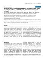
Figure 1 Result s of the gene chip microarray analysis and validation. (A) Visual display o f the cluster a nalysis for the BCL11B siRNA9 35-
transfected and control cells. (B) Principal component analysis. The closer the dots, the more similar the gene expression profiles are; the farther
apart the dots, the greater the differences are. (C) Two-dimensional scatterplot analysis of gene expression values for all genes on the BCL11B
siRNA935-transfected cells and control cells from the microarray. Yellow dots represent genes absent from both samples; blue dots represent
genes present in one sample but absent from the other sample; and red dots represent genes present in both samples. Dots outside the 2 ×
difference lines, indicated by black arrows, represent differentially expressed genes. The farther from the line, the greater the difference in gene
expression are. (D) Analysis of pathways closely related to tumor cell proliferation and apoptosis. Results are shown as fold-change in mRNA
transcripts. Genes indicated with a red star are in the apoptosis pathway; genes indicated with a blue star are in the transforming growth factor-
b pathway. (E) Gene validation by real-time PCR. Changes in TNFSF10, BCL2L1, and SPP1 expression levels agreed with the microarray results,
while those of CREBBP did not. (F) Reduced BCL-2 protein expression was confirmed by flow cytometry. BCL-2 expression in BCL11B siRNA3-
transfected cells was significantly lower, at 46% of that in SC (99.1%), MOCK (99.2%), and NC cells (99.7%).
Huang et al. Journal of Hematology & Oncology 2011, 4:23
/>Page 3 of 6
TNFSF10, BCL2L1, SPP1, and CREBBP genes were
further detected by real-time PCR (Figure 1E). The
BH3-only domain proteins BIK and BNIP3, which were
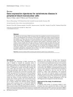
located upstream of BCL-2 (Figure 2), may enhance
their binding to BCL-2, thereby inhibiting the anti-apop-
totic function. Thus, we analyzed the BCL-2 protein
expression level by flow cytometry in Molt-4 cells at 72
hafterBCL11B-siRNA treatment (Figure 1F). A similar
altered expression pattern of these genes, as well as
expression of the BCL-2 protein, was confirmed. How-
ever, CREBBP did not show downregulation in BCL11B-
siRNA treated Molt-4 cells.
The global gene expression profile results suggest that
the molecular mechanisms of BCL11B siRNA-mediated
cell death may involve BCL-2 family genes in the intrin-
sicmitochondrialpathwayaswellastheTNFS F10 gene
in the death receptor signaling pathway (Figure 2) [20].
Upregulation of the TNFSF10 gene activated the death
rec eptor signaling pathway, whereas upregulation of the
two mitochondrial BCL-2 family genes (the BH3-only
domain proteins BIK and BNIP3) enhanced their bind-
ing to BCL-2, with a reduction in the anti-apoptotic
gene BCL2L1, thereby inhibiting the anti-apoptotic func-
tion and promoting Bax and Bak activation. This in turn
activates the downstream caspases 3, 6, and 7, leading to
increased apoptosis. Reduced expression of SPP1 corre-
lated with increased apoptosis in Molt-4 cells, suggest-
ing that the SPP1 gene may be a BCL11B gene target.
CREBBP overexpression has been detected in Jurkat
cells [21]. However, previous studies h ave not reported
achangeinCREBBP expression in T cell lines after
BCL11B-siRNA treatment. In the present study, downre-
gulation of CREBBP was identified in the microarray
analysis, but not confirmed by real-time PCR analysis.
The reason may be due to a systemic error on the
microarr ay analysis. Interestingly, unlike the result from
Molt-4 cells, the alteration of the CREBBP expression
level in primary T-All cells after BCL11B-siRNA treat-
ment was in accordance with the results from the
microarray analysis (Figure 3). Thus, the role of
CREBBP during BCL11B downregulation in malignant T
cells requires further investigation.
Expression of TNFSF10, BCL2L1, SPP1, and CREBBP genes
in BCL11B-siRNA935-treated primary leukemic T cells
After obtaining interesting data from Molt-4 cells, we
analyzed the effect of the BCL11B-siRNA in primary T-
ALL cells. We examined the expression levels of
TNFSF10, BCL2L1, SPP1, and CRE BBP in primary leu-
kemic T cells after BCL11B siRNA935 treatment.
Figure 2 Schematic model of the molecular mechanism of BCL11B-siRNA-mediated apoptosis in Molt-4 cell [modified from reference 20].
Huang et al. Journal of Hematology & Oncology 2011, 4:23
/>Page 4 of 6
BCL11B expression level decreased in primary leukemic
T cells treated with BCL11B siRNA935 (282.77 ± 247.57
copies/10
5
b2-MG)ascomparedwiththesccontrol
group (519.48 ± 303.41 copies/10
5
b2-MG). The
TNFSF10, BCL2L1, SPP1, and CREBBP expression levels
in BCL11B-siRNA935- treated primary leukemic T cells
were 2.7 ± 2.17%, 9.53 ± 15.34%, 3.5 ± 2.9 5%, and 4.25
± 5.82%, respectively, whereas the expression levels in
primary leukemic T cells in the sc control group were
1.77 ± 1.93%, 6.96 ± 9.88%, 10.23 ± 13.09%, and 4.98 ±
7.2%, respectively. The T-ALL specimen number was
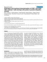
too small to perform statistical analysis. The changes in
the mRNA levels of TNFSF10, SPP1, and CREBBP in
the T cells from the four patients agreed in general with
those from the microarray analysis results (Figure 3A, C,
D). However, the changes in the BCL2L1expression
levels in the different samples varied (Figure 3B). The
reduced BCL2L1 expression rates in leukemic T cells
from patients 1 and 3 were 31.84% and 13.73%, respec-
tively, compared with the sc controls, whereas BCL2L1
expression in leukemic T cells from patients 2 and 4
was upregulated. Although BCL11B gene overexpression
occurred in all samples, it may have been due to the
heterogeneity of T-cell malignancies during apoptosis
induced by BCL11B downregulation [22], so it remains
to be determined whether apoptosis induced by BCL11B
downregulation in some cases with T-ALL involves the
BCL-2 family genes in the intrinsic mitochondrial
pathway.
A previous analysis revealed that overexpression of the
BCL11B, BCL2L1, and CREBBP genes in primary T-ALL
samples blocks apoptosis in malignant T cells [15]. This
study suggests that inhibition of BCL11B may trigger
apoptosis in leukemic T cells by downregulating the
downstream genes SPP1, CREBBP, and TNFSF10.
Conclusions
Our findings provide evidence that BCL11BsiRNA-
mediated cell apoptosis may be related to the mitochon-
drial pathway BCL-2 family ge nes and the TNFSF10
gene of the death re ceptor signaling pathway. Moreover,
the SPP1andCREBBP genes in the TGF-b pathway
may also be involved in BCL11B siRNA-mediated cell
apoptosis.
Figure 3 Expression of TNFSF10, BCL2L1, SPP1, and CREBBP genes in peripheral mononuclear cells from four patients (P1-P4) with T-
cell acute lymphoblastic leukemia at 24 h after BCL11B siRNA transfection.
Huang et al. Journal of Hematology & Oncology 2011, 4:23
/>Page 5 of 6
Acknowledgements
The authors thank Dr. Xuesong Yang for critical reading of this manuscript,
and thank Dr. Xuchao Zhang from the Cancer Center of Guangdong
Provincial Hospital for helpful analysis of the gene-chip data. This work was
supported by Grants from National Natural Science Foundation of China (no.
30771980) and the Guangdong Science & Technology Project (no.
2007B030703008; and 2009B050700029).
Author details
1
Institute of Hematology, Medical College, Jinan University, Guangzhou, PR
China.
2
Department of Hematology, Guangdong General Hospital
(Guangdong Academy of Medical Sciences), Guangzhou, PR China.
3
Department of Hematology and Oncology, Ernst-Moritz-Arndt University
Greifswald, Greifswald, Germany.
4
Institute of Human Genetics, Polish
Academy of Sciences, Poznan, Poland.
5
Key Laboratory for Regenerative
Medicine of Ministry of Education, Jinan University, Guangzhou, PR China.
Authors’ contributions
YQL contributed to concept development and study design. XH, QS, SC,
SHC, LJY performed the laboratory studies. PG, GKP and CAS provided some
materials and technical support. JYW and XD were responsible for collection
of clinical data. YQL and XH coordinated the study and helped drafting the
manuscript. All authors read and approved the final manuscript.
Competing interests
The authors declare that they have no competing interests.
Received: 21 March 2011 Accepted: 15 May 2011
Published: 15 May 2011
References
1. Aifantis I, Raetz E, Buonamici S: Molecular pathogenesis of T-cell
leukaemia and lymphoma. Nat Rev Immunol 2008, 8:380-390.
2. Budhu BA, Ji JF, Wang XW: The clinical potential of microRNAs. J Hematol
Oncol 2010, 3:37.
3. Wei GQ, Rafiyath S, Liu DL: First-line treatment for chronic myeloid
leukemia:dasatinib, nilotinib, or imatinib. J Hematol Oncol 2010, 3:47.
4. Graham DK, Salzberg DB, Kurtzberg J: Ectopic expression of the proto-
oncogene Mer in pediatric T-cell acute lymphoblastic leukemia. Clin
Cancer Res 2006, 12:2662-2669.
5. Palomero T, Ferrando A: Therapeutic targeting of NOTCH1 signaling in T-
ALL. Clin Lymphoma Myeloma 2009, 9(Suppl 3):S205.
6. Satterwhite E, Sonoki T, Willis TG, Harder L, Nowak R, Arriola EL, Liu H,
Price HP, Gesk S, Steinemann D, Schlegelberger B, Oscier DG, Siebert R,
Tucker PW, Dyer MJ: The BCL11 gene family: involvement of BCL11A in
lymphoid malignancies. Blood 2001, 98:3413-3420.
7. Avram D, Fields A, Senawong T, Topark-Ngarm A, Leid M: COUP-TF
(chicken ovalbumin upstream promoter transcription factor)-interacting
protein 1 (CTIP1) is a sequence-specific DNA binding protein. Biochem J
2002, 368:555-563.
8. Li L, Leid M, Rothenberg EV: An Early T cell lineage commitment
checkpoint dependent on the transcription factor Bcl11b. Science 2010,
329:89-93.
9. Wakabayashi Y, Watanabe H, Inoue J, Takeda N, Sakata J, Takeda N, Sakata J,
Mishima Y, Hitomi J, Yamamoto T, Utsuyama M, Niwa O, Aizawa S,
Kominami R: Bcl11b is required for differentiation and survival of αβ T
lymphocytes. Nat Immunol 2003, 4:533-539.
10. Cismasiu VB, Ghanta S, Duque J, Albu D, Chen HM, Kasturi R: BCL11B
participates in the activation of interleukin-2 gene expression in CD4+ T
lymphocytes. Blood 2006, 108:2695-2702.
11. Liu P, Li P, Burke S: Critical roles of Bcl11b in T-cell development and
maintenance of T-cell identity. Immunol Rev 2010, 238:138-149.
12. Przybylski GK, Dik WA, Wanzeck J, Grabarczyk P, Majunke S, Martin-
Subero JI, Siebert R, Dölken G, Ludwig WD, Verhaaf B, van Dongen JJ,
Schmidt CA, Langerak AW: Disruption of the BCL11B gene through inv 14
q11.2q32.31 results in the expression of BCL11B-TRDC fusion transcripts
and is associated with the absence of wild-type BCL11B transcripts in T-
ALL. Leukemia 2005, 19:201-208.
13. Karlsson A, Nordigården A, Jönsson JI, Söderkvist P: Bcl11b mutations
identified in murine lymphomas increase the proliferation rate of
hematopoietic progenitor cells. BMC Cancer 2007, 7
:195.
14. Su XY, Della-Valle V, Andre-Schmutz I, Lemercier C, Radford-Weiss I,
Ballerini P, Lessard M, Lafage-Pochitaloff M, Mugneret F, Berger R,
Romana SP, Bernard OA, Penard-Lacronique V: HOX11L2/TLX3 is
transcriptionally activated through T-cell regulatory elements
downstream of BCL11B as a result of the t(5;14) (q35;q32). Blood 2006,
108:4198-4201.
15. Huang X, Chen S, Shen Q, Yang LJ, Li B, Zhong LY, Geng SX, Du X, Li YQ:
Analysis of the expression pattern of the BCL11B gene and its relatives
in patients with T-cell acute lymphoblastic leukemia. J Hematol Oncol
2010, 3:44.
16. Nagel S, Kaufmann M, Drexler HG, MacLeod RA: The cardiac homeobox
gene NKX2-5 is deregulated by juxtaposition with BCL11B in pediatric T-
ALL cell lines via a novel t(5;14)(q35.1;q32.2). Cancer Res 2003,
63:5329-5334.
17. Oshiro A, Tagawa H, Ohshima K, Karube K, Uike N, Tashiro Y, Utsunomiya A,
Masuda M, Takasu N, Nakamura S, Morishima Y, Seto M: Identification of
subtype-specific genomic alterations in aggressive adult T-cell leukemia/
lymphoma. Blood 2006, 107:4500-4507.
18. Cismasiu VB, Ghanta S, Duque J, Albu DI, Chen HM: BCL11B participates in
the activation of IL2 gene expression in CD4+ T lymphocytes. Blood
2006, 108:2695-2702.
19. Huang X, Chen S, Chen SH, Yang LJ, Shen Q, Grabarczyk P, Przybylski GK,
Schmidt CA, Li YQ: Inhibition of BCL11B expression leads to apoptosis of
malignant T cell lines but not CD34
+
cells [abstract]. Blood 2010,
116:1539.
20. Adams JM, Cory S: The Bcl-2 apoptotic switch in cancer development
and therapy Bcl-2 apoptotic switch in cancer. Oncogene 2007,
26:1324-1337.
21. Caravatta L, Sancilio S, di Giacomo V, Rana R, Cataldi A, Di Pietro R: PI3-K/
Akt-dependent activation of cAMP-response element-binding (CREB)
protein in Jurkat T leukemia cells treated with TRAIL. J Cell Physiol 2008,
214:192-200.
22. Onciu M, Lai R, Vega F, Bueso-Ramos C, Medeiros LJ: Precursor T-cell acute
lymphoblastic leukemia in adults: age-related immunophenotypic,
cytogenetic, and molecular subsets. Am J Clin Pathol 2002, 117:252-258.
doi:10.1186/1756-8722-4-23
Cite this article as: Huang et al.: Gene expression profiles in BCL11B-
siRNA treated malignant T cells. Journal of Hematology & Oncology 2011
4:23.
Submit your next manuscript to BioMed Central
and take full advantage of:
• Convenient online submission
• Thorough peer review
• No space constraints or color figure charges
• Immediate publication on acceptance
• Inclusion in PubMed, CAS, Scopus and Google Scholar
• Research which is freely available for redistribution
Submit your manuscript at
www.biomedcentral.com/submit
Huang et al. Journal of Hematology & Oncology 2011, 4:23
/>Page 6 of 6