Báo cáo y học: "Dermoscopy as a technique for the early identification of foot melanoma" pot
Bạn đang xem bản rút gọn của tài liệu. Xem và tải ngay bản đầy đủ của tài liệu tại đây (818.5 KB, 6 trang )
BioMed Central
Page 1 of 6
(page number not for citation purposes)
Journal of Foot and Ankle Research
Open Access
Review
Dermoscopy as a technique for the early identification of foot
melanoma
Ivan R Bristow*
1
and Jonathan Bowling
2
Address:
1
School of Health Sciences, University of Southampton, UK and
2
Department of Dermatology, The Churchill Hospital, Oxford, UK
Email: Ivan R Bristow* - ; Jonathan Bowling -
* Corresponding author
Abstract
Malignant melanoma is the most common primary malignant tumour arising on the foot. Where
improvements in the prognosis have been observed for patients with melanoma elsewhere on the
skin, pedal lesions are still frequently delayed in presentation through neglect or misdiagnosis.
Detection of foot melanoma relies on the health care practitioner's skills and observations in
recognising early changes. Recent publications have documented the use a dermoscopy as a tool
to improve recognition of such suspicious lesions. This paper reviews current literature with a
special emphasis of its potential applications on plantar and nail unit melanoma. Data from these
studies suggest that the technique is a useful and significant adjunct to clinical examination, which
ultimately may lead to earlier recognition of this aggressive tumour.
Introduction
Cancers involving the skin account for a third of all
human cancers. According to the World Health Organisa-
tion, malignant melanoma (MM) accounts for an esti-
mated 132 000 new cases annually and around 66 000
deaths. Globally the incidence of the disease continues to
rise, particularly in Caucasian populations [1]. As there is
no effective treatment for the disease, improving survival
still remains around earlier detection of malignant
lesions. The thinner the lesion at diagnosis, the better the
prognosis [2]. There is some evidence to suggest that
patients are presenting earlier and that the mean
melanoma thickness at diagnosis is declining [3],
although risk factors such as older age, male gender and
low educational level still predict higher thickness at pres-
entation [4-6].
Melanoma and the foot
Malignant melanoma is the most common primary,
malignant tumour of the foot [7] accounting for between
3–15% of all cutaneous melanoma [8]. Whereas improve-
ments have been seen in the prognosis for some patients
with melanoma, pedal lesions are still a major concern.
The three most common types occurring on the foot are
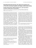
the superficial spreading (figure 1), nodular and acral len-
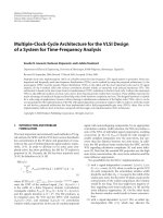
tiginous melanoma (ALM – figure 2). ALM is particularly
prevalent on the foot as it has a predilection for the soles
and nail unit [9]. In addition, it is a sub-type of melanoma
that affects all skin types [10]. Day [11] identified MM on
the foot as an independent risk factor for disease recur-
rence. This was examined further by Hsueh and colleagues
[12] who reviewed 652 cases of cutaneous melanoma and
analysed data comparing anatomical location to survival
rates. Controlling for other variables including tumour
thickness, their results confirmed that primary melanoma
on the foot had a 5 year survival rate of 77% compared
with 94% and 95% for lesions on the calf and thigh
respectively. They concluded that the prognosis deterio-
rated the further the lesion was from the trunk.
Published: 12 May 2009
Journal of Foot and Ankle Research 2009, 2:14 doi:10.1186/1757-1146-2-14
Received: 30 October 2008
Accepted: 12 May 2009
This article is available from: />© 2009 Bristow and Bowling; licensee BioMed Central Ltd.
This is an Open Access article distributed under the terms of the Creative Commons Attribution License ( />),
which permits unrestricted use, distribution, and reproduction in any medium, provided the original work is properly cited.
Journal of Foot and Ankle Research 2009, 2:14 />Page 2 of 6
(page number not for citation purposes)
From the available data, the reason for this is not clear but
is probably less likely to do with the physical nature of the
tumour and more to do with delays in presentation and
diagnosis. Prognosis, in part, is worsened in foot
melanoma as lesions frequently present later and are
therefore thicker at diagnosis [13]. Reasons for patient
delays have been well studied [5,14-17]. Richard et al
studied 590 melanoma patients and reported a number of
factors that predicted thicker lesions including melanoma
which were out of the patients view (such as the plantar
surface of the foot). From a medical perspective longer
physician delays in diagnosis have also been observed
with acral lesions [18]. Misdiagnosis could also explain a
reduced prognosis in patients with acral melanoma.
Bristow and Acland [19], reviewing 27 cases of acral len-
tiginous melanoma on the foot suggested a misdiagnosis
rate of 33% whilst other workers have reported much
higher rates of up to 60% in melanomas of the foot [20].
Metzger and co-workers [21] in a review of delayed diag-
nosis of melanoma highlighted that many acral
melanoma are initially presented to non-dermatologists
because patients do not suspect the problem to be a
melanoma. As such clinicians are less aware of the condi-
tion; mis-diagnosis would be more of an issue. Illustrating
this, many papers have been published highlighting foot
melanoma misdiagnosed as other conditions such as fun-
gal infection, onychomycosis, ulceration, haematoma and
other more common foot pathologies [20,22-27].
Detection of melanoma
The value of educating patients and practitioners through
melanoma awareness campaigns cannot be emphasized
too strongly and various initiatives have tried to heighten
the public awareness and monitoring of skin. Equally
important is the role of the practitioner in screening
patients – physician detected melanomas have been
shown to be significantly thinner at diagnosis than those
detected by patients [6]. The ABCD rule, devised in 1985
by Freidman [28] has been well used as a mnemonic in
skin assessment for recognising change in melanocytic
naevi. Its value in foot melanoma has been questioned as
acral lesions do not exhibit the typical features of malig-
nant melanoma elsewhere on the skin [19,21]. Therefore
at a clinical level, the decision to monitor, excise or refer
on a suspicious lesion can be a difficult one.
Dermoscopy
Visual examination of a suspicious skin lesion such as a
melanoma can be significantly enhanced by the addition
of surface microscopy. This was first recognised by Scot-
tish Dermatologist Rona MacKie who in 1971 published
a paper which demonstrated pre-operatively, the high pre-
dictive value of close examination of melanoma [29]. The
difficulty arises however in that evaluation of the skin
under normal conditions, with a standard magnifier, is
limited due to surface reflection and refraction. To over-
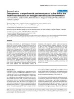
come this the dermatoscope is a simple, and relatively
cheap, hand held magnifying device (typically 10×) which
uses an oil medium or cross-polarised light allowing the
viewer to observe structures deeper in the skin, not nor-
mally visible to the naked eye (figure 3). Since the 1980's
the idea of "dermoscopy" began to gain momentum and
its popularity as a tool aiding clinical decision making
increased, particularly in Europe as more research evi-
dence was published. In 1990, around 13 papers were
published; in 2007 it had risen to over 500.
It should be emphasized that the dermatoscope itself is
not a diagnostic tool but acts to aid decision making in
when confronted with a suspicious lesion, allowing the
practitioner greater confidence when deciding whether to
refer, excise or leave a skin lesion.
Superficial spreading melanoma on the ankleFigure 1
Superficial spreading melanoma on the ankle.
Acral lentiginous melanomaFigure 2
Acral lentiginous melanoma.
Journal of Foot and Ankle Research 2009, 2:14 />Page 3 of 6
(page number not for citation purposes)
The use of the dermatoscope was initially the exclusive
realm of the dermatologist, experimental and early work
gave rise to extensive descriptions of patterns and features
visualised in melanocytic naevi, melanoma and other skin
tumours. This then moved to the formalisation of the
technique into various algorithms such as pattern analysis
[30], the 7-point technique [31], the modified ABCD tech-
nique [32] and the Menzies method [33]. Two early meta-
analyses of the dermatoscopic technique were published
concluding that it increases sensitivity and specificity for
the diagnosis of melanoma when compared to the naked
eye when in the hands of an experienced clinician [34,35].
In 2004, it was recognised that in order to achieve a
decrease in morbidity and mortality, dermoscopy should
be a screening test that is available to all practitioners
involved in skin screening providing it was accurate, easily
to apply and inexpensive. Such a test would have the aim
of highlighting suspicious lesions earlier and allow the
practitioner to refer patients onto a specialist for further
evaluation [36]. Using a randomised controlled trial
methodology Westerhoff and colleagues [37] demon-
strated it was possible to train a group of non-dermatol-
ogy expert general practitioners and significantly improve
their clinical recognition skills compared with a control
group. Argenziano et al [38] reported similar findings
with a cohort of 73 primary care physicians. In the UK,
courses have been running for a number of years and
include a range of health care practitioners. The most
recent meta analysis of dermoscopy [36] has encom-
passed a review of literature including those studies con-
ducted on practitioners with minimal training in the
technique and has still concluded a relative diagnostic
odds ratio for dermoscopy compared with naked eye
examination to be 15.6 (CI 95%; 2.9–83.7, p = 0.01). It
therefore seems pertinent to explore the technique as an
extension of scope of practice within podiatry. To date the
authors are unaware of any published literature docu-
menting its application within this profession.
The three point technique
The three point technique was developed by Soyer et al
[36] who recognised that dermoscopy could be a screen-
ing tool for all those involved in skin care. As a result it is
a simplified technique to screen suspicious lesions and it
particularly useful for the novice. Through the dermato-
scope, it assesses individual lesions on three criteria:
(i) Asymmetry of colour and dermatoscopic structures
(ii) Presence of an atypical network
(iii) Presence of blue-white structures or veil
Each criterion, if present scores 1 point. Any lesions scor-
ing two or above should be considered for biopsy and
warrant possible excision. A summary of the technique
can be found in table 1. A preliminary study of 231 pig-
mented skin lesions showed that after one hours training
six inexperienced dermatologists were able to improve
their sensitivity in recognising skin cancer from 69.7% to
96.3% [39]. In a later study with 150 participants, Soyer
[36] demonstrated 91% sensitivity, with those in the
cohort declaring no experience in dermoscopy still achiev-
ing 87% sensitivity for melanoma. Further studies are
required to confirm this finding.
Dermoscopy and the foot
The dermatoscope has been found useful for the examina-
tion of the skin, but the foot has offered a particular chal-
lenge to the technique, firstly, because of its thickened
acral plantar surface which gives an altered presentation
of pigmentation [40] and secondly the nail unit which fre-
quently presents with pigmentation due to a range of
causes including haematoma and melanoma. On plantar
DermatoscopesFigure 3
Dermatoscopes.
Table 1: The three point checklist [36]
Feature Significance
Asymmetry Examined in both axes, using the dermatoscope. Colour and structures are assessed. Significant
asymmetry of colour or structures within the lesion are recorded as a score of 1.
Atypical pigment network Many naevi have a uniform reticular pattern to the pigment distribution resembling chicken wire or a
honeycomb structure with regular brown or black lines. An atypical network is recorded as a score of
1 if the network is irregular in thickness, irregular holes, or irregular colours.
Blue structures or blue-white veil The presence of any blue structure observed including a blue-white veil scores 1.
Any lesion scoring two or more should warrant further investigation – referral/excision
Journal of Foot and Ankle Research 2009, 2:14 />Page 4 of 6
(page number not for citation purposes)
(and palmar) skin the blue-white veil is rarely observed
although asymmetry of colour and shape should still be
considered.
In addition, other dermatoscopic observations of acral
and volar skin have been reported. Saida, Myazaki and
colleagues identified 3 specific pigment patterns deter-
mined as normal in benign melanocytic naevi of plantar
skin parallel furrow, lattice-like and fibrillar pattern [41-
44] (figure 4). In each of these the pigment is located in
the furrows of the plantar dermatoglyphics. The patterns
arise as a reflection of normal melanin columns in the
stratum corneum in a vertical (parallel furrow) or slanting
fashion [40].
Malignant melanoma has been shown to exhibit different
patterns on the palmar and plantar surfaces. Saida [42]
and workers reported, in concordance with the three point
algorithm asymmetry and irregular (variegate) colour was
a common feature. Furthermore, in malignant melanoma
pigmentation is frequently accentuated on the ridges of
the dermatoglyphics and not furrows as in benign lesions
[45] (Figure 5). To test the hypothesis Saida and col-
leagues [46] reviewed 712 melanocytic lesions in acral
areas, to determine the specificity and sensitivity of these
patterns in determining the presence of malignant
melanoma. The parallel ridge pattern showed a positive
predictive value of 93.7% (the proportion of patients with
a proven melanoma who exhibited a parallel ridge pat-
tern) and in benign melanocytic lesions the positive pre-
dictive value of the parallel furrow pattern and lattice like
pattern were very high at 93.2% and 98.3% respectively
(the proportions of patients diagnosed with a benign
melanocytic naevus who showed the parallel furrow pat-
tern). The study was carried out on a Japanese cohort
although later studies have confirmed the findings in Cau-
casian populations [47,48].
Dermoscopy and its potential in assessing nail
pigmentation
In addition to the application of the dermatoscope in
assessing pigmented plantar lesions, its utility in assessing
nail pigmentation has been discussed [49]. A patient pre-
senting with longitudinal melanonychia always presents a
diagnostic challenge to Podiatrists due to its various
causes such as ethnicity, drugs, trauma and occasionally
melanoma. Biopsy of such lesions has the potential to
cause permanent scarring to the nail unit. Ronger et al
[50] discussed the role of the dermatoscope in nail pig-
mentation and suggest it as a tool to decide if a nail biopsy
should be performed. Subsequent publications have
Dermatoscopic features of benign melanocytic naevi on plantar skin (after Miyazaki et al [44])Figure 4
Dermatoscopic features of benign melanocytic naevi
on plantar skin (after Miyazaki et al [44]).
Pattern
Image Location
Parallel furrow
Observed mainly on the margins of the
weightbearing surfaces, pigmentation is
observed in the furrows
Lattice-like pattern
Arch areas and non-weightbearing volar areas,
pigmentation is observed in the furrows with
links crossing like rungs on a ladder
Fibrillar pattern
Weight bearing areas (particularly heels,
forefoot and pulps of the toe), pigmentation is
observed in the furrows with fine parallel
streaks crossing the dermatologlyphics
tangentially
Melanin distribution patterns on acral skinFigure 5
Melanin distribution patterns on acral skin.
Benign melanocytic naevus: melanoctyes are
frequently clustered in the areas below the furrows of
the plantar dermatoglyphics
Malignant melanoma: melanoctyes and pigmentation
extends and is accentuated onto the dermatopglyphic
ridges of the plantar skin
Journal of Foot and Ankle Research 2009, 2:14 />Page 5 of 6
(page number not for citation purposes)
explored this concept further. Braun and colleagues [51]
describe the dermatoscopic features of the different causes
of melanonychia and have proposed an algorithm. In a
similar manner Jellinek [52] suggests it has a role in
assessing nails prior to biopsy and again proposes an algo-
rithm. Neither of these have been formally tested to iden-
tify their true validity but with time one would expect
further development in this area as experience increases.
Conclusion
Current evidence still demonstrates a rise in the incidence
of melanoma, the most lethal form of skin cancer. With-
out an effective treatment, early detection and excision are
vital to improve the prognosis and survival. Lesions
located on the foot have been shown to be prone to more
diagnostic delays and misdiagnosis compared with
tumours elsewhere on the body, subsequently resulting in
a poorer prognosis. Dermoscopy is a simple and inexpen-
sive means of visualising pigmented lesions and has been
shown to improve diagnostic accuracy. Although origi-
nally considered a technique for specialist dermatologist,
later developments have suggested that the dermatoscope
can be a useful screening tool for health care professionals
involved in skin care. On this basis, dermoscopy is poten-
tially a new extension to the scope of practice in Podiatry.
In theory, podiatric practice would be well suited for
screening pedal lesions. Many patients are routinely seen,
particularly the elderly (the age group where most
melanoma are observed). The addition of dermoscopy at
initial patient assessment may increase not only practi-
tioner awareness but also offer an excellent opportunity to
discuss self examination with patients and reinforce the
public health message. In its short history the dermato-
scope has shown to be effective in highlighting melanoma
whilst reducing excisions of benign lesions, but its true
capabilities are still being discovered. Continued research,
in time, should uncover its true potential.
Competing interests
The authors declare that they have no competing interests.
Authors' contributions
IB designed the review, performing the literature search
and first drafts of the paper. JB undertook subsequent
drafting and the addition of clinical photographs. Both
authors read and approved the final manuscript.
References
1. Lens MB, Dawes M: Global perspectives of contemporary epi-
demiological trends of cutaneous malignant melanoma. Brit-
ish Journal of Dermatology 2004, 150:179-185.
2. Roberts D, Anstey A, Barlow R, Cox N: UK guidelines on the
management of cutaneous melanoma. British Journal of Derma-
tology 2002, 146:7-17.
3. Buettner P, Leiter U, Eigentler T, Garbe C: Development of prog-
nostic factors and survival in cutaneous melanoma over 25
years. Cancer 2005, 103:616-624.
4. Baumert J, Plewig G, Volkenandt M, Schmid-Wendtner MH: Factors
associated with a high tumour thickness in patients with
melanoma. British Journal of Dermatology 2007, 156:938-944.
5. Schmid-Wendtner MH, Baumert J, Stange J, Volkenandt M: Delay in
the diagnosis of cutaneous melanoma: an analysis of 233
patients. Melanoma Res 2002, 12:389-394.
6. Schwartz JL, Wang TS, Hamilton TA, Lowe L, Sondak VK, Johnson
TM: Thin primary cutaneous melanomas: associated detec-
tion patterns, lesion characteristics, and patient characteris-
tics. Cancer 2002, 95:1562-1568.
7. Barnes B, Seigler H, Saxby T, Kocher M, Harrelson J: Melanoma of
the foot. J Bone Joint Surg AM 1994, 76:892-898.
8. Soong SJ, Shaw HM, Balch CM, McCarthy WH, Urist MM, Lee JY:
Predicting survival and recurrence in localized melanoma: a
multivariate approach. World J Surg 1992, 16:191-195.
9. Reed R: Acral lentiginous melanoma. In New concepts in surgical
pathology of the skin Edited by: Hartmann W, Reed R. New York:
Wiley; 1976:89-90.
10. Cress R, Holly E: Incidence of cutaneous melanoma among
non-hispanic whites, hispanics, asians and blacks:an analysis
of California Cancer Registry data, 1988–1993. Cancer Causes
Control 1997, 8:246-252.
11. Day CL Jr, Sober AJ, Kopf AW, Lew RA, Mihm MC Jr, Golomb FM,
Hennessey P, Harris MN, Gumport SL, Raker JW, et al.:
A prognos-
tic model for clinical stage I melanoma of the lower extrem-
ity. Location on foot as independent risk factor for recurrent
disease. Surgery 1981, 89:599-603.
12. Hsueh E, Lucci A, Qi K, Morton D: Survival of patients with
melanoma of the lower extremity decreases with distance
from the trunk. Cancer 1999, 85(2):383-388.
13. Kuchelmeister C, Schaumburg-Lever G, Garbe C: Acral cutaneous
melanoma in caucasians: clinical features, histopathology
and prognosis in 112 patients. 2000, 143:275-280.
14. Blum A, Brand CU, Ellwanger U, Schlagenhauff B, Stroebel W, Rassner
G, Garbe C: Awareness and early detection of cutaneous
melanoma: an analysis of factors related to delay in treat-
ment. Br J Dermatol 1999, 141:783-787.
15. Demierre MF: Epidemiology and prevention of cutaneous
melanoma. Curr Treat Options Oncol 2006, 7:181-186.
16. Krige JE, Isaacs S, Hudson DA, King HS, Strover RM, Johnson CA:
Delay in the diagnosis of cutaneous malignant melanoma. A
prospective study in 250 patients. Cancer 1991, 68:2064-2068.
17. Richard MA, Grob JJ, Avril MF, Delaunay M, Gouvernet J, Wolken-
stein P, Souteyrand P, Dreno B, Bonerandi JJ, Dalac S, et al.: Delays
in diagnosis and melanoma prognosis (I): the role of patients.
Int J Cancer 2000, 89:271-279.
18. Richard MA, Grob JJ, Avril MF, Delaunay M, Gouvernet J, Wolken-
stein P, Souteyrand P, Dreno B, Bonerandi JJ, Dalac S, et al.: Delays
in diagnosis and melanoma prognosis (II): the role of doc-
tors. Int J Cancer 2000, 89:280-285.
19. Bristow I, Acland K: Acral lentiginous melanoma of the foot: a
review of 27 cases. J Foot Ankle Res 2008, 1(1):11.
20. Fortin PT, Freiberg AA, Rees R, Sondak VK, Johnson TM: Malignant
melanoma of the foot and ankle. J Bone Joint Surg Am 1995,
77:
1396-1403.
21. Metzger S, Ellwanger U, Stroebel W, Schiebel U, Rassner G, Fierlbeck
G: Extent and consequences of physician delay in the diagno-
sis of acral melanoma. Melanoma Res 1998, 8:181-186.
22. Dalmau J, Abellaneda C, Puig S, Zaballos P, Malvehy J: Acral
Melanoma Simulating Warts: Dermoscopic Clues to Pre-
vent Missing a Melanoma. Dermatologic Surgery 2006,
32:1072-1078.
23. Gregson CL, Allain TJ: Amelanotic malignant melanoma dis-
guised as a diabetic foot ulcer. Diabetic Medicine 2004,
21:924-927.
24. Kong MF, Jogia R, Jackson S, Quinn M, McNally P, Davies M: Malig-
nant melanoma presenting as a foot ulcer. Lancet 2005,
366:1750.
25. Serarslan G, Akcaly C, Atik E: Acral lentiginous melanoma mis-
diagnosed as tinea pedis: a case report. Int J Dermatol 2004,
43:37-38.
26. Soon SL, Solomon AR Jr, Papadopoulos D, Murray DR, McAlpine B,
Washington CV: Acral lentiginous melanoma mimicking
benign disease: the Emory experience. J Am Acad Dermatol
2003, 48:183-188.
Publish with Bio Med Central and every
scientist can read your work free of charge
"BioMed Central will be the most significant development for
disseminating the results of biomedical research in our lifetime."
Sir Paul Nurse, Cancer Research UK
Your research papers will be:
available free of charge to the entire biomedical community
peer reviewed and published immediately upon acceptance
cited in PubMed and archived on PubMed Central
yours — you keep the copyright
Submit your manuscript here:
/>BioMedcentral
Journal of Foot and Ankle Research 2009, 2:14 />Page 6 of 6
(page number not for citation purposes)
27. Valdes A, Kulekowskis A, Curtis L: Case Report: Amelanotic
Melanoma Located on the Lower Extremity (letter). Am Fam
Physician 2007, 76:1614.
28. Friedman RJ, Rigel DS, Kopf AW: Early detection of malignant
melanoma: the role of physician examination and self-exam-
ination of the skin. CA Cancer J Clin 1985, 35:130-151.
29. Mackie RM: An aid to perioperative assessment of pigmented
skin lesions. British Journal of Dermatology 1971, 85:232-238.
30. Pehamberger H, Steiner A, Wolff K: In vivo epiluminescence
microscopy of pigmented skin lesions. I. Pattern analysis of
pigmented skin lesions. J Am Acad Dermatol 1987, 17:571-583.
31. Bahmer FA, Fritsch P, Kreusch J, Pehamberger H, Rohrer C, Schin-
dera I, Smolle J, Soyer HP, Stolz W: [Diagnostic criteria in epilu-
minescence microscopy. Consensus meeting of the
professional committee of analytic morphology of the Soci-
ety of Dermatologic Research, 17 November 1989 in Ham-
burg]. Hautarzt 1990, 41:513-514.
32. Stolz W, Riemann A, Cognetta A: ABCD rule of dermatoscopy:
a new practical method for early recognition of malignant
melanoma. Eur J Dermatol 1994, 4:521-527.
33. Menzies SW, Ingvar C, Crotty KA, McCarthy WH: Frequency and
morphologic characteristics of invasive melanomas lacking
specific surface microscopic features. Arch Dermatol 1996,
132:1178-1182.
34. Bafounta ML, Beauchet A, Aegerter P, Saiag P: Is dermoscopy (epi-
luminescence microscopy) useful for the diagnosis of
melanoma? Results of a meta-analysis using techniques
adapted to the evaluation of diagnostic tests. Arch Dermatol
2001, 137:1343-1350.
35. Kittler H, Pehamberger H, Wolff K, Binder M: Diagnostic accuracy
of dermoscopy. Lancet Oncol 2002, 3:159-165.
36. Soyer HP, Argenziano G, Zalaudek I, Corona R, Sera F, Talamini R,
Barbato F, Baroni A, Cicale L, Di Stefani A, et al.: Three-point
checklist of dermoscopy. A new screening method for early
detection of melanoma.
Dermatology 2004, 208:27-31.
37. Westerhoff K, McCarthy WH, Menzies SW: Increase in the sensi-
tivity for melanoma diagnosis by primary care physicians
using skin surface microscopy. Br J Dermatol 2000,
143:1016-1020.
38. Argenziano G, Puig S, Zalaudek I, Sera F, Corona R, Alsina M, Barbato
F, Carrera C, Ferrara G, Guilabert A, et al.: Dermoscopy Improves
Accuracy of Primary Care Physicians to Triage Lesions Sug-
gestive of Skin Cancer. J Clin Oncol 2006, 24:1877-1882.
39. Johr R, Soyer HP, Argenziano G, Hofmann-Wellenhof R, Scalvenzi M:
Dermoscopy. The essentials London: Elsevier; 2004.
40. Kimoto M, Sakamoto M, Iyatomi H, Tanaka M: Three-Dimensional
Melanin Distribution of Acral Melanocytic Nevi Is Reflected
in Dermoscopy Features: Analysis of the Parallel Pattern.
Dermatology 2008, 216(3):205-212.
41. Saida T: Malignant melanoma in situ on the sole of the foot.
Its clinical and histopathologic characteristics. Am J Dermat-
opathol 1989, 11:124-130.
42. Saida T, Oguchi S, Ishihara Y: In vivo observation of magnified
features of pigmented lesions on volar skin using video mac-
roscope. Usefulness of epiluminescence techniques in clini-
cal diagnosis. Arch Dermatol 1995, 131:298-304.
43. Saida T, Yoshida N, Ikegawa S, Ishihara K, Nakajima T: Clinical
guidelines for the early detection of plantar malignant
melanoma. J Am Acad Dermatol 1990, 23:37-40.
44. Miyazaki A, Saida T, Koga H, Oguchi S, Suzuki T, T T: Anatomical
and histopathological correlates of the dermoscopic pat-
terns seen in melanocytic nevi on the sole: a retrospective
study. J Am Acad Dermatol 2005, 53:230-236.
45. Oguchi S, Saida T, Koganehira Y, Ohkubo S, Ishihara Y, Kawachi S:
Characteristic epiluminescent microscopic features of early
malignant melanoma on glabrous skin. A videomicroscopic
analysis. Arch Dermatol 1998, 134:563-568.
46. Saida T, Miyazaki A, Oguchi S, Ishihara Y, Yamazaki Y, Murase S,
Yoshikawa S, Tsuchida T, Kawabata Y, Tamaki K: Significance of
dermoscopic patterns in detecting malignant melanoma on
acral volar skin: results of a multicenter study in Japan. Arch
Dermatol 2004, 140:1233-1238.
47. Altamura D, Altobelli E, Micantonio T, Piccolo D, Fargnoli MC, Peris
K: Dermoscopic patterns of acral melanocytic nevi and
melanomas in a white population in central Italy. Arch Derma-
tol 2006, 142:1123-1128.
48. Malvehy J, Puig S: Dermoscopic patterns of benign volar
melanocytic lesions in patients with atypical mole syndrome.
Arch Dermatol 2004, 140:538-544.
49. Tosti A, Argenziano G: Dermoscopy allows better manage-
ment of nail pigmentation. Arch Dermatol 2002, 138:1369-1370.
50. Ronger S, Touzet S, Ligeron C, Balme B, Viallard AM, Barrut D, Colin
C, Thomas L: Dermoscopic examination of nail pigmentation.
Arch Dermatol 2002, 138:1327-1333.
51. Braun RP, Baran R, Le Gal FA, Dalle S, Ronger S, Pandolfi R, Gaide O,
French LE, Laugier P, Saurat JH, et al.: Diagnosis and management
of nail pigmentations. Journal of the American Academy of Dermatol-
ogy 2007, 56:835-847.
52. Jellinek N: Nail matrix biopsy of longitudinal melanonychia:
Diagnostic algorithm including the matrix shave biopsy. Jour-
nal of the American Academy of Dermatology 2007, 56:803-810.