báo cáo khoa học: "A patient with amyotrophic lateral sclerosis and atypical clinical and electrodiagnostic features: a case report" pptx
Bạn đang xem bản rút gọn của tài liệu. Xem và tải ngay bản đầy đủ của tài liệu tại đây (288.27 KB, 16 trang )
This Provisional PDF corresponds to the article as it appeared upon acceptance. Fully formatted
PDF and full text (HTML) versions will be made available soon.
A patient with amyotrophic lateral sclerosis and atypical clinical and
electrodiagnostic features: a case report
Journal of Medical Case Reports 2011, 5:538 doi:10.1186/1752-1947-5-538
Alexander Venizelos ()
Youngsook Park ()
Morris A Fisher ()
ISSN 1752-1947
Article type Case report
Submission date 9 May 2011
Acceptance date 2 November 2011
Publication date 2 November 2011
Article URL />This peer-reviewed article was published immediately upon acceptance. It can be downloaded,
printed and distributed freely for any purposes (see copyright notice below).
Articles in Journal of Medical Case Reports are listed in PubMed and archived at PubMed Central.
For information about publishing your research in Journal of Medical Case Reports or any BioMed
Central journal, go to
/>For information about other BioMed Central publications go to
/>Journal of Medical Case
Reports
© 2011 Venizelos et al. ; licensee BioMed Central Ltd.
This is an open access article distributed under the terms of the Creative Commons Attribution License ( />which permits unrestricted use, distribution, and reproduction in any medium, provided the original work is properly cited.
A patient with amyotrophic lateral sclerosis and atypical clinical and
electrodiagnostic features: a case report
Alexander. Venizelos
1,2
, Youngsook Park
1,2
, Morris A Fisher
1,2*
1
Department of Neurology, Neurology (127), Hines VAH, Hines, IL 60141, USA
2
Loyola University Chicago Medical Center, 2160 S. First Ave., Maywood, IL 60153,
USA
*Corresponding author
AV:
YP:
MAF:
Abstract
Introduction: Amyotrophic lateral sclerosis is a rapidly progressive, fatal
neurodegenerative disorder for which there is no effective treatment. The diagnosis is
dependent on the clinical presentation and consistent electrodiagnostic studies.
Typically, there is a combination of upper and lower motor neuron signs as well as
electrodiagnostic studies indicative of diffuse motor axonal injury. The presentation of
amyotrophic lateral sclerosis, however, may be variable. At the same time, the diagnosis
is essential for patient prognosis and management. It is therefore important to appreciate
the range of possible presentations of amyotrophic lateral sclerosis.
Case Presentation: We present the case of a 57-year-old Caucasian man with
pathological findings on postmortem examination consistent with amyotrophic lateral
sclerosis but atypical clinical and electrodiagnostic features. He died after a rapid course
of progressive weakness. The patient did not respond to immunosuppressive therapy.
Conclusion: Amyotrophic lateral sclerosis should be considered in patients with a
rapidly progressive, unexplained neuropathic process. This should be true even if there
are atypical clinical and electrodiagnostic findings. Absence of response to therapy and
the development of upper motor neuron signs should reinforce the possibility that
amyotrophic lateral sclerosis may be present. Since amyotrophic lateral sclerosis is a
fatal illness, however, the possibility of this disease in patients with atypical clinical
features should not diminish the need for a thorough diagnostic evaluation and treatment
trials.
Introduction
Amyotrophic lateral sclerosis (ALS) is a progressive neurodegenerative disorder
characterized by anterior horn cell and corticospinal degeneration, primarily involving
motor neurons in the cerebral cortex, brainstem, and spinal cord. Despite its recognition
for over 140 years, ALS remains a poorly understood, usually rapidly progressive and
fatal disease. More than 60% of patients with ALS die within three years [1]. Riluzole is
the only recognized medication treatment and its effect is modest, prolonging life for only
two to three months [2]. Given this, an accurate diagnosis is critical. A primary
responsibility for the physician caring for patients with ALS is considering other
potentially treatable illnesses including an acquired neuropathy.
Although criteria are available for diagnosing ALS [3,4], the diagnosis may be difficult
given the variability of clinical findings and absence of a biological marker. The
importance of electrodiagnostic studies in making a diagnosis of ALS has recently been
emphasized [3]. We describe he case of a patient who had clinical and electrodiagnostic
features that would be unusual for ALS, and yet was found to have pathological findings
indicative of ALS on post-mortem examination. This study emphasizes the limitations of
using currently accepted clinical and electrodiagnostic criteria in diagnosing ALS.
Case Presentation
A 57-year-old Caucasian man with no known past medical history presented two months
after the onset of bilateral lower extremity weakness. The weakness initially affected his
right leg, with subsequent progression to his left leg. He then noted ‘muscle twitching’.
There had been no preceding illness or insect bites. For several months prior to his illness
he had been painting his house and reportedly was exposed to mold. Examination showed
mild decrease in strength at the hips and knees with a somewhat more pronounced
decrease at the ankles and toes, more prominent on the right side than the left. Strength in
the arms was preserved. Fasciculations were observed in the arms and legs. Initial
sensory exam revealed decreased position in the toes and decreased vibration in the legs.
Cerebellar examination was unremarkable. Reflexes were present and symmetrical except
for absent Achilles’ reflexes. He was unable to stand from sitting without using his arms
for support, nor able to walk unassisted. Electrodiagnostic studies (initial; see below)
revealed findings consistent with a polyneuropathy, possibly multifocal. Laboratory
testing (see below) did not reveal a cause for the patient’s difficulties. An elevated
protein in the cerebrospinal fluid was thought consistent with an acquired neuropathy. He
was given a presumptive diagnosis of a chronic acquired demyelinating polyneuropathy
(CIDP) and received a course of intravenous immunoglobulin (0.4g/kg over four days)
without symptomatic relief. He was then started on prednisone (60mg daily) and
azathioprine (150mg daily). Computed tomography (CT) scans of the chest and abdomen
were unremarkable as was a gallium scan. MRI scans of the entire spine were
unrevealing except for some mild degenerative changes in the lumbar region. His
weakness worsened with progressive decreased movement in the legs as well as
weakness in the arms associated with atrophy in intrinsic hand muscles. He was admitted
for further workup. A left sural nerve biopsy revealed findings of a chronic axonal
neuropathy with active Wallerian degeneration and remyelination without evidence of
inflammation. The patient was treated with plasmapheresis (equivalent of one plasma
volume five times in 14 days) and discharged to a nursing home on prednisone and
azathioprine. He was non-ambulatory. Five months later he returned to the clinic with
worsening of his lower extremity weakness. Otherwise, his examination was the same
except that reflexes were now brisk without spread and persistently absent Achilles’
reflexes. Babinski signs were not present. A second electrodiagnostic examination five
months after the initial study (repeat; see below) showed findings consistent with a
progressive polyneuropathy. He continued to symptomatically deteriorate, and was
admitted for intravenous cyclophosphamide. He died one month later of cardiorespiratory
failure at age 58, seven months after symptom onset. There is no known family history
of similar problems in a large well, known family. Electrodiagnostic studies (Table 1)
were performed according to standard protocols used in the Clinical Neurophysiology
Laboratories at the Hines Veterans Administration Hospital. F-waves were analyzed
following 20 supramaximal stimuli. On needle electromyography (EMG) in the initial
study, fibrillations and positive sharp waves were limited to the leg muscles with
associated voluntary motor unit activation present distally. Fasciculations were present in
the proximal arms and legs. The right tibial motor conduction velocity was in a
demyelinating range and that for the right peroneal nerve was borderline [5]. There was a
meaningful difference in conduction velocities between the right and left legs [6]. Distal
motor latencies were diffusely prolonged in the feet, sensory conductions were slowed,
and F-wave latencies in the median nerve in the arms were slowed despite unremarkable
median motor conduction studies. A repeat examination three months later revealed
findings that would be consistent with progression of a peripheral neuropathic process.
Fibrillations and positive sharp waves were now present in the hands with associated
decreased motor unit activation. Voluntary motor unit activation in the legs was absent
distally and decreased proximally. Evoked motor responses were absent stimulating in
the legs including recording from the tibialis anterior. The right median conduction
velocity was in a demyelinating range with prominent prolongation of the median distal
motor latency and median F-wave latency. There was meaningful asymmetry between the
tested right median motor conduction velocity (29m/sec) and that for the ulnar nerve
(47m/sec) [6]. There was no evidence for conduction block or temporal dispersion.
Measurements for the motor conduction studies included negative peak and total
durations as well as negative peak areas (Table 1). Recording sites in parentheses, median
and ulnar Sensory conduction studies were performed orthodromically. Predicted F-wave
latencies were based on regression equations including age and limb length [7].
Extensive laboratory evaluations in this patient did not reveal a cause for his
neuropathies. A complete blood count and a basic metabolic profile were all within
normal limits as were studies of thyroid function and B12. A serum protein
electrophoresis, prostate specific antigen, and urine for heavy metals were all
unrevealing. This was also true for antibodies for Lyme disease and human
immunodeficiency virus as well cultures for campylobacter jejuni. A complete
SensoriMotor Neuropathy Profile and a NeoComplete Paraneoplastic Profile [Athena
R
,
Worcester,MA] were unremarkable. These studies included assays for anti-GM1, anti-
GD1, and myelin associated glycoprotein (MAG) antibodies as well as studies for Hu,
Ma, and Yo antigens. Cerebrospinal protein was elevated (73mg/dl; normal <45). There
were no cells or oligoclonal bands.
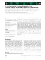
A post-mortem examination was obtained. There was loss of anterior horn cells, some
with ubiquitin positive inclusions, and associated astrocytosis of the anterior horns
throughout the entire length of the spinal cord. Lewy body-like (Figure 1) and skein-like
inclusions were present. The lateral and anterior columns showed extensive vacuolar
degeneration with macrophages and reactive gliosis. The posterior columns were
preserved. The corticospinal tracts of the medulla were mildly vacuolated. The motor
nuclei of the brainstem and cortex showed mild degenerative changes. There was no
evidence of inflammation anywhere throughout the neuraxis.
Sections of peripheral nerve revealed loss of nerve fiber with extensive axonal
degeneration and active Wallerian degeneration. There were a few CD26 positive B
lymphocytes thought to be a nonspecific finding or a reaction of extensive degeneration.
The muscle showed extensive atrophy with fiber type grouping and angulated fibers.
There was no muscle inflammation. The final diagnoses from the autopsy were spinal
cord showing extensive degeneration of anterior horn cells and tracts consistent with ALS
and axonal degeneration of peripheral nerve.
Discussion
When first seen, the patient had none of the requisite combination of upper and lower
motor neuron signs consistent with ALS [3,4]. Although he subsequently developed brisk
reflexes, at no time was there spread of the reflexes nor pathological reflexes such as
Babinski responses. The asymmetry of leg weakness would be consistent with ALS.
Asymmetric weakness, however, is also consistent with an acquired neuropathic process
with demyelinating features [8,9]. Fasciculations were present and are a characteristic
clinical finding in ALS. Fasciculations, however, are a non-specific finding related to
neuropathic dysfunction. Fasciculations are not considered of clinical significance for
ALS without associated evidence of neurogenic changes on needle EMG in the motor
units recorded as fasciculations [10]. Such motor unit abnormalities were not seen. This
patient had findings that could be consistent with a lower motor neuron variant of motor
system disease, but the sensory loss would argue for a polyneuropathy. Sensory loss
unless otherwise explained is considered inconsistent with a diagnosis of ALS [3].
More striking were the atypical electrodiagnostic findings for ALS. His electrodiagnostic
studies contained all of those features that have been reported as rare or not present at all
in ALS; namely, motor conduction velocities less than 70% of the lower limit of normal,
distal motor latencies greater than 125% of the upper limit of normal, and F-wave
latencies greater than 125% of the upper limit of normal [11]. Although a large number of
criteria sets have been proposed for defining an acquired demyelinating neuropathy
electrodiagnostically, none have really proven satisfactory [12]. Nevertheless, the pattern
of abnormalities in this patient would be compatible with an acquired neuropathy with
possible demyelinating features. Most striking were the asymmetries consistent with a
multifocal process. Large differences in motor conduction velocities between his legs
were observed on the initial electrodiagnostic examination, even allowing for differences
in evoked response amplitudes. Also, in the repeat study, there were large differences
between the median and ulnar nerves in the right arm, even allowing for the prolonged
median distal motor latency [6]. Multifocal acquired demyelinating sensory and motor
neuropathy (MADSAM; Lewis-Sumner syndrome) could be considered but characteristic
conduction block was not present. In addition, the rapid progression with ultimate death
in our patient would be inconsistent with previous reports of the natural history of
MADSAM [13].
Three patients have recently been described who had a polyneuropathy resembling CIDP
but were thought to have ALS [14]. Two patients had a family history of ALS. There was
no family history of ALS in our patient. The only post mortem examination was performed
on one of the patients with famalial ALS. The mean disease duration was 23 months with
the shortest being 13 and the longest 38, considerably longer than in our patient. There have
been two patients described in published manuscripts with multifocal motor neuropathy
(MMN) and pathological changes of ALS [cited in reference 14]. There was no evidence
for MMN in our patient including absence of conduction block and GM1 antibodies and
rapid course. Our patient is therefore arguably unique.
Electrodiagnostic studies in patients with possible ALS are critical for not only aiding in
the diagnosis but also helping to provide information that could be consistent with a
different, potentially treatable, diagnosis. A main consideration here would be a chronic
acquired demyelinating polyneuropathy [15]. Treatment of these neuropathies can be
costly and is not without risk. Studies such as electrodiagnosis that can provide
justification for such treatments are therefore important. Our patient was treated as if he
may have had a chronic acquired demyelinating neuropathy. Given the essentially
invariably fatal outcome of ALS, treatment for CIDP was probably justified.
Conclusions
This patient is a seemingly unique lesson on our limitations in making the distinction
between ALS and an acquired, treatable neuropathy. Sensitivity to these issues is
important given the serious nature of ALS and the resultant complexities in the
management of such patients.
Abbreviations
ADM, abductor digiti minimi; AH, abductor hallucis; AMP, amplitude; APB, abductor
pollicis brevis; CV, conduction velocity; DML, distal motor latency; EBD,extensor
digitorum brevis; NR, no response
Consent
Written informed consent was obtained from the patient’s next-of-kin for publication of
this case report and any accompanying images. A copy of the written consent is available
for review by the Editor-in-Chief of this journal.
Competing interests
The authors have no competing interests.
Authors’ contributions
All of the authors were involved in drafting and revising the manuscript and have given
final approval of the report. AV and MAF made substantial contributions to the
conception and design as well analysis and interpretation of data. MAF and YP made
substantial contributions to the acquisition of the data.
Acknowledgements
The authors would like to thank John M. Lee, MD, PhD for review of the pathological
material and for providing Figure 1.
References
1. Lee RJ, Annegers JF, Appel SH: Prognosis of amyotrophic lateral sclerosis
and the effect of referral selection. J Neurol Sci 1995, 132:207-215.
2. Miller RG, Jackson CE, Kasarskis EJ, .England JD, Forshew D, Johnston W,
Kalra S, Katz JS, Mitusmoto H, Rosenfeld, J, Shoesmith C, Strong MJ, Wooley SC:
Practice parameter update: The care of the patient with amyotrophic lateral
sclerosis: drug, nutritional, and respiratory therapies (an evidence-based review):
report of the Quality Standards Subcommittee of the American Academy of
Neurology. Neurology 2009, 73:1218-2126.
3. Brooks, BR: El Escorial World Federation of Neurology criteria for the
diagnosis of amyotrophic lateral sclerosis. Subcommittee on Motor Neuron
Diseases/Amyotrophic Lateral Sclerosis of the World Federation of Neurology
Research Group on Neuromuscular Diseases and the El Escorial “Clinical Limits of
Amyotrophic Lateral Sclerosis” workshop contributors. J Neurol Sci 1994, 124:96-107.
4. Brooks BR., Miller RG, Munsat TL: El Escorial revisited: revised criteria for the
diagnosis of amyotrophic lateral sclerosis. Amyotroph Lateral Scler Other Motor Neuron
Disord 2000, 1:293-299.
5. Berger AR, Bradley WG, Brannagan TH, Busis NA, Cros DP, Dalakas MC,
Danon MJ, Donofrio P, Engel WK, England JD, Feldman EL, Freeman RL, Kinsella LJ,
Lacomis D, Latov N, Menkes DL, Sander HW, Thomas FP, Triggs WJ, Windebank AJ,
Wolfe GI, The Neuropathy Association Medical Advisory Committee: Guidelines for the
diagnosis and treatment of chronic demyelinating polyneuropathy. J Peripher Nerv Syst
2003, 28:282-284.
6. Wilson JR. Park Y, Stittsworth JD, Fisher MA: Electrodiagnostic patterns in
MGUS neuropathy. Electromyogr Clin Neurophysiol 2001, 41:409-418.
7. Fisher MA: F response latency determination. Muscle Nerve 1982, 5:730-734.
8. De Sousa EA, Chin RL, Sander HW, Latov N, Brannigan III TH:
Demyelinating findings in typical and atypical chronic inflammatory
polyneuroppathy: sensitivity and specificity. J Clin Neuromuscul Dis 2009,
10:163-169.
9. Saperstein DS, Katz JS, Amato AA., and Barohn RJ: Clinical spectrum of chronic
acquired demyelinating polyneuropathies. Muscle Nerve 2001, 24:311-324.
10. Carvalho ME, Dengler R, Eisen A, England JD, Kaji R, Kimura J, Mills K.,
Mitsumoto H, Modera H, Shefner J, Swash J: Electrodiagnostic criteria for
diagnosis of ALS. Clin Neurophysiol 2008, 119:497-503.
11. Cornblath DR, Kuncl RW, Mellits ED, Quaskey SA, Clawson L, Pestronk A,
Drachman DB: Nerve conduction studies in amyotrophic lateral sclerosis.
Muscle Nerve 1992, 15:1111-1115.
12. Bromberg MB: Review of the evolution of electrodiagnostic criteria for
chronic inflammatory demyelinating polyradiculoneuropathy. Muscle Nerve 2011,
43:780-794.
13. Viala K, Renié L, Masionobe T, Béhin A, Neil J, Léger JM, Bouche P: Follow-up
study and response to treatmen in 23 patients with Lewis-Sumner syndrome. Brain
2004, 127:2010-2017.
14. Echantz-Laguna A, Degos B, Mohr M, Kessler R, Urban-Kraemer E, and Tranchnat
C: A study of three patients with amyotrophic lateral sclerosis and polyneuropathy
resembling CIDP. Muscle Nerve 2006, 33:356-362.
15. Brannagan, TH3
rd
: Current treatments of chronic-immune-mediated
demyelinating polyneuropathies. Muscle Nerve, 2009, 39:563-578.
Table 1. Electrodiagnostic Studies
Initial Repeat
Right Left Right
Left
Study Nerve
DML
(ms)
AMP
(mV)
CV
(m/sec)
DML
(ms)
AMP
(mV)
CV
(m/sec)
DML
(ms)
AMP
(mV)
CV
(m/sec)
Tibial (AH)
6.6 0.4 26 7.4 1 37 NR NR NR
Peroneal (EDB)
7.9 0.1 29 7.9 12 37 NR NR NR
Median (APB)
4.4 7 51
6.4 0.7 29.6
Motor
Ulnar (ADM)
3.4 8 56
3.5 2.4 47
Sural
21.2
31
Median(dig 2)
13 53
3.6 44
Median(dig 3)
9.4
49
Sensory
Ulnar
8.7
46
3.0 42
Mean F-wave latency (ms) Mean F-wave latency
(ms)
Tibial(soleus)
NR
F-Wave
Median (APB)
33 (predicted 28.9)
41.4 (predicted 28.9)
Normal values: DML – peroneal <6.1ms; tibial <6.4ms; median <4.5ms; ulnar <3.4ms.
MNCV (amplitudes) – tibial >39m/sec (>4mV); peroneal >39m/sec (>3mV); median
>49m/sec (>4mV); ulnar >49m/sec (>4mV). SNAP conduction velocities (amplitudes) –
sural >39m/sec (>6µ); median >49m/sec (10µV); ulnar >49m/sec (>3µV). Abnormal
values in bold.
Figure legend
Figure 1. Ubiquitin stain of the anterior horn neurons with Lewy body-like
inclusions. High powered field.
Figure 1