báo cáo khoa học: "Serum proteomic profiling and haptoglobin polymorphisms in patients with GVHD after allogeneic hematopoietic cell transplantation" pps
Bạn đang xem bản rút gọn của tài liệu. Xem và tải ngay bản đầy đủ của tài liệu tại đây (942.93 KB, 12 trang )
BioMed Central
Page 1 of 12
(page number not for citation purposes)
Journal of Hematology & Oncology
Open Access
Research
Serum proteomic profiling and haptoglobin polymorphisms in
patients with GVHD after allogeneic hematopoietic cell
transplantation
Joseph McGuirk
1,5
, Gang Hao
3
, Weijian Hou
1
, Sunil Abhyankar
1
,
Casey Williams
1
, Weisi Yan
3
, Jianda Yuan
4
, Xiuqin Guan
2
, Robert Belt
1
,
Shaun Dejarnette
1
, Jeffery Wieman*
1
and Ying Yan*
1,2
Address:
1
Blood and Marrow Transplantation Program, Saint Luke's Cancer Institute, Kansas City, Missouri, USA,
2
Department of Medicine,
School of Medicine, University Missouri-Kansas City, Kansas City, Missouri, USA,
3
Department of Pharmacology, Weill Medical College of Cornell
University, New York, New York, USA,
4
Laboratory of Cellular Immunobiology, Memorial Sloan-Kettering Cancer Center, New York, New York,
USA and
5
Blood and Marrow Transplant Program, The University of Kansas Hospital Cancer Center, 2330 Shawnee Mission Parkway, Westwood,
Kansas 66205, USA
Email: Joseph McGuirk - ; Gang Hao - ; Weijian Hou - ;
Sunil Abhyankar - ; Casey Williams - ; Weisi Yan - ;
Jianda Yuan - ; Xiuqin Guan - ; Robert Belt - ;
Shaun Dejarnette - ; Jeffery Wieman* - ; Ying Yan* -
* Corresponding authors
Abstract
We studied serum proteomic profiling in patients with graft versus host disease (GVHD) after
allogeneic hematopoietic cell transplantation (allo-HCT) by two-dimensional gel electrophoresis
(2-DE) and mass spectrometry analysis. The expression of a group of proteins, haptoglobin (Hp),
alpha-1-antitrypsin, apolipoprotein A-IV, serum paraoxonase and Zn-alpha-glycoprotein were
increased and the proteins, clusterin precursor, alpha-2-macroglobulin, serum amyloid protein
precursor, sex hormone-binding globulin, serotransferrin and complement C4 were decreased in
patients with extensive chronic GVHD (cGVHD). Serum haptoglobin (Hp) levels in patients with
cGVHD were demonstrated to be statistically higher than in patients without cGVHD and normal
controls (p < 0.01). We used immunoblotting and PCR in combination with 2-DE gel image analysis
to determine Hp polymorphisms in 25 allo-HCT patients and 16 normal donors. The results
demonstrate that patients with cGVHD had a higher incidence of HP 2-2 phenotype (43.8%), in
comparison to the patients without cGVHD (0%) and normal donors (18.7%), suggesting the
possibility that specific Hp polymorphism may play a role in the development of cGVHD after allo-
HCT. In this study, quantitative serum Hp levels were shown to be related to cGVHD
development. Further, the data suggest the possibility that specific Hp polymorphisms may be
associated with cGVHD development and warrant further investigation.
Published: 20 April 2009
Journal of Hematology & Oncology 2009, 2:17 doi:10.1186/1756-8722-2-17
Received: 9 February 2009
Accepted: 20 April 2009
This article is available from: />© 2009 McGuirk et al; licensee BioMed Central Ltd.
This is an Open Access article distributed under the terms of the Creative Commons Attribution License ( />),
which permits unrestricted use, distribution, and reproduction in any medium, provided the original work is properly cited.
Journal of Hematology & Oncology 2009, 2:17 />Page 2 of 12
(page number not for citation purposes)
Introduction
Allogeneic hematopoietic cell transplantation (Allo-HCT)
has been a potentially curative treatment approach for
patients with hematological malignancies, lympho-
hematopoietic failure, autoimmune diseases as well as
genetic disorders. Despite its curative potential, the appli-
cation of allo-HCT is limited by life-threatening complica-
tions, in particular, graft-versus-host disease (GVHD), a
highly morbid toxic complication [1,2]. As a clinical syn-
drome related to the reaction of donor-derived immuno-
competent cells against patient tissues, GVHD remains the
most frequent transplant-related complication.
GVHD is classified as a clinicopathologic syndrome
involving skin, liver, gastrointestinal tract, and/or other
organs. Currently, there are no reliable laboratory tests
that will confirm or refute its presence. Thus, GVHD is
mostly a clinical diagnosis. Diagnosis of GVHD requires
an interpretation of clinical and laboratory findings, rec-
ognizing that in some patients the differential diagnosis
may be difficult to resolve [3]. To predict development
and clinical prognosis of GVHD, several in vitro tests have
been described. However, results have been difficult to
reproduce and no assay has been widely adopted [3-7].
Studies of certain cytokine gene polymorphisms, includ-
Table 1: Clinical characteristics of the allo-HSCT patients
Sex/Age Diagnosis GVHD Grade Organ
involvement
Conditioning
Regimen
GVHD
Prophylaxis
Infections Outcome
1.* F/40 AML Chronic Extensive Skin eye oral Cy/TBI/ATG Tac/MTX None Alive, no active
GVHD
2.* M/28 CML Chronic Extensive Liver Bu/Cy Tac/MTX None Alive, no active
GVHD
3. M/48 AML Chronic Extensive Skin eyes oral gut Bu/Cy Tac/MTX None Alive, active GVHD
4.* F/42 AML Chronic Extensive Skin eye oral Cy/TBI Tac/MTX None Dead, AML relapse
5.* F/42 IMF Chronic Extensive Skin oral eye
liver
Bu/Cy Tac/MTX None Alive, active GVHD
6. M/49 CLL Chronic Limited Skin eye oral Cy/TBI Tac/MTX None Dead, GVHD
7.* F/29 CML Chronic Extensive Skin Gut Cy/TBI CSA/MTX CMV Dead, CMV
pneumonia
8.* F/58 AML Chronic Extensive Skin Gut Cy/TBI CSA/MTX None Dead, unknown
causes
9.* F/45 AML Chronic Extensive Skin Cy/TBI CSA/MTX CMV Dead, CMV/organ
failure
10.* M/38 CML Chronic Extensive Eye oral gut Bu/Cy Tac/MTX None Alive, active GVHD
11.* F/54 SAA Chronic Limited Skin, oral Cy/TBI/ATG Tac/MTX None Alive, no active
GVHD
12.* M/54 NHL Chronic Extensive Skin eye Cy/TBI Tac/MTX None Alive, active GVHD
13. M/54 NHL Chronic Limited Oral BEAC Tac/MTX None Alive, no active
GVHD
14.* M/50 NHL Chronic Extensive Skin, lung Cy/TBI/ATG Tac/MTX None Dead, GVHD
15. M/59 AML Chronic Limited Skin Gut Bu/Cy Tac/MTX CMV Alive, active GVHD
16. M/52 NHL Chronic Extensive Skin Gut Oral Cy/TBI/ATG Tac/MTX CMV Alive, active GVHD
17. M/29 CML No GVHD - - Cy/TBI Tac/MTX None Alive, no active
GVHD
18. M/61 CLL No GVHD - - Bu/Flu/ATG Tac/MTX None Alive, no active
GVHD
19.* F/40 AML No GVHD - - Cy/TBI/ATG Tac/MTX CMV, EBV Alive, no active
GVHD
20. F/58 MDS No GVHD - - Cy/TBI Tac/MTX None Dead, AML/MDS
relapsed
21.* M/38 AML No GVHD - - Cy/TBI/ATG Tac/MTX None Dead, relapsed
22. M/23 AML No GVHD - - Cy/TBI/ATG Tac/MTX None Alive, no active
GVHD
23.* F/49 AML No GVHD - - Cy/TBI/ATG Tac/MTX None Alive, no active
GVHD
24.* M/31 CML No GVHD - - Bu/Cy/ATG Tac/MTX None Alive, no active
GVHD
25.* F/54 NHL No GVHD - - Cy/TBI/ATG Tac/MTX None Dead, heart failure
AML: acute myelogenous leukemia; CML: chronic myelogenous leukemia; IMF: idiopathic myelofibrosis; CLL: chronic lymphocyte leukemia: SAA:
severe aplastic anemia; NHL: Non-Hodgkin's Lymphoma; ALL: acute lymphoblastic leukemia; MM: multiple myeloma; MDS: myelodysplastic
syndrome; * done of 2-DE gel assay.
Journal of Hematology & Oncology 2009, 2:17 />Page 3 of 12
(page number not for citation purposes)
ing tumor necrosis factor alpha, interferon gamma, inter-
leukin-1 (IL-1), IL-6 and IL-10, as well as polymorphisms
of certain adhesive molecules such as CD31 and CD54
have been extensively conducted to explore their potential
for GVHD risk prediction and the development of predict-
able genetic risk indexes. However, these efforts have not
yet resulted in reliable models [3,8-13].
Over the past decade, the study of proteomics has rapidly
evolved and developed. Proteomics studies can generate
protein expression profiles which may predict clinical
events, therapeutic response, or probe underlying mecha-
nisms of disease. Proteome analysis is emerging as an
important technology for understanding biological proc-
esses and discovery of novel biomarkers in diseases such
as autoimmune disorders, cardiovascular diseases and
cancers [14-17]. A recent study used an intact-protein-
based quantitative analysis system for determining the
plasma proteome profile of patients with acute GVHD
after transplant. The proteins, including amyloid A, apol-
ipoproteins A-I/A-IV and complement C3 were found to
be quantitatively different between the pre- and post-
GVHD samples [18]. In another report, several differen-
tially excreted polypeptides were identified from patient
urine samples by a capillary electrophoresis and mass
spectrometry (CE-MS) based technique. The peptide pro-
file displayed a pattern of early GVHD markers, allowing
discrimination of GVHD from patients without the com-
plication [19]. These reports hinted that GVHD can be
monitored by changes in protein expression patterns
detectable through proteomic methods.
Few investigations utilizing proteomic profiling in the
study of patients with and without GVHD after allo-HCT
have been reported to date. Several contributions in this
regard have recently been reported to be confirmatory of
a clinical diagnosis of acute GVHD (aGVHD) and to pro-
vide prognostic information. Paczesny et al have devel-
oped a panel consistent of 4 biomarkers which both
confirm the diagnosis of aGVHD at onset of clinical symp-
toms and provide prognostic information independent of
aGVHD severity [20]. Weissinger, et al have described an
aGVHD-specific model consisting of 31 polypeptides and
Hori et al have correlated a member of a large chemokine
family, CCL8 to be closely correlated with aGVHD sever-
ity through proteomic analysis [21,22].
In this study, we performed serum proteomic profiling in
a group of patients with and without cGVHD after allo-
HCT by 2-dimensional electrophoreses (2-DE) and mass
spectrometry based technology. Differential expression
patterns of 11 serum proteins were demonstrated in
patients before and after cGVHD development. Serum Hp
precursors, one of the 11 differentially expressed serum
proteins, were found to be significantly up-regulated dur-
ing cGVHD development. We also investigated the rela-
tionship between serum Hp quantity as well as Hp
polymorphisms and cGVHD development in this study.
Serum Hp level as well as its polymorphisms were shown
to be related to cGVHD development. Thus, Hp might
serve as a worthy future target for monitoring cGVHD and
understanding cGVHD mechanism.
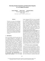
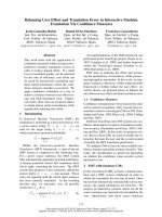
A paired 2-DE gel images from a patient with cGVHDFigure 1
A paired 2-DE gel images from a patient with cGVHD. Gel a). represents the protein 2-DE gel profile of the serum
from the patient before cGVHD development. Gel b). represents the protein 2-DE profile of the serum derived from the
patient with cGVHD. The protein spots labeled with numbers were collected, digested and analyzed by Mass spectrometry.
pH4
pH4 pH7pH7
15 -
20 -
25 -
37 -
50 -
75 -
100 -
150 -
250 -
491
488
489
513
522
528
550
552
532
585
661
663
664
726
726
537
492
547
416
526
556
a
b
Journal of Hematology & Oncology 2009, 2:17 />Page 4 of 12
(page number not for citation purposes)
Methods
Patients
Twenty-five patients who received allo-HCT at Saint
Luke's Cancer Institute were studied. The 25 patients
included 14 males and 11 females and the median age
was 48 years (range 23–61 year old). Details of diagnostic
indication for transplant are delineated in table 1. Sixteen
patients developed cGVHD and 9 patients developed no
cGVHD after allo-HCT. Samples were collected prior to
transplant from each patient and at approximately 20 and
150 days, 6 months and 1 year or at the time of initial
diagnosis of cGVHD (before initiation of steroid based
therapy) in the BMT clinic and then periodically during
follow up visits in patients with active cGVHD. Initial
therapy of cGVHD included tacrolimus continuation or
re-initiation and prednisone at 1 mg/kg daily. None of the
25 patients had a clinical diagnosis of transplant associ-
ated microangiopathy.
Sixteen normal healthy donors, 10 males and 6 females,
were included as controls in this study. Median age of the
normal donors was 38 years (range 20–55).
Serum processing
Peripheral blood samples were obtained, with informed
consent, during routine diagnostic blood studies from
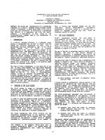
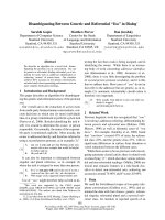
Identification of haptoglobin by LC-MS/MS analysis and database searchingFigure 2
Identification of haptoglobin by LC-MS/MS analysis and database searching. Proteins were excised from the corre-
sponding gel spots and subjected to in-gel digestion. The resulting peptides were extracted from the gel and analyzed by LC-
MS/MS. MS/MS data were searched against the human database by Spectrum Mill software to obtain protein identification infor-
mation. Upper panel: the sequence coverage of haptoglobin by LC-MS/MS analysis. Ten tryptic peptides (sequence underlined)
were matched to human haptoglobin by database searching. Lower panel: MS/MS spectra of two of the ten peptides: DIAPTLT-
LYVGK and VVLHPNYSQVDIGLIK. The peptide sequences were established by extensive b and y ions matched to sequence.
MSALGAVIAL LLWGQLFAVD SGNDVTDIAD DGCPKPPEIA HGYVEHSVRY QCKNYYKLRT EGDGVYTLND KKQWINKAVG
DKLPECEADD GCPKPPEIAH GYVEHSVRYQ CKNYYKLR
TE GDGVYTLNNE KQWINKAVGD KLPECEAVCG KPKNPANPVQ
RILGGHLDAK GSFPWQAKMV SHHNLTTGAT LINEQWLLTT AKNLFLNHSE NATAKDIAPT LTLYVGKKQL VEIEKVVLHP
NYSQVDIGLI KLKQKVSVNE RVMPICLPSK DYAEVGRVGY VSGWGRNANF KFTDHLKYVM LPVADQDQCI RHYEGSTVPE
KKTPKSPVGV QPILNEHTFC AGMSKYQEDT CYGDAGSAFA VHDLEEDTWY ATGILSFDKS CAVAEYGVYV KVTSIQDWVQ
KTIAEN
0
1
2
3
4
7
x10
Intens.
2.5 5.0 7.5 10.0 12.5 15.0 17.5 20.0 22.5 25.0
Time [min]
y7
++
b8
++
b9
++
y5
b10
++
b11
++
y6
y7
y14
++
b15
++
y8 y9
b9
0.0
0.5
1.0
1.5
2.0
4
x10
Intens.
0.0
0.2
0.4
0.6
0.8
1.0
1.2
6
x10
200 400 600 800 1000 1200
Mass (m/z)
y12
b9
-H2O
++
y4
++
b12
++
b14
++
b10
+++
VVLHPNYSQVDIGLIK
y9
y10
b10
y9
++
y10
++
b3
b2
y4
y6
DIAPTLTLYVGK
y10
-H2O
++
y8b8y7y5 b6
Journal of Hematology & Oncology 2009, 2:17 />Page 5 of 12
(page number not for citation purposes)
patients before and after allo-HSCT in Saint Luke's Cancer
Institute (SLCI). Serum samples were collected and aliq-
uoted into 300 μl per tube from whole blood specimens,
allowed to stand over night at 4°C without anti-coagulant
and stored at -80°C in a freezer. Mononuclear cells
(MNC) were isolated by Ficoll Hypaque density gradient
separation and cyropreserved in liquid nitrogen. Serum
albumin was removed by Swellgel Blue Albumin Removal
kit (Pierce biotechnology, Rockford IL) following the
instructions provided by the kit. 150 ul of serum was
loaded for each single reaction. After preparing resin disc,
binding sample and washing to release albumin-free sam-
ple, the albumin-free serum was collected for determina-
tion of protein concentration. The protein concentration
was determined by the D
c
Protein Assay kit (Bio-Rad, Her-
cules CA) and following the instructions provided by the
kit.
2-Dimensional protein gel electrophoresis
2-DE was performed as previously described with certain
modifications [23]. Briefly, 500 μg of serum protein resus-
pended in rehydration buffer (8 M urea, 2% CHAPS, 0.5%
immobilized pH gradient [IPG] buffer, 1% DTT, and trace
of bromophenol blue) was loaded into an immobiline
DryStrip (pI 4–7, 13 cm) (Amersham Biosciences) for
rehydration over 18 hr. The first dimension isoelectric
focusing was performed for 46,000 Vhr using a multiPhor
II IEF System (Amersham) at 20°C. Then, the gels were
equilibrated for 30 minutes in equilibration buffer I (50
mM Tris-HCL [pH 8.8], 6 M urea, 30% glycerol, 2% SDS,
and 0.1% DTT) and buffer II (50 mM Tris-HCl [pH 8.8], 6
M urea, 30% glycerol, 2% SDS, and 0.25% iodoaceta-
mide). The second dimension electrophoresis was con-
ducted according to the Hoefer SE 600 system operating
manual (Amersham). A gradient SDS-polyacrylamide gel
(7%–12%) was used for the second dimension gel electro-
phoresis. The IPG strips were placed on the surface of the
second dimension gel, and then the IPG strips were sealed
with 0.5% agarose in SDS electrophoresis buffer (25 mM
Tris base, 192 mM glycine, 0.1% SDS). The gels were run
over 4 hrs at 110 V.
Silver staining
Silver staining was performed according to a protocol
published previously [23]. Briefly, gels were fixed with
50% methanol/10% acetic acid for 30 minutes and 5%
methanol/1% acetic acid for 15 minutes respectively, and
then the gels were washed by distilled water 3 times for 10
Table 2: Identity of proteins in 2-DE gel with increased or decreased intensity after onset of cGVHD
Accession No. Spot No. MW (KDa) pl Identified protein (VI)/× 10
5
*(VI) Fold change
P00738 489 46 4.8 haptoglobin-β 53.4 +4.2
513 46 5.1 haptoglobin-β 47.1 +7.3
522 45 5.2 haptoglobin-β 22.4 +6.5
528 45 5.4 haptoglobin-β 6.5 +8.4
552 40 5.6 haptoglobin-β newly appeared
661 21 5.6 haptoglobin-α 22.8 +37.1
663 21 5.9 haptoglobin-α 12.3 +32.0
664 21 5.3 haptoglobin-α 7.4 +35.2
P06727 488 47 4.8 apolipoprotein A-IV 60.2 +25.5
489 46 4.8 apolipoprotein A-IV 53.4 +4.2
491 48 4.7 apolipoprotein A-IV 21.0 +15.0
P01009 489 46 4.8 α-1-antitrypsin 53.4 +4.2
513 46 5.1 α-1-antitrypsin 47.1 +7.3
528 45 5.4 α-1-antitrypsin 16.5 +8.4
P27169 488 47 4.8 serum paraoxonase 60.2 +25.5
491 48 4.7 serum paraoxonase 21.0 +15.0
P25311 488 47 4.8 Zn-α-2-glycoprotein 50.2 +25.5
491 48 4.7 Zn-α-2-glycoprotein 21.0 +15.5
P04278 492 48 5.1 sex hormone-binding globulin 1.2 -5.1
P10909 537 39 4.8 clusterin precursor 0.6 -4.5
547 38 4.9 clusterin precursor 0.5 -4.9
556 36 4.8 clusterin precursor absence
726 13 5.6 clusterin precursor 0.1 -4.5
P02735 726 13 5.6 serum amyloid A protein precursor 0.1 -4.5
P02787 416 29 6.6 serotransferrin absence
P01023 556 36 4.8 α-2-microglobulin 0.2 -7.5
P01028 526 31 6.6 complement C4 absence
Acession #: Number as listed in online protein database; VI: volume index
Journal of Hematology & Oncology 2009, 2:17 />Page 6 of 12
(page number not for citation purposes)
minutes each time. After washing, the gels were sensitized
by incubation in sensitizing solution (0.02% sodium thi-
osulphate) for 90 seconds, and then rinsed with distilled
water 3 times for 30 seconds each time. After rinsing, the
gels were incubated in 0.2% silver nitrate for 30 minutes.
The silver nitrate was then discarded and the gels were
rinsed with distilled water 3 times for 1 minute each time
and then developed with 0.02% formaldehyde and
0.0004% sodium thiosulphate in 6% sodium carbonate
with shaking. The development was terminated with 6%
acetic acid.
Image analysis
The silver-stained 2-DE gels were scanned with LabScan
software on an UMAX Powerlook III scanner (UMAX Tech
Inc, California), and the images were digitalized and ana-
lyzed with a α-GelFox 2D 3.1 (Alpha Innotech) software.
In-gel digestion and protein identification by LC-MS/MS
Protein spots were cut out from the silver-stained gels for
in-gel digestion. Proteins were reduced and alkylated
before digestion with trypsin (Promega, Madison, WI)
overnight at 37°C. The peptides were extracted from the
gel and concentrated in a vacuum centrifuge. 8 μL of con-
centrated peptide mixtures was injected to an Agilent LC-
MSD ion trap mass spectrometer (Agilent Technologies,
USA) for identification. Mass spectra were acquired in
positive-ion mode with automated data-dependent MS/
MS on the four most intense ions from precursor MS
scans. The mass spectra were extracted and searched
against the human database using Mascot software.
Serum haptoglobin determination by Elisa Assay
Serum Hp determination was use AssayMax human Hp
ELISA kit and followed the Elisa kit protocol provided by
manufacturer (Assaypro St. Charles MO). Pooled human
normal serum control (PNS), which contains serum
derived from 20 normal donors, was purchased from
George King Bio-Medical. INC. (Overland Park, KS).
Haptoglobin genotype determination by PCR
Genomic DNA was extracted from peripheral blood MNC
by the QIAamp DNA Kit as suggested by the supplier
(Qiagen). Oligonucleotide primers A (5'-GAG-
GGGAGCTTGCCTTTCCATTG-3') and B (5'-GAGATTTTT-
GAGCCCTGGCTGGT-3') were used for amplification of a
1757-bp Hp-1 allele-specific sequence and a 3481-bp Hp-
2 allele-specific sequence. Primers C (5'-CCTGCCTCG-
TATTAACTGCACCAT-3') and D (5'-CCGAGTGCTCCA-
CATAGCC ATGT-3') were used to amplify a 349-bp Hp-2
allele-specific sequence [24]. The oligonucleotide primers
were synthesized by IDT, Inc (Coralville IA). 20-μL reac-
tions contained 2 U of Taq polymerase (Promega), 1–100
ng of DNA, and 200 μM each of dATP, dCTP, dGTP and
dTTP (Promega); PCR buffer was used as suggested by the
supplier (Promega) with no supplements added. After ini-
tial denaturation at 95°C for 2 min, the two-step thermo-
cycling procedure consisted of denaturation at 95°C for 1
min and annealing and extension at 69°C for 2 min (in
the presence of primers A and B or primers A, B, C, and D)
or 1 min (in the presence of primers C and D only),
repeated for 35 cycles, and followed by a final extension
at 72°C for 7 min. The thermocycler used was Perkin
Elmer 480 PCR system. For genotype assignments, the
PCR products where primers A and B were used were sep-
arated in 1% agarose gels and products where primers C
and D were used were separated in 8% polyacrylamide
gels.
Restriction enzyme analysis was performed to verify the
identity of Hp-1- and Hp-2-specific PCR products. The
1757-and 3481-bp products were digested with restriction
enzyme MlsI, and the 349-bp product was digested with
DraI, as recommended by the supplier (MBI Fermentas).
DNA fragments were separated by gel electrophoresis.
Immunoblot
Immunoblot were performed as previously published
with modifications [25]. Briefly, 1 μL of human serum in
20 μL of sample loading buffer [10 g/L sodium dodecyl
sulfate (SDS), 100 mL/L glycerol, 25 mmol/L Tris (pH
6.8), 0.05 g/L bromphenol blue, and 50 mL/L β-mercap-
toethanol] mixture were boiled at 95°C for 5 min, then
the boiled samples were loaded on a 15% polyacrylamide
gel. Standard Hp protein (Sigma Chemical Co.) was
diluted to 1 g/L and treated in the same way as a control.
Samples were electrophoresed in 25 mmol/L Tris base-
192 mmol/L glycine-1 g/L SDS running buffer for 45 min
at 150 V and then transferred to PVDF membranes (Bio
Rad, California). The membranes were blocked in 5% Dry
milk in Tris-buffered Tween [TBST; 10 mmol/L Tris-HCl
(pH 8.0), 150 mmol/L NaCl, 0.5 mL/L Tween 20] for 1 h
Table 3: Comparison of volume index (VI) of Hp spots in 2-DE between patients with and without cGVHD
Groups Protein spot volume index (VI)/× 10
5
(Mean ± SD) p- value
cGVHD Patients (n = 6) 391.15 ± 305.38
Before allo-HCT (n = 12) 100.02 ± 108.89 0.03
After allo-HCT without GVHD (n = 4) 146.49 ± 139.44 0.045
Normal donors (n = 9) 136.71 ± 94.50 0.044
Journal of Hematology & Oncology 2009, 2:17 />Page 7 of 12
(page number not for citation purposes)
and then incubated at 4°C overnight with a 1:1000 dilu-
tion of polyclonal rabbit anti-human Hp antibody
(Sigma). After washing the membranes three times in
TBST, a second antibody, anti-rabbit IgG horse radish per-
oxidase conjugate (Santa Crus, California) was used at a
dilution 1:2000 in TBST; the membranes were then incu-
bated at room temperature for 1 h. The membranes were
washed three times in TBST and finally developed with
Luminal Reagent (Santa Crus).
Statistical analysis
Statistical comparison of the serum Hp levels and 2-DE
gel Hp protein spots VI among the different study groups
was done using the Waller-Duncan K-ratio t test.
Results
Changes of protein expression patterns before and after
cGVHD development
Thirty-six serum specimens derived from 16 patients and
9 samples from normal donors were subjected to 2-DE gel
and silver staining assays. Out of the 36 patient-derived
samples, 13 were collected before transplantation. Out of
the 23 patient derived samples collected after allo-HCT, 9
samples were collected prior to the cGVHD occurring and
9 during cGVHD development, 5 were from the patients
with no cGVHD development at all after receiving allo-
HCT. The median number of protein spots in 2-DE gels
was 709 (range 499–1012 spots) for the specimens from
patients pre-transplantation; 637 (458–806 spots) in
samples from prior to or no cGVHD development
patients; 760 (508–1031 spots) in the serum from
patients with active cGVHD and 735 (645–783 spots) in
the serum of normal donors.
Paired 2-DE gel analyses were performed using 2-DE gel
software between the serum collected prior to cGVHD and
the samples of active cGVHD phase from the same indi-
vidual patient. Protein spot patterns were significantly dif-
ferent between the gels of pre-cGVHD and active cGVHD
phase in the same patient. The median protein spot num-
bers were determined as 753 (range 610–1012) and 726
(508–1031) for the specimens pre and post-cGVHD in the
7 patients with cGVHD, respectively. Median of matched
spots between paired gels was 501 (405–725, median and
range), however, in comparison to the gels pre-cGVHD,
the medians for missing spots and newly appeared spots
were 248 (190–391) and 276 (117–373), respectively in
the gels of cGVHD.
A group of protein spots in the 2-DE gels was found signif-
icantly and consistently different between the serum col-
lected prior to and during the cGVHD development from
various patients. The protein spots, which were collected
and analyzed from a paired representative 2-DE gels by
LC-MS/MS are demonstrated in Figure 1. The sum of the
spot areas multiple spot density as the volume index (VI)
was used to determine the individual serum protein level
in 2-DE gel semi-quantitatively. Five proteins, including
Hp, apolipoprotein A-IV, α-1-antitrypsin, serum paraoxo-
nase and Zn-α-glycoprotein were found quantitatively
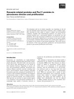
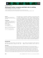
Comparison of serum Hp level in patient with and without GVHD after allo-HCT by Elisa assayFigure 4
Comparison of serum Hp level in patient with and
without GVHD after allo-HCT by Elisa assay. Serum
Hp level in GVHD group is significantly higher than all the
other 3 groups (p < 0.01)
0
1
2
3
4
012345
Hp mg/ml
Normal donors
(n=16 + 5 PNS)
Patients before
transplantation
(n=23)
Patients after
transplantation
GVHDí
(n=9)
Patients after
transplantation
GVHD+
(n=14)
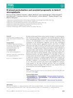
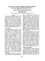
Expression patterns of Hp in paired 2-DE gels in individual patients before and after GVHDFigure 3
Expression patterns of Hp in paired 2-DE gels in indi-
vidual patients before and after GVHD. The left panel
lists the Hp 2-DE gel spots derived from patients before
GVHD occurred and the right side panel represents the 2-
DE gel spots after GVHD development.
Before GVHD
After GVHD
Hp ȕ
Hp Į-1s
a).
b).
Hp ȕ
Hp Į-2
Hp Į-1s
Hp ȕ
Hp Į-2
c).
Journal of Hematology & Oncology 2009, 2:17 />Page 8 of 12
(page number not for citation purposes)
increased or newly appearing after cGVHD development.
A group of 5 proteins were either down-regulated or
absent in the 2-DE gel of patient with cGVHD, including
sex hormone-binding globulin, clusterin precursor, serum
amyloid A protein precursor, serotransferrin and comple-
ment C4 (Table 2). As a representative of protein spots
analyzed, the mass spectrum identification of Hp by LC-
MS/MS analysis is shown in Figure 2.
Differential expression patterns of haptoglobin in cGVHD
development
As a particular example, Hp expression patterns were
demonstrated to be significantly different among the
patients with and without cGVHD development in our 2-
DE gel analysis. To quantitatively compare the differences
in Hp spot volumes between the different study groups by
2-DE gel image analyses, we used VI to determine the
serum Hp level semi-quantitatively. Since the complexity
of Hp α chain polymorphisms and that the Hp β chain are
identical in all haptoglobin phenotypes, we selected the
VI of Hp precursor β as the representative of Hp volumes
in each study groups. The VIs in the cGVHD group (n = 6),
was demonstrated to be significantly higher than the VIs
of the patients before transplantation (n = 12; p = 0.027);
patients with no cGVHD after transplantation (n = 4; p =
0.045) and normal donor group (n = 9; p = 0.044), respec-
tively (Table 3).
The differential expression patterns of Hp in individual
patients in paired 2-DE gels before and after cGVHD are
demonstrated in Figure 3. Both volume and density of the
Hp protein spots were shown to increase in the gels of
cGVHD paired-set in all 3 patients irrespective of Hp phe-
notype differences. Figure 3a is from a cGVHD patient,
who has an Hp β and an Hp α-1s chain, indicating an Hp
1-1 phenotype. The patient's Hp spot VI was 24.4 × 10
5
(Hp β) and 0.1 × 10
5
(Hp α-1s) before cGVHD and 295.1
× 10
5
(Hp β) and 29.4 × 10
5
(Hp α-1s) after cGVHD,
respectively; 3b shows the 2-DE Hp pattern of a different
cGVHD patient, who has Hp β, α-1s and α-2 chain which
indicates a Hp 2-1 phenotype. The Hp spot VI was 91.0 ×
10
5
(Hp β), 3.5 × 10
5
(α-1s) and 12.2 × 10
5
(α-2) before
cGVHD and 140.9 × 10
5
(β), 4.8 × 10
5
(α-1s) and 18.7 ×
10
5
(α-2) after cGVHD, respectively; Figure 3c represent
the Hp expression patterns from another cGVHD patient.
The patient expressed an Hp β and α-2 protein spots, sug-
gesting an Hp 2-2 phenotype. The VI of 3c was 5.4 × 10
5
(Hp β) and 2.7 × 10
5
(α-2) before cGVHD and 274.9 × 10
5
(Hp β) and 67.7 × 10
5
(α-2) after cGVHD.
Serum Hp levels in the patients before and after cGVHD
development
We performed Elisa assays to confirm the prior finding
and compared serum Hp levels in patients before and
after cGVHD. Hp levels were examined in the serum from
the same patient before and after cGVHD development, as
well as normal donors. The mean of Hp concentration
was 1.97 ± 0.99 mg/ml (mean ± SD) in the patients with
cGVHD (n = 14); 0.83 ± 0.40 in the patients before trans-
plantation (n = 23); 0.74 ± 0.51 in the patients with no
Serum Hp phenotype determination of patients by immuoblotFigure 5
Serum Hp phenotype determination of patients by immuoblot. Polyclonal antibody against human Hp was used to
binding the Hp on the blots. Lane 1 is the Hp protein standard containing Hp β, α-1 and α-2 chains. Lane 2–25 shows the
serum Hp phenotype from patient 1 to patient 24 listed in Table 1. Patients who have Hp β and α-1 chains indicate a Hp 1-1
type; patients with Hp β and α-2 chains are Hp 2-2 type and patients with all the Hp β, α-1 and α-2 chains indicate a Hp 2-1
type.
Hp ȕ chain
1-1 2-1
2-2
1-1
2-2
Hp Į-2 chain
Hp Į-1 chain
2-1
2-11-1
2-1
123456 7 8910111213141516171819202122232425
Journal of Hematology & Oncology 2009, 2:17 />Page 9 of 12
(page number not for citation purposes)
cGVHD development after transplantation (n = 6) and
0.82 ± 0.31 mg/ml in the control group of normal donors
(n = 16 and 5 PNS). Statistical analysis demonstrated that
the serum Hp level in the cGVHD group was significantly
higher than all the other 3 groups (p < 0.01). The differ-
ences of the serum Hp level were insignificant between
the 3 cGVHD-negative groups (Figure 4).
Determination of Hp polymorphisms in the patients
In humans, Hp is characterized by a molecular heteroge-
neity with three main genotypes/phenotypes: Hp 1-1, Hp
2-1, and Hp 2-2. These different proteins have distinctive
efficiencies and it has been suggested that the polymor-
phism may have important biological consequences in
several diseases [26]. To probe the possible relationship
between cGVHD and Hp polymorphism, we used immu-
noblot and PCR in combination with 2-DE gel image
analysis to determine Hp polymorphisms in the 24 allo-
HCT patients of this study group as well as 12 normal
donors. Serum Hp phenotype was determined by immu-
noblot using polyclonal antibody, which recognize Hp β,
α-1 and α-2 chains and the Hp phenotypes in the patients
are shown in Figure 5. To verify the Hp phenotype in these
patients, we examined Hp genotypes of the patients by
PCR assay (figure 6), and the Hp PCR products were fur-
ther confirmed by restriction enzyme analysis (figure 7
and 8). All the DNA typing of the samples derived from 25
patients were matched with their phenotypes determined
by immunoblot.
Out of 16 patients with cGVHD, 2 (12.5%, 1 limited and
1 extensive) had an Hp 1-1 type; 7 (43.8%, 4 extensive
and 3 limited) were 2-1 and 7 (43.8%, 6 extensive and 1
limited) were Hp 2-2 type (Table 4). In comparison with
normal donors, patients with cGVHD had a higher inci-
dence of Hp 2-2 phenotype (43.8%) and a lower inci-
dence of Hp 2-1 (43.8%) type than the normal donors
(18.7%, p < 0.01) and (75.0%, p < 0.05), respectively. In
addition, out of 9 patients in whom no cGVHD occurred,
8 (88.9%, p < 0.01) were Hp 2-1 type and 1 (11.1%) was
1-1 type, but no 2-2 type was detected (Table 4), suggest-
ing that the patients with Hp 2-2 phenotype might have
more genetic susceptibility or tendency for cGVHD devel-
opment.
Discussion
Proteome analysis is now emerging as an important tech-
nology for deciphering biological processes and is aiding
in the discovery of biomarkers for diseases from tissues
and body fluids. In this study, we examined serum pro-
teomic profiles in a group of patients with cGVHD after
allo-HCT by two-dimensional gel electrophoresis (2-DE)
and mass spectrometry based technology. A panel of pro-
teins, Hp alpha-1-antitrypsin, apolipoprotein A-IV, serum
paraoxonase and Zn-alpha-glycoprotein were demon-
strated to be up-regulated and clusterin precursor, alpha-
2-macroglobulin, serum amyloid protein precursor, sex
Analysis of DNA amplification products representing geno-types with restriction enzymes MlsIFigure 7
Analysis of DNA amplification products representing
genotypes with restriction enzymes MlsI. Agarose gel
showing experiments with MlsI. Lane 1, DNA size marker;
lane 2, 1757-bp PCR product (Hp 1-specific), undigested; lane
3, 1757-bp product, digested with MlsI; lane 4, 3481-bp prod-
uct (Hp 2-specific), undigested; lane 5, 3481-bp product,
digested with MlsI; lane 6, DNA size marker.
1
2
3
45
MlsI
6
-
++
-
3481 bp
1715 bp
1215 bp
551 bp
1757 bp
1206 bp
551 bp
Genotype:
1-1
1-1
2-2
2-2
Haptoglobin genotypingFigure 6
Haptoglobin genotyping. Hp genotyping based on combi-
nation of the results of two separate DNA amplification
reactions involving primers A and B in the first reaction
(lanes 2, 4, 6) and primers C and D in the second reaction
(lanes 3, 5, 7). The reactions in lanes 2 and 3 contained DNA
from the individual with genotype Hp 1-1; the reactions in
lanes 4 and 5 contained DNA from the individual with geno-
type Hp 2-1; the reactions in lanes 6 and 7 contained DNA
from the individual with genotype Hp 2-2. Lane 1, DNA size
marker 100 – 1500 bp (Genscrip); lane 2, allele Hp 1; lane 3,
no amplification product was obtained with the Hp 2-specific
primer pair C/D because this sample was homozygous for
allele Hp 1; lane 4, allele Hp 1; lane 5, allele Hp 2; lane 6,
allele Hp 2; lane 7, allele Hp 2; lane 8, DNA size marker 1 kb
plus (Gibco).
3481 bp
1757 bp
349 bp
1
2
3
4
56
7
8
1-1 2-1 2-2
Genotype:
Allele:
11
12
2
2
Journal of Hematology & Oncology 2009, 2:17 />Page 10 of 12
(page number not for citation purposes)
hormone-binding globulin, serotransferrin and comple-
ment 4 were found down-regulated in the patients with
cGVHD.
Medical literature in the area of proteomic profiling in
GVHD is scarce [18,19,27]. One study has used an intact-
protein-based quantitative analysis combined with pro-
tein tagging and immunodepletion of abundant proteins
to quantitatively profile the plasma proteome in the
patients with acute GVHD after transplant [18]. In this
study, plasma samples were subjected to immunodeple-
tion chromatography to remove six of the most-abundant
plasma proteins (albumin, transferrin, IgG, IgA, Hp and
α-1-antitrypsin) to increase the sensitivity of serum low
abundant protein detection. However, it is not clear
whether or not serum high abundant proteins such as Hp,
transferrin and immunoglobin, est., are involved in the
pathophysiology of cGVHD. In our study, high abundant
proteins including Hp, alpha-1-antitrypsin and transferrin
exhibited quantitative differences between the pre- and
post-GVHD samples, which suggest that those proteins
might be importantly involved in the pathophysiologic
processes of cGVHD. Therefore, the potential role of these
high abundant proteins in the development and propaga-
tion of cGVHD should be fully assessed before being
methodically eliminated in proteomic profiling studies.
Increased serum Zn-alpha-glycoprotein and decreased
complement C4 in patients with cGVHD in our study
were in agreement with this report [18]. In contrast, serum
amyloid protein and alpha-2-macroglobulin, which were
increased in their study, were down-regulated in our study
[18]. One possible explanation might be that the immun-
odepletion process affected their results.
In our study, Hp was identified as one of the increased
proteins after cGVHD onset. Both the results of Hp vol-
ume index in 2-DE gel image analysis and serum Elisa
assay demonstrated a significant increase of Hp in
patients with cGVHD. Hp is an acute-phase response
serum protein that has been known to play an important
inhibitory role in inflammation and the Hp plasma con-
centration may increase in response to a variety of stimuli,
such as: infection, neoplasia, and other inflammatory and
immune reactions [28-30]. In this study, we report for the
first time that an increase of serum Hp concentration is
observed in patients with cGVHD after allo-HCT. The
quantitative changes of serum Hp, as well as the well
known acute-phase reactants found in this study, such as
apolipoproteins A-IV, complement C4 and serum amy-
loid A thus might reflect changes in these proteins as man-
ifestation of their roles in the pathophysiologic
development and propagation of cGVHD or, alterna-
tively, simply a nonspecific manifestation of an inflam-
matory state. Other investigators have described results
that differ from the data reported here in terms of some
acute phase reactants such as apolipoproteins in the set-
ting of GVHD [31]. However, these data were derived
from patients undergoing cord blood transplantation in
contrast to our data set which is derived from patients
receiving only adult derived hematopoietic stem cell
transplantation. Additionally, the samples were collected
within the first 100 days of transplant, before cGVHD
could have developed in the report of Harvey, et al.
Finally, the subtype of apolipoprotein measured differed
from our study. Hp levels may increase in response to var-
ious stimuli, a further well designed study with more cases
included would be necessary for ruling out the Hp
changes secondary to transplantation-related infection,
lung injury, and other possible complications post-trans-
Table 4: Serum Hp polymorphism in the patients and normal
controls
Hp phenotype
Patients 1-1 2-1 2-2
Chronic GVHD n = 16 2(12.5%) 7(43.8%) 7(43.8%)
no GVHD n = 9 1(11.1%) 8(88.9%) 0
Normal Donor n = 16 1(6.3%) 12(75.0%) 3(18.7%)
Analysis of DNA amplification products representing geno-types Hp 2-2 with restriction enzymes DraIFigure 8
Analysis of DNA amplification products representing
genotypes Hp 2-2 with restriction enzymes DraI. poly-
acrylamide gel showing experiment with DraI. Lane 1, DNA
size marker; lane 2, 349-bp product (Hp 2-specific), undi-
gested; lane 3, 349-bp product, digested with DraI.
1
23
-
+
4
349 bp
193 bp
156 bp
DraI:
Genotype:
2-2
2-2
Journal of Hematology & Oncology 2009, 2:17 />Page 11 of 12
(page number not for citation purposes)
plant. Additionally, haptoglobin levels may be signifi-
cantly changed in patients experiencing GVHD associated
microangiopathy [32]. Although, no patient in our data
set met criteria for transplant associated microangiopathy,
this represents a well described GVHD associated clinical
syndrome and will need to be closely evaluated in future
studies concerning the association of haptoglobin and
cGVHD.
Hp is characterized by molecular heterogeneity with three
major phenotypes: Hp 1-1, Hp 2-2, and the heterozygous
Hp 2-1 [33-35]. Hp is synthesized as a single polypeptide
chain and is proteolytically cleaved to a short α-chain and
a long β-chain that remains connected through a disulfide
bond. Although Hp is found in serum of all mammals,
this polymorphism exists only in humans [36,37]. These
Hp phenotypes have different biologic activities, which
include a stronger anti-oxidation, hemoglobin banding
and anti-·OH production activities derived from Hp 1-1
and otherwise a stronger activity of macrophage activation
for Hp 2-2 [23,27,36,37]. The functional differences
between Hp phenotypes may play a role in determining
the severity and extent of myocardial damage in the set-
ting of myocardial infarction; Hp 2-2 is considered an
independent predictor of myocardial infarction [38,39].
The Hp 1-1 phenotype was reported to be protective in the
setting of two critical vascular complications of diabetes
mellitus: diabetic nephropathy and restenosis after percu-
taneous transluminal coronary angioplasty [40]. In addi-
tion, Hp 2-2 was reported to be overrepresented in
autoimmune diseases, such as rheumatoid arthritis and
systemic lupus erythematosus [41,42]. In our small case
study, 43.8% of patients with cGVHD had an Hp 2-2 phe-
notype, higher than the normal donor group (Hp 2-2
18.7%). In addition, no Hp 2-2 type was found in the 9
patients with no clinical cGVHD presentation after allo-
HCT (Table 4). Based on our preliminary results and the
characteristics of the lower anti-oxidation activity and
higher potential of APC cell activation by Hp 2-2, we may
suspect that the patients with Hp 2-2 phenotype might
have more genetic susceptibility or tendency for cGVHD
development than the patients with Hp 1-1 or 2-1 type. To
further confirm this hypothesis, a prospective analysis of
correlations of Hp phenotype and the subsequent devel-
opment of cGVHD is now being conducted.
In conclusion, we found that an increase in Hp expression
is associated with cGVHD development. Further, the Hp
2-2 phenotype is present in patients who develop cGVHD
more commonly than in those who do not develop this
immunologic complication after allo-HCT. These findings
might establish Hp as a valuable protein candidate for
early cGVHD prediction and diagnosis. Several recently
reported studies have utilized proteomic profiling in the
development of predictive models of aGVHD [20-22].
Optimally, further studies utilizing proteomic profiling in
patients with cGVHD will eventually lead to predictive
models as well. Finally, further studies involving many
more patients regarding the possible effects of Hp on T
cell function during cGVHD are highly desirable.
Competing interests
The authors declare that they have no competing interests.
Authors' contributions
JM carried out the clinical research and participated in the
design of the study. SA, CW and RB carried out the clinical
research and participated in clinical patient care. GH and
WY participated in In-gel digestion and protein identifica-
tion by LC-MS/MS. WH and JY carried out 2-Dimensional
protein gel electrophoresis, silver staining, Haptoglobin
genotype determination by PCR and immnoblot. XG per-
formed Serum haptoglobin determination by Elisa Assay.
SD carried out clinical date collection and coordinated
patient specimen collection. JW and YY participated and
conceived in the design of the study and coordination. All
authors read and approved the final manuscript.
Acknowledgements
This study is supported by the grants from Saint Luke's Research Founda-
tion and Glass Family Cancer Research Foundation. We thank Dr. Jianfeng
Liu for statistics assistance and Sue Latham for the research project coor-
dination.
References
1. Copelan EA: Hematopoietic stem-cell transplantation. N Engl
J Med 2006, 354:1813-1826.
2. Leisenring WM, Martin PJ, Petersdorf EW, Regan AE, Aboulhosn N,
Stern JM, Aker SN, Salazar RC, McDonald GB: An acute graft-ver-
sus-host disease activity index to predict survival after
hematopoietic cell transplantation with myeloablative con-
ditioning regimens. Blood 2006, 108(2):749-755.
3. Deeg HJ, Antin JH: The Clinical Spectrum of Acute Graft-Ver-
sus-Host Disease. Semin Hematol 2006, 43:24-31.
4. Vogelsang GB, Hess AD, Berkman AW, Tutschka PJ, Farmer ER, Con-
verse PJ, Santos GW: An in vitro predictive test for graft versus
host disease in patients with genotypic HLA-identical bone
marrow transplants. N Engl J Med 1985, 313:645-650.
5. Dickinson AM, Sviland L, Wang XN, Jackson G, Taylor PR, Dunn A,
Proctor SJ: Predicting graft-versus-host disease in HLA-identi-
cal bone marrow transplant: a comparison of T-cell fre-
quency analysis and a human skin explant model.
Transplantation 1998, 66:857-863.
6. Theobald M, Nierle T, Bunjes D, Arnold R, Heimpel H: Host-spe-
cific interleukin-2-secreting donor T-cell precursors as pre-
dictors of acute graft-versus host disease in bone marrow
transplantation between HLA-identical siblings. N Engl J Med
1992, 327:1613-1617.
7. Wang XN, Taylor PR, Skinner R, Jackson GH, Proctor SJ, Hedley D,
Dickinson AM: T-cell frequency analysis does not predict the
incidence of graft-versus-host disease in HLA-matched sib-
ling bone marrow transplantation. Transplantation 2000,
70:488-493.
8. Dickinson AM, Holler E: Polymorphisms of cytokine and innate
immunity genes and GVHD. Best Prac & Res Clin Haematol 2008,
21:149-164.
9. Rocha V, Franco RF, Porcher R, Bittencourt H, Silva WA Jr, Latouche
A, Devergie A, Esperou H, Ribaud P, Socie G, Zago MA, Gluckman E:
Host defense and inflammatory gene polymorphisms are
associated with outcomes after HLA-identical sibling bone
marrow transplantation. Blood 2002, 100:3908-3918.
Publish with BioMed Central and every
scientist can read your work free of charge
"BioMed Central will be the most significant development for
disseminating the results of biomedical research in our lifetime."
Sir Paul Nurse, Cancer Research UK
Your research papers will be:
available free of charge to the entire biomedical community
peer reviewed and published immediately upon acceptance
cited in PubMed and archived on PubMed Central
yours — you keep the copyright
Submit your manuscript here:
/>BioMedcentral
Journal of Hematology & Oncology 2009, 2:17 />Page 12 of 12
(page number not for citation purposes)
10. Cullup H, Dickinson AM, Cavet J, Jackson GH, Middleton PG: Poly-
morphisms of interleukin-1alpha constitute independent
risk factors for chronic graft-versus-host disease after alloge-
neic bone marrow transplantation. Br J Haematol 2003,
122(5):778-87.
11. Lin MT, Storer B, Martin PJ, Tseng LH, Gooley T, Chen PJ, Hansen JA:
Relation of an interleukin-10 promoter polymorphism to
graft-versus-Host disease and survival after hematopoietic-
Cell transplantation. N Engl J Med 2003, 349:2201-10.
12. Behar E, Chao NJ, Hiraki DD, Krishnaswamy S, Brown BW, Zehnder
JL, Grumet FC: Polymorphism of adhesion molecule CD31 and
its role in acute graft-versus-host disease. N Engl J Med 1996,
334(5):286-91.
13. Cavet J, Dickinson AM, Norden J, Taylor PRA, Jackson GH, Middleton
PG: Interferon-γ and interleukin-6 gene polymorphisms asso-
ciate with graft-versus-host disease in HLA-matched sibling
bone marrow transplantation. Blood 2001, 98:1594-1600.
14. Mateos-Caceres PJ, Garcia-Mendez A, Lopez Farre A, Macaya C,
Nunez A, Gomez J, Alonso-Orgaz S, Carrasco C, Burgos ME, de
Andres R, Granizo JJ, Farre J, Rico LA: Proteomic analysis of
plasma from patients during an acute coronary syndrome. J
Am Coll Cardiol 2004, 44(8):1578-83.
15. Hershko AY, Naparstek Y: Autoimmunity in the era of genom-
ics and proteomics. Autoimmun Rev 2006, 5(4):230-233.
16. Albitar M, Potts SJ, Giles FJ, O'Brien S, Keating M, Thomas D, Clarke
C, Jilani I, Aguilar C, Estey E, Kantarjian H: Proteomic-based pre-
diction of clinical behavior in adult acute lymphoblastic
leukemia. Cancer 2006, 106(7):1587-1594.
17. Hudelist G, Singer CF, Pischinger KI, Kaserer K, Manavi M, Kubista E,
Czerwenka KF: Proteomic analysis in human breast cancer:
identification of a characteristic protein expression profile of
malignant breast epithelium. Proteomics 2006, 6(6):1989-2002.
18. Wang H, Clouthier SG, Galchev V, Misek DE, Duffner U, Min CK,
Zhao R, Tra J, Omenn GS, Ferrara JL, Hanash SM: Intact-protein-
based high-resolution three-dimensional quantitative analy-
sis system for proteome profiling of biological fluids. Mol Cell
Proteomics 2005, 4(5):
618-625.
19. Kaiser T, Kamal H, Rank A, Kolb HJ, Holler E, Ganser A, Hertenstein
B, Mischak H, Weissinger EM: Proteomics applied to the clinical
follow-up of patients after allogeneic hematopoietic stem
cell transplantation. Blood 2004, 104(2):340-349.
20. Paczesny S, Krijanovski OI, Braun TM, Choi SW, Clouthier SG, Kuick
R, Misek DE, Cooke KR, Kitko CL, Weyand A, Bickley D, Jones D,
Whitfield J, Reddy P, Levine JE, Hanash SM, Ferrara JLM: A biomar-
ker panel for acute graft-versus-host disease. Blood 2009,
113:273-278.
21. Weissinger EM, Schiffer E, Hertenstein B, Ferrara JL, Holler E, Stadler
M, Kolb HJ, Zander A, Zürbig P, Kellmann M, Ganser A: Proteomic
patterns predict acute graft-versus-host disease after alloge-
neic hematopoietic stem cell transplantation. Blood 2007,
109:5511-5519.
22. Hori T, Naishiro Y, Sohma H, Suzuki N, Hatakeyama N, Yamamoto
M, Sonoda T, Mizue Y, Imai K, Tsutsumi H, Kokai Y: CCL8 is a
potential molecular candidate for the diagnosis of graft-ver-
sus-host disease. Blood 2008, 111:4403-4412.
23. Westereier R: Electrophoresis in practice. In Part II WILEY-VCH;
2001:123-286.
24. Koch W, Latz W, Eichinger M, Roguin A, Levy AP, Schomig A, Kastrati
A: Genotyping of the common haptoglobin Hp 1/2 polymor-
phism based on PCR. Clin Chem 2002, 48(9):1377-82.
25. Beutler E, Gelbart T, Lee P: haptoglobin polymorphism and iron
homeostasis. Clin Chem 2002, 48:2232-2235.
26. Sadrzadeh SMH, Bozorgmehr J: Haptoglobin phenotypes in
health and disorders. Am J Clin Pathol 2004, 121 Suppl:S97-S104.
27. Srinivasan R, Daniels J, Fusaro V, Lundqvist A, Killian JK, Geho D,
Quezado M, Kleine D, Rucker S, Espina V, Whiteley G, Liotta L, Pet-
ricoin E, Pittaluga E, Hitt B, Barrett AJ, Rosenblattc K, Childs RW:
Accurate diagnosis of acute graft-versus-host disease using
serum proteomic pattern analysis. Expe Hematol 2006,
34:796-801.
28. Baumann H, Gauldie J: The acute phase response. Immunol Today
1994,
15:74-80.
29. Gabay C, Kushner I: Acute-phase proteins and other systemic
responses to inflammation. N Engl J Med 1999, 340:448-454.
30. Wang Y, Kinzie E, Berger FG, Lim SK, Baumann H: Haptoglobin, an
inflammation-inducible plasma protein. Redox Rep 2001,
6:379-385.
31. Harvey SB, Zhang Y, Wilson-Grady J, Monkkonen T, Nelsestuen JL,
Kasthuri JS, Verneris MR, Lund TC, Ely EW, GR Bernard, Zeisler H,
Homoncik M, Jilma B, Swan T, Kellogg TA: O-Glycoside Biomar-
ker of Apolipoprotein C3: Responsiveness to Obesity, Bari-
atric Surgery, and Therapy with Metformin, to Chronic or
Severe Liver Disease and to Mortality in Severe Sepsis and
Graft vs Host Disease. J Proteome Res 2009, 8:603-612.
32. Ruutu T, Barosi G, Benjamin RJ, Clark RE, George JN, Gratwohl A,
Holler E, Iacobelli M, Kentouche K, Lämmle B, Moake JL, Richardson
P, Socié G, Zeigler Z, Niederwieser D, Barbui T: Diagnostic crite-
ria for hematopoietic stem cell transplant-associated micro-
angiopathy: results of a consensus process by an
International Working Group. Haematologica 2007, 92:95-100.
33. Langlois MR, Delanghe JR: Biological and clinical significance of
haptoglobin polymorphism in humans. Clin Chem 1996,
42(10):1589-1600.
34. Wicher KB, Fries E: Prohaptoglobin is proteolytically cleaved in
the endoplasmic reticulum by the complement C1r-like pro-
tein. PNAS 2004, 101:14390-14395.
35. Wejman JC, Hovsepian D, Wall JS, Hainfeld JF, Greer J: Structure
and assembly of haptoglobin polymers by electron micros-
copy. J Mol Biol 1984, 174:343-368.
36. Lange V: Haptoglobin polymorphism: not only a genetic
marker. Anthropol Anz 1992, 50:281-302.
37. Gutteridge JM: The antioxidant activity of haptoglobin
towards haemoglobin-stimulated lipid peroxidation. Biochim
Biophys Acta 1987, 917:219-223.
38. Gogishvili AV, Kavtaradze VG, Mamaladze GT: Haptoglobin phe-
notype distribution in patients at high risk of developing
myocardial infarct.
Kardiologiia 1985, 25:55-58.
39. Chapelle JP, Albert A, Smeets JP, Heusghem C, Kulbertus HE: Effect
of the haptoglobin phenotype on the size of a myocardial inf-
arct. N Engl J Med 1982, 307:457-463.
40. Levy AP, Roguin A, Hochberg I, Herer P, Marsh S, Nakhoul FM, et al.:
Haptoglobin phenotype and vascular complications in
patients with diabetes. N Engl J Med 2000, 343:969-70.
41. Rantapãã Dahlqvist S, FrÖhlander N: Haptoglobin groups and
rheumatoid arthritis. Hum Hered 1985, 35:207-11.
42. Rantapãã , Dahlqvist , Beckman G, Beckman L: Serum protein
markers in systemic lupus erythematosus. Hum Hered 1988,
38:44-7.