báo cáo khoa học: "Adult B lymphoblastic leukaemia/lymphoma with hypodiploidy (-9) and a novel chromosomal translocation t(7;12)(q22;p13) presenting with severe eosinophilia – case report and review of literature" pdf
Bạn đang xem bản rút gọn của tài liệu. Xem và tải ngay bản đầy đủ của tài liệu tại đây (947.95 KB, 6 trang )
BioMed Central
Page 1 of 6
(page number not for citation purposes)
Journal of Hematology & Oncology
Open Access
Case report
Adult B lymphoblastic leukaemia/lymphoma with hypodiploidy (-9)
and a novel chromosomal translocation t(7;12)(q22;p13) presenting
with severe eosinophilia – case report and review of literature
Farhat Abbas Bhatti*, Iftikhar Hussain and Muhammad Zafar Ali
Address: Department of Haematology, PNS Shifa Hospital, Karachi, Pakistan
Email: Farhat Abbas Bhatti* - ; Iftikhar Hussain - ;
Muhammad Zafar Ali -
* Corresponding author
Abstract
Patients suffering from adult acute lymphoblastic leukemia are acutely ill and present most
commonly with fever, pallor, bleeding, lymphadenopathy, hepatosplenomegaly and presence of
lymphoblasts in the peripheral blood and bone marrow. We describe a rare presentation of acute
lymphoblastic leukemia, in a young adult male who had vague and minimal symptoms with mild
splenomegaly. There was severe eosinophilia along with absence of blasts in the peripheral blood,
and 40% blasts with increase in eosinophils in the bone marrow. The blasts were positive for
common precursor B cell markers on flow cytometry. The patient had a unique cytogenetic
abnormality t(7;12)(q22;p13),-9, not previously described in acute lymphoblastic leukemia. He was
categorized as poor risk due to failure to achieve complete remission after induction with UK ALL
XII chemotherapy.
Introduction
Severe eosinophilia, defined as eosinophil count > 5000/
μl, can be seen in helminthic infections, allergic disorders,
lymphoproliferative disorders, chronic myeloid leukemia,
and chronic eosinophilic leukaemia[1]. A history of aller-
gic disorders, exposure to helminthic infestations, passage
of worms in feces, drug intake, weight loss, fever, cough,
diarrhoea and skin rash need to be complemented with
proper clinical examination to delineate the likely cause
of eosinophilia. Extensive investigations, which include
stool examination, chest X Ray, ultrasound abdomen, CT
scan, bone marrow aspiration/biopsy and cytogenetic
studies, are required to know the etiology and differenti-
ate between 'reactive' or 'clonal' eosinophilia.' Severe eosi-
nophilia may occur several years before the onset of
haematological malignancy, like in Hodgkin lym-
phoma[2], and may pose a diagnostic dilemma.
Precursor B acute lymphoblastic leukemia with exagger-
ated eosinophilia is a rare entity with less than 50 cases
reported since 1973, when it was first described by Spitzer
and Garson [3,4]. In most patients, the characteristic fea-
ture of ALL with eosinophilia is the absence of blasts in
the peripheral blood film. This could lead to delay in the
diagnosis, if bone marrow aspiration is not done and the
patient is started on steroid therapy. The most common
cytogenetic abnormality encountered in acute lymphob-
lastic leukemia with eosinophilia is t(5;14), and is charac-
terized by overproduction of IL-3 [5]. The latter entity is
now included as 'B lymphoblastic leukemia/lymphoma
Published: 21 June 2009
Journal of Hematology & Oncology 2009, 2:26 doi:10.1186/1756-8722-2-26
Received: 22 April 2009
Accepted: 21 June 2009
This article is available from: />© 2009 Bhatti et al; licensee BioMed Central Ltd.
This is an Open Access article distributed under the terms of the Creative Commons Attribution License ( />),
which permits unrestricted use, distribution, and reproduction in any medium, provided the original work is properly cited.
Journal of Hematology & Oncology 2009, 2:26 />Page 2 of 6
(page number not for citation purposes)
with t(5;14); IL3-IGH' in new WHO classification of lym-
phoid neoplasms published in 2008 [6].
In the following case report, diagnosis and management
of a young male is discussed who suffered from precursor
B acute lymphoblastic leukemia with severe eosinophilia,
and a unique cytogenetic abnormality
45,XY,t(7;12)(q22;p13),-9, reported for the first time.
Case Description
A 31 years old male presented with history of aches and
pains in whole body especially marked in temporoman-
dibular joints, lower legs and both hip joints lasting for 1
month. He was also suffering from fatigue and general-
ized weakness for the same duration. There was no history
of fever, allergies, skin rash, cough, urinary and bowel
complaints. He is employed in Navy as a marine, and is a
non-smoker, non-diabetic and non-hypertensive. He had
received anti-tuberculosis treatment 3 years ago for pul-
monary Koch's. At the time of his present illness, he was
not taking any medications. He was living in the sailors'
accommodation with his colleagues, and there was no
history of handling of any pets. Both his parents and his 5
siblings were healthy, and did not have history of major
illness in the past.
On physical examination he was comfortable, afebrile,
and did not have any bone tenderness. There was no pal-
lor, jaundice or lymphadenopathy. Pulse was 78/minute
and blood pressure was 110/75 mmHg. The heart and
lungs were normal on auscultation, and there were no
murmurs or added sounds. On abdominal examination
liver was not palpable, while spleen was enlarged and pal-
pable 3 cm below left costal margin. Neurological exami-
nation did not show any abnormality.
His complete blood counts showed Hb: 13.6 g/dl, total
leucocyte count 48 × 10
9
/l with 72% eosinophils, 21%
neutrophils, 7% lymphocytes; and platelet count 167 ×
10
9
/l. The absolute eosinophil count was 34.5 × 10
9
/l
(34,560/cmm), and the eosinophils had heterogenous
morphology in peripheral blood film (Fig 1). His ultra-
sound abdomen revealed splenomegaly, while there was
no enlargement of para-aortic lymph nodes, or presence
of abdominal/pelvic mass and abscess. 2-D echocardiog-
raphy showed normal sized cardiac chambers with good
left ventricular contraction. There were no vegetations on
the valves, no left ventricular hypertrophy and ejection
fraction was >65%. Electrocardiography revealed sinus
rhythm and no evidence of any abnormality including
axis deviation, ischaemia, previous infarction or heart
block. Chest X-Ray showed normal lung fields and cardiac
shadow. Serum bilirubin, ALT, alkaline phosphatase,
urea, creatinine, sodium, potassium, uric acid and blood
glucose were within normal limits. Stool routine examina-
tion did not show any ova or cysts.
His bone marrow examination showed a hyperplastic
marrow with depressed erythropoiesis and reduced meg-
akaryocytes. Myelopoiesis showed increase in eosinophils
and their precursors. There was infiltration by 40% blasts
with high nucleo-cytoplasmic ratio, homogenous nuclear
chromatin pattern and a thin rim of light basophilic cyto-
plasm (Fig 2). The blasts were negative for Sudan Black B,
acid phosphatase but displayed occasional block positiv-
ity with Periodic Acid Schiff stain. Flow cytometric analy-
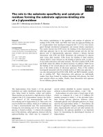
Increased eosinophils with heterogenous features in periph-eral blood filmFigure 1
Increased eosinophils with heterogenous features in
peripheral blood film.
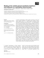
Bone marrow aspirate of patient showing presence of blasts with high nucleocytoplasmic ratio and eosinophilic precur-sorsFigure 2
Bone marrow aspirate of patient showing presence
of blasts with high nucleocytoplasmic ratio and eosi-
nophilic precursors.
Journal of Hematology & Oncology 2009, 2:26 />Page 3 of 6
(page number not for citation purposes)
sis (FC500 Beckman Coulter) was done after incubation
of bone marrow mononuclear cells with a panel of fluo-
rescence labeled antibodies (obtained from Becton Dick-
inson, USA). The blast cell population showed strong
reactivity (>95%) with B-cell markers including CD10,
CD19, CD20, CD22 and cCD79a.
The blasts did not show expression of lymphoid T markers
(CD3, 5 & 7) and myeloid markers (CD 13, 33 & cMPO).
HLA-DR and cTdT also showed strong positivity. There
were no detectable surface immunoglobulins or cytoplas-
mic light chains (IgM, IgG, kappa, lambda). The immu-
nophenotype was consistent with common precursor B-
lymphoblastic leukaemia. Cytogenetic analysis (Fig 3)
revealed karyotype 45 XY, t(7;12)(q22;p13),-9[15]/46,XY
[05]. Routine examination of cerebrospinal fluid showed
a protein level: 0.33 g/l, glucose: 4.4 mmol/l and occa-
sional mature lymphocytes on Leishman's stain (There
were no blasts seen in the cytospin smear).
The height and weight of the patient was 170 cm and 70
kg, respectively, and the body surface area was 1.7 m
2
. He
was started on induction therapy based on UK ALL XII
consisting of four drugs regimen, which included Inj vin-
cristine 1.5 mg/m
2
IV on day 1, 8, 16 and 24; Inj daunoru-
bicin 60 mg/m
2
IV on day 1, 8, 16 and 24; tab
prednisolone 60 mg/m
2
/day × 28 days and Inj asparagi-
nase 6000 units/m
2
IM days 18, 19, 20, 21, 22, 23 and 24.
He was also given intrathecal methotrexate 12.5 mg on
days 1 and 8. The patient tolerated chemotherapy well,
except that he developed an episode of gastroenteritis on
12
th
day of chemotherapy. He was rehydrated with IV flu-
ids, and was administered Inj ciprofloxacin 400 mg b.d.
and Inj Metronidazole 500 mg IV 8 hourly. For pneumo-
cystis prophylaxis, he was given Co-trimoxazole 960 mg
b.d 3 times a week. On the 20
th
day of the induction ther-
apy, the complete blood counts showed correction of
eosinophilia with a total leucocyte count of 8.1 × 10
9
/l
with 81% neutrophils, 12% lymphocytes, 05% eosi-
nophils and 02% monocytes; Hb: 11.7 g/dl and platelet
count: 170 × 10
9
/l. His repeat bone marrow biopsy which
was done on the 28
th
day of induction therapy showed
active hematopoiesis with increase in eosinophil precur-
sors and presence of 10% blasts in the marrow. Biochem-
ical profile including serum urea, creatinine, electrolytes,
bilirubin, ALT and uric acid were within normal limits.
Serum LDH was 1254 U/ml (normal range: 250–500 u/
ml). Due to his partial response to chemotherapy, re-
induction therapy was given to him with the following
drug combination: Inj vincristine 1.5 mg/m
2
day 1, Inj
daunorubicin 45 mg/m
2
days 1 & 2; Inj etoposide 100
mg/m
2
days 1–5; and Inj cytosine arabinoside 100 mg/m
2
IV infusion 12 hourly days 1–5 and oral prednisolone 60
mg daily day 1–5 gradually to be tapered off in next two
weeks. He was also given intrathecal Inj methotrexate 12.5
mg, on day 1 of second induction. The aforementioned
chemotherapy regimen was repeated 4 weeks after the end
of the cycle, when hematopoeitic recovery was adequate
and the patient was stable. On 28
th
day of the intensifica-
tion, the patient bone marrow was done which showed
normal maturation of erythroid and myeloid series with
less than 5% blasts, and normal number of megakaryo-
cytes. The patient is scheduled for evaluation at Armed
Forces Bone Marrow Transplant Centre, Rawalpindi, Paki-
stan for stem cell transplantation from matched sibling
donor.
Discussion and Evaluation
The eosinophil count in a normal adult ranges from 0.02–
0.5 × 10
9
/l (20–500/μl) [7]. Eosinophilia is classified as
mild (0.5–1.5 10
9
/l), moderate (1.5–5 × 10
9
/l) and severe
[8] when the eosinophil count is more than 5 × 10
9
/l.
Although, the patient presented with severe eosinophilia
(34.5 × 10
9
/l), there was no organ involvement, as shown
by normal ECG, echocardiography, chest X-Ray, and no
evidence of nervous system dysfunction or skin rash.
Different causes of eosinophilia, as shown in table 1, were
excluded based upon clinical evaluation and investiga-
tions of the patient. In Pakistan, infestation with Ascaris
lumbricoides, Ancylostoma duodenale, Necator americanus
and Echinococcus are common causes of eosinophilia.
However, there were no ova detected on stool examina-
tion and ultrasound of abdomen did not show any abnor-
mality. This patient, diagnosed as acute lymphoblastic
leukaemia, had unremarkable clinical presentation at
onset, with vague symptoms and absence of fever or lym-
Cytogenetics of the patient's bone marrow showing 45,XY,t(7;12)(q22;p13),-9 (20 cells were counted; 15 cells showed 45 chromosomes, while 5 cells showed 46 chromo-somes)Figure 3
Cytogenetics of the patient's bone marrow showing
45,XY,t(7;12)(q22;p13),-9 (20 cells were counted; 15
cells showed 45 chromosomes, while 5 cells showed
46 chromosomes).
Journal of Hematology & Oncology 2009, 2:26 />Page 4 of 6
(page number not for citation purposes)
phadenopathy. His complete blood counts revealed
marked eosinophilia, and interestingly absence of blasts
in the peripheral blood film. The bone marrow biopsy of
this patient showed presence of 40% blasts, which stained
negative with Sudan Black B, and were of common precur-
sor B-cell origin on flow cytometric analysis.
A variant of acute myelomonocytic leukaemia (AML-M4
Eo) [9] is associated with presence of increased number of
abnormal eosinophil precursors in the bone marrow.
Such patients usually have blasts in peripheral blood film
and a characteristic cytogenetic abnormality of inversion
of chromosome 16. Wynn et al [10] described B-cell line-
age acute lymphoblastic leukaemia in a 5 year old girl,
with peripheral hypereosinophilia, absence of blasts in
peripheral blood film and hyperdiploid blast cell popula-
tion with 5q deletion. Hypereosinophilia has also been
reported in a 4 year old boy who had granular acute lym-
phoblastic leukaemia with a normal karyotype [11]. One
case of acute lymphoblastic leukaemia with peripheral
hypereosinophilia was found to have 9p21 deletion [12],
on cytogenetic analysis. As discussed earlier, a common
cytogenetic abnormality detected in most cases of precur-
sor B acute lymphoblastic leukaemia with hypereosi-
nophilia is t(5;14), which disappears during remission
and reappears in relapse [13,14]. In the latter transloca-
tion, eosinophilia was secondary to overproduction of
interleukin-3 by the blasts, due to activation of inter-
leukin-3 gene on chromosome 5 after its translocation
adjacent to the immunoglobulin heavy chain gene on
chromosome 14 [5].
Eosinophilia may antedate the development of acute lym-
phoblastic leukemia by several months to 2 years, and the
patients may present with urticarial lesions and other
non-haematological features of hypereosnophilic syn-
drome (hepatosplenomegaly, cardiac lesions, CNS
involvement) during this period [15,16]. Hypereosi-
nophilia was present in our patient at the time of diagno-
sis of his disease, and was associated with a unique
cytogenetic abnormality of t(7;12)(q22;p13),-9, present
in majority of the metaphases. To the best of our knowl-
edge, the latter translocation has not been reported earlier
in adult B lymphoblastic leukemia/lymphoma. The
t(7;12)(q22;p13) previously reported in infant leukemia
[17], did not occur as a sole abnormality, and was accom-
panied by deletion of 7(q22q36). Two rare recurrent
translocations {t(7;12)(q36;p13) and t(7;12)(q32;p13)}
have been identified in 5/125 children, less than 18
months of age, and who were suffering from acute mye-
Table 1: Differential diagnosis of eosinophilia
Parasitic Infestations:
Ankylostoma duodenale
Necator americanus
Toxoplasma gondii
Strongyloides stercoralis
Ascaris lumbricoides
Hydatid disease
Allergic diseases
Bronchial asthma
Urticaria
Allergic rhinitis
Atopic dermatitis
Haematological malignancies
Hodgkin lymphoma
Chronic myeloid leukemia
Acute myeloid leukemia with inv (16) or t(16;16)
B lymphoblastic leukemia/lymphoma with t(5;14); IL3-IGH
Chronic eosinophilic leukemia
Myeloid & lymphoid neoplasms with eosinophilia and PDGFRA, PDGFRB and FGFR1
abnormalities
Connective tissue disorders
Rheumatoid arthritis
Serum sickness
Scleroderma
Others
Loeffler syndrome
Graft versus host disease
Journal of Hematology & Oncology 2009, 2:26 />Page 5 of 6
(page number not for citation purposes)
loid leukemia [18]. Involvement (deletions or transloca-
tions) of chromosome 7, especially in the region 7q22,
predominantly occurs in myelodysplastic syndrome [19]
and myeloid leukemias [20]. Translocation and deletion
of a segment of chromosome 7 (7q22 or 7q32-q35), or
loss of chromosome 7 may lead to inactivation (position
effect) of tumour suppressor gene(s) or their dysfunction
(at the breakpoint region), leading to transformation to
myeloid leukemias [20].
The other chromosomal gene affected in this case is
12p13. More than forty translocations involving 12p13,
or the ETV6 gene, and different partner chromosomes
have been described in various haematopoeitic malignan-
cies. t(5;12)(q31;p13) is a recurrent mutation which has
been reported in refractory anaemia with excess blasts
(RAEBt) with basophilia, acute myeloid leukemia with
eosinophilia and acute eosinophilic leukemia [21]. Chro-
mosomal abnormality affecting 12p13 and associated
with severe eosinophilia, has also been described in Phil-
adelphia negative myeloproliferative disorder, eosi-
nophilic leukemia and acute lymphoblastic leukemia
[22]. Similarly, a chimeric gene ETV6/ACS2 due to
t(9;12)(q22;p13) has been reported in myelodysplastic
syndrome associated with eosinophilia [23]. The ETV6
gene codes for a transcription activator, and has a span of
240 kb with eight exons [24,25]. Different segments of the
ETV6 gene act as fusion partners with other genes affected
in various translocations, and can result in the production
of a fusion protein having a ligand-independent tyrosine
kinase activity [26]. In our case, the events responsible for
leukemogenesis may be related to hypodiploidy, loss of
suppressor gene at 7q22, or activation of a proto-onco-
gene after translocation of 7q22 to 12p13 (ETV6 gene).
Hypodiploidy (loss of chromosome 9) is likely responsi-
ble for poor response to chemotherapy and adverse prog-
nosis, while eosinophilia seen in this case is most likely
secondary to t(7;12). The patient was categorized in the
poor risk category because of late remission (>6 weeks
after induction therapy). He is thus planned to be referred
for bone marrow transplantation from matched sibling
with a curative intent.
Conclusion
Marked increase of eosinophils in the blood and bone
marrow can occur in precursor B-acute lymphoblastic
leukemia, as a result of different cytogenetic abnormali-
ties. In majority of the cases there is absence of blasts in
the peripheral blood film. Adult B lymphoblastic leuke-
mia/lymphoma with hypodiploidy and severe eosi-
nophilia in blood and bone marrow, having cytogenetic
abnormality t(7;12)(q22;p13),-9, is a distinct entity with
poor response to chemotherapy, and a bad prognosis.
Individuals with this disease should be selected for bone
marrow transplantation from matched sibling/unrelated
donor, once remission is achieved, for best chances of dis-
ease free survival.
Abbreviations
ALL: Acute lymphoblastic leukemia
Competing interests
The authors declare that they have no competing interests.
Authors' contributions
All authors were involved in management of the patient
and preparation of this manuscript. All authors read and
approved the final manuscript.
Consent
The patient has provided informed consent for the publi-
cation of this case report and accompanying images. A
copy of the written consent is available for review by the
Editor-in-Chief of this journal.
References
1. Pardanani A, Brockman SR, Patemoster SF, Flynn HC, Ketterling RP,
Lasho TL, Ho CL, Li CY, Dewald GW, Tefferi A: FIP1L1-PDGFRA
fusion: prevalence of clinicopathologic correlates in 89 con-
secutive patients with moderate to severe eosinophilia. Blood
2004, 104(10):3038-3045.
2. Ayyub M, Anwar M, Luqman M, Ali W, Bashir M: A case of hypere-
osinophilic syndrome developing Hodgkin's disease after 4
years. Br J Haematol 2003, 123(5):955-956.
3. Spitzer G, Garson OM: Lymphoblastic leukemia with marked
eosinophilia: a report of two cases. Blood 1973, 42:377-384.
4. D'Angelo G, Hotz AM, Todeschin P: Acute lymphoblastic leuke-
mia with hypereosinophilia and 9p21 deletion: Case report
and review of literature. Lab Hematol 2008, 14:7-9.
5. Meeker TC, Hardy D, William C, Hogan T, Abrams J: Activation of
interleukin-3 gene by chromosome translocation in acute
lymphocytic leukemia with eosinophilia. Blood 1990, 76:285-9.
6. Swerdlow SH, Campo E, Harris NL, Jaffe ES, et al.: WHO classifica-
tion of tumours of haematopoietic and lymphoid tissues. (4
th
edition). Lyon, France: IARC Press; 2008:109-138.
7. Lewis M: Reference ranges and normal values. In Dacie and
Lewis Practical Haematology 10th edition. Edited by: Lewis SM, Bain BJ,
Bates I. Churchill Livingstone; 2006:11-24.
8. Rothenberg ME: Eosinophilia. N Eng J Med 1998, 338:1592-1600.
9. Haferlach T, Winkemann M, Loffler H, Schoch R, Gassmann W,
Fonatsch C, Schoch C, Poetsch M, Weber-Matthiesen K, Schlegel-
berger B: The abnormal eosinophils are part of the leukaemic
cell population in acute myelomonocytic leukemia with
abnormal eosinophils (AML M4Eo) and carry pericentric
inversion 16: a combination of May Grunwald Geimsa stain-
ing and fluorescence in situ hybridization. Blood 1996,
87(6):2459-2463.
10. Wynn TT, Heerema NA, Hammond S, Ranalli M, Kahwash SB: Acute
lymphoblastic leukaemia with hypereosinophilia: report of a
case with 5q deletion and review of literature. Pediatr Dev
Pathol 2003, 6(6):558-63.
11. Jain P, Kumar R, Gujral S, Kumar S, Singh A, Jain Y, Dubey S, Anand
M, Arya LS: Granular acute lymphoblastic leukaemia with
hypereosinophilic syndrome. Ann Hematol 2000, 79:272-274.
12. D'Angelo G, Hotz AM, Todeschin P: Acute lymphoblastic leukae-
mia with hypereosinophilia and 9p21 deletion: Case report
and review of literature. Laboratory Hematology 2008, 14(1):7-9.
13. Hogan TF, Koss W, Murgo AJ, Amato RS, Fontana JA, VanScoy FL:
Acute lymphoblastic leukemia with chromosomal 5;14
translocation and hypereosinophilia: case report and litera-
ture review. J Clin Oncol 1987, 5(3):382-90.
14. Baumgarten E, Wegner RD, Fengler R, Ludwig WD, Schulte-Over-
berg U, Domeyer C, Schüürmann J, Henze G: Calla-positive acute
Publish with BioMed Central and every
scientist can read your work free of charge
"BioMed Central will be the most significant development for
disseminating the results of biomedical research in our lifetime."
Sir Paul Nurse, Cancer Research UK
Your research papers will be:
available free of charge to the entire biomedical community
peer reviewed and published immediately upon acceptance
cited in PubMed and archived on PubMed Central
yours — you keep the copyright
Submit your manuscript here:
/>BioMedcentral
Journal of Hematology & Oncology 2009, 2:26 />Page 6 of 6
(page number not for citation purposes)
leukaemia with t(5q;14q) translocation and hypereosi-
nophilia – a unique entity? Acta haematologica 1989, 82(2):85-90.
15. Hill A, Metry D: Urticarial lesions in a child with acute lym-
phoblastic leukaemia and eosinophilia. Pediatr Dermatol 2003,
20(6):502-5.
16. Blatt J, Proujansky R, Horn M, Phebus C, Longworth D, Penchansky
L: Idiopathic hypereosinophilic syndrome terminating in
acute lymphoblastic leukemia. Pediatr Hematol Oncol 1992,
9:151-155.
17. Tosi S, Hughes J, Scherer SW, Nakabayashi K, Harbott J, Haas OA,
Cazzaniga G, Biondi A, Kempski H, Kearney L: Heterogeneity of
the 7q36 breakpoints in the t(7;12) involving ETV6 in infant
leukemia. Genes Chromosomes Cancer 2003, 38(2):191-200.
18. Slater RM, Drunen EV, Kroes WG, et al.: t(7;12)q36;p13) and
t(7;12)(q32;p13)-translocations involving ETV6in children 18
months of or younger with myeloid disorders. Leukemia 2001,
16:915-20.
19. Johnson EJ, Scherer SW, Osborne L, Tsui LC, Oscier D, Mould S,
Cotter FE: Molecular definition of a narrow interval at 7q22.1
associated with myelodysplasia. Blood 1996, 87:3579-86.
20. Fischer K, Frohling S, Scherer S, Brown JM, Scholl C, Stilgenbauer S,
Tsui LC, Lichter P, Döhner H: Molecular cytogenetic delineation
of deletions and translocations involving chromosome band
7q22 in myeloid leukemias. Blood 1997, 89:2036-41.
21. Odero MD: t(5;12)(q31;p13) in MDS, AML and AEL. Atlas Genet
Cytogenet Oncol Haematol 2007.
22. Keene P, Mendelow B, Pinto MR, Bezwoda W: Abnormalities of
chromosome 12p 13 and malignant proliferation of eosi-
nophils: a nonrandom association. Br J Haematol 2008,
67(1):25-31.
23. Kuno Y, Abe A, Emi N, Iida M, Yokozawa T, Towatari M, Tanimoto
M, Saito H: Constitutive kinase activation of the TEL-SYK
fusion gene in myelodysplstic syndrome with
t(9;12)(q22;p12). Blood 2001, 97:1050-55.
24. Wlodarska I, Mecucci C, Baens M, Marynen P, Berghe H van den:
ETV6 gene rearrangementsin haemopoietic malignant dis-
orders. Leuk Lymphoma 1996, 23(3–4):287-95.
25. Gao NA, Li ZH, Ding BT, Chen Y, Wang YS, Qiao Y, Guo NJ:
Expression of ETV6 rearrangement in a subject with acute
myeloid leukemia-M4Eo. Chin Med J 2008, 121(17):1744-46.
26. Wai DH, Knezevich SR, Lucas T, Jansen B, Kay RJ, Sorensen PH: The
ETV6-NTRK3 gene fusion encodes a chimeric protein tyro-
sine kinase that transforms NIH3T3 cells. Oncogene 2000,
19(7):906-915.