báo cáo khoa học: "Mechanism of action of lenalidomide in hematological malignancies" pptx
Bạn đang xem bản rút gọn của tài liệu. Xem và tải ngay bản đầy đủ của tài liệu tại đây (948.11 KB, 10 trang )
BioMed Central
Page 1 of 10
(page number not for citation purposes)
Journal of Hematology & Oncology
Open Access
Review
Mechanism of action of lenalidomide in hematological malignancies
Venumadhav Kotla
†1
, Swati Goel
†1
, Sangeeta Nischal
1
, Christoph Heuck
1
,
Kumar Vivek
2
, Bhaskar Das
3
and Amit Verma*
1
Address:
1
Department of Medicine, Albert Einstein College of Medicine, Bronx, USA,
2
Harrison Department of Surgical Research, University of
Pennsylvania, Philadelphia, USA and
3
Developmental and Molecular Biology, Albert Einstein College of Medicine, Bronx, USA
Email: Venumadhav Kotla - ; Swati Goel - ; Sangeeta Nischal - ;
Christoph Heuck - ; Kumar Vivek - ; Bhaskar Das - ;
Amit Verma* -
* Corresponding author †Equal contributors
Abstract
Immunomodulatory drugs lenalidomide and pomalidomide are synthetic compounds derived by
modifying the chemical structure of thalidomide to improve its potency and reduce its side effects.
Lenalidomide is a 4-amino-glutamyl analogue of thalidomide that lacks the neurologic side effects
of sedation and neuropathy and has emerged as a drug with activity against various hematological
and solid malignancies. It is approved by FDA for clinical use in myelodysplastic syndromes with
deletion of chromosome 5q and multiple myeloma. Lenalidomide has been shown to be an
immunomodulator, affecting both cellular and humoral limbs of the immune system. It has also been
shown to have anti-angiogenic properties. Newer studies demonstrate its effects on signal
transduction that can partly explain its selective efficacy in subsets of MDS. Even though the exact
molecular targets of lenalidomide are not well known, its activity across a spectrum of neoplastic
conditions highlights the possibility of multiple target sites of action.
Thalidomide is the first immunomodulatory
drug with multiple effects on the immune
system
Immunomodulatory drugs (IMiDs) CC-5013 (Revlimid
TM, Lenalidomide) and CC-4047 (ActimidTM, Pomalid-
omide) are a series of synthetic compounds derived using
structural modifications of the chemical structure of tha-
lidomide. Thalidomide (a-(N-phthalimido) glutaramide)
was synthesized in Germany, in 1954, from glutamic acid,
to be used as a sedative and hypnotic anti-emetic drug,
indicated to treat morning sickness in the first trimester of
gestation. Thalidomide was banned in the 1960s because
of the reports of congenital malformations like phocome-
lia associated with its use in pregnant women. One of the
possible hypothesis to explain this teratogenecity is that
thalidomide creates oxidative stress by with subsequent
downregulation of Wnt and Akt survival pathways which
induces apoptosis during early embryonic limb develop-
ment resulting in limb truncations[1]. Following an
observation in 1965 that thalidomide administration
improved the inflammatory lesions of erythema nodo-
sum leprosum (ENL) in a patient suffering from sleep dif-
ficulty, the use of thalidomide continued. Eventually in
1998, FDA approved the drug for the treatment of ENL,
with tight restrictions on its marketing. ENL is an immune
complex mediated inflammatory reaction that occurs dur-
ing therapy in lepromatous leprosy patients. It is com-
monly associated with systemic symptoms, and
constitutes a medical emergency with urgent need of ther-
apy with anti-inflammatory/immunomodulatory drugs
Published: 12 August 2009
Journal of Hematology & Oncology 2009, 2:36 doi:10.1186/1756-8722-2-36
Received: 24 March 2009
Accepted: 12 August 2009
This article is available from: />© 2009 Kotla et al; licensee BioMed Central Ltd.
This is an Open Access article distributed under the terms of the Creative Commons Attribution License ( />),
which permits unrestricted use, distribution, and reproduction in any medium, provided the original work is properly cited.
Journal of Hematology & Oncology 2009, 2:36 />Page 2 of 10
(page number not for citation purposes)
to prevent long term disabilities. Research into the mech-
anism of action of thalidomide unraveled an immunolog-
ical and immunomodulatory basis for the effect, notably
inhibition of denovo IgM antibody synthesis[2] by possi-
bly affecting the macrophages, B-cells, helper or suppres-
sor lymphocytes, decreasing TNF-α synthesis and
modulating the T cell subsets by increasing the T-helper
population after therapy[3]. TNF-α is a potent pro inflam-
matory cytokine, and is also involved in the pathogenesis
of neural damage in leprosy. The inhibitory effect of tha-
lidomide on TNF-α is a consequence of increased degra-
dation of its mRNA due to the drug [4]. Thalidomide also
regulates the levels of IL-6 and IFN-γ in ENL patients, fur-
ther contributing to the immunomodulatory mechanism
of action. Interest in thalidomide as a neoplastic agent
intensified after the demonstration of antiangiogenic
activity in animal models. The recognition that angiogen-
esis plays an important pathogenic role in multiple mye-
loma as reflected by increased bone marrow
microvascular density and VEGF (vascular endothelial
growth factor) levels, prompted the clinical use of thalid-
omide in relapsed/refractory multiple myeloma. With the
recognition of adverse effects like neuropathy, deep vein
thrombosis, and sedation, more potent and safer ana-
logues were developed by Celgene. Lenalidomide is one
such analogue which has been extensively tested and
proven to be more potent than thalidomide and has fewer
adverse effects compared to thalidomide. Another newer
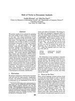
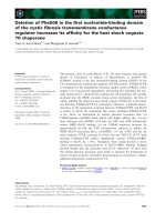
thalidomide analogue is pomalidomide. Figure 1 consists
of the chemical structures and names of these three com-
pounds and Table 1 illustrates the differences amongst
them.
Mechanism of action of Lenalidomide
The clinical evidence for therapeutic potential of lenalid-
omide in various malignant conditions is consistent with
the multitude of pharmacodynamic effects that have been
shown in vitro and in animal models. Studies have shown
that lenalidomide may work through various mechanisms
in different hematologic malignancies. These mechanism
involved direct cytotoxicity as well as through indirect
effects on tumor immunity. Thus the differential efficacy
noted with lenalidomide therapy among various disease
states can possibly be explained individual's immune sta-
tus and disease specific pathophysiology. Following are
the different mechanisms explained by which lenalido-
mide acts in the body.
Immunomodulation
The immune system is comprised of cellular (macro-
phages, dendritic cells, NK cells, T cells and B cells), and
humoral components (antibodies, cytokines). The
immune system can prevent development of cancers by
eliminating or suppressing oncogenic viral infections,
altering the inflammatory milieu conducive to tumor gen-
esis, and by immune surveillance by identifying and
destroying transformed cells before they can cause
harm[5].
Lenalidomide has been shown to modulate different
components of the immune system by altering cytokine
production, regulating T cell co stimulation and augment-
ing the NK cell cytotoxicity. Immunomodulatory proper-
ties of Lenalidomide are implicated in its clinical efficacy
in multiple myeloma, CLL and myelodysplastic syn-
dromes; where the disease pathogenesis involves in part a
deregulated immune system in the form of altered
cytokine networks in tumor microenvironment, defective
Table 1: Differences between thalidomide, lenalidomide and pomalidomide
Name Thalidomide Lenalidomide Pomalidomide
Empirical Formula C
13
H
10
N
2
O
4
C
13
H
13
N
3
O
3
C
13
H
11
N
3
O
4
Molecular weight 258.2 259.3 273.2
Chemical Structural Thalidomide has two oxo groups
in Phthaloyl ring
Lenalidomide has amino group at
4th position and single oxo group
in Phthaloyl ring
Pomalidomide has amino group at
4th position and two oxo groups in
Phthaloyl ring
Effects on T-cell proliferation Thalidomide stimulates T cell
proliferation and increases IFN-γ
and IL-2 production
Lenalidomide is 100–1000 times
more potent in stimulating T cell
proliferation and IFN-γ and IL-2
production than thalidomide
Pomalidomide is similar to
lenalidomide, in addition, it also
enhances transcription factor T-
bet, which reverts Th2 cells into
Th1 like effector cells in vitro
Adverse Effects Thalidomide has higher incidence
of side effects like sedation,
neuropathy and constipation.
Lenalidomide has lower incidence
of adverse effects namely sedation,
constipation and neuropathy than
thalidomide.
Pomalidomide has lower incidence
of adverse effects like sedation,
constipation and neuropathy than
thalidomide.
Teratogenecity Thalidomide is a known teratogen. Lenalidomide is not teratogenic in
rabbit models
Pomalidomide is a known
teratogen.
Journal of Hematology & Oncology 2009, 2:36 />Page 3 of 10
(page number not for citation purposes)
T cell regulation of host-tumor immune interactions, and
diminished NK cell activity.
Altering cytokine production
Cytokines are soluble proteins secreted by hematopoietic
and non hematopoietic cell types and are critical for both
innate and adaptive immune responses. The expression of
cytokines by cells may be altered in immunological,
inflammatory, infectious and neoplastic disease states.
Cytokines in turn exert their effects by influencing gene
activation, growth, differentiation, functional cell surface
molecule expression and cellular effector function. A
coordinated cellular and humoral (cytokines, antibodies)
interactions facilitate tumor destruction.
Lenalidomide has been shown to inhibit production of
pro inflammatory cytokines TNF-α, IL-1, IL-6, IL-12 and
elevate the production of anti-inflammatory cytokine IL-
10 from human PBMCs[6]. The downregulation of TNF-α
secretion is particularly striking and is up to 50,000 times
more when compared to thalidomide[7]. TNF-α is a
highly pleiotropic cytokine produced primarily by mono-
cytes and macrophages and plays an important role in
protective immune responses against bacterial and viral
infections. Elevated TNF-α production is implicated in the
pathogenesis of various hematologic malignancies and
may be partly responsible for stem cell apoptosis and inef-
fective hematopoiesis seen in MDS [8]. TNF-α levels in
CLL patients are also elevated and exhibit a significant
decrease as early as 7 days after lenalidomide treatment.
These reductions correlate with cytoreduction suggesting a
casual relationship with tumor growth [9].
Similarly, reduction in IL-6 and TNF-α levels could
explain the action of lenalidomide in multiple myeloma.
IL-6 inhibits the apoptosis of malignant myeloma cells
and helps in their proliferation[10]. Lenalidomide down-
regulates the production of IL-6 directly and also by inhib-
iting multiple myeloma (MM) cells and bone marrow
stromal cells (BMSC) interaction [11,12], which aug-
ments the apoptosis of myeloma cells[13]. The precise
mechanism of TNF-α downregulation by lenalidomide is
not known, however thalidomide has been shown to
increase the degradation of TNF-α mRNA [4,14]. It is pos-
sible that lenalidomide may work through similar mech-
anisms.
T cell activation
T cells are important effectors of immune response and
their activation is tightly regulated to prevent auto reactiv-
ity. T cell activation involves the presentation of the pep-
tide fragments displayed by antigen presenting cells
(APCs) to the T cell receptor (TCR) and it is this interac-
tion that gives specificity to the response. However this
interaction alone is not sufficient if a T cell has to generate
an effective response against the antigen. A secondary
interaction of B7 molecule on APC and CD28 on the T cell
surface provides the co stimulatory signal that augments
the T cell response and aids in T cell proliferation, differ-
entiation, and survival followed by a cascade of cytokine
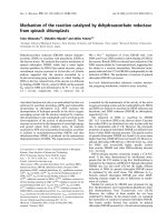
and cellular responses[15].(Figure 2). IMiDs, including
lenalidomide act on T cells via B7-CD28 co stimulatory
pathway. Blockade of this interaction using the CTLA-4-Ig,
B7 blocking antibody, is partially overcome by IMiDs.
IMiDs do not up regulate expression of CD28 and B7 on
T cells and APCs respectively but they can directly induce
tyrosine phosphorylation of CD28 on T cells leading to
activation of downstream targets such as PI3K, GRB-2-OS,
and NF-κb. This might explain their ability to partially
overcome CTLA4 Ig blockade[16]. T cell co-stimulation by
lenalidomide leads to an increased Th1 type cytokine
response resulting in increased secretion of IFN-γ and IL-2
that in turn stimulate clonal T cell proliferation and NK
cell activity[6,17].
IMiDs have been shown to stimulate both cytotoxic CD8+
as well as helper CD4+ cells[18]. Their effects on T helper
cells can potentially mediate Th1 type antitumor immu-
nity in response to tumor cell vaccination in animal mod-
els[17]. The IMiD, CC-4047 (pomalidomide) enhanced
partially protective antitumor effect of whole tumor cell
vaccination in mice and generated long term immunity
against subsequent live tumor challenge[17]. In vivo pro-
Chemical structures of thalidomide, lenalidomide and pomal-idomideFigure 1
Chemical structures of thalidomide, lenalidomide
and pomalidomide.
Journal of Hematology & Oncology 2009, 2:36 />Page 4 of 10
(page number not for citation purposes)
duction of IFN-γ correlated with the tumor protection.
When nude mice lacking T cells were exposed to IMiD and
tumor cells during the priming phase, they did not dem-
onstrate protection from the tumor, demonstrating that T
cells are needed for tumor immunity. The IMiD drug itself
was shown to have no direct anti tumor effect on growth
inhibition or expression of co stimulatory molecules, rul-
ing out direct cytotoxic effects. These effects can also partly
explain the beneficial effects of lenalidomide in MDS.
Clonal expansion of abnormal hematopoietic suppressive
T cells are believed to have a pathogenic role in ineffective
erythropoiesis of patients with MDS and 50% of the
patients with MDS were shown to have clonal T cells com-
pared to 5% in age matched controls[19]. It is possible
that lenalidomide may affect certain T cell subsets and
result in hematologic improvements in MDS patients.
Augmentation of NK cell function
Natural Killer (NK) Cells comprise 2% of the circulating
lymphocytes and are an important component of innate
immunity. NK cells are not driven by specificity to anti-
gens unlike T cells or B cells and are able to respond rap-
idly on contact with the target cell (cancer, viral infected)
and kill the cell with antibody dependent cell mediated
cytotoxicity(ADCC) and natural cytotoxicity. Natural
killer cells also contribute to immunoregulation by secret-
T cell activationFigure 2
T cell activation. B7-CD28 co-stimulation pathway is needed for T cell activation and CTLA4 Ig blocks this pathway leading
to T cell inactivation. Lenalidomide acts by directly inducing tyrosine phosphorylation of CD28 on T cells leading to activation
of downstream targets such as PI3K, GRB-2-OS, and NF-κb, thus partially overcoming CTLA4 Ig blockade and leading to T cell
clonal proliferation.
Journal of Hematology & Oncology 2009, 2:36 />Page 5 of 10
(page number not for citation purposes)
ing cytokines like IFN-γ and TNF-α. Modulation of NK cell
function is also believed to contribute to the anti tumor
activity of Lenalidomide in MDS, MM and CLL.
Davies et al examined the potential immunomodulatory
effects of thalidomide and its analogues in patients with
multiple myeloma. The in vitro/in vivo role of NK cell
cytotoxicity of MM cells in thalidomide treated patient
was supported by the observation that the cell killing was
not MHC restricted and CD56(NK cell) depletion in vitro
inhibited killing of drug treated multiple myeloma
cells[20]. Furthermore, treatment with Thalidomide was
also accompanied by increased NK cell numbers and IL-2
levels. The precise mechanism whereby IMIDS increase
the NK cell number or augment its cytotoxicity is not well
known and it is possible that these effects may be indirect.
Hayashi et al in their study of IMiDs in MM cell lines have
demonstrated that when culturing PBMC with IMiDs
leads to 1.2–1.3 fold increase in the percentage of CD56
cells. IMiDs enhanced ADCC when 51 Cr-labelled MM
cells that express CD40 were incubated with rhuCD40
and then subsequently treated with PBMC cells incubated
in the presence of IMiDs for 5 days. The increase in NK cell
function may be related to the increase in IL-2 production
by the T cells as the presence of a monoclonal Ab against
IL-2 R blocked the NK cell cytotoxicity. IMiDs also were
shown not to directly activate the NK cells, as evidenced
by lack of phosphorylation of signaling molecules (ERK/
p38MAPK/Akt/PKC) in NK cells[21].
Lenalidomide also enhanced the NK cell mediated ADCC
in a series of functional in vitro studies using Rituximab
coated NHL cell lines, Trastuzumab coated breast cancer
cells expressing Her2 and cetuximab coated colon cancer
cells positive for EGFR expression. The cell killing was
increased in a dose dependent manner and presence of IL-
2 was required to achieve cell killing[22]. In another study
[23], IFN-γ production by NK cell in rituximab coated
NHL cell lines pretreated with lenalidomide, was induced
with the interaction of Ig G with FC-γ receptors in the pres-
ence of IL-2 or IL-12. Thus, lenalidomide enhanced Fc-γ
receptor signaling may also play a role in increasing the
potency of NK cells.
Anti-angiogenesis activity
The growth of the primary and metastatic tumors requires
the development of new blood vessels, a process
described as angiogenesis. Tumors possess the ability to
promote the formation of new blood vessels from preex-
isting host capillaries at a critical phase of the tumor
development when the balance of pro- angiogenic and
anti-angiogenic factors is altered. Vascular endothelial
growth factor (VEGF) and its receptors are required for the
formation of blood vessels during embryonic develop-
ment, wound healing, and carcinogenesis. Tumors are
more dependent on the VEGF-Receptor signaling for
growth and survival compared to normal endothelial cells
[24]. Early studies showed that Thalidomide had anti ang-
iogenic activity in a rabbit model of corneal neovasculari-
zation that was induced as a response to bFGF[25]. This
report led to its use in Multiple Myeloma, where it dem-
onstrated clinical benefit and was approved for use by the
FDA. Thalidomide and the newer IMiDs have also been
shown to significantly decrease the expression of ang-
iogenic factors VEGF and Interleukin-6 (IL-6) in multiple
myeloma; thereby reducing angiogenesis and hence con-
tributing to clinical activity in multiple myeloma[26]. The
newer IMiDs were found to be 2–3 times more potent
compared to thalidomide in antiangiogenic activity in
various vivo assays [27] The antiangiogenic activity of
both thalidomide and IMiDs has also been shown to be
independent of immunomodulatory effects[28].
VEGF receptors are overexpressed on blast cells in dysplas-
tic marrows in MDS patients [29]. Increased plasma levels
of VEGF R have also been correlated with lower remission
rate in patients with myelodysplastic syndromes. A recent
study in 35 MDS patients with del 5 q showed a marked
decrease in bone marrow vascularity subsequent to lenal-
idomide therapy. This reduction in vascularity correlated
with clinical responses. However VEGF levels and VEGFR
levels did not change significantly even though vasculari-
zation was decreased, supporting the notion that lenalid-
omide may uncouple angiogenesis from the effect of
VEGF[30]. Apart from alteration in the levels of VEGF,
analysis of signal transduction events show that lenalido-
mide partially inhibits Akt phosphorylation after VEGF
stimulation in endothelial cells and also has inhibitory
effects on phosphorylation of Gab1, a protein upstream of
Akt 1[31,32]. These observations demonstrate that IMiDs
may affect angiogenesis by multiple mechanisms.
Direct anti tumor activity
Lenalidomide treatment has also shown anti proliferative
activity against MDS and MM cells in the absence of
immune effector cells[33]. Malignant plasma cells derived
from refractory cases of myeloma were shown to be sus-
ceptible to IMiD induced growth arrest. Lenalidomide has
also been shown to inhibit proliferation in Burkitt's Lym-
phoma cell lines by causing dose dependant cell cycle
arrest in G0-G1 phase[34]. Lenalidomide upregulated
Cyclin dependant kinase (CDK) Inhibitor, p21 waf-1, a
key cell cycle regulator that modulates the activity of
CDKs. Similar reductions in CDK2 activity have been
demonstrated in myeloma derived cell lines, U266 and
LP-1[34]. In contrast, the normal B cells obtained from
healthy donors were immune from growth inhibition and
did not show any upregulation of p21 expression after 3
days of lenalidomide treatment. In other studies, thalido-
mide and its analogues have also been shown to induce
Journal of Hematology & Oncology 2009, 2:36 />Page 6 of 10
(page number not for citation purposes)
apoptosis in MM cell lines[35]. Effects on apoptosis in
MM cells is secondary to increased potentiation of TNF
related Apoptosis inducing ligand (TRAIL), inhibition of
apoptosis protein-2, increased sensitivity to Fas mediated
cell death, and up regulation of caspase-8 activation,
down regulation of caspase-8 inhibitors (FLIP, cIAP2),
down regulation of NF-κb activity and inhibition of pro-
survival effects of IGF-1[36]. The proapoptotic activity of
IMiDs has also been demonstrated in CLL. Lenalidomide
was shown to induce apoptosis and affect the Phosphoti-
dylinositol pathway in CLL cells by decreasing activation
of pro-survival kinases, erk1/2 and Akt2[37].
Interestingly, lenalidomide has shown opposite effects on
the growth of normal progenitors. When cord derived
CD34+ progenitors cells were cultured in expansion
medium supplemented with lenalidomide, there was a
dose dependent increase in the total number of CD34
cells after 6 days of culture [34]. p21 was upregulated in
normal Cd34 cells, but did not affect the CDK2 activity in
contrast to Nawalma cells (Burkitt's lymphoma cells).
While the transfusion independence seen with lenalido-
mide use in MDS can be explained by the normal progen-
itor expansion, the dose dependent cytopenias that are
common with early treatment cycles of lenalidomide may
be a result of inhibition of proliferation of abnormal
clonal cell populations in the marrow.
Effects on multiple myeloma microenvironment
Lenalidomide exerts its distinct anti myeloma effects by
altering the myeloma microenvironment. In multiple
myeloma, osteoclasts lead to bone resorption and secrete
survival factors for MM cells. The interaction between MM
cells and BMSC in turn leads to increased production of
IL-6 and other growth factors for MM cells and osteo-
clasts[38]. Lenalidomide directly decreases the formation
of tartrate- resistant acid phosphatase(TRAP)- positive
cells which form osteoclasts [11]. Additionally, it
decreases αVβ3-integrin levels, an adhesion molecule
needed for osteoclast activation and downregulates cathe-
psin K, a major cysteine protease expressed in osteoclasts,
pertinent for matrix degradation in the resorption proc-
ess[11]. It downregulates the important mediators of oste-
oclastogenesis such as transcription factor PU.1 and MAP
kinase pERK and reduces the levels of bone remodeling
factor -receptor activator of nuclear factor-kappaB ligand.
Immunomodulators are also known to decrease the cell
surface adhesion molecules such as ICAM-1, VCAM-1 and
E -selectin [12] and inhibit the adhesion of MM cells to
BMSC. Thus, lenalidomide interferes with the synergism
amongst the osteoclasts, MM cells and BMSC and
decreases osteoclastogenesis by acting at various levels.
Selective efficacy in cells with deletion of
chromosome 5q
The del 5q syndrome is now recognized as a distinct path-
ologic subtype of MDS with markedly better clinical
responses with lenalidomide treatment compared to non
del 5q MDS patients. The exact mechanism of action of
lenalidomide on del 5q clones is not known, but there
appears to be several candidate genes (tumor suppressor)
whose expression may be modulated by lenalidomide
treatment. Hellstrom et al [39] studied the effect of lenal-
idomide on isolated differentiating erythroblasts from del
5q MDS patients and healthy controls. The addition of
lenalidomide significantly inhibited the invitro prolifera-
tion of erythroblasts harboring del 5q while the prolifera-
tion of cells from normal controls and cells without 5q
deletion was not affected. Gene expression profiling was
performed at day 7 when a median of 97% cells in culture
from MDS patients with del5q still possess del 5q, and
thus any difference in gene expression deemed to be
reflective of del 5q cells. There was altered gene expression
in many genes, but a set of 4 genes was consistently upreg-
ulated (VSIG4, PPIC, TPBG and SPARC) by more than 2
fold in all samples. The upregulation of SPARC (Secreted
Protein Acidic and Rich in Cysteine) after treatment with
lenalidomide is particularly interesting given its location
at 5q 31–32 and its role as a tumor suppressor with its
anti-proliferative, anti adhesion, anti-angiogenic proper-
ties. The levels of activin -A increased 4 fold and analysis
of global gene expression revealed significant deregula-
tion of genes involved in extracellular matrix interactions,
erythropoiesis relative to healthy control.
Another recent study compared gene expression profile of
CD34 stem cells of 5q del MDS patients to healthy con-
trols and MDS patients with normal karyotype using
Affymetrix arrays. Approximately 40% of the probe sets
showing reduced expression levels localized to the del 5q
region. The commonly deleted region (CDR) region is
thought to comprise of approximately 40 genes that are
hypothesized to have a tumor suppressive role given the
observation that deletion of the 5q region leads to clonal
proliferation of myelodysplastic clone. Majority of the
genes associated with CDR showed lower expression but
several candidate genes (RBM22 and CSNK1A1, SPARC
and RPS14) associated with CDR of the 5 q syndrome
showed marked down regulation[40]. RBM22 is a highly
conserved ribosomal protein, and the effects of downreg-
ulation may include deregulated apoptosis by its action
on ALG-2(apoptosis linked gene). CSNK1A1 has recently
been shown to be important in Hedgehog signaling that
governs cell growth and a deregulation is observed in can-
cers. Downregulation of CSNK1A1 may contribute to
MDS by altering the Hh signaling. RPS14 is related to the
40S subunit of the ribosome that is downregulated in
Cd34 cells from MDS patients with del 5q[40]. Recent
Journal of Hematology & Oncology 2009, 2:36 />Page 7 of 10
(page number not for citation purposes)
work shows that downregulation of RPS14 leads to defec-
tive erythropoiesis and increased apoptosis in erythroid
progenitors [41].
Another candidate gene in the CDR region is Early growth
response gene (EGR-1), that encodes a transcription factor
involved in the regulation of cell proliferation and apop-
tosis[42]. The effect of lenalidomide treatment on expres-
sion of EGR-1 was studied in del 5q Burkitt's lymphoma
and del 5q multiple myeloma cell line. It was observed
that lenalidomide treatment did not influence the tran-
scriptional activity of EGR-1 gene, but increased the
nuclear export of EGR-1 in a dose dependent manner,
especially in those with a single copy of EGR-1 gene.
When the gene expression was blocked with an EGR1
siRNA, Burkitt's cells proliferated more than normal cells,
supporting the tumor suppressor role of EGR-1 in
Burkitt's cells. Thus, lenalidomide increases the nuclear
transport of the pro apoptotic and tumor suppressor EGR-
1, which could explain its cytotoxic effects on del 5q31
myelodysplastic clones.
In an effort to identify molecular markers of response to
lenalidomide, Ebert et al [43] collected bone marrow aspi-
rates of non 5 q del MDS patients before and after treat-
ment with lenalidomide and studied the difference in
gene expression between responders and non responders.
Differential expression of the genes that needed for eryth-
roid differentiation was noted in non responders than
responders. In patients who responded to lenalidomide,
they found that the bone marrow aspirates before treat-
ment showed decreased expression of the set of the genes
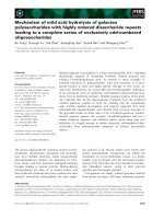
Mechanism of action of lenalidomideFigure 3
Mechanism of action of lenalidomide. Various mechanisms by which lenalidomide achieves clinical efficacy in hematologi-
cal malignancies.
Journal of Hematology & Oncology 2009, 2:36 />Page 8 of 10
(page number not for citation purposes)
needed for erythroid differentiation. The thinking is that
lenalidomide helps to overcome this differentiation block
and hence the clinical response is seen in that subset of
patients with decreased gene expression compared to the
non responders. This was thought have potential predict-
ability for benefit from lenalidomide therapy in non 5 q
del patients.
A recent study by Wei et al [44] demonstrates that the hap-
lodeficient enzymatic targets of lenalidomide within the
CDR are dual specificity phosphatases, Cdc25C and
PP2Acα. These phosphatases are coregulators of G2-M
checkpoint in the cell cycle and thus, their inhibition by
lenalidomide leads to G2 arrest and apoptosis. Since,
most MDS patients including those with deletion 5q
become refractory to Erythropoietin, the authors exam-
ined the molecular mechanisms by which lenalidomide
may modulate this effect. They observed that the CD45
phosphatase is overactivated in MDS and may inhibit Epo
receptor stimulated phosphorylation of stat5. Further-
more, they observed that lenalidomide is a Protein Tyro-
sine Phosphatase inhibitor of CD45 leading to reversal of
CD45 induced inhibition of EPO-R/STAT5 signaling
essential for hematopoiesis. The authors hypothesized
that lenalidomide may thus be able to restore sensitivity
to MDS by this mechanism. These concepts have led to
clinical trial effort using lenalidomide in combination
with erythropoietin in low grade MDS [45].
Conclusion
Lenalidomide has shown clinical efficacy in myelodyspla-
sia [46-50], multiple myeloma [51-56], chronic lym-
phocytic leukemia [9,57-59], primary systemic
amyloidosis [60,61], Non-Hodgkin's lymphoma [62],
solid tumors [63-70], myelofibrosis with myeloid meta-
plasia [71] and Waldenstrom Macroglobulinemia [72]. It
is also being increasedly used in combination with other
chemotherapeutic agents. In relapsed multiple myeloma,
it was combined with liposomal doxorubicin, vincristine
and dexamethasone[53] as well as with adriamycin and
dexamethasone[73]. Another combination being tested is
lenalidomide with melphalan and dexamethsaone in
treatment naïve myeloma[56]. A regimen combining
lenalidomide with docetaxel and carboplatin has been
tested in a phase 1 trial in advanced solid tumors[70].
Another very interesting combination is lenalidomide
and rituximab in diseases such as NHL[74], CLL[9] and
Waldenstrom Macroglobulinemia[72]. Preliminary
results from some of these trials appear encouraging and
final results are awaited. Even though various mecha-
nisms have been proposed to explain its efficacy, as a sin-
gle agent or in combination, in these conditions, the exact
molecular and cellular targets of lenalidomide are not
very well defined. It is possible that its efficacy is a result
of its effects on the immune system, angiogenesis and sig-
nal transduction or a combination of all of these. Figure 3
summarizes the mechanism of action of lenalidomide as
we know so far. Future studies will assess these mecha-
nisms as well as direct actions on the malignant cells.
These studies may uncover newer targets and lead to
efforts to enhance the efficacy of this interesting new
agent. These studies may also lead to development of
newer IMiDs that may target specific mechanisms of
action more potently, to further enhance their clinical
activity and may provide an important biologic rationale
to combine therapies with distinct, yet well defined site of
action.
Competing interests
The authors declare that they have no competing interests.
Authors' contributions
Venumadhav Kotla and Swati Goel equally contributed to
the extensive literature review and manuscript drafting.
Sangeeta Nischal and Christoph Heuck participated in the
literature review. Kumar Vivek participated in the litera-
ture review and designed the figures. Bhaskar Das pro-
vided the chemical names and the structures of the
different compounds. Amit Verma conceived of the
review, and participated in its design and coordination.
All authors read and approved the final manuscript.
Acknowledgements
Supported by NIH 1R01HL082946-01, NIH RO1AG02913801, Gabrielle
Angel Foundation, Hershaft family Foundation, Leukemia and Lymphoma
society and an American cancer society grant
References
1. Knobloch J, Ruther U: Shedding light on an old mystery: thalid-
omide suppresses survival pathways to induce limb defects.
Cell Cycle 2008, 7(9):1121-7.
2. Shannon EJ, Miranda RO, Morales MJ, Hastings RC: Inhibition of de
novo IgM antibody synthesis by thalidomide as a relevant
mechanism of action in leprosy. Scand J Immunol 1981,
13(6):553-62.
3. Moncada B, Baranda ML, Gonzalez-Amaro R, Urbina R, Loredo CE:
Thalidomide–effect on T cell subsets as a possible mecha-
nism of action. Int J Lepr Other Mycobact Dis 1985, 53(2):201-5.
4. Moreira AL, Sampaio EP, Zmuidzinas A, Frindt P, Smith KA, Kaplan G:
Thalidomide exerts its inhibitory action on tumor necrosis
factor alpha by enhancing mRNA degradation. J Exp Med
1993, 177(6):1675-80.
5. Swann JB, Smyth MJ: Immune surveillance of tumors. J Clin Invest
2007, 117(5):1137-46.
6. Corral LG, Haslett PA, Muller GW, Chen R, Wong LM, Ocampo CJ,
Patterson RT, Stirling DI, Kaplan G: Differential cytokine modu-
lation and T cell activation by two distinct classes of thalido-
mide analogues that are potent inhibitors of TNF-alpha. J
Immunol 1999, 163(1):380-6.
7. Muller GW, Chen R, Huang SY, Corral LG, Wong LM, Patterson RT,
Chen Y, Kaplan G, Stirling DI: Amino-substituted thalidomide
analogs: potent inhibitors of TNF-alpha production. Bioorg
Med Chem Lett 1999, 9(11):1625-30.
8. Symeonidis A, Kourakli A, Katevas P, Perraki M, Tiniakou M,
Matsouka P, Georgoulias V, Zoumbos N: Immune function
parameters at diagnosis in patients with myelodysplastic
syndromes: correlation with the FAB classification and prog-
nosis. Eur J Haematol 1991, 47(4):277-81.
Journal of Hematology & Oncology 2009, 2:36 />Page 9 of 10
(page number not for citation purposes)
9. Chanan-Khan A, Miller KC, Musial L, Lawrence D, Padmanabhan S,
Takeshita K, Porter CW, Goodrich DW, Bernstein ZP, Wallace P,
Spaner D, Mohr A, Byrne C, Hernandez-Ilizaliturri F, Chrystal C, Star-
ostik P, Czuczman MS: Clinical efficacy of lenalidomide in
patients with relapsed or refractory chronic lymphocytic
leukemia: results of a phase II study. J Clin Oncol 2006,
24(34):5343-9.
10. Lichtenstein A, Tu Y, Fady C, Vescio R, Berenson J: Interleukin-6
inhibits apoptosis of malignant plasma cells. Cell Immunol 1995,
162(2):248-55.
11. Breitkreutz I, Raab MS, Vallet S, Hideshima T, Raje N, Mitsiades C,
Chauhan D, Okawa Y, Munshi NC, Richardson PG, Anderson KC:
Lenalidomide inhibits osteoclastogenesis, survival factors
and bone-remodeling markers in multiple myeloma. Leuke-
mia 2008, 22(10):1925-1932.
12. Geitz H, Handt S, Zwingenberger K: Thalidomide selectively
modulates the density of cell surface molecules involved in
the adhesion cascade. Immunopharmacology 1996, 31(2–
3):213-21.
13. Richardson PG, Schlossman RL, Weller E, Hideshima T, Mitsiades C,
Davies F, LeBlanc R, Catley LP, Doss D, Kelly K, McKenney M,
Mechlowicz J, Freeman A, Deocampo R, Rich R, Ryoo JJ, Chauhan D,
Balinski K, Zeldis J, Anderson KC: Immunomodulatory drug CC-
5013 overcomes drug resistance and is well tolerated in
patients with relapsed multiple myeloma. Blood 2002,
100(9):3063-7.
14. Melchert M, List A: The thalidomide saga. Int J Biochem Cell Biol
2007, 39:7-8.
15. Sharpe AH, Abbas AK: T-cell costimulation–biology, therapeu-
tic potential, and challenges. N Engl J Med 2006, 355(10):973-5.
16. LeBlanc R, Hideshima T, Catley LP, Shringarpure R, Burger R, Mitsia-
des N, Mitsiades C, Cheema P, Chauhan D, Richardson PG, Anderson
KC, Munshi NC: Immunomodulatory drug costimulates T
cells via the B7-CD28 pathway. Blood 2004, 103(5):1787-90.
17. Dredge K, Marriott JB, Todryk SM, Muller GW, Chen R, Stirling DI,
Dalgleish AG: Protective antitumor immunity induced by a
costimulatory thalidomide analog in conjunction with whole
tumor cell vaccination is mediated by increased Th1-type
immunity. J Immunol 2002, 168(10):4914-9.
18. Stirling D: Thalidomide: a novel template for anticancer
drugs. Semin Oncol 2001, 28(6):602-6.
19. Epling-Burnette PK, Painter JS, Rollison DE, Ku E, Vendron D, Widen
R, Boulware D, Zou JX, Bai F, List AF: Prevalence and clinical
association of clonal T-cell expansions in Myelodysplastic
Syndrome. Leukemia 2007, 21(4):659-67.
20. Davies FE, Raje N, Hideshima T, Lentzsch S, Young G, Tai YT, Lin B,
Podar K, Gupta D, Chauhan D, Treon SP, Richardson PG, Schlossman
RL, Morgan GJ, Muller GW, Stirling DI, Anderson KC: Thalidomide
and immunomodulatory derivatives augment natural killer
cell cytotoxicity in multiple myeloma. Blood 2001, 98(1):210-6.
21. Hayashi T, Hideshima T, Akiyama M, Podar K, Yasui H, Raje N, Kumar
S, Chauhan D, Treon SP, Richardson P, Anderson KC: Molecular
mechanisms whereby immunomodulatory drugs activate
natural killer cells: clinical application. Br J Haematol 2005,
128(2):192-203.
22. Bartlett JB: Lenalidomide enhances tumor killing invitro dur-
ing ADCC mediated by Trastuzumab, Cetuximab, and
Rituximab. Journal of Clinical Oncology 2007, 25(18S):3023.
23. Wu L, Adams M, Carter T, Chen R, Muller G, Stirling D, Schafer P,
Bartlett JB: lenalidomide enhances natural killer cell and
monocyte-mediated antibody-dependent cellular cytotoxic-
ity of rituximab-treated CD20+ tumor cells. Clin Cancer Res
2008, 14(14):4650-7.
24. Shadduck RK, Latsko JM, Rossetti JM, Haq B, Abdulhaq H: Recent
advances in myelodysplastic syndromes. Exp Hematol 2007,
35(4 Suppl 1):137-43.
25. D'Amato RJ, Loughnan MS, Flynn E, Folkman J: Thalidomide is an
inhibitor of angiogenesis. Proc Natl Acad Sci USA 1994,
91(9):4082-5.
26. Gupta D, Treon SP, Shima Y, Hideshima T, Podar K, Tai YT, Lin B,
Lentzsch S, Davies FE, Chauhan D, Schlossman RL, Richardson P,
Ralph P, Wu L, Payvandi F, Muller G, Stirling DI, Anderson KC:
Adherence of multiple myeloma cells to bone marrow stro-
mal cells upregulates vascular endothelial growth factor
secretion: therapeutic applications. Leukemia 2001,
15(12):1950-61.
27. Teo SK: Properties of thalidomide and its analogues: implica-
tions for anticancer therapy. Aaps J 2005, 7(1):E14-9.
28. Dredge K, Marriott JB, Macdonald CD, Man HW, Chen R, Muller
GW, Stirling D, Dalgleish AG: Novel thalidomide analogues dis-
play anti-angiogenic activity independently of immunomod-
ulatory effects. Br J Cancer 2002, 87(10):1166-72.
29. Giagounidis AA, Germing U, Haase S, Hildebrandt B, Schlegelberger
B, Schoch C, Wilkens L, Heinsch M, Willems H, Aivado M, Aul C:
Clinical, morphological, cytogenetic, and prognostic fea-
tures of patients with myelodysplastic syndromes and
del(5q) including band q31. Leukemia 2004, 18(1):113-9.
30. Buesche G: Anti-Angiogenic in vivo effect of lenalidomide in
MDS with del 5 q chromosome abnormality. Oral Abstratc Ses-
sion ASCO 2007 2007.
31. Gandhi AK, Kang J, Naziruddin S, Parton A, Schafer PH, Stirling DI:
Lenalidomide inhibits proliferation of Namalwa CSN.70 cells
and interferes with Gab1 phosphorylation and adaptor pro-
tein complex assembly. Leuk Res 2006, 30(7):849-58.
32. Dredge K, Horsfall R, Robinson SP, Zhang LH, Lu L, Tang Y, Shirley
MA, Muller G, Schafer P, Stirling D, Dalgleish AG, Bartlett JB: Orally
administered lenalidomide (CC-5013) is anti-angiogenic in
vivo and inhibits endothelial cell migration and Akt phospho-
rylation in vitro. Microvasc Res 2005, 69(1–2):56-63.
33. Bartlett JB, Dredge K, Dalgleish AG: The evolution of thalidomide
and its IMiD derivatives as anticancer agents. Nat Rev Cancer
2004, 4(4):314-22.
34. Verhelle D, Corral LG, Wong K, Mueller JH, Moutouh-de Parseval L,
Jensen-Pergakes K, Schafer PH, Chen R, Glezer E, Ferguson GD,
Lopez-Girona A, Muller GW, Brady HA, Chan KW: Lenalidomide
and CC-4047 inhibit the proliferation of malignant B cells
while expanding normal CD34+ progenitor cells. Cancer Res
2007, 67(2):746-55.
35. Hideshima T, Chauhan D, Shima Y, Raje N, Davies FE, Tai YT, Treon
SP, Lin B, Schlossman RL, Richardson P, Muller G, Stirling DI, Ander-
son KC: Thalidomide and its analogs overcome drug resist-
ance of human multiple myeloma cells to conventional
therapy. Blood 2000, 96(9):2943-50.
36. Mitsiades N, Mitsiades CS, Poulaki V, Chauhan D, Richardson PG,
Hideshima T, Munshi NC, Treon SP, Anderson KC:
Apoptotic sig-
naling induced by immunomodulatory thalidomide analogs
in human multiple myeloma cells: therapeutic implications.
Blood 2002, 99(12):4525-30.
37. Chanan-Khan A: Pro-Apoptotic effect of Lenalidomide in
patients with Chronic Lymphocytic Leukemia is possibly
mediated through interruption of Phosphatidylinositol path-
way. Blood(ASH annual meeting abstracts) 2006, 108:.
38. Mitsiades CS, Mitsiades NS, Richardson PG, Munshi NC, Anderson
KC: Multiple myeloma: a prototypic disease model for the
characterization and therapeutic targeting of interactions
between tumor cells and their local microenvironment. J Cell
Biochem 2007, 101(4):950-68.
39. Pellagatti A, Jadersten M, Forsblom AM, Cattan H, Christensson B,
Emanuelsson EK, Merup M, Nilsson L, Samuelsson J, Sander B, Wain-
scoat JS, Boultwood J, Hellstrom-Lindberg E: Lenalidomide inhib-
its the malignant clone and up-regulates the SPARC gene
mapping to the commonly deleted region in 5q- syndrome
patients. Proc Natl Acad Sci USA 2007, 104(27):11406-11.
40. Boultwood J, Pellagatti A, Cattan H, Lawrie CH, Giagounidis A, Mal-
covati L, Porta MG, Jadersten M, Killick S, Fidler C, Cazzola M, Hell-
strom-Lindberg E, Wainscoat JS: Gene expression profiling of
CD34(+) cells in patients with the 5q- syndrome. Br J Haematol
2007, 139(4):578-89.
41. Ebert BL, Pretz J, Bosco J, Chang CY, Tamayo P, Galili N, Raza A, Root
DE, Attar E, Ellis SR, Golub TR: Identification of RPS14 as a 5q-
syndrome gene by RNA interference screen. Nature 2008,
451(7176):335-9.
42. Gandhi AK, Kang J, Verhelle D, Stirling DI, Schafer PH: Inhibition of
cell proliferation by lenalidomide is associated with stimula-
tion of Egr1 transcriptional activity in a chromosome 5
deleted Burkitt's lymphoma and multiple myeloma cell line.
J Clin Oncol (Meeting Abstracts) 2007, 25(18_suppl):.
43. Ebert BL, Galili N, Tamayo P, Bosco J, Mak R, Pretz J, Tanguturi S,
Ladd-Acosta C, Stone R, Golub TR, Raza A: An erythroid differen-
tiation signature predicts response to lenalidomide in myel-
odysplastic syndrome. PLoS Med 2008, 5(2):e35.
Journal of Hematology & Oncology 2009, 2:36 />Page 10 of 10
(page number not for citation purposes)
44. Wei S, Chen X, Rocha K, Epling-Burnette PK, Djeu JY, Liu Q, Byrd J,
Sokol L, Lawrence N, Pireddu R, Dewald G, Williams A, Maciejewski
J, List A: A critical role for phosphatase haplodeficiency in the
selective suppression of deletion 5q MDS by lenalidomide.
Proc Natl Acad Sci USA 2009 in press.
45. List A: Lenalidomide promotes erythropoiesis in Myelodys-
plastic syndromes by CD45 Protein tyrosine Phosphatase
inhibition. Blood 2006, 108:. abstract 1360
46. List A, Kurtin S, Roe DJ, Buresh A, Mahadevan D, Fuchs D, Rimsza L,
Heaton R, Knight R, Zeldis JB: Efficacy of lenalidomide in myelo-
dysplastic syndromes. N Engl J Med 2005, 352(6):549-57.
47. List AF: Lenalidomide: from bench to bedside (part 1). Cancer
Control 2006, 13(Suppl):2-3.
48. List AF, Baker AF, Green S, Bellamy W: Lenalidomide: targeted
anemia therapy for myelodysplastic syndromes. Cancer Con-
trol 2006, 13(Suppl):4-11.
49. Raza A, Reeves JA, Feldman EJ, Dewald GW, Bennett JM, Deeg HJ,
Dreisbach L, Schiffer CA, Stone RM, Greenberg PL, Curtin PT, Klimek
VM, Shammo JM, Thomas D, Knight RD, Schmidt M, Wride K, Zeldis
JB, List AF: Phase 2 study of lenalidomide in transfusion-
dependent, low-risk, and intermediate-1 risk myelodysplas-
tic syndromes with karyotypes other than deletion 5q. Blood
2008, 111(1):86-93.
50. List A, Dewald G, Bennett J, Giagounidis A, Raza A, Feldman E, Powell
B, Greenberg P, Thomas D, Stone R, Reeder C, Wride K, Patin J,
Schmidt M, Zeldis J, Knight R: Lenalidomide in the myelodysplas-
tic syndrome with chromosome 5q deletion. N Engl J Med
2006, 355(14):1456-65.
51. Tariman JD: Lenalidomide: a new agent for patients with
relapsed or refractory multiple myeloma. Clin J Oncol Nurs
2007, 11(4):569-74.
52. Weber DM, Chen C, Niesvizky R, Wang M, Belch A, Stadtmauer EA,
Siegel D, Borrello I, Rajkumar SV, Chanan-Khan AA, Lonial S, Yu Z,
Patin J, Olesnyckyj M, Zeldis JB, Knight RD: Lenalidomide plus
dexamethasone for relapsed multiple myeloma in North
America. N Engl J Med 2007, 357(21):2133-42.
53. Baz R, Walker E, Karam MA, Choueiri TK, Jawde RA, Bruening K,
Reed J, Faiman B, Ellis Y, Brand C, Srkalovic G, Andresen S, Knight R,
Zeldis J, Hussein MA:
Lenalidomide and pegylated liposomal
doxorubicin-based chemotherapy for relapsed or refractory
multiple myeloma: safety and efficacy. Ann Oncol 2006,
17(12):1766-71.
54. Niesvizky R, Jayabalan DS, Christos PJ, Furst JR, Naib T, Ely S, Jalbr-
zikowski J, Pearse RN, Zafar F, Pekle K, Larow A, Lent R, Mark T, Cho
HJ, Shore T, Tepler J, Harpel J, Schuster MW, Mathew S, Leonard JP,
Mazumdar M, Chen-Kiang S, Coleman M: BiRD (Biaxin [clarithro-
mycin]/Revlimid [lenalidomide]/dexamethasone) combina-
tion therapy results in high complete- and overall-response
rates in treatment-naive symptomatic multiple myeloma.
Blood 2008, 111(3):1101-9.
55. Rajkumar SV, Hayman SR, Lacy MQ, Dispenzieri A, Geyer SM, Kabat
B, Zeldenrust SR, Kumar S, Greipp PR, Fonseca R, Lust JA, Russell SJ,
Kyle RA, Witzig TE, Gertz MA: Combination therapy with lena-
lidomide plus dexamethasone (Rev/Dex) for newly diag-
nosed myeloma. Blood 2005, 106(13):4050-3.
56. Palumbo A, Falco P, Corradini P, Falcone A, Di Raimondo F, Giuliani
N, Crippa C, Ciccone G, Omede P, Ambrosini MT, Gay F, Bringhen
S, Musto P, Foa R, Knight R, Zeldis JB, Boccadoro M, Petrucci MT:
Melphalan, prednisone, and lenalidomide treatment for
newly diagnosed myeloma: a report from the GIMEMA–Ital-
ian Multiple Myeloma Network. J Clin Oncol 2007,
25(28):4459-65.
57. Chanan-Khan A, Porter CW: Immunomodulating drugs for
chronic lymphocytic leukaemia. Lancet Oncol 2006, 7(6):480-8.
58. Ramsay AG, Johnson AJ, Lee AM, Gorgun G, Le Dieu R, Blum W,
Byrd JC, Gribben JG: Chronic lymphocytic leukemia T cells
show impaired immunological synapse formation that can
be reversed with an immunomodulating drug. J Clin Invest
2008, 118(7):2427-37.
59. Ferrajoli A, Lee BN, Schlette EJ, O'Brien SM, Gao H, Wen S, Wierda
WG, Estrov Z, Faderl S, Cohen EN, Li C, Reuben JM, Keating MJ:
Lenalidomide induces complete and partial remissions in
patients with relapsed and refractory chronic lymphocytic
leukemia. Blood 2008, 111(11):5291-7.
60. Dispenzieri A, Lacy MQ, Zeldenrust SR, Hayman SR, Kumar SK,
Geyer SM, Lust JA, Allred JB, Witzig TE, Rajkumar SV, Greipp PR,
Russell SJ, Kabat B, Gertz MA: The activity of lenalidomide with
or without dexamethasone in patients with primary sys-
temic amyloidosis. Blood 2007, 109(2):465-70.
61. Gertz MA, Comenzo R, Falk RH, Fermand JP, Hazenberg BP, Hawkins
PN, Merlini G, Moreau P, Ronco P, Sanchorawala V, Sezer O, Solo-
mon A, Grateau G: Definition of organ involvement and treat-
ment response in immunoglobulin light chain amyloidosis
(AL): a consensus opinion from the 10th International Sym-
posium on Amyloid and Amyloidosis, Tours, France, 18–22
April 2004. Am J Hematol 2005, 79(4):319-28.
62. Wiernik PH, Lossos IS, Tuscano JM, Justice G, Vose JM, Cole CE, Lam
W, McBride K, Wride K, Pietronigro D, Takeshita K, Ervin-Haynes A,
Zeldis JB, Habermann TM: Lenalidomide Monotherapy in
Relapsed or Refractory Aggressive Non-Hodgkin's Lym-
phoma. J Clin Oncol 2008, 26(30):4952-7.
63. Sonpavde G, Hutson TE: Recent advances in the therapy of
renal cancer. Expert Opin Biol Ther 2007, 7(2):233-42.
64. Sharma RA, Steward WP, Daines CA, Knight RD, O'Byrne KJ, Dal-
gleish AG: Toxicity profile of the immunomodulatory thalido-
mide analogue, lenalidomide: phase I clinical trial of three
dosing schedules in patients with solid malignancies. Eur J
Cancer 2006, 42(14):2318-25.
65. Amato RJ, Hernandez-McClain J, Saxena S, Khan M: Lenalidomide
therapy for metastatic renal cell carcinoma. Am J Clin Oncol
2008, 31(3):244-9.
66. Choueiri TK, Dreicer R, Rini BI, Elson P, Garcia JA, Thakkar SG, Baz
RC, Mekhail TM, Jinks HA, Bukowski RM: Phase II study of lenal-
idomide in patients with metastatic renal cell carcinoma.
Cancer 2006, 107(11):2609-16.
67. Miller AA, Case D, Harmon M, Savage P, Lesser G, Hurd D, Melin SA:
Phase I study of lenalidomide in solid tumors. J Thorac Oncol
2007, 2(5):445-9.
68. Zhang MM, Chan JK, Husain A, Guo HY, Teng NN: Safety and effi-
cacy of lenalidomide (Revlimid) in recurrent ovarian and pri-
mary peritoneal carcinoma. Gynecol Oncol 2007, 105(1):194-8.
69. Fine HA, Kim L, Albert PS, Duic JP, Ma H, Zhang W, Tohnya T, Figg
WD, Royce C: A phase I trial of lenalidomide in patients with
recurrent primary central nervous system tumors. Clin Can-
cer Res 2007, 13(23):7101-6.
70. Kalmadi S, Davis M, Dowlati A, O'Keefe S, Cline-Burkhardt M, Pelley
RJ, Borden E, Dreicer R, Bukowski R, Mekhail T:
Phase I trial of
three-weekly docetaxel, carboplatin and oral lenalidomide
(Revlimid) in patients with advanced solid tumors. Invest New
Drugs 2007, 25(3):211-6.
71. Tefferi A, Cortes J, Verstovsek S, Mesa RA, Thomas D, Lasho TL,
Hogan WJ, Litzow MR, Allred JB, Jones D, Byrne C, Zeldis JB, Ketter-
ling RP, McClure RF, Giles F, Kantarjian HM: Lenalidomide ther-
apy in myelofibrosis with myeloid metaplasia. Blood 2006,
108(4):1158-64.
72. Treon SP, Patterson CJ, Hunter ZR, Branagan AR: Phase II Study of
CC-5013 (Revlimid) and Rituximab in Waldenstrom's Mac-
roglobulinemia: Preliminary Safety and Efficacy Results. ASH
Annual Meeting Abstracts 2005, 106(11):. p. publication no.2443
73. Knop S, Gerecke C, Liebisch P, Topp MS, Platzbecker U, Sezer O,
Vollmuth C, Falk K, Glasmacher A, Maeder U, Einsele H, Bargou RC:
Lenalidomide, adriamycin, and dexamethasone (RAD) in
patients with relapsed and refractory multiple myeloma: a
report from the German Myeloma Study Group DSMM
(Deutsche Studiengruppe Multiples Myelom). Blood 2009,
113(18):4137-43.
74. Zhang L, Qian Z, Cai Z, Sun L, Wang H, Bartlett JB, Yi Q, Wang M:
Synergistic antitumor effects of lenalidomide and rituximab
on mantle cell lymphoma in vitro and in vivo. Am J Hematol
2009 in press.