báo cáo khoa học: "Lymphomatoid granulomatosis masquerading as interstitial pneumonia in a 66-year-old man: a case report and review of literature" pps
Bạn đang xem bản rút gọn của tài liệu. Xem và tải ngay bản đầy đủ của tài liệu tại đây (3.89 MB, 6 trang )
BioMed Central
Page 1 of 6
(page number not for citation purposes)
Journal of Hematology & Oncology
Open Access
Case report
Lymphomatoid granulomatosis masquerading as interstitial
pneumonia in a 66-year-old man: a case report and review of
literature
Ashima Makol
1
, Kalyan Kosuri
2
, Deimante Tamkus
3
, Wanderley de M Calaca
4
and Howard T Chang*
5
Address:
1
Department of Internal Medicine, Michigan State University, East Lansing, MI, USA,
2
Division of Pulmonary & Critical Care, Department
of Internal Medicine, Wayne State University, Detroit, MI, USA,
3
Division of Hematology/Oncology, Michigan State University, East Lansing, MI,
USA,
4
Department of Pathology, Sparrow Health System, Lansing, MI, USA and
5
Department of Neurology and Ophthalmology, Michigan State
University, East Lansing, MI, USA
Email: Ashima Makol - ; Kalyan Kosuri - ; Deimante Tamkus - ; Wanderley de M
Calaca - ; Howard T Chang* -
* Corresponding author
Abstract
Lymphomatoid granulomatosis (LG) is a rare, Epstein-Barr virus (EBV)-associated systemic
angiodestructive lymphoproliferative disorder that may progress to a diffuse large B cell lymphoma.
Pulmonary involvement may mimic other more common lung pathologies including pneumonias.
Therapeutic standards have not been established for LG, but rituximab, interferon-α2b (INF-α2b),
and chemotherapy have shown to improve symptoms and long term prognosis.
We report a case of rapid respiratory deterioration in a 66-year-old man with clinical presentation,
chest radiography, pulmonary function testing and high resolution computed tomography (HRCT)
findings consistent with idiopathic interstitial pneumonia, but very poor response to antibiotics and
low dose steroids. Lung biopsy showed histopathology consistent with LG that was confirmed by
a positive in situ hybridization for Epstein - Barr virus encoded RNA (EBER). The patient was
treated with rituximab and combination chemotherapy and showed significant initial clinical
improvement with gradual resolution of abnormal findings on imaging. However, the patient
developed pancytopenia as a complication of chemotherapy and died secondary to septic shock and
renal failure that were refractory to medical management. Autopsy showed diffuse alveolar damage
but no evidence of any residual LG within the lungs.
This case demonstrates that an open lung biopsy or video-assisted thoracoscopic surgical (VATS)
biopsy is often necessary to rule out the presence of LG in order to determine the appropriate
therapeutic strategy early in the course of illness to improve prognosis.
Background
Lymphomatoid granulomatosis (LG) was first described
as a clinicopathological entity in 1972 by Liebow et al [1].
It is a rare, angiocentric and angiodestructive, Epstein-Barr
virus (EBV)-driven, T cell rich- B cell lymphoproliferative
disorder (LPD) with clinical presentation varying widely
Published: 4 September 2009
Journal of Hematology & Oncology 2009, 2:39 doi:10.1186/1756-8722-2-39
Received: 24 July 2009
Accepted: 4 September 2009
This article is available from: />© 2009 Makol et al; licensee BioMed Central Ltd.
This is an Open Access article distributed under the terms of the Creative Commons Attribution License ( />),
which permits unrestricted use, distribution, and reproduction in any medium, provided the original work is properly cited.
Journal of Hematology & Oncology 2009, 2:39 />Page 2 of 6
(page number not for citation purposes)
from an indolent process to an aggressive B cell lym-
phoma. It usually presents in the fifth-sixth decade of life
and is often associated with immunosuppression or
immunodeficiency states. Men are affected twice as often
as women (2:1) [2].
Lungs are most commonly involved, with less frequent
involvement of skin, kidney, liver and central nervous sys-
tem (Table 1) [1,3]. Pulmonary LG may present with
cough, dyspnea or chest pain, and constitutional symp-
toms of fever and weight loss are common [4]. Less than
5% are asymptomatic but delay in diagnosis is common.
Braham et al reported a patient whose presentation mim-
icked interstitial lung disease clinically [5]. Our case rep-
resents another presentation of LG masquerading as
interstitial pneumonitis clinically.
Chest radiographs are usually non-specific. Pulmonary
nodules of varying sizes ranging from 1 to 9 cm are the
most common findings (80% cases), but hilar adenopa-
thy, pleural effusion, pneumothorax, pneumomediasti-
num and abscesses [4] also have been reported. Studies
containing large radiographic series often report presence
of diffuse bilateral nodules in the lower and peripheral
lung fields with mass-like opacities [1-3,6-8]. Broncho-
scopic biopsy is positive in up to 27% cases, but definitive
diagnosis requires tissue biopsy obtained by open lung
biopsy or video assisted thoracoscopic surgery (VATS) (3).
Gross pathology of the lung lesions may consist of multi-
ple yellow-white spherical masses with central necrosis
with a solid or granular cheesy appearance [2]. Histologi-
cally, it is characterized by large atypical CD20+ B cells in
a polymorphous inflammatory milieu of small lym-
phocytes, plasma cells, histiocytes (with karyorrhectic
debris), and numerous CD3+ T cells (quantitatively out-
numbering the B cells) with the infiltrate centered around
bronchovascular and perivascular regions [8]. Epithelioid
granulomas and giant cells are almost always absent
despite the name 'granulomatosis'. EBV is demonstrable
in the large atypical B cells by in situ hybridization of EBV
encoded RNA. LG has been classified into 3 histological
grades depending on the number of atypical large EBV-
Table 1: Clinical presentation of Lymphomatoid Granulomatosis-a review of literature
Organ System Involved Clinical Features Diagnosis Treatment and Prognosis
1. Pulmonary
(lung and mediastinal lymph nodes)
-Dyspnea, Cough, Chest pain,
Fatigue, Non-productive cough
-Constitutional symptoms
-rarely, asymptomatic
-Underlying Immunodeficiency e.g.
AIDS
- may clinically mimic pneumonia
or interstitial lung disease [5]
-Chest Radiograph-non specific
Differential Diagnosis:
Pseudolymphoma, Interstitial
Pneumonia, Wegener's
Granulomatosis, Sarcoidosis,
Metastasis [8]
-High Resolution CT chest-
peribronchovascular distribution
of nodules and coarse irregular
opacities, small thin walled cysts,
and conglomerating small nodules
[8]
- Gold standard-Histopathology
and Immuno-histochemical staining
with EBV RNA in situ
hybridization.
Progresses to malignant lymphoma
in 13-47% cases [3,8]
Mortality ranges from 53-63.5%
[3,8]
Treatment modalities-combination
chemotherapy, Rituximab,
Interferon-α2b, Autologous stem
cell transplantation [10-13]
2. Central Nervous System
(in 20% cases)
[14,15]
-Spastic Paraparesis
-Gait disturbances
-Neurogenic Bladder
-Central Diabetes Insipidus
-Peripheral neuropathy
-Concomitant Pulmonary
involvement
-Elevated soluble IL-2 receptor
level (normal 167-497 U/ml)
-CSF-elevated protein,
lymphocytic pleocytosis
-MRI-spotty high intensity lesions
on T2 imaging and enhancement
with gadolinium contrast
-PET scan-increased uptake of
FDG
-Gold standard-Biopsy and
immuno-histochemical staining
studies, EBV RNA in situ
hybridization.
No well established treatment.
CNS involvement is a marker of
poor prognosis.
Whole brain irradiation,
chemotherapy, stem cell
transplantation tried without much
efficacy.
Rituximab monotherapy
demonstrating efficacy [14].
3. Others-skin, liver, kidney,
spleen, mesenteric lymph nodes,
etc
-Rash, subcutaneous nodules,
ulceration. Usually non tender but
occasionally pruritic
-Usually associated with
pulmonary or CNS LG
Work up as above Treatment is along lines of
systemic LG.
Journal of Hematology & Oncology 2009, 2:39 />Page 3 of 6
(page number not for citation purposes)
infected cells [9]. Grade 3 LG lesions most closely resem-
ble clinically and pathologically the more conventional
forms of diffuse large B cell lymphoma (DLBCL) and are
treated in a similar manner. Most patients have grade 1-2
LG lesions at presentation.
Outcomes are variable and correlate with the histological
grade. About one third of grade 1 lesions and two-thirds
of grade 2 lesions progress to lymphoma [9]. The course
of LG tends to be fulminant with a median survival of 14
months and mortality of 65-90%, with death resulting
from progressive pulmonary involvement, extrapulmo-
nary disease (particularly neurological) and/or complica-
tions of therapy [6].
Due to its rarity, standard treatment has not been estab-
lished but it is important to diagnose and intervene early
because of rapid progression. Therapy has ranged from
observation to treatment with steroids or aggressive chem-
otherapy in various case series [10]. Low grade lesions
may be treated with steroids alone. In view of similarity to
EBV associated post transplant lymphoma, Interferon-
α2b has often been used to treat LG due to its antiviral,
antiproliferative and immunomodulatory properties,
with good response [11]. Grade 1 and 2 diseases are often
treated by interferon-α2b in combination with a human-
ized monoclonal anti-CD20 antibody (rituximab) [10]
while Grade 3 lesions are treated like high grade lympho-
mas with aggressive chemotherapy. Combination chemo-
therapy, usually, R-CHOP regimen (rituximab,
cyclophosphamide, doxorubicin, vincristine, prednisone)
is used but response rates are poor at this grade [12].
Autologous stem cell transplantation has also been
reported to be successful in refractory cases but the clinical
implications of this modality have not been reported in
large studies [13].
Case Presentation
A 66-year-old Caucasian man with no significant past
medical history presented with flu-like symptoms, pro-
gressively worsening shortness of breath, difficulty in
breathing (NYHA class III) and dry cough over the past 2
weeks prior to presentation. He denied any fever, chills,
sputum production, orthopnea or paroxysmal nocturnal
dyspnea, anorexia, weight loss, recent or past exposure to
tuberculosis, sick contacts, pets, recent travel or past expo-
sure to cigarette smoke (active or passive), asbestos, silica,
coal dust or chemicals. He maintained an active lifestyle
walking 5 miles three times a week without overt dyspnea.
He had no prior history of connective tissue disease or
HIV and denied any history of skin rash or joint pains.
Past surgical, social and family histories were non-contrib-
utory. He also had no history of prior use of any long term
medications. He had presented to the hospital 4 days
prior to present admission and a provisional diagnosis of
community acquired pneumonia was made based on
chest radiograph findings and he was discharged home on
2L of oxygen and Levofloxacin, but came back to the hos-
pital due to lack of obvious improvement.
On examination, he appeared tired with shortness of
breath. The temperature was 99.9°F, the pulse 124/min,
respiratory rate 26/min and oxygen saturation of 86% on
2L. Skin was diaphoretic. There was no evidence of rash,
pallor, lymphadenopathy, clubbing, joint swelling or
edema. Auscultation of chest revealed diminished breath
sounds with bilateral velcro-like fine inspiratory crackles.
Laboratory studies showed a normal complete blood
count, metabolic panel, liver function tests, cardiac
enzymes, BNP, lactate, PT/INR, aPTT, fibrinogen and TSH.
D-dimer was elevated at 15.4 (normal <1.6). ESR was 52
mm/hour. Rheumatoid factor was <5 (negative). ANA, p-
ANCA and c-ANCA were negative. Blood cultures (bacte-
rial/fungal/mycobacterial), urine legionella and strepto-
coccal antigen were negative. Viral respiratory panel,
including cytomegalovirus & Herpes Simplex Virus PCR
were also negative. ABG showed hypoxemia with respira-
tory alkalosis. Pulmonary function tests showed restrictive
pattern of lung disease. Transthoracic echocardiogram
showed normal cardiac chamber sizes and left ventricular
ejection fraction of 81%.
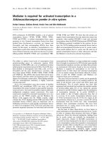
Plain chest radiograph (Fig. 1A) showed bilateral basilar
infiltrates and a peripheral reticulonodular pattern super-
imposed on generalized interstitial changes, involving the
upper lobes as well as lung bases. High Resolution Com-
puted Tomography (HRCT) of the chest (Fig. 1B and 1C)
revealed moderate to severe thickening of intralobular
septa, septal line formation, parenchymal band formation
and peribronchial thickening, ground glass opacities and
mild mediastinal lymphadenopathy (likely reactive, larg-
est lymph node being 1.1 cm) was noted. This was con-
sistent with idiopathic interstitial pneumonia (IIP)
without a specific pattern. At this time, treatment with lev-
ofloxacin was continued and solumedrol was added for
empirical therapy. Computed Tomogram (CT) of the
abdomen/pelvis was normal with no evidence of retro-
peritoneal lymphadenopathy. Despite steroid therapy, the
patient's respiratory status deteriorated over the next day
requiring intubation and mechanical ventilation. Conse-
quently, consent for wedge biopsy of the lung was
obtained for a pathological diagnosis to guide further
therapy.
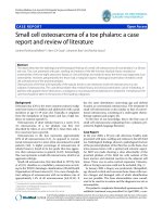
A right lung wedge biopsy was obtained by video-assisted
thoracoscopic surgery (VATS). A polymorphic lymphoid
infiltrate composed of large atypical cells, small lym-
phocytes and many plasma cells was noted, with lym-
phoid cells infiltrating blood vessels and bronchial walls
(Fig. 2A). Multinucleated large Reed Sternberg-like cells
Journal of Hematology & Oncology 2009, 2:39 />Page 4 of 6
(page number not for citation purposes)
were also present along with foci of necrosis. Bronchial
washings showed atypical benign bronchial cells and pul-
monary macrophages with a few rare atypical Reed-Stern-
berg-like cells. Bone marrow biopsy was normal.
Immuno-histochemical studies of lung tissue showed pre-
dominance of T cells expressing CD3, CD5 and CD43
with a smaller population of large atypical cells expressing
CD20 and CD79a (B cell markers) (Fig. 2B). Many
CD138+ plasma cells exhibiting polyclonal staining pat-
tern for kappa and lambda immunoglobulin light chains
were also seen. Bone marrow biopsy revealed normal cel-
lularity with no evidence of lymphoma. In situ hybridiza-
tion for EBV encoded RNA (EBER) was positive within
scattered large lymphoid cells throughout the biopsy spec-
imen (Fig. 2C).
Based on the pathological and immuno-histochemical
findings, a diagnosis of Lymphomatoid Granulomatosis
was made. Treatment with high dose steroids and rituxi-
mab showed significant clinical improvement and he was
extubated 4 days after starting therapy. Treatment was
continued with cyclophosphamide, vincristine, doxoru-
bicin and prednisolone chemotherapy and he showed
gradual but slow resolution of clinical and x-ray findings.
However, he subsequently developed pancytopenia con-
sequent to chemotherapy, septic shock requiring increas-
ing doses of pressors and acute renal failure which did not
respond to aggressive management and he was terminally
weaned per family wishes 4 weeks later. A chest-only
autopsy revealed diffuse alveolar damage, likely second-
ary to sepsis or chemotherapy or both, but no evidence of
residual LG was noted within the lungs (Fig. 2D).
Discussion and Conclusion
This case illustrates that LG can clinically and radiograph-
ically mimic idiopathic interstitial pneumonia on presen-
tation. However, rapid respiratory deterioration, without
any other obvious etiology as in our patient, must prompt
physicians to consider additional differential diagnoses.
Although HRCT is an excellent modality in diagnosing
interstitial pathology, we must be aware of potential mim-
ics and proceed with open or VATS biopsy to obtain a
pathological diagnosis in all patients who do not respond
to empirical therapy. Early diagnosis and aggressive inter-
vention, with interferon therapy, rituximab and chemo-
therapy in high grade LG can be life-saving for a patient
with this rare yet treatable disease.
Abbreviations
LG: Lymphomatoid granulomatosis; LPD: Lymphoprolif-
erative disorder; HRCT: High Resolution Computed Tom-
ography; VATS: Video Assisted Thoracoscopic surgery;
EBV: Epstein-Barr Virus; EBER: Epstein-Barr Virus encoded
RNA; R-CHOP: Rituximab, cyclophosphamide, doxoru-
bicin, vincristine, prednisone combination chemother-
apy; DLBCL: Diffuse large B cell lymphoma; IIP:
Idiopathic Interstitial Pneumonia.
Competing interests
The authors declare that they have no competing interests.
Authors' contributions
AM was involved in conception and writing the manu-
script. AM and KK participated in collection of clinical
data and writing the manuscript. WDC made the patho-
logical diagnosis on the biopsy and performed the
A. Plain chest radiograph shows bibasilar infiltrates with a peripheral reticulonodular pattern superimposed on generalized interstitial changes involving the upper lobes and lung basesFigure 1
A. Plain chest radiograph shows bibasilar infiltrates with a peripheral reticulonodular pattern superimposed
on generalized interstitial changes involving the upper lobes and lung bases. B. (Coronal view) and C. (Axial
View) High Resolution Computed Tomography of the chest shows thickening of intralobular septa, septal line formation,
parenchymal band formation and peribronchial thickening. Mild mediastinal lymphadenopathy is also noted.
Journal of Hematology & Oncology 2009, 2:39 />Page 5 of 6
(page number not for citation purposes)
autopsy. DT was the treating oncologist. HTC was
involved in reviewing the pathology, preparing figures,
and critical appraisal of the manuscript. All authors read
and approved the final manuscript.
Authors' Information
AM is an Internal Medicine Resident at Michigan State
University. KK is a Pulmonary and Critical Care fellow at
Wayne State University. DT is a hematology/oncology
professor at Michigan State University. WC and HTC are
pathologists in the Department of Pathology at Sparrow
Health System, and HTC and DT are also associate profes-
sors at the Michigan State University.
Consent
Written informed consent was obtained from the patient's
next-of-kin for publication of this case report and accom-
panying images. A copy of the written consent is available
for review by the Editor-in-Chief of this journal.
References
1. Liebow AA, Carrington CR, Friedman PJ: Lymphomatoid granulo-
matosis. Hum Pathol 1972, 3:457.
2. Jaffe ES, Wilson WH: Lymphomatoid granulomatosis. In World
Health Organization Classification of tumors. Pathology and Genetics of
Haemotopoietic and Lymphoid Tissues Edited by: Jaffe ES, Harris NL,
Stein H, Vardiman JW. IARC Press, Lyon; 2001:185.
3. Katzenstein ALA, Carrington CB, Liebow AA: Lymphomatoid
granulomatosis: A clinicopathological study of 152 cases.
Cancer 1979, 43:360.
A. Hematoxylin and eosin stain of the lung biopsy shows a polymorphic lymphoid infiltrate composed of large atypical cells, small lymphocytes and many plasma cells, with lymphoid cells infiltrating blood vessels and bronchial wallsFigure 2
A. Hematoxylin and eosin stain of the lung biopsy shows a polymorphic lymphoid infiltrate composed of large
atypical cells, small lymphocytes and many plasma cells, with lymphoid cells infiltrating blood vessels and bron-
chial walls. (Scale bar in A also applies to B-D, original magnification 200×) B. Immunohistochemistry on a section adjacent to
A shows that that many large atypical cells are positive for CD20 (B cell marker). C. In situ hybridization for Epstein-Barr virus
(EBV) encoded RNA (EBER) on a section adjacent to A shows that many large lymphocytes are positive for EBER (stained
blue). D. A chest-only autopsy revealed diffuse alveolar damage in both lungs, with areas of edema, fibrin deposition, and hya-
line membrane formation. There is no evidence of residual lymphomatoid granulomatosis.
Publish with BioMed Central and every
scientist can read your work free of charge
"BioMed Central will be the most significant development for
disseminating the results of biomedical research in our lifetime."
Sir Paul Nurse, Cancer Research UK
Your research papers will be:
available free of charge to the entire biomedical community
peer reviewed and published immediately upon acceptance
cited in PubMed and archived on PubMed Central
yours — you keep the copyright
Submit your manuscript here:
/>BioMedcentral
Journal of Hematology & Oncology 2009, 2:39 />Page 6 of 6
(page number not for citation purposes)
4. McCloskey M, Catherwood M, McManus D, Todd G, Cuthbert R, et
al.: A case of Lymphomatoid granulomatosis masquerading a
lung abscess. Thorax 2004, 59:818-19.
5. Braham E, Ayadi-Kaddour A, Smati B, Ben Mrad S, Besbes M, El Mezni
F: Lymphomatoid granulomatosis mimicking interstitial lung
disease. Respirology 2008, 13:1085-1088.
6. Sheehy N, Bird B, O'Briain DS, Daly P, Wilson G: Synchronous
regression and progression of pulmonary nodules on chest
CT in untreated lymphomatoid granulomatosis. Clin Radiol
2004, 59:451-4.
7. Guinee D, Jaffe E, Kingma D, Wallberg K, Krishnan J, et al.: Pulmo-
nary lymphomatoid granulomatosis. Evidence for a prolifer-
ation of Epstein Barr virus infected B-lymphocytes with a
predominant T-cell component and vasculitis. Am J Surg Pathol
1994, 18:753-64.
8. Lee JS, Tuder R, Lynch DA: Lymphomatoid granulomatosis:
radiological features and pathologic correlations. AJR 2000,
175:1335-39.
9. Jaffe ES, Wilson WH: Lymphomatoid granulomatosis: Patho-
genesis, pathology and clinical implications. Cancer Surv 1997,
30:233.
10. Jordan K, Grothey A, Grothe W, Kegel T, Wolf H-H, et al.: Success-
ful treatment of mediastinal Lymphomatoid granulomatosis
with Rituximab monotherapy. Eur J Haemotol 2005, 74:263-6.
11. Wilson WH, Kingma DW, Raffeld M, Wittes RE, Jaffe ES: Associa-
tion of Lymphomatoid granulomatosis with Espstein-Barr
Viral Infection of B lymphocytes and Response to Interferon-
α2b. Blood 1996, 87:4531-37.
12. Pisani RJ, DeRemee RA: Clinical implications of the histopatho-
logical diagnosis of pulmonary Lymphomatoid granulomato-
sis. Mayo Clin Proc 1990, 65:151.
13. Lemieux J, Bernier V, Martel N, Delage R: Autologous hematopoi-
etic stem cell transplantation for refractory Lymphomatoid
granulomatosis. Hematology 2002, 7:355.
14. Ishiura H, Morikawa M, Hamada M, Watanabe T, Kako S, et al.: Lym-
phomatoid Granulomatosis Involving Central Nervous Sys-
tem Successfully treated With Rituximab alone. Arch Neurol
2008, 65:662-665.
15. Mizuno T, Takanashi Y, Onodera H, Shigeta M, Tanaka N, Yuya H, et
al.: A case of Lymphomatoid granulomatosis/angocentric
immunoproliferative leson with long clinical course and dif-
fuse brain involvement. J Neurol Sci 2003, 213:67-76.