báo cáo khoa học: "Treatment options for patients with triple-negative breast cancer" docx
Bạn đang xem bản rút gọn của tài liệu. Xem và tải ngay bản đầy đủ của tài liệu tại đây (408.76 KB, 11 trang )
Santana-Davila and Perez Journal of Hematology & Oncology 2010, 3:42
/>
REVIEW
JOURNAL OF HEMATOLOGY
& ONCOLOGY
Open Access
Treatment options for patients with triple-negative
breast cancer
Rafael Santana-Davila1, Edith A Perez2*
Abstract
Breast cancer is a heterogeneous disease composed of different subtypes, characterized by their different clinicopathological characteristics, prognoses and responses to treatment. In the past decade, significant advances have
been made in the treatment of breast cancer sensitive to hormonal treatments, as well as in patients whose malignant cells overexpress or amplify HER2. In contrast, mainly due to the lack of molecular targets, little progress has
been made in the treatment of patients with triple-negative breast cancer. Recent improved understanding of the
natural history, pathophysiology, and molecular features of triple-negative breast cancers have provided new
insights into management and therapeutic strategies for women affected with this entity. Ongoing and planned
translational clinical trials are likely to optimize and improve treatment of women with this disease.
Introduction
Breast cancer affected an estimated 192,370 women and
men in 2009, and was responsible for 40,170 deaths during the same year [1]. It is now clear that it is a disease
composed of multiple subgroups characterized by their
pathophysiological features, outcomes, and responses to
treatment. The heterogeneity of this disease underscores
the need for treatments to be tailored for a specific
patient, depending on the molecular characteristics of
their malignancy.
An initial subdivision of patients with breast cancer
can be done by immunohistochemical techniques separating those whose malignant cells express either estrogen (ER) or progesterone receptors (PgR) and those that
do not, as the first two can be treated with endocrine
therapy. Immunohistochemistry (IHC) or fluorescence
in situ hybridization (FISH) can also detect the overexpression (or amplification) of the human epidermal
growth factor receptor 2 (HER2), which can also be targeted therapeutically with antibodies or small molecule
tyrosine kinase inhibitors. Tumors that do not express
ER, PgR, or HER2 are commonly referred to as triplenegative breast cancer (TNBC).
Further understanding of the biology of breast cancer
comes from studies that have identified gene expression
* Correspondence:
2
Division of Hematology and Oncology Mayo Clinic, 4500 San Pablo Road,
Jacksonville, Florida. 32224. USA
Full list of author information is available at the end of the article
profiles that provide insight into therapeutic strategies,
although more work remains to be done [2-6]. Perou
and colleagues [4,5] proposed an initial classification in
which breast cancer was subdivided into four groups:
Luminal types A and B, HER2 positive cancer and
basal-like subset. Luminal type A is characterized by
neoplasms that express ER and have a low-grade histology. Luminal type B is composed mostly of tumors with
low ER expression and a higher grade compared to
those with type A. HER2 positive cancers are distinguished by the amplification of the HER2 gene. Finally,
the basal-like subset, which is composed mostly of ER
and HER2 negative cancers. This is, of course, an oversimplification of the heterogeneity of breast cancer,
albeit helpful based on the current status of knowledge.
TNBC and Basal-like Cancer
Although the terms TNBC and basal-like cancer are
often used interchangeably, it is important to clarify
that not all TNBCs belong to the basal-like subtype
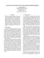
(Figure 1). Although one of the key features of most
basal-like cancers is the low expression of hormonal
receptors and HER2 related genes, they are also characterized by other features. This was illustrated in the
study by Parker and collaborators who, in an attempt to
incorporate gene expression based “intrinsic” molecular
subtypes for prognosis and prediction of chemotherapy
benefit, applied a 50 gene expression signature (PAM50)
to a cohort of 1,004 patients, of which 626 had ER
© 2010 Santana-Davila and Perez; licensee BioMed Central Ltd. This is an Open Access article distributed under the terms of the
Creative Commons Attribution License ( which permits unrestricted use, distribution, and
reproduction in any medium, provided the original work is properly cited.
Santana-Davila and Perez Journal of Hematology & Oncology 2010, 3:42
/>
Page 2 of 11
express some of the basal cell markers such as cytokeratin 5 (CK5) and 17 (CK17), as well as caveolin-1, EGFR,
B-crystallin, P-cadherin, and c-KIT [15-17]. This does
not necessarily imply that basal-like tumors arise from
the myoepithelial layer; this area remains the focus of
intensive investigation[18].
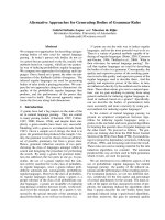
Figure 1 Schematic diagram the represents the significant
overlap that exists between triple-negative (TNBC), basal-like
breast cancer (BLBC) and breast cancer that arises in patients
who have a BRCA mutation. While the majority of cancers that
are TNBC are also BLBC. Non-basal triple-negative breast cancer also
exists. Similarly most breast cancers that occur in women with BRCA
mutations are TNBC and of the BLBC subtype, however this overlap
is not complete.
positive disease. In this group the majority (73%) were
luminal (A or B), but 11% were HER2-enriched, 5%
were basal-like, and 12% were normal-like [7]. Similarly,
in the ER negative group, 11% of the tumors were found
to be luminal, 32% HER2-enriched, 50% basal-like, and
7% normal-like. Their work, and that of others, demonstrated that ER and HER2 status is not an accurate surrogate for true intrinsic subtype status (differentiation
between luminal A, luminal B, HER2 and basal-like) [8].
As we wait for validation and further research related
to several proposed gene profiles, several investigators
have used expression of basal/myoepithelial cell proteins
identified by immunohistochemical staining, as a surrogate of gene expression [9-11]. The most widely used
panel is based on the expression of cytokeratin 5/6
(CK5/6) and/or the epidermal growth factor receptor
(EGFR) in tumors that are triple-negative [12]; however,
no uniform consensus exists as to what is the optimal
immunnohistochemical panel to identify basal-like
breast cancer. Thus TNBC, despite having an imperfect
correlation [9,13,14], is generally used clinically as a
marker of being a basal-like cancer.
Rationale for the Term Basal-like Breast Cancer
The normal human breast ducts and acini are composed
of two cell layers, which include an inner luminal cell
population and a distinct outer cell layer juxtaposed to
the basement membrane, named the myoepithelial or
basal layer. Cells from each layer have a distinct immunophenotypic profile. Basal-like cancer cells commonly
Clinicopathological Characteristics
Approximately 15-20% of breast cancers are TNBC
[19,20], the majority of which are from the basal-like
subtype. Basal-like cancers are typically associated with
a higher histological grade, marked cellular pleomorphism, a high Ki67 index, increase mitotic activity and atypical mitotic figures[9,21-24]. At the genomic level, in
comparison with other subtypes, the basal-like subtype
is distinguished by genomic instability, an increase in
DNA copy number changes, and frequent low-level
gains and deletions [25,26]. This subtype is also characterized by deregulation of important components of the
cell cycle process, such as the RB pathway [27] and frequent p53 abnormalities [3,21,28]. Mutations in this
gene have been reported in up to 82% of patients, compared to only 13% in the luminal A group [3].
Relationship with BRCA-related Cancers
Patients with germline mutations in the BRCA genes are
at risk of developing breast, ovarian, pancreatic, and
prostate cancers, among other malignancies. The products of the BRCA genes have a variety of roles, including those relating to DNA-repair mechanisms. Cells that
lack a functional BRCA1 or BRCA2 have a deficiency in
the repair of DNA double-strand breaks, which is probably one of the mechanisms behind their association
with increased cancer predisposition [29]. There are
interesting and relevant similarities between cancers that
arise in carriers of BRCA gene mutations and basal-like
breast cancer that have led to the hypothesis that they
share defects of the BRCA or related pathways. When
breast cancer arises in patients with BRCA mutations,
the majority are triple negative [30], and of the basallike subtype in 80-90% of the cases [2,31-33]. BRCA1related cancers similar to basal-like breast cancers tend
to be characterized by a high frequency of p53 mutations [3,34] and genomic instability [26,32].
Mutations in the BRCA genes are found to be rare in
sporadic breast cancers [35,36], however, recent studies
have suggested that alteration in the expression or function of these or related DNA-pathway repair genes is
important in the development of sporadic breast cancer
[32]. Methylation of the BRCA1 promoter, which leads to
a reduced expression of BRCA1, has been reported to be
present in 11 to 14% of sporadic breast cancers [37,38],
where it is associated with a higher histological grade and
a triple-negative phenotype [37-39]. In basal-like breast
Santana-Davila and Perez Journal of Hematology & Oncology 2010, 3:42
/>
cancer, the overexpression of ID4, a negative regulator of
BRCA1, appears to also play a significant role in the
deregulation of BRCA1 [40], but further studies are
needed to confirm these findings. Other genes associated
with BRCA1 in DNA repair by homologous recombination, such as RAD51, Fanconi’s anemia proteins, CHEK2
and ATM, have also been found to be implicated in
breast carcinogenesis. Whether alterations in these genes
also have a role in the development of basal-like breast
cancer is currently unknown and poses an interesting
question for further study.
Patients’ Characteristics and Prognosis
TNBC and basal-like cancers are associated with a
younger age at presentation, having a mean age of 53
years old, compared to 58 years old for other subgroups
in one study [20]. Race also appears to be a risk factor,
as it is more frequent in premenopausal patients of
African-American heritage [19,41]. Patients with these
subtypes generally present at a similar stage compared
to other tumors [19], but appear to have an inferior outcome [42,43]. This inferior prognosis has been found to
be independent of several other factors such as tumor
grade, size and nodal status [44].
Basal-like cancers are characterized by a distinct pattern of metastasis with a predilection to metastasize to
brain and lungs and less incidence of metastases to
bone, liver and non-regional lymph nodes [45]. Patients
with basal-like breast cancer appear have a higher incidence of locoregional failures after initial surgical treatment when compared with Luminal type A patients
[46]. Interestingly, in the study by Voduc and colleagues
which used IHC to determine subtype, those cancers
that were triple-negative and negative for the expression
of EGFR and CK5/6 (non-basal triple-negatives), had a
lower incidence of locoregional relapse when compared
to the basal-like subtype [46].
Therapy
As stated above, there is no currently accepted specific
molecular targeted agent against TNBC; however, they
do appear to be responsive to chemotherapy [47]. Posthoc analysis of several studies with diverse chemotherapy agents have shown that it is TNBC patients who
seem to benefit the most from cytotoxic agents in the
adjuvant setting [48-50]. Similarly, when neoadjuvant
chemotherapy is administered, patients with TNBC and
HER2 amplification have better response rates, as well
as more frequent incidence of a pathological complete
response (pCR) [51-53]; as high as 45% in a study that
used 5-fluorouracil (5-FU), doxorubicin and cyclophosphamide [51]. Unfortunately, this does not translate
into a better overall survival, mostly because those
patients who did not achieve a complete response (CR)
Page 3 of 11
tend to relapse sooner than patients with other breast
cancer subtypes.
There is no preferred agent in the neoadjuvant setting,
although more data are definitely needed related to
whether anthracycline/taxane based therapies should
remain the standard approach [63].
Platinum Agents
A group of agents particularly interesting for management of patients with TNBC are the platinum compounds, partially based on their ability to bind directly
to DNA. This causes the DNA to crosslink, resulting in
double-strand DNA breakage[54,55]. It has been theorized and shown in preclinical models, that neoplastic
cells harboring BRCA mutations, and thus lacking one
of the mechanisms to repair damaged DNA, are consequently more susceptible to agents that induce DNA
damage [56]. A very small retrospective study that
included women with BRCA mutations who received
neo-adjuvant treatment demonstrated that patients who
received cisplatin had a higher degree of pCR (83% vs.
less than 22% with other regimens, not including platins) [57]. Although these data are intriguing, they
should be taken with caution as the study only had 12
patients in the cisplatin cohort and it was retrospective.
In the neoadjuvant setting, single agent cisplatin was
evaluated in 28 patients with TNBC which led to a pCR
in six (22%) women[58]. This same group of investigators conducted a separate neoadjuvant study, this time
adding bevacizumab to cisplatin. Preliminary results
indicated that this combination led to a pCR in 15% (7
out of 46 patients) [59]. These results are somewhat disappointing, as the proportions of complete responses
are significantly less than that achieved with multiagent
neoadjuvant chemotherapy (30 - 45% in other studies)
[51,52]. Because of the biochemical similarities between
BRCA related breast cancers and TNBC, it has been
hypothesized that TNBCs are also specifically sensitive
to platinum agents. This remains a controversial topic,
as to date there is no randomized, controlled study that
has demonstrated the benefit of platinum versus other
agents.
Cisplatin has also been coupled with other cytotoxic
agents for neoadjuvant treatment; when used with epirubicin and 5-FU a pCR of 40% was achieved [60]. In a
similar study of 74 patients treated with cisplatin, epirubicin and paclitaxel with G-CSF support, a remarkably
high rate of pCR (65%) was seen [61]. These are
encouraging results that merit further validation and
testing. At the current time, however, platinum agents
in the neoadjuvant setting cannot be recommended over
established regimens outside of a clinical trial. Two current neoadjuvant randomized studies should help clarify
the role of platinum agents in the these situations,
Santana-Davila and Perez Journal of Hematology & Oncology 2010, 3:42
/>
CALGB40603 (described below in the bevacizumab
section), and a Spanish Breast Cancer Research Group
study [62]. In both of these trials, patients will be randomized to receive carboplatin as part of their preoperative
treatment, in the Spanish study patients will receive
epirubicin and cyclophosphamide for 4 cycles and then
be randomized to receive docetaxel or carboplatin
(NCT00432172).
In patients with metastatic disease, two clinical trials
will help clarify the role of platinum agents. First, the
Phase II Translational Breast Cancer Research Consortium 009 trial (NCT00483223) is evaluating the
response rate of metastatic breast cancer patients treated
with cisplatin or carboplatin. This trial will also evaluate,
prospectively, the expression of p63/p73 as a potential
biomarker of platinum sensitivity. These proteins are
part of the p53 family. They are expressed in approximately one-third of patients with TNBC, and their coexpression in breast cancer cell lines results in 10-fold
to 100-fold greater sensitivity to platinum chemotherapy
[63]. The second study is a phase III trial currently
underway in the UK, which will randomize 400 women
with TNBC to carboplatin or docetaxel with crossover
at progression (NCT00532727).
Anti-tubulin Agents
A new agent that has recently been added to the armamentarium of drugs available for the treatment of breast
cancer is ixabepilone. Similar to taxanes, ixabepilone stabilizes microtubules and causes cell cycle arrest and
apoptosis [64]. It has the advantage of bypassing the
resistance mechanisms associated with drug efflux
pumps and specific paclitaxel resistance associated with
b-tubulin [64]. Its use has been studied as a single agent
in four distinct clinical trials that included 288 patients,
of whom 113 (39%) had TNBC. Two phase III clinical
trials have also compared ixabepilone coupled with capecitabine versus capecitabine alone. A subset analysis of
women with TNBC identified an improved overall
response for this combination of 31% versus 15% and a
progression free survival (PFS) of 4.2 months versus 1.7
months (HR 0.63; 95% CI 0.52-0.77) [65]. In the neoadjuvant setting, treatment with ixabepilone led to a pCR in
26% of the 42 women with TNBC [66]. A retrospective
analysis of this study analyzed the expression of bIIItubulin, a b-tubulin, whose expression is correlated with
resistance to taxanes. Patients with a basal-like phenotype
had a higher expression of bIII-tubulin, and its expression was predictive of response to therapy in the overall
population (area under the curve of 0.66) [67]. Further
studies of the potential role of this as a predictive marker
are needed before conclusions can be reached.
Another novel mitotic inhibitor currently being studied for the treatment of breast cancer is eribulin. A
Page 4 of 11
recently reported phase III trial compared eribulin
against several investigator-chosen regimens for the
treatment of women with refractory metastatic breast
cancer. An improved survival in favor of those women
taken eribulin was demonstrated (Median OS was 13.1
vs. 10.7, HR 0.81; 95% CI 0.66-0.99) [68]. Of the patients
enrolled in this trial, 20% had TNBC. The subset analysis for this trial has not been yet reported.
Targeted Therapies
Poly(ADP)ribose polymerase 1 (PARP1) is a nuclear protein that is recruited to the site of damage after the
induction of both single and double stranded DNA
breaks. PARP1 catalyzes the transfer of ADP-ribose
polymers from NAD+ to target proteins, which in turn
modulate DNA restoration by activating and recruiting
important components of base excision repair pathway,
such as XRCC1 [69,70]. PARP1 also contributes to the
modification of histones, which leads to local chromatin
remodeling, allowing access of DNA repair proteins to
the repair site [71]. The inhibition of PARP1 potentiates
the effects of ionizing radiation, DNA methylating
agents, topoisomerase I inhibitors, and platinum compounds [70,72]. When PARP1 is inhibited in normal
cells, DNA repair is done through the homologousrecombination pathway, a process for which BRCA is a
key factor [73]. Cells that are deficient in BRCA are
more dependent on PARP1 to maintain genomic integrity. Its inhibition thus leads to synthetic lethality
[32,74,75], a process that occurs when inactivation of
either of the two genes individually has no effect but
combining the mutations is deadly to the cell [69,73].
Several PARP1 inhibitors are at different stages of clinical development, olaparib (previously known as
AZD2281) has been evaluated in a phase 1 study where
60 patients with breast cancer were enrolled, of these,
nine patients had an objective response. In addition, all
the responders had abnormalities in one of the BRCA
genes. Of the women with breast cancer, three had a
BRCA2 mutation. A complete response that lasted in
excess of 60 weeks also occurred in one of the BRCA
carriers and another one had stable disease for 7
months. Olaparib was further evaluated in a phase II
study that enrolled 54 patients with known BRCA mutations and breast cancer. The first 27 women enrolled
received 400 mg twice per day, of which 11 (41%)
experienced a response with a median PFS of 5.7
months. A second cohort of 27 women received 100 mg
of olaparib twice per day (the lowest dose that showed
an active pharmacodynamic profile in the phase 1
study). In this group, 6 patients (22%) experienced a
response with a median PFS of 3.8 months. This agent
was fairly well tolerated, with nausea and fatigue being
the most common adverse events[76]. A recent phase I
Santana-Davila and Perez Journal of Hematology & Oncology 2010, 3:42
/>
study reported by Dent et al. at the 2010 American
Society of Clinical Oncology (ASCO) meeting demonstrated that it was not feasible to administer the 200 mg
daily dose of olaparib in combination with weekly paclitaxel due to significant myelosuppression, in spite of
prophylaxis with growth factor support [77]. Several
clinical trials using olaparib in women with BRCA deficient cancers are in different stages of development (see
table 1)
The similarities described above between the breast
cancers that arise in patients with BRCA mutations and
basal-like cancer have led to the hypothesis that a deficiency in a component of the BRCA pathway plays an
important role in basal-like cancers, thus inhibition of
PARP1 could also be an important therapeutic strategy.
In a phase 2 study, 120 patients were randomized (1:1)
to gemcitabine and carboplatin alone or the same combination plus the intravenous PARP1 inhibitor, iniparib
(BSI-201). Gemcitabine (1000 mg/m2; IV) and carboplatin (AUC = 2; IV) were given on days 1 and 8, and iniparib (5.6 mg/kg; IV) on days 1, 4, 8, and 11 every 21
days. The addition of iniparib led to an improved
response rate, (16% vs. 48%; p = 0.002) as well as PFS
(3.3 vs. 6.9 months; p < 0.0001) and overall survival
(12.2 vs. 7.7 months; p = 0.005; HR = 0.5; 95% CI, 0.30
- 0.82)[78-80]. The addition of iniparib was well tolerated, with no evidence of neither incremental nor new
adverse effects compared to the standard arm. A confirmatory phase III clinical trial using the same regimen
has completed accrual in February 2010, with data
expected in 2011(NCT00938652). Iniparib is also being
evaluated in 2 neoadjuvant clinical trials, NCT00813956
Page 5 of 11
is a single arm trial that is studying the combination of
iniparib, carboplatin and gemcitabine. The other one is
a Spanish study in which patients will be randomize to
received either iniparib plus paclitaxel versus placlitaxel
alone (NCT01204125).
Veliparib (ABT-888) is another PARP1 inhibitor
being evaluated in breast cancer. A recently reported
study where it was used with temozolamide enrolled
41 women with metastatic disease, of which 23 (56%)
had TNBC. The dose of veliparib was reduced from
40 mg to 30 mg BID due to thrombocytopenia
encountered during the first cycle. In this study the
activity of this combination was limited to those
women who were deficient for BRCA1 (1 partial
response (PR)) and BRCA2 (1 CR and 1 PR). Stable
disease lasting more than 4 months was seen in
4 patients, 2 of who had a BRCA2 mutation. Median
PFS was 1.9 months in all patients and 5.5 months in
those with BRCA mutations[81].
It is intriguing why patients treated with oral PARP1
inhibitors had increased toxicity when these agents were
used with cytotoxic chemotherapy when in contrast
those patients treated with iniparib, an IV PARP1 inhibitor, had no increase toxicity.
Of note is that several studies suggest that PARP1
inhibitors may also be beneficial in other subtypes of
breast cancer beyond TNBC. Analysis of PARP1 expression via IHC was done in tissue microarrays from core
biopsies of 582 patients recruited to the phase III taxane-anthracycline neoadjuvant, GeparTrio trial. PARP1
expression was found to be present in 20% of patients
with hormone receptor positive tumors, 34.4% of
Table 1 Active Clinical Trials with the 3 most developed PARP1 inhibitors
Number
Population studied
Description
Olaparib
NCT01116648 Metastatic TNBC
Phase I/II study to evaluate optimal drug dose and establish activity of cediranib and olaparib.
NCT01078662 BRCA associated breast cancer
Phase II in patients with BRCA related cancers
NCT01115829 Metastatic TNBC
Phase 1 of the combination of cediranib plus olaparib followed by a randomized phase 2 that
evaluates cediranib with or without olaparib.
NCT00516724 Metastatic TNBC
Multi-arm Phase I study that evaluates the safety and efficacy of the combination of olaparib with
carboplatin, olaparib with paclitaxel or olaparib with carboplatin and paclitaxel.
NCT00647062 Metastatic TNBC or BRCA
associated breast cancer
Phase 1 of the combination of olaparib and carboplatin
Veliparib
NCT01104259 Metastatic TNBC or BRCA
associated cancer
Phase I study of the combination of veliparib, vinorelbin and cisplatin
NCT01149083 BRCA associated breast cancer
Randomized phase 2 evaluating veliparib with or without carboplatin.
NCT01042379 Neoadjuvant TNBC
Multi-arm study that evaluates several regimens, One arm contains the combination of paclitaxel,
carboplatin and veliparib
Iniparib
NCT00813956 Neoadjuvant TNBC
Phase 2 study of the combination of gemcitabine, carboplatin and iniparib.
NCT01204125 Neoadjuvant TNBC
Randomized phase 2 study of paclitaxel with or without iniparib
NCT01173497 TNBC with brain metastasis
Phase 2 study of iniparib and irinotecan in women with CNS metastasis.
Santana-Davila and Perez Journal of Hematology & Oncology 2010, 3:42
/>
hormone receptor negative and HER2 positive tumors
and 34.2% of TNBC. A high PARP1 expression was
associated with higher incidence of pCR (26.5%) in
patients in with high PARP1 expression compared to
19.1% and 8.9% in patients with medium or low expression respectively (p < 0.0005) [82]. Another clue that
PARP1 inhibition might be beneficial in other breast
cancer subtypes relates to its relationship with phosphatase and tensin homolog (PTEN), a phosphatase that
contributes to the regulation of cell cycle progression,
cell proliferation and DNA repair. Cell lines deficient in
PTEN have an impaired homologous DNA recombination and increased cytotoxicity with PARP1 inhibition
both in vitro and in vivo An estimated 50% of breast
cancers, irrespective of their triple-receptor negativity,
have a mutation in, or loss of, at least one copy of the
PTEN gene [83,84]. Lastly, deregulation of DNA repair
mechanisms and genomic instability is not exclusive of
triple-negative or basal-like breast cancers, and is also
commonly present in Luminal B and HER2 amplified
tumors [85]. Whether using a PARP1 inhibitor will lead
to synthetic lethality in other breast cancer subtypes is
an intriguing question that is worth exploring.
The use of PARP1 inhibitors is at its infancy and
many questions remain, such as the following: Which
patients are most likely to benefit from this therapy?
Are there any biomarkers that predict response to
PARP1 inhibition besides BRCA mutations? What are
the best cytotoxic agents to use with PARP1 inhibitors?
What are the mechanisms of resistance to these therapies? Should PARP1 inhibitors be continued upon progression of the disease when introducing another
cytotoxic agent? To answer such questions, new translational clinical trials are being designed and conducted.
Other Targeted Agents
Some studies suggest that TNBC expresses EGFR in
nearly half of the cases [9,21]. Its expression is found to
be associated with an inferior outcome. A phase II study
randomized patients to receive either cetuximab, an
EGFR monoclonal antibody, alone followed by carboplatin upon progression versus concomitant cetuximab and
carboplatin. Cetuximab by itself has little activity as a single agent with only 2 of 31 patients achieving a PR. When
used in combination with carboplatin, it led to a PR in 13
patients and overall clinical benefit (CR+PR+ Standard
disease for more than 6 months) in 19 of the 71 patients
enrolled [86]. In a separate randomized phase II study,
the addition of cetuximab to irinotecan and carboplatin
increased RR from 30% to 49% [87]. Samples from
patients enrolled in both of these trials are being studied
to identify biomarkers that correlate with response to
this agent [88]. A fully humanized antibody against
EGFR, panitumumab [89], is currently being evaluated in
Page 6 of 11
combination with gemcitabine and carboplatin in TNBC
(NCT00894504). Another approach to inhibit EGFR
receptor signaling is with the use of small molecules that
inhibit the tyrosine kinase domain of this receptor. Erlotinib, an agent of this kind, is currently being evaluated in
combination with docetaxel and carboplatin in patients
with metastatic TNBC (NCT00491816).
The SRC tyrosine kinase is a non-receptor signaling
kinase that functions downstream of several growth factor receptors including PDGFR, EGFR, IGF-1R, and
HGFR. It plays an important role in cancer cell proliferation and invasion through multiple pathways [90,91].
SRC has been found to be deregulated in breast cancer
[92,93] making it a potentially important therapeutic
target. Using gene expression profiling of breast cancer
cell lines, two groups independently identified a gene
expression pattern that was predictive of sensitivity to
dasatinib, a mutitargeted thyrosine kinase that targets
important oncogenic pathways, including the SRC family
kinases [94,95]. This gene signature was present more
commonly in both cell lines and in patients who had a
triple-negative profile. However, dasatinib has now been
studied as a single agent in TNBC with disappointing
results, with only two out of 43 patients achieving a PR
[96]. A currently ongoing study (NCT00780676) is evaluating whether a gene expression pattern, if present, can
predict a response to dasatinib as a single agent in different subsets of breast cancers [97].
Anti-angiogenic Agents
Angiogenesis is required for tumor growth, invasion and
metastasis in several malignancies, including breast cancer. This process can be targeted with therapeutic purposes through several mechanisms. The vascular
endothelial growth factor (VEGF) is a key mediator of
angiogenesis. Its intratumoral expression has been found
to be markedly elevated in patients with TNBC, compared to other subtypes [98]. Bevacizumab, a humanized
monoclonal antibody against VEGF-A, has proven to be
a valuable agent in metastatic breast cancer in several
phase III clinical trials. In the E2100 study that evaluated this agent along with paclitaxel, patients who were
randomized to the bevacizumab arm had an improved
overall response rate of 48% versus 33% in those who
received paclitaxel alone. The median PFS was significantly longer in those who received bevacizumab (11.4
versus 5.8 months, hazard ratio, 0.42; P < 0.0001), but
the overall survival rate was similar in both groups
(median, 26.7 vs. 25.2 months; hazard ratio, 0.88; p =
0.16)[99]. TNBC was present in 233 of the 763 (31%)
patients enrolled in the E2100 trial. In this group, the
PFS was increased to 10.2 months compared to 4.7
months in the paclitaxel alone arm (HR = 0.45; 95%, CI,
0.33-0.61) [100]. The AVADO trial evaluated docetaxel
Santana-Davila and Perez Journal of Hematology & Oncology 2010, 3:42
/>
alone or with two different doses of bevacizumab (7.5
and 15 mg/kg every 3 weeks). Compared to placebo,
PFS was superior in both bevacizumab arms, the 15 mg/
kg arm was more favorable than the 7.5 mg/kg arm
(median 10.0 months (15 mg/kg), HR = 0.67; P = 0.0002
and 9.0 months (7.5 mg/kg), HR = 0.80; P = 0.0450 versus 8.1 months in the docetaxel alone arm) [101]. There
were 167 women with TNBC (22%), in this subgroup
the addition of bevacizumab at 15 mg/kg led to an
improvement in PFS from 6.0 to 8.1 months (HR =
0.60, 95%; CI, 0.39-0.92) [100]. This occurred even
though the design of this study did not take full advantage of the interaction of chemotherapy plus bevacizumab, as the docetaxel was only used for a pre-set
number of cycles per patient.
The RIBBON-1 trial proved that bevacizumab
increased PFS and overall response rate when compared
to placebo when this agent was used with single agent
taxanes, anthracycline-based regimes, and capecitabine
[102]. A subset analysis of patients with TNBC demonstrated an improvement in PFS when bevacizumab was
used both with capecitabine (6.1 vs. 4.2 months, HR =
0.72, 95% CI, 0.49-1.06). This was also found in the taxane/anthracycline cohort (8.2 to 14.5 months, HR = 0.78,
95% CI, 0.53-1.15) [100]. A recently reported meta analysis of these 3 trials showed, as expected, a PFS advantage
for patients on bevacizumab (HR = 0.64, 95% CI, 0.580.71)[103]. This was also true in a subset analysis of
patients with TNBC (HR = 0.63, 95% CI, 0.52-0.76).
However, no survival advantage was seen in the whole
population or in those with triple-negative disease, which
may be partially explained by the fact that there was a
60% crossover to adding bevacizumab for patients who
developed tumor progression after receiving chemotherapy plus placebo. Moreover, it is important to document
that this meta-analysis did demonstrate a statistically significant improvement in one-year survival for patients
assigned to chemotherapy and bevacizumab versus chemotherapy and placebo. Bevacizumab is currently being
evaluated in TNBC by several independent studies.
CALGB 40603 (NCT00861705) is a phase II neoadjuvant
study in which patients will undergo two randomizations
in order to receive paclitaxel with or without carboplatin
and this combination with or without bevacizumab. The
second study, BEATRICE (NCT00528567) is a phase III
adjuvant study where several chemotherapy regimens
and different doses of bevacizumab are being evaluated
in patients with TNBC. This trial recently completed
accrual and the results are eagerly awaited.
Page 7 of 11
response in 11% of a heavily pretreated cohort of metastatic breast cancer patients [104]. Unfortunately, two
phase III studies have now shown that combining sunitinib with docetaxel or capecitabine does not offer any
benefit in prolonging PFS compared to the cytotoxic
regimen alone in patients with advanced breast cancer
[105,106]. This agent is currently being evaluated in
addition to carboplatin and paclitaxel as adjuvant treatment for TNBC (NCT00887575).
The mammalian target of rapamycin (mTOR) is a protein that is downstream of the PI3K/AKT pathway and,
when activated, promotes protein synthesis and angiogenesis [107]. Everolimus, an mTOR inhibitor, has a
12% overall RR when used as a single agent in heavily
pretreated patients with metastatic breast cancer [108].
It is currently being evaluated as a single agent in a
phase II clinical trial in patients with metastatic TNBC
(NCT00827567), and in a placebo controlled neoadjuvant randomized phase II study along with cisplatin and
paclitaxel in patients with stages II and III TNBC
(NCT00930930).
Therapy Based on the Androgen Receptor
In an effort to further study the heterogeneity of TNBC,
Doane and colleagues [109] conducted a genome wide
gene expression profiling study of 99 patients with
breast cancer, 41 of whom had triple negative disease.
They noticed that nine of the patients with TNBC clustered together with the ER positive group. When focusing on only those patients with TNBC, the nine ERdiscordant samples closely correlated with each other
and were contained in a single cluster with only one
additional case. Further characterization of this subtype
of TNBC showed that it had a molecular resemblance
to ER positive tumors and expressed genes that are targets of the ER. Half of the tumors in this group
expressed the androgen receptor. Subsequently, these
investigators identified MDA-MB-453 as a cell line that
had a molecular phenotype similar to the previously
described subtype of TNBC. This cell line, as expected,
did not respond to estrogen administration but in contrast had a proliferative effect with androgen stimulation
in an ER-independent but AR-dependent manner. Several studies have established that between 10-35% of
TNBC express the androgen receptor [110-112]. These,
and other, preclinical data have given support to the
development of a phase II trial using bicalutamide, an
antiandrogen, in the treatment of TNBC that are androgen receptor positive (NCT00468715).
Other Antiangiogenic and Multikinase Inhibitors
Other Targets
Another multikinase inhibitor with antiangiogenic properties, sunitinib, has been evaluated as a single agent in
a phase II study, where it was found to induce a
New studies that utilize high throughput technologies to
assess gene expression and genomic copy number variations have provided insight into the heterogeneity of
Santana-Davila and Perez Journal of Hematology & Oncology 2010, 3:42
/>
TNBC and have successfully identified potential new
targets [113]. Among the targets is the fibroblast growth
receptor (FGFR), which is part of an important signaling
pathway found to be deregulated in several malignancies
[114]. FGFR1 is overexpressed in up to 5.5% of patients
with TNBC [115]. The FGFR2 gene has alleles that have
been associated with risk of developing postmenopausal
breast cancer [116]. This gene has also been found to be
overexpressed in 5% of patients with TNBC[114]. Several tyrosine kinase inhibitors that target the FGFR
receptor are currently in different stages of development
[114]. One of these agents, TKI258, is currently being
evaluated in a phase II study of women with HER2
negative breast cancer (NCT00958971).
Another potential target is the RAS-mitogen activated
protein kinase (MAPK) signaling pathway, as it plays a
central role in regulating the growth and survival of
neoplastic cells. The inhibition of this pathway has been
a sought after target in cancer drug development for
several years. Several inhibitors of the mitogen-activated
protein kinase (MEK), an essential component of this
pathway, are in clinical trials for multiple malignancies
including breast cancer [117]. Preclinical studies have
demonstrated that the inhibition of MEK leads to the
activation of the phosphatidylinositol 3-kinase (PI3K)
pathway, a pathway that is also found to be deregulated
in 30% of patients with basal-like breast cancer [84,118].
This feedback counteracts the effects of MEK inhibition
on cell cycle and apoptosis induction [119,120]. Dual
blockade, with inhibitors of both PI3K and MEK, synergistically inhibits growth of basal-like breast cancer cells
in vitro and in vivo [119,120]. This combination needs
to be evaluated in women with TNBC.
Finally, Speers and colleagues have used transcriptional
profiling data to evaluate the expression of the human
kinome. They were able to identify a set of kinases differentially expressed and critical for the growth of ER negative breast cancer [121]. In this study, two groups of
TNBC were identified, a subset defined by kinases
involved in cell cycle checkpoint control and mitogenesis
such as CHK1, BUB1, TTK, and AK2 and another subset
defined by kinases involved in the S6 kinase-signaling
pathway, which includes the RPS6KA3, SMG-1, and
RPS6KA1 kinases. The authors performed siRNA knockdown experiments to downregulate the expression of several of the kinases of interest and established that of the
20 kinases evaluated, 14 were critical for the growth of
ER-negative breast cancer cell lines. The majority of
these kinases are “druggable” targets that could be potentially used for therapeutic purposes.
Conclusion
TNBC, of which the majority of cases belong to the
basal-cell like phenotype of breast cancer, is a
Page 8 of 11
heterogeneous group. Although very likely to change in
the near future, at this time, we still recommend the
combination of doxorubicin plus cyclophosphamide followed by paclitaxel for patients with TNBC, in the adjuvant setting. For patients with metastatic disease, there is
no standard first line agent to recommend, although the
results of the ongoing phase III trial of iniparib may
change the recommended standard of care, therapy
should be individualized for each patient and enrollment
into clinical trials is strongly encouraged. Established
agents such as platinums, ixabepilone, and the antiangiogenic monoclonal antibody bevacizumab are under evaluation in both the adjuvant and the metastatic setting.
The result of studies using new agents, such as inhibitors
of PARP1, tyrosine kinases, and mTOR are currently in
different phases of development and will hopefully
change the paradigm of how we treat patients affected
with TNBC. As new discoveries are being made, current
clinical trials have translational components that we
expect will provide biomarkers useful to effectively discriminate patients into those who are more likely to
respond to certain therapies. The use of newer molecular
techniques have and will continue to be very valuable in
indentifying potential new molecules important for survival of neoplastic cells and that could potentially be targeted in the treatment of women with TNBC.
List of abbreviations
ASCO: American Society of Clinical Oncology; CK: Cytokeratin; EGFR:
epidermal growth factor receptor; ER: Estrogen Receptors; 5-FU: 5fluorouracil; FISH: fluorescence in situ hybridization; HER2: human epidermal
growth factor receptor 2; IHC: Immunohistochemistry; PgR: Progesterone
Receptors; TNBC: Triple-negative Breast Cancer; CR: complete response; PR:
partial response.
Author details
1
Division of Neoplastic Diseases and Related Disorders Medical College of
Wisconsin, 9200 W. Wisconsin Ave, Milwaukee, WI 53226 USA. 2Division of
Hematology and Oncology Mayo Clinic, 4500 San Pablo Road, Jacksonville,
Florida. 32224. USA.
Authors’ contributions
RSD and EAP both made substantial contributions to conception and
design, have both been involved in the drafting of the manuscript and both
have given final approval of the version to be published.
Competing interests
Dr. Rafael Santana-Davila has no competing interest. Dr. Edith A. Perez has
research funding from Genentech, Sanofi-Aventis, and Roche.
Received: 22 July 2010 Accepted: 27 October 2010
Published: 27 October 2010
References
1. Jemal A, Siegel R, Ward E, Hao Y, Xu J, Thun MJ: Cancer statistics, 2009. CA
Cancer J Clin 2009, 59:225-249.
2. Sorlie T, Tibshirani R, Parker J, Hastie T, Marron JS, Nobel A, Deng S,
Johnsen H, Pesich R, Geisler S, et al: Repeated observation of breast
tumor subtypes in independent gene expression data sets. Proc Natl
Acad Sci USA 2003, 100:8418-8423.
Santana-Davila and Perez Journal of Hematology & Oncology 2010, 3:42
/>
3.
4.
5.
6.
7.
8.
9.
10.
11.
12.
13.
14.
15.
16.
17.
18.
19.
20.
21.
22.
23.
24.
25.
Sorlie T, Perou CM, Tibshirani R, Aas T, Geisler S, Johnsen H, Hastie T,
Eisen MB, van de Rijn M, Jeffrey SS, et al: Gene expression patterns of
breast carcinomas distinguish tumor subclasses with clinical
implications. Proc Natl Acad Sci USA 2001, 98:10869-10874.
Perou CM, Sorlie T, Eisen MB, van de Rijn M, Jeffrey SS, Rees CA, Pollack JR,
Ross DT, Johnsen H, Akslen LA, et al: Molecular portraits of human breast
tumours. Nature 2000, 406:747-752.
Hu Z, Fan C, Oh DS, Marron JS, He X, Qaqish BF, Livasy C, Carey LA,
Reynolds E, Dressler L, et al: The molecular portraits of breast tumors are
conserved across microarray platforms. BMC Genomics 2006, 7:96.
Sotiriou C, Pusztai L: Gene-expression signatures in breast cancer. N Engl J
Med 2009, 360:790-800.
Parker JS, Mullins M, Cheang MC, Leung S, Voduc D, Vickery T, Davies S,
Fauron C, He X, Hu Z, et al: Supervised risk predictor of breast cancer
based on intrinsic subtypes. J Clin Oncol 2009, 27:1160-1167.
Weigelt B, Mackay A, A’Hern R, Natrajan R, Tan DS, Dowsett M, Ashworth A,
Reis-Filho JS: Breast cancer molecular profiling with single sample
predictors: a retrospective analysis. Lancet Oncol 2010, 11:339-349.
Nielsen TO, Hsu FD, Jensen K, Cheang M, Karaca G, Hu Z, HernandezBoussard T, Livasy C, Cowan D, Dressler L, et al: Immunohistochemical and
clinical characterization of the basal-like subtype of invasive breast
carcinoma. Clin Cancer Res 2004, 10:5367-5374.
Banerjee S, Reis-Filho JS, Ashley S, Steele D, Ashworth A, Lakhani SR,
Smith IE: Basal-like breast carcinomas: clinical outcome and response to
chemotherapy. J Clin Pathol 2006, 59:729-735.
Rakha EA, El-Sayed ME, Green AR, Paish EC, Lee AH, Ellis IO: Breast
carcinoma with basal differentiation: a proposal for pathology definition
based on basal cytokeratin expression. Histopathology 2007, 50:434-438.
Cheang MC, Voduc D, Bajdik C, Leung S, McKinney S, Chia SK, Perou CM,
Nielsen TO: Basal-like breast cancer defined by five biomarkers has
superior prognostic value than triple-negative phenotype. Clin Cancer Res
2008, 14:1368-1376.
Bertucci F, Finetti P, Cervera N, Esterni B, Hermitte F, Viens P, Birnbaum D:
How basal are triple-negative breast cancers? International Journal of
Cancer 2008, 123:236-240.
Rakha EA, Tan DS, Foulkes WD, Ellis IO, Tutt A, Nielsen TO, Reis-Filho JS: Are
triple-negative tumours and basal-like breast cancer synonymous? Breast
Cancer Res 2007, 9:404, author reply 405.
Rakha EA, Elsheikh SE, Aleskandarany MA, Habashi HO, Green AR, Powe DG,
El-Sayed ME, Benhasouna A, Brunet JS, Akslen LA, et al: Triple-negative
breast cancer: distinguishing between basal and nonbasal subtypes. Clin
Cancer Res 2009, 15:2302-2310.
Turner N, Lambros MB, Horlings HM, Pearson A, Sharpe R, Natrajan R,
Geyer FC, van Kouwenhove M, Kreike B, Mackay A, et al: Integrative
molecular profiling of triple negative breast cancers identifies amplicon
drivers and potential therapeutic targets. Oncogene 2010, 29:2013-2023.
Weigelt B, Reis-Filho JS: Histological and molecular types of breast
cancer: is there a unifying taxonomy? Nat Rev Clin Oncol 2009, 6:718-730.
Stingl J, Caldas C: Molecular heterogeneity of breast carcinomas and the
cancer stem cell hypothesis. Nat Rev Cancer 2007, 7:791-799.
Carey LA, Perou CM, Livasy CA, Dressler LG, Cowan D, Conway K, Karaca G,
Troester MA, Tse CK, Edmiston S, et al: Race, breast cancer subtypes, and
survival in the Carolina Breast Cancer Study. JAMA 2006, 295:2492-2502.
Dent R, Trudeau M, Pritchard KI, Hanna WM, Kahn HK, Sawka CA, Lickley LA,
Rawlinson E, Sun P, Narod SA: Triple-negative breast cancer: clinical
features and patterns of recurrence. Clin Cancer Res 2007, 13:4429-4434.
Rakha EA, El-Sayed ME, Green AR, Lee AH, Robertson JF, Ellis IO: Prognostic
markers in triple-negative breast cancer. Cancer 2007, 109:25-32.
Livasy CA, Karaca G, Nanda R, Tretiakova MS, Olopade OI, Moore DT,
Perou CM: Phenotypic evaluation of the basal-like subtype of invasive
breast carcinoma. Mod Pathol 2006, 19:264-271.
Rakha EA, Putti TC, Abd El-Rehim DM, Paish C, Green AR, Powe DG, Lee AH,
Robertson JF, Ellis IO: Morphological and immunophenotypic analysis of
breast carcinomas with basal and myoepithelial differentiation. J Pathol
2006, 208:495-506.
Dawson SJ, Provenzano E, Caldas C: Triple negative breast cancers: clinical
and prognostic implications. Eur J Cancer 2009, 45(Suppl 1):27-40.
Chin K, DeVries S, Fridlyand J, Spellman PT, Roydasgupta R, Kuo WL,
Lapuk A, Neve RM, Qian Z, Ryder T, et al: Genomic and transcriptional
aberrations linked to breast cancer pathophysiologies. Cancer Cell 2006,
10:529-541.
Page 9 of 11
26. Andre F, Job B, Dessen P, Tordai A, Michiels S, Liedtke C, Richon C, Yan K,
Wang B, Vassal G, et al: Molecular characterization of breast cancer with
high-resolution oligonucleotide comparative genomic hybridization
array. Clin Cancer Res 2009, 15:441-451.
27. Gauthier ML, Berman HK, Miller C, Kozakeiwicz K, Chew K, Moore D,
Rabban J, Chen YY, Kerlikowske K, Tlsty TD: Abrogated response to cellular
stress identifies DCIS associated with subsequent tumor events and
defines basal-like breast tumors. Cancer Cell 2007, 12:479-491.
28. Troester MA, Herschkowitz JI, Oh DS, He X, Hoadley KA, Barbier CS,
Perou CM: Gene expression patterns associated with p53 status in breast
cancer. BMC Cancer 2006, 6:276.
29. Narod SA, Foulkes WD: BRCA1 and BRCA2: 1994 and beyond. Nat Rev
Cancer 2004, 4:665-676.
30. Foulkes WD, Metcalfe K, Sun P, Hanna WM, Lynch HT, Ghadirian P, Tung N,
Olopade OI, Weber BL, McLennan J, et al: Estrogen receptor status in
BRCA1- and BRCA2-related breast cancer: the influence of age, grade,
and histological type. Clin Cancer Res 2004, 10:2029-2034.
31. Lakhani SR, Reis-Filho JS, Fulford L, Penault-Llorca F, van der Vijver M,
Parry S, Bishop T, Benitez J, Rivas C, Bignon YJ, et al: Prediction of BRCA1
status in patients with breast cancer using estrogen receptor and basal
phenotype. Clin Cancer Res 2005, 11:5175-5180.
32. Turner N, Tutt A, Ashworth A: Hallmarks of ‘BRCAness’ in sporadic
cancers. Nat Rev Cancer 2004, 4:814-819.
33. Foulkes WD, Stefansson IM, Chappuis PO, Begin LR, Goffin JR, Wong N,
Trudel M, Akslen LA: Germline BRCA1 mutations and a basal epithelial
phenotype in breast cancer. J Natl Cancer Inst 2003, 95:1482-1485.
34. Hartman AR, Ford JM: BRCA1 and p53: compensatory roles in DNA repair.
J Mol Med 2003, 81:700-707.
35. Futreal PA, Liu Q, Shattuck-Eidens D, Cochran C, Harshman K, Tavtigian S,
Bennett LM, Haugen-Strano A, Swensen J, Miki Y, et al: BRCA1 mutations in
primary breast and ovarian carcinomas. Science 1994, 266:120-122.
36. Teng DH, Bogden R, Mitchell J, Baumgard M, Bell R, Berry S, Davis T, Ha PC,
Kehrer R, Jammulapati S, et al: Low incidence of BRCA2 mutations in
breast carcinoma and other cancers. Nat Genet 1996, 13:241-244.
37. Esteller M, Silva JM, Dominguez G, Bonilla F, Matias-Guiu X, Lerma E,
Bussaglia E, Prat J, Harkes IC, Repasky EA, et al: Promoter hypermethylation
and BRCA1 inactivation in sporadic breast and ovarian tumors. J Natl
Cancer Inst 2000, 92:564-569.
38. Catteau A, Harris WH, Xu CF, Solomon E: Methylation of the BRCA1
promoter region in sporadic breast and ovarian cancer: correlation with
disease characteristics. Oncogene 1999, 18:1957-1965.
39. Rice JC, Ozcelik H, Maxeiner P, Andrulis I, Futscher BW: Methylation of the
BRCA1 promoter is associated with decreased BRCA1 mRNA levels in
clinical breast cancer specimens. Carcinogenesis 2000, 21:1761-1765.
40. Turner NC, Reis-Filho JS, Russell AM, Springall RJ, Ryder K, Steele D,
Savage K, Gillett CE, Schmitt FC, Ashworth A, Tutt AN: BRCA1 dysfunction
in sporadic basal-like breast cancer. Oncogene 2007, 26:2126-2132.
41. Morris GJ, Naidu S, Topham AK, Guiles F, Xu Y, McCue P, Schwartz GF,
Park PK, Rosenberg AL, Brill K, Mitchell EP: Differences in breast carcinoma
characteristics in newly diagnosed African-American and Caucasian
patients: a single-institution compilation compared with the National
Cancer Institute’s Surveillance, Epidemiology, and End Results database.
Cancer 2007, 110:876-884.
42. Rakha EA, Reis-Filho JS, Ellis IO: Basal-like breast cancer: a critical review. J
Clin Oncol 2008, 26:2568-2581.
43. Schneider BP, Winer EP, Foulkes WD, Garber J, Perou CM, Richardson A,
Sledge GW, Carey LA: Triple-negative breast cancer: risk factors to
potential targets. Clin Cancer Res 2008, 14:8010-8018.
44. Gluz O, Liedtke C, Gottschalk N, Pusztai L, Nitz U, Harbeck N: Triplenegative breast cancer–current status and future directions. Ann Oncol
2009, 20:1913-1927.
45. Fulford LG, Reis-Filho JS, Ryder K, Jones C, Gillett CE, Hanby A, Easton D,
Lakhani SR: Basal-like grade III invasive ductal carcinoma of the breast:
patterns of metastasis and long-term survival. Breast Cancer Res 2007, 9:R4.
46. Voduc KD, Cheang MC, Tyldesley S, Gelmon K, Nielsen TO, Kennecke H:
Breast cancer subtypes and the risk of local and regional relapse. J Clin
Oncol 2010, 28:1684-1691.
47. Kriege M, Seynaeve C, Meijers-Heijboer H, Collee JM, Menke-Pluymers MB,
Bartels CC, Tilanus-Linthorst MM, Blom J, Huijskens E, Jager A, et al:
Sensitivity to first-line chemotherapy for metastatic breast cancer in
BRCA1 and BRCA2 mutation carriers. J Clin Oncol 2009, 27:3764-3771.
Santana-Davila and Perez Journal of Hematology & Oncology 2010, 3:42
/>
48. Citron ML, Berry DA, Cirrincione C, Hudis C, Winer EP, Gradishar WJ,
Davidson NE, Martino S, Livingston R, Ingle JN, et al: Randomized trial of
dose-dense versus conventionally scheduled and sequential versus
concurrent combination chemotherapy as postoperative adjuvant
treatment of node-positive primary breast cancer: first report of
Intergroup Trial C9741/Cancer and Leukemia Group B Trial 9741. J Clin
Oncol 2003, 21:1431-1439.
49. Hayes DF, Thor AD, Dressler LG, Weaver D, Edgerton S, Cowan D,
Broadwater G, Goldstein LJ, Martino S, Ingle JN, et al: HER2 and response
to paclitaxel in node-positive breast cancer. N Engl J Med 2007,
357:1496-1506.
50. Ellis P, Barrett-Lee P, Johnson L, Cameron D, Wardley A, O’Reilly S, Verrill M,
Smith I, Yarnold J, Coleman R, et al: Sequential docetaxel as adjuvant
chemotherapy for early breast cancer (TACT): an open-label, phase III,
randomised controlled trial. Lancet 2009, 373:1681-1692.
51. Rouzier R, Perou CM, Symmans WF, Ibrahim N, Cristofanilli M, Anderson K,
Hess KR, Stec J, Ayers M, Wagner P, et al: Breast cancer molecular
subtypes respond differently to preoperative chemotherapy. Clin Cancer
Res 2005, 11:5678-5685.
52. Carey LA, Dees EC, Sawyer L, Gatti L, Moore DT, Collichio F, Ollila DW,
Sartor CI, Graham ML, Perou CM: The triple negative paradox: primary
tumor chemosensitivity of breast cancer subtypes. Clin Cancer Res 2007,
13:2329-2334.
53. Liedtke C, Mazouni C, Hess KR, Andre F, Tordai A, Mejia JA, Symmans WF,
Gonzalez-Angulo AM, Hennessy B, Green M, et al: Response to
neoadjuvant therapy and long-term survival in patients with triplenegative breast cancer. J Clin Oncol 2008, 26:1275-1281.
54. Martin LP, Hamilton TC, Schilder RJ: Platinum resistance: the role of DNA
repair pathways. Clin Cancer Res 2008, 14:1291-1295.
55. Kelland L: The resurgence of platinum-based cancer chemotherapy. Nat
Rev Cancer 2007, 7:573-584.
56. Kennedy RD, Quinn JE, Mullan PB, Johnston PG, Harkin DP: The role of
BRCA1 in the cellular response to chemotherapy. J Natl Cancer Inst 2004,
96:1659-1668.
57. Byrski T, Gronwald J, Huzarski T, Grzybowska E, Budryk M, Stawicka M,
Mierzwa T, Szwiec M, Wisniowski R, Siolek M, et al: Pathologic complete
response rates in young women with BRCA1-positive breast cancers
after neoadjuvant chemotherapy. J Clin Oncol 2010, 28:375-379.
58. Silver DP, Richardson AL, Eklund AC, Wang ZC, Szallasi Z, Li Q, Juul N,
Leong CO, Calogrias D, Buraimoh A, et al: Efficacy of neoadjuvant
Cisplatin in triple-negative breast cancer. J Clin Oncol 2010, 28:1145-1153.
59. Ryan PD, Tung NM, Isakoff SJ, Golshan M, Richardson A, Corben AD,
Smith BL, Gelman R, Winer EP, Garber JE: Neoadjuvant cisplatin and
bevacizumab in triple negative breast cancer (TNBC): Safety and efficacy
[abstract 551]. J Clin Oncol 2009, 27:551.
60. Torrisi R, Balduzzi A, Ghisini R, Rocca A, Bottiglieri L, Giovanardi F,
Veronesi P, Luini A, Orlando L, Viale G, et al: Tailored preoperative
treatment of locally advanced triple negative (hormone receptor
negative and HER2 negative) breast cancer with epirubicin, cisplatin,
and infusional fluorouracil followed by weekly paclitaxel. Cancer
Chemother Pharmacol 2008, 62:667-672.
61. Frasci G, Comella P, Rinaldo M, Iodice G, Di Bonito M, D’Aiuto M, Petrillo A,
Lastoria S, Siani C, Comella G, D’Aiuto G: Preoperative weekly cisplatinepirubicin-paclitaxel with G-CSF support in triple-negative large
operable breast cancer. Ann Oncol 2009, 20:1185-1192.
62. Isakoff SJ: Triple-negative breast cancer: role of specific chemotherapy
agents. Cancer J 2010, 16:53-61.
63. Leong CO, Vidnovic N, DeYoung MP, Sgroi D, Ellisen LW: The p63/p73
network mediates chemosensitivity to cisplatin in a biologically defined
subset of primary breast cancers. J Clin Invest 2007, 117:1370-1380.
64. Kavallaris M: Microtubules and resistance to tubulin-binding agents. Nat
Rev Cancer 2010, 10:194-204.
65. Rugo HS, Thomas ES, Lee RK, Fein LE, Peck R, Verrill M: Combination
therapy with the novel epothilone B analog, ixabepilone, plus
capecitabine has efficacy in ER/PR/HER2-negative breast cancer resistant
to anthracyclines and taxanes. Breast Cancer Res Treat 2007, 106:
S270-S270.
66. Baselga J, Zambetti M, Llombart-Cussac A, Manikhas G, Kubista E,
Steger GG, Makhson A, Tjulandin S, Ludwig H, Verrill M, et al: Phase II
genomics study of ixabepilone as neoadjuvant treatment for breast
cancer. J Clin Oncol 2009, 27:526-534.
Page 10 of 11
67. Horak CE, Lee FY, Xu L, Galbraith S, Baselga J: High {beta}-III tubulin
expression in triple-negative (TN) breast cancer (BC) subtype and
correlation to ixabepilone response: A retrospective analysis[abstract
3587]. J Clin Oncol 2009, 27:3587.
68. Twelves C, Loesch D, Blum JL, Vahdat LT, Petrakova K, Chollet PJ,
Akerele CE, Seegobin S, Wanders J, Cortes J: A phase III study (EMBRACE)
of eribulin mesylate versus treatment of physician’s choice in patients
with locally recurrent or metastatic breast cancer previously treated with
an anthracycline and a taxane. [abstract 1004]. J Clin Oncol 2010, 28.
69. Ashworth A: A synthetic lethal therapeutic approach: poly(ADP) ribose
polymerase inhibitors for the treatment of cancers deficient in DNA
double-strand break repair. J Clin Oncol 2008, 26:3785-3790.
70. Rouleau M, Patel A, Hendzel MJ, Kaufmann SH, Poirier GG: PARP inhibition:
PARP1 and beyond. Nat Rev Cancer 2010, 10:293-301.
71. Kraus WL: Transcriptional control by PARP-1: chromatin modulation,
enhancer-binding, coregulation, and insulation. Curr Opin Cell Biol 2008,
20:294-302.
72. Curtin NJ: PARP inhibitors for cancer therapy. Expert Rev Mol Med 2005,
7:1-20.
73. Iglehart JD, Silver DP: Synthetic lethality–a new direction in cancer-drug
development. N Engl J Med 2009, 361:189-191.
74. Farmer H, McCabe N, Lord CJ, Tutt AN, Johnson DA, Richardson TB,
Santarosa M, Dillon KJ, Hickson I, Knights C, et al: Targeting the DNA repair
defect in BRCA mutant cells as a therapeutic strategy. Nature 2005,
434:917-921.
75. McCabe N, Turner NC, Lord CJ, Kluzek K, Bialkowska A, Swift S, Giavara S,
O’Connor MJ, Tutt AN, Zdzienicka MZ, et al: Deficiency in the repair of
DNA damage by homologous recombination and sensitivity to poly
(ADP-ribose) polymerase inhibition. Cancer Res 2006, 66:8109-8115.
76. Tutt A, Robson M, Garber JE, Domchek SM, Audeh MW, Weitzel JN,
Friedlander M, Arun B, Loman N, Schmutzler RK, et al: Oral poly(ADPribose) polymerase inhibitor olaparib in patients with BRCA1 or BRCA2
mutations and advanced breast cancer: a proof-of-concept trial. Lancet
2010, 376:235-244.
77. Dent R, Lindeman G, Clemons M, Wildiers H, Chan A, McCarthy N, Singer C,
Lowe E, Kemsley K, Carmichael J: Safety and efficacy of the oral PARP
inhibitor olaparib (AZD2281) in combination with paclitaxel for the firstor second-line treatment of patients with metastatic triple-negative
breast cancer: Results from the safety cohort of a phase I/II multicenter
trial. [abstract 1018]. J Clin Oncol 2010, 28:118s.
78. O’Shaughnessy J, Osborne C, Pippen J, Patt D, Rocha C, Ossovskaya V,
Sherman B, Bradley C: Final Results of a Randomized Phase II Study
Demonstrating Efficacy and Safety of BSI-201, a Poly (ADP-Ribose)
Polymerase (PARP) Inhibitor, in Combination with Gemcitabine/
Carboplatin (G/C) in Metastatic Triple Negative Breast Cancer (TNBC).
Cancer Res 2009, 69:686S-687S.
79. O’Shaughnessy J, Osborne C, Pippen J, Yoffe M, Patt D, Monaghan G,
Rocha C, Ossovskaya V, Sherman B, Bradley C: Efficacy of BSI-201, a poly
(ADP-ribose) polymerase-1 (PARP1) inhibitor, in combination with
gemcitabine/carboplatin (G/C) in patients with metastatic triple-negative
breast cancer (TNBC): Results of a randomized phase II trial. J Clin Oncol
2009, 27:3-3.
80. von Minckwitz G, Müller B, Loibl S, Blohmer JU, du Bois A, Huober J,
Kandolf R, Budczies J, Denkert C: PARP is expressed in all subtypes of
early breast cancer and is a predictive factor for response to
neoadjuvant chemotherapy. European Journal of Cancer Supplements 2010,
8.
81. Isakoff SJ, Overmoyer B, Tung NM, Gelman RS, Giranda VL, Bernhard KM,
Habin KR, Winer EP, Goss PE: A phase II trial of the PARP inhibitor
veliparib (ABT888) and temozolomide for metastatic breast cancer. J Clin
Oncol (Meeting Abstracts) 2010, 28:1019.
82. Loibl S, Mueller B, von Minckwitz G, Blohmer JU, du Bois A, Huober J,
Fend F, Budczies J, Denkert C: PARP expression in early breast cancer and
its predictive value for response to neoadjuvant chemotherapy[abstract
10511. J Clin Oncol 2010, 28.
83. Pandolfi PP: Breast cancer–loss of PTEN predicts resistance to treatment.
N Engl J Med 2004, 351:2337-2338.
84. Saal LH, Johansson P, Holm K, Gruvberger-Saal SK, She QB, Maurer M,
Koujak S, Ferrando AA, Malmstrom P, Memeo L, et al: Poor prognosis in
carcinoma is associated with a gene expression signature of aberrant
Santana-Davila and Perez Journal of Hematology & Oncology 2010, 3:42
/>
85.
86.
87.
88.
89.
90.
91.
92.
93.
94.
95.
96.
97.
98.
99.
100.
101.
102.
103.
PTEN tumor suppressor pathway activity. Proc Natl Acad Sci USA 2007,
104:7564-7569.
Kwei KA, Kung Y, Salari K, Holcomb IN, Pollack JR: Genomic instability in
breast cancer: Pathogenesis and clinical implications. Mol Oncol 2010.
Carey LA, Rugo HS, Marcom PK, Irvin W Jr, Ferraro M, Burrows E, He X,
Perou CM, Winer EP, on behalf of the Translational Breast Cancer Research
C: TBCRC 001: EGFR inhibition with cetuximab added to carboplatin in
metastatic triple-negative (basal-like) breast cancer[abstract 1009]. J Clin
Oncol 2008, 26:1009.
O’Shaughnessy J, Weckstein DJ, Vukelja SJ, McIntyre K, Krekow L, Holmes FA,
Asmar L, Blum JL: Preliminary results of a randomized phase II study of
weekly irinotecan/carboplatin with or without cetuximab in patients with
metastatic breast cancer. Breast Cancer Res Treat 2007, 106:S32-S33.
Carey LA, O’Shaughnessy JA, Hoadley K, Khambata-Ford S, Horak CE, Xu LA,
Awad M, Brickman D, Muller S, Donato J, et al: Potential Predictive Markers
of Benefit from Cetuximab in Metastatic Breast Cancer: An Analysis of
Two Randomized Phase 2 Trials. Cancer Res 2009, 69:596S-596S.
Weiner LM: Building better magic bullets–improving unconjugated
monoclonal antibody therapy for cancer. Nat Rev Cancer 2007, 7:701-706.
Yeatman TJ: A renaissance for SRC. Nat Rev Cancer 2004, 4:470-480.
Bromann PA, Korkaya H, Courtneidge SA: The interplay between Src family
kinases and receptor tyrosine kinases. Oncogene 2004, 23:7957-7968.
Ottenhoff-Kalff AE, Rijksen G, van Beurden EA, Hennipman A, Michels AA,
Staal GE: Characterization of protein tyrosine kinases from human breast
cancer: involvement of the c-src oncogene product. Cancer Res 1992,
52:4773-4778.
Xu J, Wu RC, O’Malley BW: Normal and cancer-related functions of the
p160 steroid receptor co-activator (SRC) family. Nat Rev Cancer 2009,
9:615-630.
Huang F, Reeves K, Han X, Fairchild C, Platero S, Wong TW, Lee F, Shaw P,
Clark E: Identification of candidate molecular markers predicting
sensitivity in solid tumors to dasatinib: rationale for patient selection.
Cancer Res 2007, 67:2226-2238.
Finn RS, Dering J, Ginther C, Wilson CA, Glaspy P, Tchekmedyian N,
Slamon DJ: Dasatinib, an orally active small molecule inhibitor of both
the src and abl kinases, selectively inhibits growth of basal-type/"triplenegative” breast cancer cell lines growing in vitro. Breast Cancer Res Treat
2007, 105:319-326.
Finn RS, Bengala C, Ibrahim N, Strauss LC, Fairchild J, Sy O, Roche H,
Sparano J, Goldstein LJ: Phase II trial of dasatinib in triple-negative breast
cancer: results of study CA 180059. Cancer Res 2009, 69:237S-237S.
Moulder S, Yan K, Huang F, Hess KR, Liedtke C, Lin F, Hatzis C,
Hortobagyi GN, Symmans WF, Pusztai L: Development of candidate
genomic markers to select breast cancer patients for dasatinib therapy.
Mol Cancer Ther 2010, 9:1120-1127.
Linderholm BK, Hellborg H, Johansson U, Elmberger G, Skoog L, Lehtio J,
Lewensohn R: Significantly higher levels of vascular endothelial growth
factor (VEGF) and shorter survival times for patients with primary
operable triple-negative breast cancer. Ann Oncol 2009, 20:1639-1646.
Miller K, Wang M, Gralow J, Dickler M, Cobleigh M, Perez EA, Shenkier T,
Cella D, Davidson NE: Paclitaxel plus bevacizumab versus paclitaxel alone
for metastatic breast cancer. N Engl J Med 2007, 357:2666-2676.
O’Shaughnessy J, Dieras V, Glaspy J, Brufsky A, Miller K, Miles D,
Koralewski P, Phan S, Bhattacharya S: Comparison of Subgroup Analyses
of PFS from Three Phase III Studies of Bevacizumab in Combination with
Chemotherapy in Patients with HER2-Negative Metastatic Breast Cancer
(MBC). Cancer Res 2009, 69:207.
Chan A, Miles DW, Pivot X: Bevacizumab in combination with taxanes for
the first-line treatment of metastatic breast cancer. Ann Oncol 2010.
Robert NJ, Dieras V, Glaspy J, Brufsky A, Bondarenko I, Lipatov O, Perez E,
Yardley D, Zhou X, Phan S: RIBBON-1: Randomized, double-blind,
placebo-controlled, phase III trial of chemotherapy with or without
bevacizumab (B) for first-line treatment of HER2-negative locally
recurrent or metastatic breast cancer (MBC)[abstract 1005]. J Clin Oncol
2009, 27:1005.
O’Shaughnessy J, Miles D, Gray RJ, Dieras V, Perez E, Zon J, Cortes J,
Zhou X, Phan S, Miller K: A meta-analysis of overall survival data from
three randomized trials of bevacizumab (BV) and first-line
chemotherapy as treatment for patients with metastatic breast cancer
(MBC)[abstract 1005]. J Clin Oncol 2010, 7S.
Page 11 of 11
104. Burstein HJ, Elias AD, Rugo HS, Cobleigh MA, Wolff AC, Eisenberg PD,
Lehman M, Adams BJ, Bello CL, DePrimo SE, et al: Phase II study of
sunitinib malate, an oral multitargeted tyrosine kinase inhibitor, in
patients with metastatic breast cancer previously treated with an
anthracycline and a taxane. J Clin Oncol 2008, 26:1810-1816.
105. Berg J, Greil R, Voytjo N, Makhson A, Cortes J, Lortholary A, Huang X,
Giorgetti C, Kern KA, Lichinister M: Sunitinib (SU) in combination with
docetaxel (D) versus D alone for the first-line treatment of advanced
breast cancer (ABC)[abstract 1010]. J Clin Oncol 2010, 28.
106. Crown J, Dieras V, Starolawska DA, Yardley D, Davidson NE, Bachelot VR,
Tassell VR, Huang X, Kern KA, Romieu G: Phase III trial of sunitinib (SU) in
combination with capecitabine (C) versus C in previously treated
advanced breast cancer (ABC)[abstract 1011 ]. J Clin Oncol 2010, 28.
107. Meric-Bernstam F, Gonzalez-Angulo AM: Targeting the mTOR signaling
network for cancer therapy. J Clin Oncol 2009, 27:2278-2287.
108. Ellard SL, Clemons M, Gelmon KA, Norris B, Kennecke H, Chia S, Pritchard K,
Eisen A, Vandenberg T, Taylor M, et al: Randomized phase II study
comparing two schedules of everolimus in patients with recurrent/
metastatic breast cancer: NCIC Clinical Trials Group IND.163. J Clin Oncol
2009, 27:4536-4541.
109. Doane AS, Danso M, Lal P, Donaton M, Zhang L, Hudis C, Gerald WL: An
estrogen receptor-negative breast cancer subset characterized by a
hormonally regulated transcriptional program and response to
androgen. Oncogene 2006, 25:3994-4008.
110. Gonzalez-Angulo AM, Stemke-Hale K, Palla SL, Carey M, Agarwal R, MericBerstam F, Traina TA, Hudis C, Hortobagyi GN, Gerald WL, et al: Androgen
receptor levels and association with PIK3CA mutations and prognosis in
breast cancer. Clin Cancer Res 2009, 15:2472-2478.
111. Niemeier LA, Dabbs DJ, Beriwal S, Striebel JM, Bhargava R: Androgen
receptor in breast cancer: Expression in estrogen receptor-positive
tumors and in estrogen receptor-negative tumors with apocrine
differentiation. Modern Pathology 2010, 23:205-212.
112. Park S, Koo J, Park HS, Kim JH, Choi SY, Lee JH, Park BW, Lee KS: Expression
of androgen receptors in primary breast cancer. Annals of Oncology 2009,
21:488-492.
113. Di Cosimo S, Baselga J: Management of breast cancer with targeted
agents: importance of heterogenicity. Nat Rev Clin Oncol 2010, 7:139-147.
114. Turner N, Grose R: Fibroblast growth factor signalling: from development
to cancer. Nat Rev Cancer 2010, 10:116-129.
115. Elbauomy Elsheikh S, Green AR, Lambros MB, Turner NC, Grainge MJ,
Powe D, Ellis IO, Reis-Filho JS: FGFR1 amplification in breast carcinomas: a
chromogenic in situ hybridisation analysis. Breast Cancer Res 2007, 9:R23.
116. Hunter DJ, Kraft P, Jacobs KB, Cox DG, Yeager M, Hankinson SE,
Wacholder S, Wang Z, Welch R, Hutchinson A, et al: A genome-wide
association study identifies alleles in FGFR2 associated with risk of
sporadic postmenopausal breast cancer. Nat Genet 2007, 39:870-874.
117. Sebolt-Leopold JS, Herrera R: Targeting the mitogen-activated protein
kinase cascade to treat cancer. Nat Rev Cancer 2004, 4:937-947.
118. Saal LH, Gruvberger-Saal SK, Persson C, Lovgren K, Jumppanen M, Staaf J,
Jonsson G, Pires MM, Maurer M, Holm K, et al: Recurrent gross mutations
of the PTEN tumor suppressor gene in breast cancers with deficient DSB
repair. Nat Genet 2008, 40:102-107.
119. Mirzoeva OK, Das D, Heiser LM, Bhattacharya S, Siwak D, Gendelman R,
Bayani N, Wang NJ, Neve RM, Guan Y, et al: Basal subtype and MAPK/ERK
kinase (MEK)-phosphoinositide 3-kinase feedback signaling determine
susceptibility of breast cancer cells to MEK inhibition. Cancer Res 2009,
69:565-572.
120. Hoeflich KP, O’Brien C, Boyd Z, Cavet G, Guerrero S, Jung K, Januario T,
Savage H, Punnoose E, Truong T, et al: In vivo antitumor activity of MEK
and phosphatidylinositol 3-kinase inhibitors in basal-like breast cancer
models. Clin Cancer Res 2009, 15:4649-4664.
121. Speers C, Tsimelzon A, Sexton K, Herrick AM, Gutierrez C, Culhane A,
Quackenbush J, Hilsenbeck S, Chang J, Brown P: Identification of novel
kinase targets for the treatment of estrogen receptor-negative breast
cancer. Clin Cancer Res 2009, 15:6327-6340.
doi:10.1186/1756-8722-3-42
Cite this article as: Santana-Davila and Perez: Treatment options for
patients with triple-negative breast cancer. Journal of Hematology &
Oncology 2010 3:42.