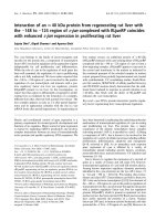
báo cáo khoa học: "Analysis of the expression pattern of the BCL11B gene and its relatives in patients with T-cell acute lymphoblastic leukemia" doc
Bạn đang xem bản rút gọn của tài liệu. Xem và tải ngay bản đầy đủ của tài liệu tại đây (583.44 KB, 7 trang )
RESEARC H Open Access
Analysis of the expression pattern of the BCL11B
gene and its relatives in patients with T-cell acute
lymphoblastic leukemia
Xin Huang
1,2
, Shaohua Chen
1
, Qi Shen
1
, Lijian Yang
1
,BoLi
1
, Liye Zhong
1,2
, Suxia Geng
2
, Xin Du
2
, Yangqiu Li
1,3*
Abstract
Background: In a human T-cell acute lymphoblastic leukemia (T-ALL) cell line (Molt-4), siRNA-mediated
suppression of BCL11B expression was shown to inhibit proliferation and induce apoptosis, functions which may be
related to genes involved in apoptosis (such as TNFSF10 and BCL2L1) and TGF-b pathways (such as SPP1and
CREBBP).
Methods: The expression levels of the above mentioned genes and their correlation with the BCL11B gene were
analyzed in patients with T-ALL using the TaqMan and SYBR Green I real-time polymerase chain reaction
technique.
Results: Expression levels of BCL11B, BCL2L1 , and CREBBP mRNA in T-ALL patients were significantly higher than
those from healthy controls (P<0.05). In T-ALL patients, the BCL11B expression level was negatively correlated with
the BCL2L1 expression level (r
s
= -0.700; P<0.05), and positively correlated with the SPP1 expression level (r
s
=
0.683; P<0.05). In healthy cont rols, the BCL11B expression level did not correlate with the TNFSF10, BCL2L1, SPP1,or
CREBBP expression levels.
Conclusions: Over-expression of BCL11B might play a role in anti-apoptosis in T-ALL cells through up-regulation of
its downstream genes BCL2L1 and CREBBP.
Background
T-cell acute lymphoblastic leukemia (T-ALL) accounts
for 15% of newly diagnosed ALL cases in children and
20-25% of ALL cases in adults [1,2]. Overall, these are
aggressive malignancies that do not respond well to che-
motherapy and have a poorer prognosis than their B-cell
counterparts [3]. The development of targeted therapies,
including monoclonal antibodies and gene therapy, con-
tinues. Small interfering RNA (siRNA) is a promising
gene-targeting agent that has shown great potential, par-
ticularly in the field of cancer treatment [4-6].
The B-cell chronic lymphocytic leukemia (CLL)/lym-
phoma 11B (BCL11B) gene plays a crucial role in T-cell
development, differentiation, and proliferation [7], and
altered expression, mutation, disruption, or rearrange-
ment of BCL11B have been associated with T-cell
malignancies [8-11]. BCL11B over-expression has been
observed primarily in T-cell malignancies [8,12].
BCL11B has b een hypothesized to act as a tumor sup-
pressor gene [9,13], but its precise function remains
unclear.
BCL2-like 1 (BCL2L1; Bcl-xL) is similar to Bcl-2
because i t restrains the apoptosis induction of multiple
stimuli, and is a key factor in the terminal step of apop-
tosis regulation. Studies have shown that BCL2L1 parti-
cipates in v arious protein-protein interactions, playing a
role in inhibiting apoptosis. In the endogenous apoptosis
pathway, BCL2L1 of the BCL-2 family in hibits apoptosis
by blocking the translocation of Bax to the mitochon-
drial outer membrane [14]. cAMP-response element
binding protein (CREBBP) plays a critical role in
embry onic development , growth control, and homeosta-
sis by coupling chromatin remodeling t o transcription
factor recognition. A CREBBP gene rearrangement with
chromosomal t ranslocation has been identified in acute
myeloid leukemia [15,16] and over-expression of
* Correspondence:
1
Institute of Hematology, Medical College, Jinan University, Guangzhou,
510632, PR China
Full list of author information is available at the end of the article
Huang et al. Journal of Hematology & Oncology 2010, 3:44
/>JOURNAL OF HEMATOLOGY
& ONCOLOGY
© 2010 Huang et al; licensee BioMed Central Ltd. This is an Open Access article distribute d under the terms of the Creative Commons
Attribution License ( which permits unrestricted use, di stribution, and rep roduction in
any medium, provid ed the original work i s properly cited.
CREBBP was found in Jurkat cells. Additionally,
enhancement of apoptotic cell death occurred in the
presence of CREB1 siRNA [17]. Tumor necrosis factor
(lig and) superfamily, member 10 (TNFSF10; TRAIL)isa
tumor necrosis factor superfamily member, and induces
apoptosis through its interaction with death receptors.
BCL-2 family genes and TNFSF10 probably act together
through crosstalk between the intrinsic and death recep-
tor-mediated apoptosis pathways [18]. Secr eted phos-
phoprotein 1 (SPP1) is also known as OPN and its
abnormal activation can stimulate tumor growth, inva-
sion, angiogenesis, and immune suppression, with wide-
ranging effects on cell proliferation, apoptosis, differen-
tiation, and migration [19,20].
Previous studies [21,22] showed that the inhibition of
BCL11B expression by siRNA selectively inhibited prolif-
eration and e ffectively induced apoptosis in human
T-cell acute lymphoblastic leukemia (T-ALL) cell lines
(Jurkat, Molt-4). Additionally, global gene expression
profiling revealed that BCL11B siRNA-mediat ed cell
apoptosis may be related to BCL-2 family genes of the
mitochondrial pathway, and the TRAIL (TNFSF10)gene
of the death receptor signaling pathway [22], further-
more, in our previous study, the genes (SPP1 and
CREBBP) of the TGF-b pathway (unpublished data). Lit-
tle is known about the expression pattern of these genes
in T-ALL. Thus, analyzing the expression pattern of
these genes in malign ant T-cells is important because
BCL11B disruption and disturbed expression may con-
tribute to the development of T-cell malignancies in
humans [8]. In the present study, we further analyzed
expression levels of TNFSF10, BCL2L1, SPP1,and
CREBBP, and their correlation with BCL11B in male
patients with T-ALL, to clarify the role of BCL11B in
T-cell malignancies.
Methods
Samples
Nine newly diagnosed T-ALL patients (male, 6-28 years
old; median age, 20 years; white blood cell count
(WBC), 1.8-293.5 × 10
9
/L; bone marrow blast percen-
tage: 65-93%; were recruited. The di agnosis of T-ALL
was based on cytomorphology, immunohistochemistry,
and cytoimmunological analysis. Peripheral blood mono-
nuclear cells (PBMCs) from nine healthy volunteers
served as control s (five males and four females, 20 -45
years old; median age, 28 years). Peripheral blood was
collected by heparin anticoagulation and PBMCs were
separated using the Ficoll-Hypaque gradient centrifuga-
tion method. The percentage of CD3+cells in PBMCs
were detected, there are 75.30 ± 26.77% (range 21.2-
97.8%) in PBMCs from T-ALL samples and 59.66 ±
4.75% (range 52.4-65.8%) in PBMCs from he althy con-
trol samples.
All procedures were conducted in accordance with the
guidelines of the Medical Ethics committees of the
health bureau of Guangdong Province, PR China.
RNA extraction and cDNA synthesis
RNA was extracted using the Trizol kit (Invitrogen,
Carlsbad, CA, USA) and reverse transcribed into the
first-strand cDNA using random hexamer primers and
the reverse transcriptase Superscript II Kit (Invitrogen),
according to the manufacturer’s instructions.
Real-time quantitative reverse transcription-polymerase
chain reaction (qRT-PCR)
Quantitative detection of the BCL11B gene expression
level in cDNA from PBMCs was performed using Taq-
Man real-time PCR. PCR was performed as described
previously [8]. To precisely determine the copy numbers
of BCL11B, a duplex vector, including a fragment of the
BCL11B and the b2 microglobulin (b2M) genes was con-
structed and used as a reference (the duplex vector was
a gift from Prof. C.A. Schmidt, Ernst-Moritz-Arndt Uni-
versity Greifswald, Germany). Based on the DNA con-
centration, measured by spectrophotometry and
confirmed by quantitative gel eletrophoresis, standard
dilutions of the vector from 10
7
to 10
1
copies were pre-
pared [8]. Briefly, PCR was performed in a 25-μLtotal
volume containing 2 μL of cDNA, 25 pmol of each pri-
mer (BCL11B-f and BCL11B-b for BCL11B gene amplifi-
cation; b2Mf and b2Mb for b2M gene amplification), 10
nmolofeachdNTP,1.5UAmpliTaqGold(Applied
Biosystems, Branchburg, NJ, USA), 5 pmol of 6FAM-
TAMRA probe, and PCR buffer containing 4.5 mM
MgCl
2
. After an initial denaturation at 95°C for 5 min,
50 cycles consisting of 95°C for 15 s and 64°C for 1 min
were performed. Primers and probes for BCL11B and
b2M gene amplification were synthesized by TIB Mol-
biol Co. (Berlin, Germany; Table 1).
The absolute amounts of BCL11B and b2M were mea-
sured in tw o independent assays and BCL11B content
per 100,000 b2M copies was c alculated using the for-
mula: n = 100000 × BCL11B/b2M.
Expression levels of TNFSF10, BCL2L1, SPP1,
CREBBP, and the reference gene b2-MG were deter-
mined by SYBR Green I real-time PCR. Briefly, PCR was
performed in a 25-μL total volume containing 1 μLof
cDNA, 9 μL of 2.5× SYBR Green mix (Tiangen, Beijing,
PR China), and 10 μmol/L primer pairs. The following
cycling conditions were used: initial denaturation at
95°C for 2 min, followed by 44 cycles at 95°C for 15 s,
and 81°C (TNFSF10, SPP1, CREBBP,andb-2-MG)or
84°C (BCL2L1) for 1 min. The relative amounts of the
genes of interest and the b2M reference gene were mea-
suredintwoindependentassays.The2
(-ΔΔCT)
method
was used to present the data of the genes o f interest
Huang et al. Journal of Hematology & Oncology 2010, 3:44
/>Page 2 of 7
relative to an internal control gene [23,24]. The efficien-
cies of real-time PCR for expression analysis of different
genes were evaluated using diluted Molt-4 cDNA (1, 5
-1
,
5
-2
,5
-3
,5
-4
) a s templates to construct relative standard
curves. Additionally, the specific amplification of PCR
products was analyzed by melting curve analysis and
agarose electrophoresis. Primers used in the S YBR
Green I real-time PCR for all four gene amplifications
were synthesized by Shanghai Biological Engineering
Technology Services Co., Ltd. (Table 2).
RT-PCR for TNFSF10, BCL2L1, SPP1,andCREBBP
genes was performed using the same primers as
described above, and the PCR products were sent to
Shanghai Invitrogen Biotechnology Co. for DNA
sequence analysis.
Statistical analyses
Independent-sample t -test ana lysis was used for the
BCL11B gene mRNA levels in different samples, while
the Mann-Whitney U test and Spearman’s rank correla-
tion analyses were used for non-normally distributed
data using the SPSS 13.0 statistical software. Differences
were considered statistically significant at P < 0.05.
Results
Over-expression of BCL11B gene in T-ALL
The expression level of BCL11B mRNA in PBMCs from
patients with T-ALL (1821.81 ± 1896.58 copies/10
5
b2M
copies) was significantly higher than that from healthy
controls (259.71 ± 182.72 copie s/10
5
b2M copies; t =
2.46; P = 0.039; Figure 1). PCR products from b2M and
BCL11B genes were confirmed by 2.5% gel electrophor-
esis (Figure 2D, E).
Expression of TNFSF10, BCL2L1, SPP1, and CREBBP genes
in T-ALL
The high amplification efficiency of the four genes of
interest (TNFSF10, BCL2L1, SPP1,andCREBBP)was
consistent with that of the b2M reference gene. For
example, the accurate standard curve graphs of BCL2L1
and b
2
M control gene amplification are illustrated in
Figure 2A and 2B (r
2
= 0.995). The amplification effi-
ciencies of BCL2L1 and the b2M control gene were
95.30% and 95.16%, respectively, and the melting curves
are shown in Figure 2C. PCR products from the b2M
control gene and genes of interest were confirmed using
2.5% gel electrophoresis (Figure 2D, E), followed by
sequence confirmation (data not shown).
Relative expression levels of BCL2L1 mRNA (397. 82 ±
565.98%) and CREBBP mRNA (53.28 ± 39.21%) in
patients with T-ALL were significantly higher than
those from healthy controls (BCL2L1: 10.83 ± 11.18%;
CREBBP: 20.80 ± 13.50%; P < 0.05), whereas the relative
expression levels of TNFSF10 and SPP1 mRNA showed
no significant differenc e between T-ALL and healthy
groups (Figure 2F).
In T-ALL patients, Spearman’s rank correlation analyses
revealed that the BCL11B expression level was negativel y
Table 1 Sequences of primers and probes for real-time PCR (TaqMan method)
primers/probes sequence function
BCL11Bf 5’-CACCCCCGACGAAGATGACCAC forward primer
BCL11Bb 5’-CGGCCCGGGCTCCAGGTAGATG backward primer
BCL11Bp 5’-6FAM-TCACCCACGAAAGGCATCTGTCCCAAGCA-TAMRA probe
b2Mf 5’-CTCGCGCTACTCTCTCTTTCT forward primer
b2Mb 5’-TACATGTCTCGATCCCACTTAACTAT backward primer
b2Mp 5’-6FAM-CTCACGTCATCCAGCAGAGAATGGAAAGTCA-TAMRA probe
Table 2 Sequences of primers for real-time PCR (SYB
Green I method)
primers sequence function
TNFSF10 5’-GAGTATGAACAGCCCCT-3’ forward primer
TNFSF10 5’-GTTGCTTCTTCCTCTGGT-3’ backward primer
BCL2L1 5’-AAACTGGGTCGCATTGTGG-3’ forward primer
BCL2L1 5’-TCTCGGCTGCTGCATTGTTC-3’ backward primer
SPP1 5’-ACAGCCAGGACTCCATTGA-3’ forward primer
SPP1 5’-TCAGGTCTGCGAAACTTCTTAG-3’ backward primer
CREBBP 5’-CGGTTTCTCGGCGAATGAC-3’ forward primer
CREBBP 5’-CATTTCCTATTCCTGGGTTGAT-3’ backward primer
Figure 1 Expression levels of the BCL11B gene in PBMCs from
T-ALL and healthy controls.
Huang et al. Journal of Hematology & Oncology 2010, 3:44
/>Page 3 of 7
Figure 2 Features of the expression of TNFSF10, BCL2L1, SPP1, and CREBBP genes in T-ALL and healthy groups. A, B: Accurate standard
curve graphs of BCL2L1 and the b2M control gene are shown using diluted Molt-4 cDNA as the template. The amplification efficiency of
BCL2L1-related genes was more than 95%, and consistent with the high amplification efficiency of the b2M reference gene. C: Melting curves of
the BCL2L1 and b2M genes from nine patients. #: Specific peak of the b2M reference gene begins at 81°C. ##: Specific peak of the BCL2L1 gene
begins at 84°C. D: PCR products of the b2M gene by 2.5% agarose gel electrophoresis analysis. The size of the PCR products of the b2M gene
used for the BCL11B reference is 332 bp (line 1, 2) and that used for the four genes of interest is 145 bp (line 4-11). Line 3: DNA ladder. E: PCR
products analyzed by 2.5% agarose gel electrophoresis. Line 1-2: BCL11B (193bp), line 3: DNA ladder, line 4-5: BCL2L1 (202 bp), line 6-7: CREBBP
(206 bp), line 8-9: SPP1 (241 bp), line 10-11: TNFSF10 (190 bp). F: Relative expression levels of the four genes of interest in T-ALL and healthy
groups.
Huang et al. Journal of Hematology & Oncology 2010, 3:44
/>Page 4 of 7
correlated with the BCL2L1 relative expression level (r
s
=
-0.700; P = 0.036; Figure 3A), and positively correlated
with the SPP1 relative expression level (r
s
= 0.683; P =
0.042; Figure 3B). The BCL11B expression level did not
exhibit an obvious correlation with TNFSF10 or CREBBP
relative expression levels. No significant correlation was
found between the BCL11B gene and the other four genes
of interest in the healthy controls.
Discussion
Increasing numbers of translocations involving the
BCL11B locus [8,10,11] or high levels of BCL11B mRNA
expression in most T-ALL cases [8,12] have been
repo rted; however, the mechanism of BCL11B-mediated
oncogenesis remains unknown. To clarify the role of
BCL11B in T -cell malignancies, we further analyzed the
expression levels of TNFSF10, BCL2L1, SPP1,and
CREBBP genes and their correlations with BCL11B in
patients with T-ALL and controls. Over-expression of
the BCL11B gene, as well as BCL2L1 and CREBBP
mRNA, were characteristic features of T-ALL.
Recent evidence has suggested that multiple mechan-
isms may regulate the release of mitochondrial factors,
some of which depend on the action of caspases. BCL2L1
may inactivate caspase-8 by decreasing death-inducing sig-
naling complex (DISC) formation in the plasma mem-
brane, nucleus, and Golgi complex while diverting DISC
formation to the mitochondria. The inhibitory effects of
BCL2L1 on DISC formation may play a significant role in
protecting endothelial cells from hypoxia/reoxygenation
(H/R)-induced cell death [25].Thus,over-expressionof
the BCL2L1 gene suggests that it might be related to the
occurrence of T-ALL by defective regulation of apoptosis.
During the pro cess of T-ALL, over-expressed BCL2L1 is
thought to suppress the activity of caspase-8; thus, as a
kind of protection mechanism, the TNFSF10 gene of some
patients is highly expressed, promoting caspase-8 activity
in response to this abnormal cell proliferation. However,
the low expression level of SPP1 in untreated Molt-4 cells
differed from the high expression levels found in mostly
solid tumors [26]. Additionally, our findings indicated no
significant difference in SPP1 gen e expression in the T-
ALL group. Comprehensive analysis revealed that T-ALL
occurred in the presence o f BCL11B, BCL2L1,and
CREBBP gene over-expression, which was closely related
to blocking apoptosis of malignant T cell, whereas the
TNFSF10 gene was also highly expressed in some patients,
which may partly correct the imbalance.
Correlation analysis of BCL11B in the T-ALL group
revealed that the BCL11B expression level was nega-
tively correlated with that of BCL2L1 (Bcl-xL),
although over-expression of both genes was found in
T-ALL samples. This suggested that BCL2L1 was
affected by the BCL11B gene in transcr iptional regula-
tion, and both participated in the s ame protein-protein
interactions, acting as apoptosis regulators along with
a competitive target protein downstream. In BCL 11B-
knockdown T-cell lines, when exposed to growth sti-
muli, T cells exhibit apoptosis in S phase with conco-
mitant decreases in the cell-cycle inhibitor p27 and the
anti-apoptotic protein Bcl-xL, due t o transcriptional
repression [13]. However, BCL11B and BCL2L1 protein
levels in the T-ALL group still remain to be validated.
Correlation analysis of BCL11B in the T-ALL group
revealed that the BCL11B expression level was posi-
tively correlated with the relative SPP1 expression
level. The expression of
SPP1 was significantly down-
regulated with BCL11B silencing by RNA interference,
suggesting that the SPP1 gene may be a target of the
BCL11B gene in transcriptional regulation (unpub-
lished data). SPP1 gene silencing in vitro significantly
increased mitochondrial cytochrome c rele ase, and the
Figure 3 Linear correlation analyses of the BCL11B and BCL2L1 genes (A) and SPP1 gene (B) in T-ALL samples.
Huang et al. Journal of Hematology & Oncology 2010, 3:44
/>Page 5 of 7
inhibitory action of the Wnt target gene osteopontin
(SPP1) on mitochondrial cytochrome c release deter-
mines renal ischemic resistance [27]. Thus, the SPP1
gene may play a consistent role in anti-apoptotic
effects with the BCL11B gene, by decreasing mitochon-
drial cytochrome c release. The hypothetical regulatory
network of apoptosis in BCL11B and related genes is
shown in Figure 4. However, the role of the SPP1 gene
in T-cell malignancies is unclear, because low expres-
sion of SPP1 was detected in T-ALL.
Conclusions
The expression pattern of the BCL11B gene and four of
its related genes (TNFSF10, BCL2L1, SPP1,and
CREBBP) was characterized in T-ALL. Over-expression
ofBCL11Bmayplayaroleinanti-apoptosisinT-ALL
cells through up-regulation of its downstream genes
BCL2L1 and CREBBP.
Acknowledgements
The project was sponsored by grants from National Natural Science
Foundation of China (No. 30771980), the Fundamental Research Funds for
the Central Universities (No. 21610604) and the Guangdong Science &
Technology Project (No. 2007B030703008; and 2009B050700029).
Author details
1
Institute of Hematology, Medical College, Jinan University, Guangzhou,
510632, PR China.
2
Department of Hematology, Guangdong General Hospital
(Guangdong Academy of Medical Sciences), Guangzhou, 510080, PR China.
3
Key Laboratory for Regenerative Medicine of Ministry of Education, Jinan
University, Guangzhou, 510632, PR China.
Authors’ contributions
YQL made contributions to conception and design laboratory study. XH,
SHC, QS, LJY, and BL performed the laboratory technique process and the
laboratory analyses. LYZ, SXG and XD were responsible of the patient’s
treatment and carried out acquisition of clinical data. YQL and XH
coordinated the study and helped to draft the manuscript. All authors read
and approved the final manuscript.
Competing interests
The authors declare that they have no competing interests.
Received: 19 October 2010 Accepted: 16 November 2010
Published: 16 November 2010
References
1. Rivera GK, Crist WM: Acute lymphoblastic leukemia. In Principles and
Practice of Hematology. Blood Edited by: Handin RI, Stossel TP, Lux SE 1995,
743-759.
2. Uckun FM, Sensel MG, Sun L, Steinherz PG, Trigg ME, Heerema NA: Biology
and treatment of childhood T-lineage acute lymphoblastic leukemia.
Blood 1998, 91:735-746.
3. Morris JC, Waldmann TA, Janik JE: Receptor-Directed Therapy of T-Cell
Leukemias and Lymphomas. J Immunotoxicol 2008, 5:235-248.
Figure 4 Schematic representation of the regulatory network of apoptosis in BCL11B and its related genes. (a) BCL2L1 is affected by the
BCL11B gene in transcriptional regulation. (b, d) BCL11B and BCL2L1 participate in the same protein-protein interactions, along with competitive
downstream target proteins. BCL2L1 (Bcl-xL) normally interferes with the mitochondrial programmed cell death pathway by sequestering
proapoptotic proteins such as BCL2-associated × protein (BAX) and BCL2-antagonist/killer 1 (BAK1; BAK), suggesting that BAX/BAK may be
competitive target proteins downstream of BCL11B. (c) The SPP1 gene may be a target of the BCL11B gene in transcriptional regulation: it plays a
consistent role in anti-apoptotic effects with the BCL11B gene by decreasing mitochondrial cytochrome c release.
Huang et al. Journal of Hematology & Oncology 2010, 3:44
/>Page 6 of 7
4. Oh YK, Park TG: siRNA delivery systems for cancer treatment. Adv Drug
Deliv Rev 2009, 61:850-862.
5. Devi RS: siRNA-based approaches in cancer therapy. Cancer Gene Therapy
2006, 13:819-829.
6. Whitehead KA, Langer R, Anderson DG: Knocking down barriers: Advances
in siRNA delivery. Nat Rev Drug Discov 2009, 8:129-138.
7. Liu P, Keller JR, Ortiz M, Tessarollo L, Rachel RA, Nakamura T, Jenkins NA,
Copeland NG: Bcl11a is essential for normal lymphoid development. Nat
Immunol 2003, 4:525-532.
8. Przybylski GK, Dik WA, Wanzeck J, Grabarczyk P, Majunke S, Martin-
Subero JI, Siebert R, Dölken G, Ludwig WD, Verhaaf B, van Dongen JJ,
Schmidt CA, Langerak AW: Disruption of the BCL11B gene through inv 14
q11.2q32.31 results in the expression of BCL11B-TRDC fusion transcripts
and is associated with the absence of wild-type BCL11B transcripts in T-
ALL. Leukemia 2005, 19:201-208.
9. Karlsson A, Nordigården A, Jönsson JI, Söderkvist P: Bcl11b mutations
identified in murine lymphomas increase the proliferation rate of
hematopoietic progenitor cells. BMC Cancer 2007, 7:195.
10. Strehl S, Konig M, Spath K: Juxtaposition of the BCL11B gene to a novel
region at 17q by a t(14;17) (q32; Q21) in childhood T-Cell lymphoblastic
lymphoma [abstract]. Blood 2007, 110:101B.
11. Su XY, Della-Valle V, Andre-Schmutz I, Lemercier C, Radford-Weiss I,
Ballerini P, Lessard M, Lafage-Pochitaloff M, Mugneret F, Berger R,
Romana SP, Bernard OA, Penard-Lacronique V: HOX11L2/TLX3 is
transcriptionally activated through T-cell regulatory elements
downstream of BCL11B as a result of the t(5;14) (q35;q32). Blood 2006,
108:4198-4201.
12. Oshiro A, Tagawa H, Ohshima K, Karube K, Uike N, Tashiro Y, Utsunomiya A,
Masuda M, Takasu N, Nakamura S, Morishima Y, Seto M: Identification of
subtype-specific genomic alterations in aggressive adult T-cell leukemia/
lymphoma. Blood 2006, 107:4500-4507.
13. Kamimura K, Mishima Y, Obata M: Lack of Bcl11b tumor suppressor
results in vulnerability to DNA replication stress and damages. Oncogene
2007, 26:5840-5850.
14. Breckenridge DG, Xue D: Regulation of mitochondrial membrane
permeabilization by BCL-2 family proteins and caspases. Curr Opin Cell
Biol 2004, 16:647-652.
15. Borrow J, Stanton VP Jr, Andresen JM, Becher R, Behm FG, Chaganti RS,
Civin CI, Disteche C, Dubé I, Frischauf AM, Horsman D, Mitelman F,
Volinia S, Watmore AE, Housman DE: The translocation t(8;16)(p11;13) of
acute myeloid leukaemia fuses a putative acetyltransferase to the CREB-
binding protein. Nature Genet 1996, 14:33-41.
16. Giles RH, Dauwerse JG, Higgins C, Petrij F, Wessels JW, Beverstock GC,
Döhner H, Jotterand-Bellomo M, Falkenburg JH, Slater RM, van Ommen GJ,
Hagemeijer A, van der Reijden BA, Breuning MH: Detection of CBP
rearrangements in acute myelogenous leukemia with t(8;16). Leukemia
1997, 11:2087-2096.
17. Caravatta L, Sancilio S, di Giacomo V, Rana R, Cataldi A, Di Pietro R: PI3-K/
Akt-dependent activation of cAMP-response element-binding (CREB)
protein in Jurkat T leukemia cells treated with TRAIL. J Cell Physiol 2008,
214:192-200.
18. Adams JM, Cory S: The Bcl-2 apoptotic switch in cancer development
and therapy Bcl-2 apoptotic switch in cancer. Oncogene 2007,
26:1324-1337.
19. Standal T, Borset M, Sundan A: Role of osteopontin in adhesion,
migration, cell survival, and bone remodeling. Exp Oncol 2004,
26:179-184.
20. Rangaswami H, Bulbule A, Kundu GC: Osteopontin: role in cell signaling
and cancer progression. Trends Cell Biol 2006, 16:79-87.
21. Huang X, Chen S, Yang LJ, Chen SH, Zhou YB, Schmidt CA, Li YQ: Effects of
down-regulating BCL11B expression on the proliferation, apoptosis and
global gene expression profiling of Molt-4 cells [Abstract]. Blood 2009,
114:4505.
22. Grabarczyk P, Przybylski GK, Depke M, Völker U, Bahr J, Assmus K,
Bröker BM, Walther R, Schmidt CA: Inhibition of BCL11B expression leads
to apoptosis of malignant but not normal mature T cells. Oncogene 2007,
26:3797-3810.
23. Livak KJ, Schmittgen TD: Analysis of relative gene expression data using
real-time quantitative PCR and the 2(-Delta Delta C(T)) Method. Methods
2001, 25:402-408.
24. Pfaffl MW, Horgan GW, Dempfle L: Relative expression software tool
(REST) for group-wise comparison and statistical analysis of relative
expression results in real-time PCR. Nucleic Acids Res 2002, 30:e36.
25. Wang X, Zhang J, Kim HP: Bcl-XL disrupts death-inducing signal complex
formation in plasma membrane induced by hypoxia/reoxygenation.
FASEB J 2004, 18:1826-1833.
26. Rodrigues LR, Teixeira JA, Schmitt FL, Paulsson M, Lindmark-Mänsson H: The
role of osteopontin in tumor progression and metastasis in breast
cancer. Cancer Epidemiol Biomarkers Prev 2007, 16:1087-1097.
27. Viñas JL, Sola A, Jung M, Mastora C, Vinuesa E, Pi F, Hotter G: Inhibitory
action of Wnt target gene osteopontin on mitochondrial cytochrome c
release determines renal ischemic resistance. Am J Physiol Renal Physiol
2010, 299:F234-242.
doi:10.1186/1756-8722-3-44
Cite this article as: Huang et al.: Analysis of the expression pattern of
the BCL11B gene and its relatives in patients with T-cell acute
lymphoblastic leukemia. Journal of Hematology & Oncology 2010 3:44.
Submit your next manuscript to BioMed Central
and take full advantage of:
• Convenient online submission
• Thorough peer review
• No space constraints or color figure charges
• Immediate publication on acceptance
• Inclusion in PubMed, CAS, Scopus and Google Scholar
• Research which is freely available for redistribution
Submit your manuscript at
www.biomedcentral.com/submit
Huang et al. Journal of Hematology & Oncology 2010, 3:44
/>Page 7 of 7